Embed Size (px)
Citation preview

ARTICLE
A non-classical view on calcium oxalateprecipitation and the role of citrateEncarnación Ruiz-Agudo1, Alejandro Burgos-Cara 1, Cristina Ruiz-Agudo2,3, Aurelia Ibañez-Velasco1,
Helmut Cölfen 3 & Carlos Rodriguez-Navarro1
Although calcium oxalates are relevant biominerals, their formation mechanisms remain
largely unresolved. Here, we investigate the early stages of calcium oxalate formation in pure
and citrate-bearing solutions. Citrate is used as a well-known oxalate precipitation
inhibitor; moreover, it resembles the functional domains of the biomolecules that
modulate biomineralization. Our data suggest that calcium oxalate forms after Ca2+ and
C2O42− association into polynuclear stable complexes that aggregate into larger assemblies,
from which amorphous calcium oxalate nucleates. Previous work has explained citrate
inhibitory effects according to classical theories. Here we show that citrate interacts with all
early stage CaC2O4 species (polynuclear stable complexes and amorphous precursors),
inhibiting calcium oxalate nucleation by colloidal stabilization of polynuclear stable
complexes and amorphous calcium oxalate. The control that citrate exerts on calcium oxalate
biomineralization may thus begin earlier than previously thought. These insights provide
information regarding the mechanisms governing biomineralization, including pathological
processes (e.g., kidney stone formation).
DOI: 10.1038/s41467-017-00756-5 OPEN
1 Department of Mineralogy and Petrology, University of Granada, Fuentenueva s/n, 18071 Granada, Spain. 2 Institut für Mineralogie, Universität Münster,Corrensstrasse 24, 48149 Münster, Germany. 3 Physical Chemistry, Department of Chemistry, University of Konstanz, Universitätsstraße 10, 78457Konstanz, Germany. Correspondence and requests for materials should be addressed to E.R.-A. (email: [email protected])
NATURE COMMUNICATIONS |8: 768 |DOI: 10.1038/s41467-017-00756-5 |www.nature.com/naturecommunications 1

Calcium oxalates are one of the most common biomineralsin nature, and the most abundant group of organicminerals found in sediments and hydrothermal veins1.
Calcium oxalate may represent up to 80% of the dry weight ofsome plants2. In higher plants, calcium oxalate is formed withinspecialized cells3 displaying a wide variety of shapes and sizes,and acting mainly as structural support or protection againstpredators4, 5. Additionally, the precipitation of crystallinecalcium oxalate monohydrate (COM, CaC2O4·H2O, whewellite)or calcium oxalate dihydrate (COD, CaC2O4·2H2O, weddellite)in plants may serve to store calcium and maintain a lowconcentration in the cytosol in order to prevent interferences withcell processes2.
However, calcium oxalate mineralization is typically patholo-gical in vertebrates. In humans, calcium oxalate is associated withbenign breast tissue calcifications6 and is commonly found inkidney stones5, 7. In healthy individuals, urine is typicallysupersaturated with respect to COM, but the developmentof stone disease is prevented by biological mechanisms. Thepresence of urinary proteins and small molecules such as thecarboxylate-rich molecule citrate that act as COM nucleationand growth inhibitors prevents oxalate stone formation7. Theconcentration of citrate in the urine of individuals that developkidney stones is commonly below the normal physiological rangeof 1–2 mM7. Thus, this molecule is used as a common therapeuticagent for treating stone disease. A sound knowledge of thephysical–chemical mechanisms governing the role of citrate atmodulating COM nucleation and early growth is critical forimproving therapies for stone disease. Moreover, because theacidic residues of organic (macro)molecules are known to governbiomineralization in a wide range of organisms and minerals,analysis of the effects exerted by citrate on the early stages ofCOM formation may also provide insights on the molecularcontrol of biomineralization8 and help to define better strategiesfor the synthesis of biomimetic materials. Note also that citratehas been reported to play a key role in the biomineralization ofcollagen by calcium phosphates during bone development9.
Unlike calcium carbonate or calcium phosphatesunderstanding the early stages of calcium oxalate precipitationhas been elusive. It is only very recently that evidence hasbeen presented that amorphous phases precede the formationof crystalline calcium oxalate2, 5, despite the fact that itwas suggested that calcium oxalate may form via non-classicalcrystallization pathways10. Moreover, most studies have approa-ched the analysis of CaC2O4 formation and inhibition by citrateor nucleation from a classical point of view11–13, and none ofthem, to our knowledge, has explored the effect of citrate on theearly formation stages of calcium oxalate minerals. Here we showhow under the conditions of this study, association of Ca2+
and C2O42– into negatively charged pre-nucleation species and
clusters (PNCs) occurs prior to nucleation of amorphous calciumoxalate (ACO). This raises the possibility that the control thatdifferent organic additives exert on calcium oxalate (pathological)biomineralization may begin even earlier than previouslythought. Indeed, our results suggest that citrate actsstabilizing simultaneously pre-nucleation ion associates andACO nanoparticles, delaying both ACO formation and itstransformation into crystalline phase(s).
ResultsTitration experiments. Figure 1a shows the time evolution ofturbidity and free Ca2+ concentration in a 2 mM oxalate solutionfor different titration experiments performed in the absence andin the presence of different amounts of citrate and their replicates.The measured free calcium concentration increases with time up
to a point when solid calcium oxalate precipitates and the freeCa2+ concentration in solution drops and then graduallyapproaches a constant level related to the solubility of theprecipitated phase. Both in the presence and in the absence ofcitrate, the free Ca2+ concentration detected in solution rises at asignificantly lower rate than the added amount. As it has beenreported previously for other sparingly soluble minerals such asBaSO4, CaCO3, and BaCO3
10, 14–17, this suggests that Ca2+ andC2O4
2− ions associate into stable complexes prior to the onset ofliquid or solid Ca-oxalate formation. Our study provides anotherexample of such a mechanism for mineral formation, confirmingthat this may indeed be a general pathway for crystallization.
The concentration of these complexes in solution can bedetermined independently from ion-selective electrode (ISE) andconductivity measurements. A theoretical conductivity (kcal) forthe control titration run (i.e., no citrate in the reaction media) canbe calculated assuming that the activity coefficients of all ions insolution equal 1 (i.e., an ideal solution) and assuming that all ionsare free in solution. This seems reasonable considering that the
0.10
0.08
0.06
0.04
0.02
0.00
7×10⁻⁵
6×10⁻⁵
5×10⁻⁵
4×10⁻⁵S
lope
(m
M s
–1)
3×10⁻⁵
2×10⁻⁵
1×10⁻⁵
0
0 1 5 10Citrate concentration (mM)
0 2,000 4,000
Ca added
400
350
300
250
Tran
smitt
ance
(m
V)
200
150
100
50
0
0 mM citrate1 mM5 mM10 mM
6,000
Time (s)
8,000 10,000
Ca2+
(m
M)
free
a
b
Fig. 1 Results of titration experiments. a Time development of free Ca2+
concentration in the presence of different amounts of citrate at pH 6.2. Bluelines: control runs; green lines: runs performed in the presence of 1 mMcitrate; dark red lines: runs performed in the presence of 5 mM citrate;orange lines: runs performed in the presence of 10 mM citrate. The black linerefers to the amount of Ca2+ added. Evolution of transmittance is alsorepresented (dotted lines). Two replicates are presented for each run. b Barplot illustrating the effect of added citrate on the slope of t-dependent freeCa2+ concentration prior to nucleation. Error bars show S.D.
ARTICLE NATURE COMMUNICATIONS | DOI: 10.1038/s41467-017-00756-5
2 NATURE COMMUNICATIONS |8: 768 |DOI: 10.1038/s41467-017-00756-5 |www.nature.com/naturecommunications

ionic strength of the solution is never higher than 5×10⁻³. kcal ishigher than the actual measured conductivity (k), due to theassociation of ions in solution prior to nucleation:
k ¼X
i
ci�λi ¼ kcal � cCaC2O4 clusters�λCaC2O4 clusters; ð1Þ
ci (M) and λi (S cm2 mol−1) are respectively the concentration andthe molar conductivity of ion i; cCaC2O4 clusters is the concentrationof pre-nucleation CaC2O4 associates in solution and λCaC2O4 clustersis the molar conductivity of CaC2O4 associates, calculated withthe Kohlrausch law:
λCaC2O4 clusters ¼ λCa2þ þ n � λC2O2�4; ð2Þ
A perfect matching between bound oxalate determined fromconductivity measurements and the bound calcium (from ISE)was achieved considering a C2O4:Ca ratio in the ion associates, n,of 2 (Supplementary Fig. 1), thus indicating that pre-nucleationassociates are negatively charged. Notably, this is different to anyother system previously studied (e.g., CaCO3 or BaSO4), in whichprenucleation associates are neutral species, and indicates that inour system there are species different to ion pairs (e.g., CaHC2O4
+
or CaC2O40), which are at least trimers (that can be expressed as
n{[Ca(C2O4)2]2−}) and, as such, are considered as prenucleationclusters.
The well-known inhibitory effect of citrate on calcium oxalateprecipitation is seen in our experiments as a delay in the onset ofprecipitation from 760 s (control runs, no citrate in the media) upto 7,500–9,400 s (10 mM citrate). Interestingly, clouding of thesolution (indicated by a drop in the transmittance of the solution)occurs before the drop of the free Ca2+ curve, an effect that isenhanced in the presence of citrate (Fig. 1a). Carboxylate groupsin citrate are expected to bind calcium ions, an effect that isconfirmed by free calcium measurements when citrate isdissolved in pure water (Supplementary Fig. 2). In the case ofoxalate-free solutions, citrate binds about 0.12 Ca2+ ions percarboxylate group (for a citrate concentration of 10 mM). In theoxalate solution, however, the extent of Ca binding by citrate wasfound to be significantly lower, and each carboxylate group bindsan average of only 0.02 calcium ions. The slightly curved initialpart is related to a weak binding of Ca2+ ions to the citrate ions inthe presence of oxalate15. The difference in calcium bindingbetween water and oxalate solution could be related to areduction in the binding sites for calcium due to the adsorptionof pre-nucleation clusters15.
Moreover, the presence of citrate also affects the slope of thelinear part of the plot. The higher the citrate content, the flatterthe slope of the free Ca2+ ions vs. time curve is (Fig. 1b). Thisindicates that more calcium ions are bound in solution clusterswith increasing citrate concentration. The slope of the linear partof the measured free Ca2+ vs. time plot in the pre-nucleation stagereflects the stability of ion associates in solution15, 16. A simplifiedmultiple binding equilibrium was applied for the linear part of thefree-calcium concentration curve in the pre-nucleation regime inorder to quantify cluster formation10, 15. The macroscopicequilibrium constant for the formation of calcium/oxalate ion
associates, K′, can be expressed as:
Ca2þaq þ C2O2�4;aq !
KCaC2O4½ �ion associate;aq
c CaC2O4½ �ion associate;aq
� �
c Ca2þaq� �
� c C2O2�4;aq
� � ¼ K 0 ¼ x � K;
ð3Þ
For a constant, macroscopic C2O4:Ca ratio of 2:1 (as foundabove), the slope of a plot of v (see below) vs. the free calciumconcentration, cfree(Ca2+), gives the reciprocal of the microscopicequilibrium constant, K, for the formation of calcium/oxalate ionpairs and the averaged, dynamic coordination number of a singleoxalate, x10, 15:
v ¼ 2þ nfree C2O2�4
� �
nbound Ca2þð Þ ¼1xþ 1x � K � cfree Ca2þð Þ ; ð4Þ
cfree(Ca2+), nfree(C2O42−), and nbound(Ca2+) can be determined
from calcium potential measurements. This allows the determi-nation of average equilibrium constants for the formation of ionassociates in solution (pairs and/or bigger associates), K′, andtheir standard free energy of formation, ΔGion_pair:
�RT lnK 0 ¼ ΔGion pair; ð5ÞThese parameters relate to the binding strength in clusters.
From these calculations, it can be concluded that citrate stabilizesthe pre-nucleation species (i.e., more negative values of ΔGion_pair
are found in the presence of citrate; Supplementary Fig. 3).Additionally, the value of 0.31± 0.03 found for x in the controlruns (i.e., averaged, dynamic number of calcium ions coordinatedto a single oxalate ion when citrate is absent in the precipitationmedia) is in agreement with the C2O4:Ca ratio significantly higherthan 1 determined from independent conductivity measurements,indicating that indeed CaC2O4 pre-nucleation species arenegatively charged.
This behavior is very similar to that found for citrate on calciteprecipitation15. However, while the post-nucleation Ca2+-levelsand the concentration of free Ca2+-ions at the point of CaCO3
nucleation were not significantly affected by the presenceof citrate, both parameters are substantially increased withincreasing citrate concentration in this case (Fig. 1a). Notably,this represents a significant difference between the effect of thisadditive in the oxalate and carbonate systems.
Characterization of precipitates. In the control experiments,droplets quenched once both the transmittance and the free Ca2+
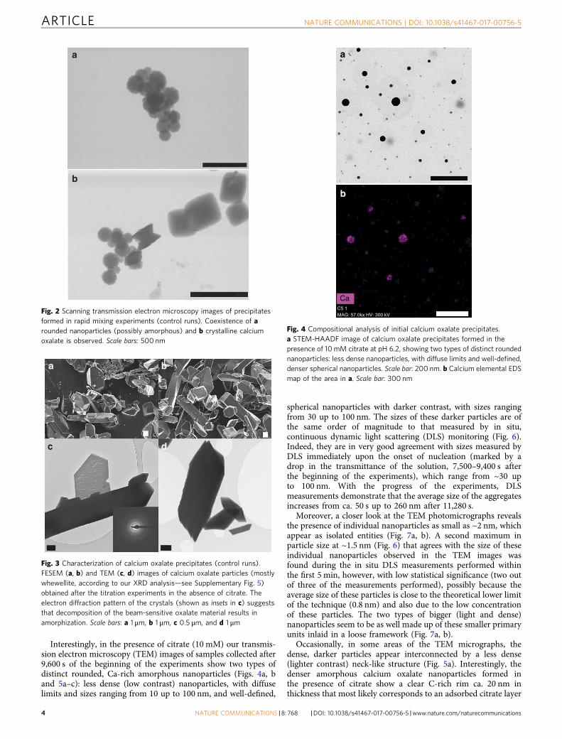
concentration are decreasing in solution (ca. 1,600 s after thebeginning of the experiment) show scarce spherical calciumoxalate nanoparticles of sizes ranging between 50 and 200 nm(Supplementary Fig. 4). These particles are initially amorphous,although their exposure to the electron beam results in theirpartial transformation into crystalline calcium oxalate (inset inSupplementary Fig. 4). Similarly, wet-scanning transmissionelectron microscopy (STEM) observations of precipitatesformed in control solutions (no ethanol quenching) showed theformation of rounded aggregates of nanoparticles (Fig. 2a)coexisting with crystalline calcium oxalate (Fig. 2b). Uponstabilization of the measured free calcium concentration(ca. 2,000 s after the beginning of the experiment), they transforminto crystalline calcium oxalate (whewellite, Fig. 3a–d andSupplementary Fig. 5).
NATURE COMMUNICATIONS | DOI: 10.1038/s41467-017-00756-5 ARTICLE
NATURE COMMUNICATIONS |8: 768 |DOI: 10.1038/s41467-017-00756-5 |www.nature.com/naturecommunications 3
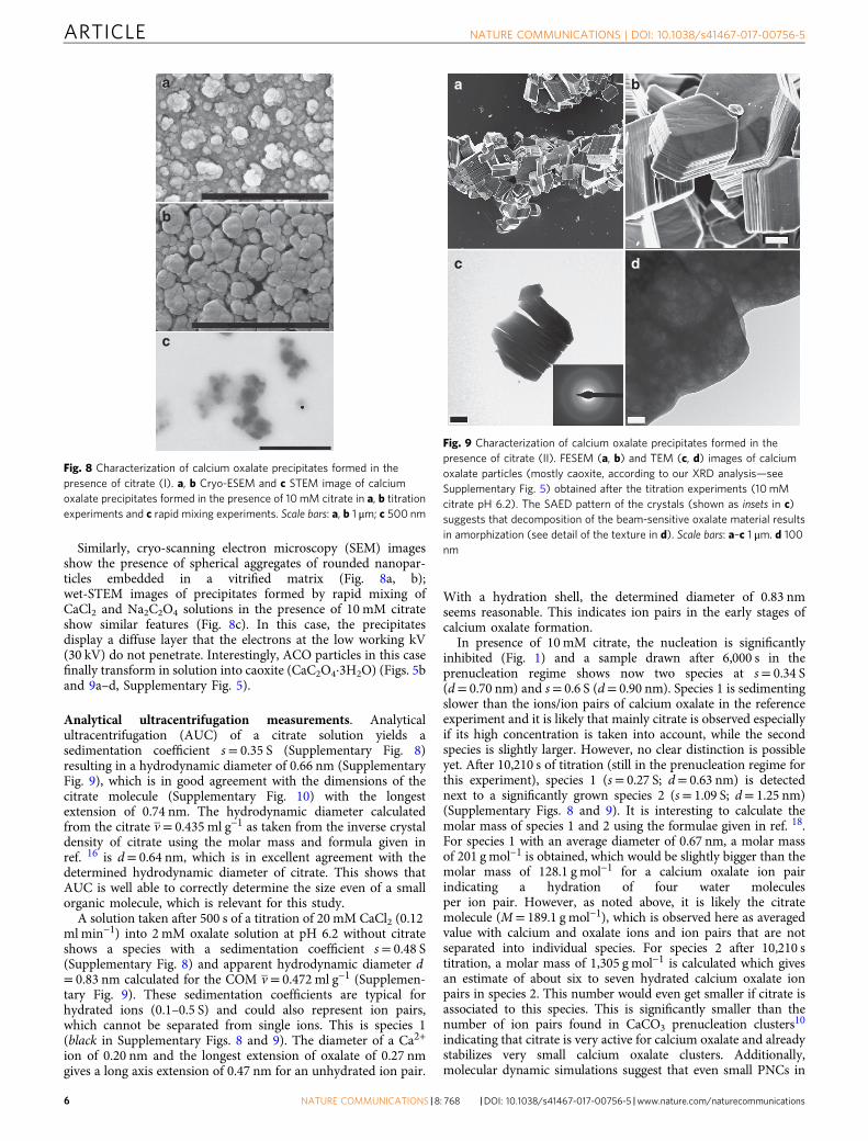
Interestingly, in the presence of citrate (10 mM) our transmis-sion electron microscopy (TEM) images of samples collected after9,600 s of the beginning of the experiments show two types ofdistinct rounded, Ca-rich amorphous nanoparticles (Figs. 4a, band 5a–c): less dense (low contrast) nanoparticles, with diffuselimits and sizes ranging from 10 up to 100 nm, and well-defined,
spherical nanoparticles with darker contrast, with sizes rangingfrom 30 up to 100 nm. The sizes of these darker particles are ofthe same order of magnitude to that measured by in situ,continuous dynamic light scattering (DLS) monitoring (Fig. 6).Indeed, they are in very good agreement with sizes measured byDLS immediately upon the onset of nucleation (marked by adrop in the transmittance of the solution, 7,500–9,400 s afterthe beginning of the experiments), which range from ~30 upto 100 nm. With the progress of the experiments, DLSmeasurements demonstrate that the average size of the aggregatesincreases from ca. 50 s up to 260 nm after 11,280 s.
Moreover, a closer look at the TEM photomicrographs revealsthe presence of individual nanoparticles as small as ~2 nm, whichappear as isolated entities (Fig. 7a, b). A second maximum inparticle size at ~1.5 nm (Fig. 6) that agrees with the size of theseindividual nanoparticles observed in the TEM images wasfound during the in situ DLS measurements performed withinthe first 5 min, however, with low statistical significance (two outof three of the measurements performed), possibly because theaverage size of these particles is close to the theoretical lower limitof the technique (0.8 nm) and also due to the low concentrationof these particles. The two types of bigger (light and dense)nanoparticles seem to be as well made up of these smaller primaryunits inlaid in a loose framework (Fig. 7a, b).
Occasionally, in some areas of the TEM micrographs, thedense, darker particles appear interconnected by a less dense(lighter contrast) neck-like structure (Fig. 5a). Interestingly, thedenser amorphous calcium oxalate nanoparticles formed inthe presence of citrate show a clear C-rich rim ca. 20 nm inthickness that most likely corresponds to an adsorbed citrate layer
a
b
Fig. 2 Scanning transmission electron microscopy images of precipitatesformed in rapid mixing experiments (control runs). Coexistence of arounded nanoparticles (possibly amorphous) and b crystalline calciumoxalate is observed. Scale bars: 500 nm
a b
c d
Fig. 3 Characterization of calcium oxalate precipitates (control runs).FESEM (a, b) and TEM (c, d) images of calcium oxalate particles (mostlywhewellite, according to our XRD analysis—see Supplementary Fig. 5)obtained after the titration experiments in the absence of citrate. Theelectron diffraction pattern of the crystals (shown as insets in c) suggeststhat decomposition of the beam-sensitive oxalate material results inamorphization. Scale bars: a 1 μm, b 1 μm, c 0.5 μm, and d 1 μm
a
b
C5 1MAG: 57.0kx HV: 300 kV
Ca
Fig. 4 Compositional analysis of initial calcium oxalate precipitates.a STEM-HAADF image of calcium oxalate precipitates formed in thepresence of 10 mM citrate at pH 6.2, showing two types of distinct roundednanoparticles: less dense nanoparticles, with diffuse limits and well-defined,denser spherical nanoparticles. Scale bar: 200 nm. b Calcium elemental EDSmap of the area in a. Scale bar: 300 nm
ARTICLE NATURE COMMUNICATIONS | DOI: 10.1038/s41467-017-00756-5
4 NATURE COMMUNICATIONS |8: 768 |DOI: 10.1038/s41467-017-00756-5 |www.nature.com/naturecommunications

(Supplementary Fig. 6). This is further corroborated by themarkedly negative values (−39.2± 2.0 mV vs. 1.4± 0.1 mV in theabsence of citrate) of measured zeta potential. Per definition, thezeta potential is the potential between the shear (slipping) planeand the bulk solution (Supplementary Fig. 7). A zeta potential canbe measured for micelles or polyelectrolytes. Similarly, we couldmeasure zeta potential values of PNCs. The negative valuemeasured in the presence of citrate seems reasonable if we assumethe presence of a large number of Cit3– and HCit2− ions (majorspecies at pH 6.2) distributed in the slipping layer and alsoaccessed into the adsorbed layer by H-bonds between the citrateand oxalate ions.
The nearly neutral zeta potential values found in the absence ofcitrate may appear counterintuitive considering that the clustersseem to be negatively charged. Note that the zeta potential relieson the concentration-dependent attraction of the positive ionspresent in solution. If our PNCs are negatively charged, they
would attract the positive ions in solution (Na+ ions from theoxalate at pH 6.2, in excess in the early stages of the titration withrespect to other positively charged cations such as Ca2+) and forman electrochemical double layer (the Stern layer). Therefore, andaccording to the above-described structure of PNCs and thesolution around them, it is a rational result that the zeta potentialof PNCs was 1.4± 0.1 mV. A similar description may be valid forACO nanoparticles. Furthermore, this result also indicates thatPNCs (and ACO nanoparticles) are colloidally unstable andwould quickly coagulate to grow and form larger clusters orparticles due to nearly neutral zeta potential.
a
b
c
Fig. 5 TEM images of calcium oxalate precipitates formed in titrationexperiments in the presence of 10 mM citrate at pH 6.2. a TEM imageshowing particles with dark contrast and well-defined sphericalmorphology, which are connected with a less dense (lighter contrast)shapeless structure (quenching time: 10,000 s). Scale bar: 500 nm.b General overview (STEM-HAADF image) of dried precipitates formed at15,000 s. Nanometer-sized crystalline calcium oxalate crystals coexist withrounded nanoparticles that seem to grow by aggregation of smallernanoparticles. Scale bar: 200 nm. c Detail of b showing the growth of ACOnanoparticles by aggregation or attachment of smaller units. Scale bar: 10nm
40
7,500 s8,760 s10,020 s10,560 s
35
30
25
Inte
nsity
(%
)
20
15
10
5
0
1 10 100 1,000
Size (nm)
Fig. 6 In situ analysis of particle size distribution of calcium oxalateprecipitates. Evolution of particle size distribution in titration experimentsperformed in the presence of 10mM citrate, obtained by in situ dynamiclight scattering (DLS)
a
b
Fig. 7 TEM images of calcium oxalate nanoclusters. a, b Citrate-stabilizedCaC2O4 nanoclusters in dry state, formed at 10,000 s, showing isolatedtiny species (average size 1.9± 0.2 nm, indicated by black arrows) scatteredall over the image area as well as forming part of bigger nanometer-sizeaggregates. Scale bars: 50 nm
NATURE COMMUNICATIONS | DOI: 10.1038/s41467-017-00756-5 ARTICLE
NATURE COMMUNICATIONS |8: 768 |DOI: 10.1038/s41467-017-00756-5 |www.nature.com/naturecommunications 5
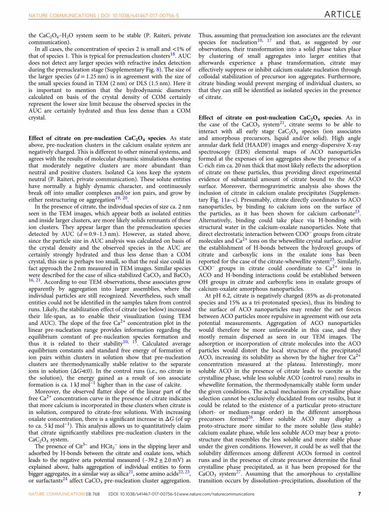
Similarly, cryo-scanning electron microscopy (SEM) imagesshow the presence of spherical aggregates of rounded nanopar-ticles embedded in a vitrified matrix (Fig. 8a, b);wet-STEM images of precipitates formed by rapid mixing ofCaCl2 and Na2C2O4 solutions in the presence of 10 mM citrateshow similar features (Fig. 8c). In this case, the precipitatesdisplay a diffuse layer that the electrons at the low working kV(30 kV) do not penetrate. Interestingly, ACO particles in this casefinally transform in solution into caoxite (CaC2O4·3H2O) (Figs. 5band 9a–d, Supplementary Fig. 5).
Analytical ultracentrifugation measurements. Analyticalultracentrifugation (AUC) of a citrate solution yields asedimentation coefficient s= 0.35 S (Supplementary Fig. 8)resulting in a hydrodynamic diameter of 0.66 nm (SupplementaryFig. 9), which is in good agreement with the dimensions of thecitrate molecule (Supplementary Fig. 10) with the longestextension of 0.74 nm. The hydrodynamic diameter calculatedfrom the citrate v = 0.435 ml g−1 as taken from the inverse crystaldensity of citrate using the molar mass and formula given inref. 16 is d= 0.64 nm, which is in excellent agreement with thedetermined hydrodynamic diameter of citrate. This shows thatAUC is well able to correctly determine the size even of a smallorganic molecule, which is relevant for this study.
A solution taken after 500 s of a titration of 20 mM CaCl2 (0.12ml min−1) into 2 mM oxalate solution at pH 6.2 without citrateshows a species with a sedimentation coefficient s= 0.48 S(Supplementary Fig. 8) and apparent hydrodynamic diameter d= 0.83 nm calculated for the COM v = 0.472 ml g−1 (Supplemen-tary Fig. 9). These sedimentation coefficients are typical forhydrated ions (0.1–0.5 S) and could also represent ion pairs,which cannot be separated from single ions. This is species 1(black in Supplementary Figs. 8 and 9). The diameter of a Ca2+
ion of 0.20 nm and the longest extension of oxalate of 0.27 nmgives a long axis extension of 0.47 nm for an unhydrated ion pair.
With a hydration shell, the determined diameter of 0.83 nmseems reasonable. This indicates ion pairs in the early stages ofcalcium oxalate formation.
In presence of 10mM citrate, the nucleation is significantlyinhibited (Fig. 1) and a sample drawn after 6,000 s in theprenucleation regime shows now two species at s= 0.34 S(d= 0.70 nm) and s= 0.6 S (d= 0.90 nm). Species 1 is sedimentingslower than the ions/ion pairs of calcium oxalate in the referenceexperiment and it is likely that mainly citrate is observed especiallyif its high concentration is taken into account, while the secondspecies is slightly larger. However, no clear distinction is possibleyet. After 10,210 s of titration (still in the prenucleation regime forthis experiment), species 1 (s= 0.27 S; d= 0.63 nm) is detectednext to a significantly grown species 2 (s= 1.09 S; d= 1.25 nm)(Supplementary Figs. 8 and 9). It is interesting to calculate themolar mass of species 1 and 2 using the formulae given in ref. 18.For species 1 with an average diameter of 0.67 nm, a molar massof 201 gmol−1 is obtained, which would be slightly bigger than themolar mass of 128.1 g mol−1 for a calcium oxalate ion pairindicating a hydration of four water moleculesper ion pair. However, as noted above, it is likely the citratemolecule (M= 189.1 g mol−1), which is observed here as averagedvalue with calcium and oxalate ions and ion pairs that are notseparated into individual species. For species 2 after 10,210 stitration, a molar mass of 1,305 g mol−1 is calculated which givesan estimate of about six to seven hydrated calcium oxalate ionpairs in species 2. This number would even get smaller if citrate isassociated to this species. This is significantly smaller than thenumber of ion pairs found in CaCO3 prenucleation clusters10
indicating that citrate is very active for calcium oxalate and alreadystabilizes very small calcium oxalate clusters. Additionally,molecular dynamic simulations suggest that even small PNCs in
a
b
c
Fig. 8 Characterization of calcium oxalate precipitates formed in thepresence of citrate (I). a, b Cryo-ESEM and c STEM image of calciumoxalate precipitates formed in the presence of 10 mM citrate in a, b titrationexperiments and c rapid mixing experiments. Scale bars: a, b 1 μm; c 500 nm
c d
a b
Fig. 9 Characterization of calcium oxalate precipitates formed in thepresence of citrate (II). FESEM (a, b) and TEM (c, d) images of calciumoxalate particles (mostly caoxite, according to our XRD analysis—seeSupplementary Fig. 5) obtained after the titration experiments (10mMcitrate pH 6.2). The SAED pattern of the crystals (shown as insets in c)suggests that decomposition of the beam-sensitive oxalate material resultsin amorphization (see detail of the texture in d). Scale bars: a–c 1 μm. d 100nm
ARTICLE NATURE COMMUNICATIONS | DOI: 10.1038/s41467-017-00756-5
6 NATURE COMMUNICATIONS |8: 768 |DOI: 10.1038/s41467-017-00756-5 |www.nature.com/naturecommunications
the CaC2O4–H2O system seem to be stable (P. Raiteri, privatecommunication).
In all cases, the concentration of species 2 is small and <1% ofthat of species 1. This is typical for prenucleation clusters18. AUCdoes not detect any larger species with refractive index detectionduring the prenucleation stage (Supplementary Fig. 8). The size ofthe larger species (d= 1.25 nm) is in agreement with the size ofthe small species found in TEM (2 nm) or DLS (1.5 nm). Here itis important to mention that the hydrodynamic diameterscalculated on basis of the crystal density of COM certainlyrepresent the lower size limit because the observed species in theAUC are certainly hydrated and thus less dense than a COMcrystal.
Effect of citrate on pre-nucleation CaC2O4 species. As stateabove, pre-nucleation clusters in the calcium oxalate system arenegatively charged. This is different to other mineral systems, andagrees with the results of molecular dynamic simulations showingthat moderately negative clusters are more abundant thanneutral and positive clusters. Isolated Ca ions keep the systemneutral (P. Raiteri, private communication). These solute entitieshave normally a highly dynamic character, and continuouslybreak off into smaller complexes and/or ion pairs, and grow byeither restructuring or aggregation19, 20.
In the presence of citrate, the individual species of size ca. 2 nmseen in the TEM images, which appear both as isolated entitiesand inside larger clusters, are more likely solids remnants of theseion clusters. They appear larger than the prenucleation speciesdetected by AUC (d= 0.9–1.3 nm). However, as stated above,since the particle size in AUC analysis was calculated on basis ofthe crystal density and the observed species in the AUC arecertainly strongly hydrated and thus less dense than a COMcrystal, this size is perhaps too small, so that the real size could infact approach the 2 nm measured in TEM images. Similar specieswere described for the case of silica-stabilized CaCO3 and BaCO316, 21. According to our TEM observations, these associates growapparently by aggregation into larger assemblies, where theindividual particles are still recognized. Nevertheless, such smallentities could not be identified in the samples taken from controlruns. Likely, the stabilization effect of citrate (see below) increasedtheir life-span, as to enable their visualization (using TEMand AUC). The slope of the free Ca2+ concentration plot in thelinear pre-nucleation range provides information regarding theequilibrium constant of pre-nucleation species formation andthus it is related to their stability10, 15. Calculated averageequilibrium constants and standard free energy of formation ofion pairs within clusters in solution show that pre-nucleationclusters are thermodynamically stable relative to the separateions in solution (ΔG≪0). In the control runs (i.e., no citrate inthe solution), the energy gained as a result of ion associateformation is ca. 1 kJ mol−1 higher than in the case of calcite.
Moreover, the observed flatter slope of the linear part of thefree Ca2+ concentration curve in the presence of citrate indicatesthat more calcium is incorporated in these clusters when citrate isin solution, compared to citrate-free solutions. With increasingoxalate concentration, there is a significant increase in ΔG (of upto ca. 5 kJ mol−1). This analysis allows us to quantitatively claimthat citrate significantly stabilizes pre-nucleation clusters in theCaC2O4 system.
The presence of Cit3− and HCit2− ions in the slipping layer andadsorbed by H-bonds between the citrate and oxalate ions, whichleads to the negative zeta potential measured (−39.2± 2.0mV) asexplained above, halts aggregation of individual entities to formbigger aggregates, in a similar way as silica21, some amino acids22, 23,or surfactants24 affect CaCO3 pre-nucleation cluster aggregation.
Thus, assuming that prenucleation ion associates are the relevantspecies for nucleation10, 17 and that, as suggested by ourobservations, their transformation into a solid phase takes placeby clustering of small aggregates into larger entities thatafterwards experience a phase transformation, citrate mayeffectively suppress or inhibit calcium oxalate nucleation throughcolloidal stabilization of precursor ion aggregates. Furthermore,citrate binding would prevent merging of individual clusters, sothat they can still be identified as isolated species in the presenceof citrate.
Effect of citrate on post-nucleation CaC2O4 species. As inthe case of the CaCO3 system21, citrate seems to be able tointeract with all early stage CaC2O4 species (ion associatesand amorphous precursors, liquid and/or solid). High angleannular dark field (HAADF) images and energy-dispersive X-rayspectroscopy (EDS) elemental maps of ACO nanoparticlesformed at the expenses of ion aggregates show the presence of aC-rich rim ca. 20 nm thick that most likely reflects the adsorptionof citrate on these particles, thus providing direct experimentalevidence of substantial amount of citrate bound to the ACOsurface. Moreover, thermogravimetric analysis also shows theinclusion of citrate in calcium oxalate precipitates (Supplemen-tary Fig. 11a–c). Presumably, citrate directly coordinates to ACOnanoparticles, by binding to calcium ions on the surface ofthe particles, as it has been shown for calcium carbonate23.Alternatively, binding could take place via H-bonding withstructural water in the calcium-oxalate nanoparticles. Note thatdirect electrostatic interaction between COO− groups from citratemolecules and Ca2+ ions on the whewellite crystal surface, and/orthe establishment of H-bonds between the hydroxyl groups ofcitrate and carboxylic ions in the oxalate ions has beenreported for the case of the citrate-whewellite system25. Similarly,COO− groups in citrate could coordinate to Ca2+ ions inACO and H-bonding interactions could be established betweenOH groups in citrate and carboxylic ions in oxalate groups ofcalcium-oxalate amorphous nanoparticles.
At pH 6.2, citrate is negatively charged (85% as di-protonatedspecies and 15% as a tri-protonated species), thus its binding tothe surface of ACO nanoparticles may render the net forcesbetween ACO particles more repulsive in agreement with our zetapotential measurements. Aggregation of ACO nanoparticleswould therefore be more unfavorable in this case, and theymostly remain dispersed as seen in our TEM images. Theadsorption or incorporation of citrate molecules into the ACOparticles would distort the local structure of the precipitatedACO, increasing its solubility as shown by the higher free Ca2+
concentration measured at the plateau. Interestingly, moresoluble ACO in the presence of citrate leads to caoxite as thecrystalline phase, while less soluble ACO (control runs) results inwhewellite formation, the thermodynamically stable form underthe given conditions. The actual mechanism for crystalline phaseselection cannot be exclusively elucidated from our results, but itcould be related to the existence of a particular proto-structure(short- or medium-range order) in the different amorphousprecursors formed26. More soluble ACO may display aproto-structure more similar to the more soluble (less stable)calcium oxalate phase, while less soluble ACO may bear a proto-structure that resembles the less soluble and more stable phaseunder the given conditions. However, it could be as well that thesolubility differences among different ACOs formed in controlruns and in the presence of citrate precursor determine the finalcrystalline phase precipitated, as it has been proposed for theCaCO3 system27. Assuming that the amorphous to crystallinetransition occurs by dissolution–precipitation, dissolution of the
NATURE COMMUNICATIONS | DOI: 10.1038/s41467-017-00756-5 ARTICLE
NATURE COMMUNICATIONS |8: 768 |DOI: 10.1038/s41467-017-00756-5 |www.nature.com/naturecommunications 7

more soluble ACO precursor formed in the presence of citratewould result in higher supersaturation locally in the solution(compared to the case of the amorphous-to-crystalline transitionof citrate-free ACO) that would allow the formation of a lessstable and more soluble crystalline phase (caoxite), according tothe Ostwald’s step rule28, 29. Parameters controlling the selectionof the final crystal phase during precipitation of calcium oxalateswill be the topic of future investigations.
Evidence of a liquid–liquid separation in the CaC2O4 system.Large, dense Ca-oxalate ion aggregates are solute entities, i.e., theydo not show interfaces with the precipitating solution. It has beensuggested that, once these aggregates reach a certain size, theycould develop interfacial surfaces, and become dense-liquidnanodroplets20. This may indeed be regarded as a liquid–liquidseparation, which has been recently reported to precede theformation of solid phases during crystallization processes30, 31. Inthis way, it would be explained how a dense-liquid precursorphase forms from a molecular point of view20. We find evidencein our system that suggests that such a liquid–liquid separation
could potentially take place in the presence of citrate prior to solidamorphous calcium oxalate formation. First, the transmittancestarts decreasing before the free Ca2+ concentration in solutionreaches a maximum (Fig. 1a). It has been suggested that theinitially formed nanodroplets can aggregate and form largerspecies with a liquid-like character as well18; the formation of ahigh concentration of liquid-like structures of big size, higherdensity and, consequently, higher refractive index, before theactual nucleation of a solid phase would more likely affect thetransmittance of the solution. Nevertheless, given that this newlyformed phase is in the liquid state, the mobility of calcium ionswould not be restricted so that the formation of this phase wouldnot imply a drop in the measured free calcium concentration.Additionally, TEM observations reveal the presence in some areasof the samples of particles with strong contrast and well-definedrounded morphologies interconnected by less dense (weakercontrast) neck-like structures. Similar structures have beenreported32, 33 for the case of calcium carbonate, and are suggestedto develop upon the initial formation of a dense-liquid phase thatsubsequently transforms into an amorphous solid phase bydensification via expulsion of water.
+ Citrate
Ca2+
C2O42–
Transformationinto crystalline Ca-oxalate Nucleation of ACO
Ion pair ClusterGrowth by
aggregationCitrate-stabilized clusters
Citrate-stabilized ACO
Liquid–liquid separation??
Reaction coordinate
Ions
CaC2O4ion pairs
LP
ACO
Whewellite
PNC
ΔG
Caoxite
ACOstabilized
Classical pathway
a
b
Fig. 10 Non-classical pathways during calcium oxalate precipitation in the presence of citrate. a Schematic drawing illustrating the sequence of stagesduring calcium oxalate formation in the presence of citrate. b An overview of the energetics of calcium oxalate crystallization pathways fromsupersaturated solutions under thermodynamic (classical pathway, black line) and kinetic control (this study, blue line in the absence of citrate and red line inthe presence of citrate)
ARTICLE NATURE COMMUNICATIONS | DOI: 10.1038/s41467-017-00756-5
8 NATURE COMMUNICATIONS |8: 768 |DOI: 10.1038/s41467-017-00756-5 |www.nature.com/naturecommunications

We suggest that inhibition of ACO nucleation frompre-nucleation clusters allows the system to reach the point atwhich a liquid phase may form. However, this aspect needsfurther investigation to unambiguously demonstrate that suchphase separation occurs in our system. As stated above, at somepoint these larger liquid entities will densify to give roundedparticles, undergo progressive dehydration and, eventually, solid(amorphous) nanoparticles nucleate from this dense-liquidprecursor phase.
DiscussionThe results of our study allow proposing a mechanism for theearly stages of calcium oxalate formation and the role that citrateplays at modulating this process, resembling that proposed for theeffect of silica on calcium and barium carbonate formation16, 21
(Fig. 10a). The crystallization pathway in the calcium oxalatesystem can be understood considering the free-energy landscapefrom supersaturated solution to crystallinity, shown in Fig. 10b34–36. This energy landscape summarizes the complex pathway offormation of crystalline calcium oxalate described in this paper,based on attachment of higher-order species (different to the ionsbuilding the crystals). It shows local minima corresponding tothe different intermediates identified in this work, includingprenucleation ion aggregates and dense-liquid or amorphousprecursors. Because the free energy barriers for the formation ofintermediate phases are smaller than the free energy barrier forthe direct formation of the final crystalline phase, the nucleationrate of the former would be higher than that of the latter. This canhelp to explain why intermediate phases form earlier (at a higherrate) than the final crystalline one under kinetically controlledcrystallization. Addition of citrate may lead to the kineticstabilization of the different intermediates identified in this workreflected in an increase of the free-energy barrier of nucleation.
Under the conditions of our study, the experimentalobservations performed suggest that calcium oxalate forms uponCa2+ and C2O4
2− association into polynuclear stable complexesprior to (possibly) liquid–liquid separation and amorphouscalcium oxalate nucleation. Our findings show that the controlthat citrate exerts on calcium oxalate mineralization begins beforesolid ACO formation; citrate acts stabilizing pre-nucleation ionassociates and preventing their aggregation, which ultimatelyinhibits ACO formation, and, simultaneously, stabilizingACO nanoparticles, preventing their subsequent crystallization.Moreover, the presence of citrate changes the crystal phaseformed, an effect that could be related either to a change inthe short range order of the precursor ACO precipitated in thepresence of citrate or differences in solubility of citrate-bearingand citrate-free ACO. Thus, our study shed light on themechanism governing calcium oxalate biomineralization, being ofparticular relevance to understand the role of citrate in preventingpathological mineralization of calcium-oxalates by influencingearly pre- and post-nucleation stages.
MethodsTitration experiments. Precipitation experiments were performed using acommercially available setup Titrino 905 manufactured by Methrom. Reactantswere purchased from Sigma-Aldrich, all with ACS grade (> 99% purity). Forexperiments, 20 mM CaCl2 solutions were continuously added to a 2 mM H2C2O4
solutions at a rate of 0.12 ml min−1. Concentrations of trisodium citrateranging from 0 up to 10 mM were added to the H2C2O4 solutions. Duringboth experiments, the Ca2+ potential was continuously monitored using an ISE(Mettler-Toledo, DX337-Ca). This electrode was calibrated for each experiment bytitration of a 10 mM CaCl2 into a NaCl solution of the same ionic strength of thecorresponding oxalate-citrate-bearing solution37. The pH was measured using aglass electrode from Metrohm, which served as well as the reference for the Ca ISE.Solution conductivity and transmittance were also monitored. This titration-basedsetup has been recently used to gain detailed insights into the nucleation ofsparingly soluble sulfates and carbonates (BaSO4, CaCO3 and BaCO3) and to
quantitatively assess the multiple effects of additives on the early stages ofnucleation and growth of such minerals10, 14–17. Ca-citrate binding was as wellinvestigated by the slow addition of 20 mM CaCl2 solution into an aqueoussolution of citric acid in concentrations ranging from 0 up to 10 mM.
Particles formed in the presence of citrate (10 mM) were in situ investigatedconcerning their size and its evolution with time during titration experiments bymeans of DLS using the controlled reference heterodyne method. Experimentswere conducted at a scattering angle of 180°, using a Microtrac NANO-flex particlesize analyzer equipped with a diode laser (λ = 780 nm, 5 mW) and a 1 m-longflexible measuring probe (diameter = 8 mm) with sapphire window as sampleinterface. Scattering was continuously monitored in situ during titrationexperiments, with an acquisition time per run of 45 s. The waiting period betweenindividual runs was 30 s. Size distributions were computed with the MicrotracFLEX application software package (v.11.1.0.1).
Finally, AUC measurements were performed with the aim of detectingany-prenucleation species forming in solution using a Beckman-Coulter XL-Iultracentrifuge equipped with Rayleigh interference optics and operated at aconstant speed of 60,000 rpm at 25 °C. Samples were drawn from the titrationexperiments performed in the presence of 10 mM citrate at 500, 6,000, and 10,210s. Data were acquired overnight for at least 8 h. Distinct sedimentation (s) anddiffusion coefficients (D) were obtained by fitting experimental data to the Lammequation using a non-interacting species model with the Sedfit software package.
Characterization of precipitates. To gain additional insights into the actualnucleation process of calcium oxalate in the presence of citrate, samples werecollected from the reaction media at different reaction times, quenched in ethanol,transferred onto carbon-coated copper grids and observed under TEM using a FEITitan, operated at 300 kV. TEM observations were performed using a 30 µmobjective aperture. Selected area electron diffraction (SAED) patterns werecollected using a 10-µm aperture, which allowed collection of diffraction data froma circular area ca. 0.2 µm in diameter. Compositional maps of selected areas wereacquired in STEM mode using a Super X EDS detector (FEI), formed by four SSDdetectors with no window surrounding the sample. STEM images in the FEI TitanTEM of the areas analyzed by EDS were collected with a HAADF detector. At theend of each titration experiment, the solution was filtered and the precipitatesformed were analyzed by means of X-ray diffraction, thermogravimetry, high-resolution scanning (FESEM; Zeiss SUPRA40VP) and TEM (FEI Titan). Also,aliquots from the reaction media were collected after the onset of nucleation inselected titration experiments (control and 10 mM citrate) and immediatelytransferred into the sample cell of a Microtrac Stabino equipment for zeta potentialmeasurement using the streaming potential method.
Finally, two additional experiments were performed to discard potentialartefacts related to ethanol quenching. First, drops of the precipitating solution incontrol runs and in the presence of 10 mM citrate were frozen in liquid nitrogenupon the onset of nucleation (detected by a drop in both the transmittance of thesolution and cryo-SEM mode by placing a solid piece of the frozen solution in themicroscope holder and sublimating the solvent. Additionally, precipitates formedby rapid mixing of 100 mM CaCl2 and Na2C2O4 solutions with and without 10 mMcitrate were collected with a TEM grid and observed in wet-STEM mode (noethanol quenching).
Data availability. The data that support the findings of this study are availablefrom the corresponding author (E.R.-A.) upon reasonable request.
Received: 11 October 2016 Accepted: 26 July 2017
References1. Echigo, T. & Kimata, M. Crystal chemistry and genesis of organic minerals: a
review of oxalate and polycyclic aromatic hydrocarbon minerals. Can. Mineral.48, 1329–1358 (2010).
2. Hajir, M., Graf, R. & Tremel, W. Stable amorphous calcium oxalate: synthesisand potential intermediate in biomineralization. Chem. Commun. 50,6534–6536 (2014).
3. Webb, M. A. Cell-mediated crystallization of calcium oxalate in plants. PlantCell 11, 751–761 (1999).
4. Zindler-Frank, E., Hönow, R. & Hesse, A. Calcium and oxalate content of theleaves of Phaseolus vulgaris at different calcium supply in relation to calciumoxalate crystal formation. Plant Physiol. 158, 139–144 (2001).
5. Ihli, J. et al. Precipitation of amorphous calcium oxalate in aqueous solution.Chem. Mater. 27, 3999–4007 (2015).
6. Winston, J. S., Yeh, I. T., Evers, K. & Friedman, A. K. Calcium oxalate isassociated with benign breast tissue. Can we avoid biopsy? Am. J. Clin. Pathol.100, 488–492 (1993).
NATURE COMMUNICATIONS | DOI: 10.1038/s41467-017-00756-5 ARTICLE
NATURE COMMUNICATIONS |8: 768 |DOI: 10.1038/s41467-017-00756-5 |www.nature.com/naturecommunications 9

7. Weaver, M. L. et al. Inhibition of calcium oxalate monohydrate growth bycitrate and the effect of the background electrolyte. J. Cryst. Growth 306,135–145 (2007).
8. Qiu, S. R. et al. Molecular modulation of calcium oxalate crystallization byosteopontin and citrate. Proc. Natl Acad. Sci. USA 101, 1811–1815 (2004).
9. Hu, Y.-Y., Rawal, A. & Schmidt-Rohr, K. Strongly bound citrate stabilizes theapatite nanocrystals in bone. Proc. Natl Acad. Sci. USA 107, 22425–22429 (2010).
10. Gebauer, D., Völkel, A. & Cölfen, H. Stable prenucleation calcium carbonateclusters. Science 322, 1819–1822 (2008).
11. Wang, L. et al. Inhibition of calcium oxalate monohydrate crystallization by thecombination of citrate and osteopontin. J. Cryst. Growth 291, 160–165 (2006).
12. Qiu, S. R. & Orme, C. A. Dynamics of biomineral formation at the near-molecular level. Chem. Rev. 108, 4784–4822 (2008).
13. Chung, J. et al. Molecular modifiers reveal a mechanism of pathological crystalgrowth inhibition. Nature 536, 446–450 (2016).
14. Gebauer, D., Cölfen, H., Verch, A. & Antonietti, M. The multiple roles ofadditives in CaCO3 crystallization: a quantitative case study. Adv. Mater. 21,435–439 (2009).
15. Verch, A., Gebauer, D., Antonietti, M. & Cölfen, H. How to control the scalingof CaCO3: a “fingerprinting technique” to classify additives. Phys. Chem. Chem.Phys. 13, 16811–16820 (2011).
16. Eiblmeier, J. et al. New insights into the early stages of silica-controlled bariumcarbonate crystallisation. Nanoscale 6, 14939–14949 (2014).
17. Ruiz-Agudo, C. et al. Exploring the effect of poly(acrylic acid) on pre- and post-nucleation BaSO4 species: new insights into the mechanisms of crystallizationcontrol by polyelectrolytes. CrystEngComm 18, 2830–2842 (2016).
18. Kellermeier, M. et al. Amino acids form prenucleation clusters: ESI-MS as a fastdetection method in comparison to analytical ultracentrifugation. FaradayDiscuss. 159, 23–45 (2012).
19. Demichelis, R., Raiteri, P., Gale, J., Quigley, D. & Gebauer, D. Stableprenucleation mineral clusters are liquid-like ionic polymers. Nat. Commun. 2,590 (2011).
20. Gebauer, D., Kellermeier, M., Gale, J. D., Bergström, L. & Cölfen, H.Pre-nucleation clusters as solute precursors in crystallisation. Chem. Soc. Rev.43, 2348–2371 (2014).
21. Kellermeier, M. et al. Colloidal stabilization of calcium carbonate prenucleationclusters with silica. Adv. Funct. Mater. 22, 4301–4311 (2012).
22. Finney, A. R. & Rodger, P. M. Probing the structure and stability of calciumcarbonate pre-nucleation clusters. Faraday Discuss. 159, 47–60 (2012).
23. Raiteri, P. et al. Exploring the influence of organic species on pre- andpost-nucleation calcium carbonate. Faraday Discuss. 159, 61–85 (2012).
24. Sun, S., Gebauer, D. & Cölfen, H. A solvothermal method for synthesizingmonolayer protected amorphous calcium carbonate clusters. Chem. Commun.52, 7036–7038 (2016).
25. Qiu, S. R. et al. Modulation of calcium oxalate monohydrate crystallization bycitrate through selective binding to atomic steps. J. Am. Chem. Soc. 127,9036–9044 (2005).
26. Gebauer, D. et al. Proto-calcite and proto-vaterite in amorphous calciumcarbonates. Angew. Chem. Int. Ed. Engl. 122, 9073–9075 (2010).
27. Zou, Z. et al. Opposite particle size effect on amorphous calcium carbonatecrystallization in water and during heating in air. Chem. Mater. 27, 4237–4246(2015).
28. Stranski, I. N. & Totomanov, D. Rate of formation of (crystal) nuclei and theOstwald step rule. Z. Phys. Chem. 163, 399–408 (1993).
29. Colfen, H. & Mann, S. Higher-order organization by mesoscale self-assemblyand transformation of hybrid nanostructures. Angew. Chem. Int. Ed. Engl. 42,350–2365 (2003).
30. Wolf, S. E. et al. Carbonate-coordinated metal complexes precede the formationof liquid amorphous mineral emulsions of divalent metal carbonates. Nanoscale3, 1158–1165 (2011).
31. Wallace, A. F. et al. Microscopic evidence for liquid-liquid separation insuperpersaturated CaCO3 solutions. Science 341, 885–889 (2013).
32. Rieger, J. et al. Precursor structures in the crystallization/precipitation processesof CaCO3 and control of particle formation by polyelectrolytes. FaradayDiscuss. 136, 265–277 (2007).
33. Rodriguez-Navarro, C., Kudłacz, K., Cizer, Ö. & Ruiz-Agudo, E. Formation ofamorphous calcium carbonate and its transformation into mesostructuredcalcite. CrystEngComm 17, 58–72 (2015).
34. Bewernitz, M. A., Gebauer, D., Long, J., Cölfen, H. & Gower, L. B. A metastableprecursor phase of calcium carbonate and its interactions with polyaspartate.Faraday Discuss. 199, 291–312 (2012).
35. Harding, J. H., Freeman, C. L. & Duffy, D. M. Oriented crystal growth onorganic monolayers. CrystEngComm 16, 1430–1438 (2014).
36. De Yoreo, J. J. et al. Crystallization by particle attachment in synthetic, biogenicand geologic environments. Science 349, aaa6760–aaa6761 (2015).
37. Kellermeier, M., Picker, A., Kempter, A., Cölfen, H. & Gebauer, D. A.Straightforward treatment of activity in aqueous caco3 solutions and theconsequences for nucleation theory. Adv. Mater. 26, 752–757 (2014).
AcknowledgementsThis research was funded by the Spanish Government (grants CGL2015-70642-R,CGL2015-73103-EXP), the European Commission (ERDF funds), the University ofGranada (“Unidad Científica de Excelencia” UCE-PP2016-05) and the Junta de Anda-lucía (P11-RNM-7550). E.R.-A. acknowledges the receipt of a Ramón y Cajal grant fromthe Spanish Government (Ministerio de Economía y Competitividad) and funding fromthe research group RNM-179 of the Junta de Andalucía. The authors thank CIC-UGR,M. Abad and I. Sanchez for assistant during microscopy studies and R. Rosenberg forperforming the AUC measurements.
Author contributionsE.R.-A., C.R.-N. and H.C. conceived the concept and designed the experiments. Allauthors (E.R.-A., A.B.-C., C.R.-A., A.I.-V., C.R.-N. and H.C.) contributed to perform theexperiments and the analysis of results. E.R.-A. wrote the paper with contributions fromall authors.
Additional informationSupplementary Information accompanies this paper at doi:10.1038/s41467-017-00756-5.
Competing interests: The authors declare no competing financial interests.
Reprints and permission information is available online at http://npg.nature.com/reprintsandpermissions/
Publisher's note: Springer Nature remains neutral with regard to jurisdictional claims inpublished maps and institutional affiliations.
Open Access This article is licensed under a Creative CommonsAttribution 4.0 International License, which permits use, sharing,
adaptation, distribution and reproduction in any medium or format, as long as you giveappropriate credit to the original author(s) and the source, provide a link to the CreativeCommons license, and indicate if changes were made. The images or other third partymaterial in this article are included in the article’s Creative Commons license, unlessindicated otherwise in a credit line to the material. If material is not included in thearticle’s Creative Commons license and your intended use is not permitted by statutoryregulation or exceeds the permitted use, you will need to obtain permission directly fromthe copyright holder. To view a copy of this license, visit http://creativecommons.org/licenses/by/4.0/.
© The Author(s) 2017
ARTICLE NATURE COMMUNICATIONS | DOI: 10.1038/s41467-017-00756-5
10 NATURE COMMUNICATIONS |8: 768 |DOI: 10.1038/s41467-017-00756-5 |www.nature.com/naturecommunications