Embed Size (px)
Citation preview

American Mineralogist, Volume 84, pages 1661–1673, 1999
0003-004X/99/0010–1661$05.00 1661
A new complex sheet of uranyl polyhedra in the structure of wölsendorfite
PETER C. BURNS*
Department of Civil Engineering and Geological Sciences, University of Notre Dame, Notre Dame, Indiana 46556-0767, U.S.A.
INTRODUCTION
Approximately 200 minerals contain U6+ as a necessarystructural constituent (Fleischer and Mandarino 1995), andthese minerals are significant components of the oxidized por-tions of U deposits (Frondel 1958; Finch and Ewing 1992).Uranyl minerals are currently receiving considerable attentionbecause of their significance to the environment (e.g., Burns1998a, 1998b, 1998c, 1997; Burns et al. 1997a, 1997b, 1997c;Finch et al. 1992, 1996, 1998; Finch and Ewing 1992; Milleret al. 1996; Murakami et al. 1997; Sowder et al. 1996; Vochtenet al. 1995, 1997). Uranyl minerals are present in the mill tail-ings that result from the mining of U and are precipitated insoils that are contaminated with actinide elements. Uranyl min-erals will be the dominant products of alteration of spent nuclearfuel in a geological repository if the conditions are oxidizingand the fuel comes in contact with water, as is likely to occurat the proposed repository at Yucca Mountain, Nevada (Finn etal. 1996; Wronkiewicz et al. 1996). Uranyl minerals may im-pact upon the mobility of various radionuclides under reposi-tory conditions by incorporating them directly into their crystal
ABSTRACT
The structure of wölsendorfite, Pb6.16Ba0.36[(UO2)14O19(OH)4](H2O)12, Z = 8, orthorhombic, a =14.131(1), b = 13.885(1), c = 55.969(4) Å, V = 10,982 Å3, space group Cmcm, was solved by directmethods and refined by full-matrix least-squares techniques to an agreement factor (R) of 6.4% anda goodness-of-fit (S) of 1.13 using 6215 unique observed reflections (|Fo| ≥ 4σF) collected with MoKαX-radiation and a CCD (charge-coupled device) detector. The structure contains eight unique U6+
positions, each of which is part of a nearly linear (UO2)2+ uranyl ion. The uranyl ions (Ur) are furthercoordinated by four or five anions (φ) arranged at the equatorial corners of square and pentagonalbipyramids, respectively. The structure contains two unique Urφ4 square bipyramids and six uniqueUrφ5 pentagonal bipyramids that link by the sharing of equatorial corners and edges to form infinitesheets that are parallel to (100). The sheets have a primitive repeat distance of c = 55.969 Å, and areby far the most complex sheet of uranyl polyhedra yet observed. The interlayer between the uranylsheets contains Pb2+ and Ba cations, as well as H2O groups that are either bonded to the interlayercations or are held in the structure by H bonding only. There are eight unique cation positions in theinterlayer that are coordinated by six to ten anions.
The structure of wölsendorfite is remarkable both in the complexity of the sheets of uranyl poly-hedra and the connectivity of the interlayer. By using the sheet anion-topology approach, it is shownthat the wölsendorfite sheet is composed of slabs of the simpler α-U3O8 and β-U3O8-type sheets. It ispossible that many as yet unknown complex sheets of uranyl polyhedra exist that are based uponanion topologies that are combinations of slabs of simpler topologies, but it is currently not possibleto distinguish which of these may be energetically favorable.
structures as they grow (Burns et al. 1997b) or by the exchangeof ions between earlier-formed crystals and radionuclide-bear-ing solutions (Burns 1999).
Despite the obvious importance of uranyl minerals, and theconsiderable attention that they have garnered, our understand-ing of their structures and crystal chemistry lags well behindmany other mineral groups. To date, the structures have beendetermined and refined for only ~60 uranyl minerals, less than1/3 of the known species. The disparity of structural informa-tion is due to experimental difficulties; all are extremely highabsorbers of X-rays, many do not form crystals of a size suit-able for structure analysis using conventional means, and somesuffer from dehydration or other effects that cause the deterio-ration of crystals.
Uranyl minerals are structurally very complex. The U6+ cat-ion invariably occurs as part of a nearly linear (UO2)2+ uranylion (Ur) in minerals (Evans 1963; Burns et al. 1997a), whichis in turn coordinated by four, five, or six additional anionsthat are arranged at the equatorial corners of square, pentago-nal, and hexagonal bipyramids, respectively. The O atoms ofthe uranyl ion (OUr) receive ~1.7 valence units (v.u.) from thebond to the U6+ cation, whereas considerably less of the bond-valence requirements of the equatorial ligands are provided bythe bond to the U6+ cation at the center of the polyhedron. Po-lymerization of uranyl polyhedra with other uranyl polyhedra,*E-mail: [email protected]

BURNS: NEW SHEET IN WÖLSENDORFITE1662
as well as other cation polyhedra of higher bond-valence, iscommon in mineral structures, as this permits the bonding re-quirements of the equatorial ligands to be met. The stronglyasymmetric distribution of bond valences in the uranyl polyhe-dra dictates that the structures of uranyl minerals are domi-nated by sheets of polymerized polyhedra (Burns et al. 1996,1997a).
An interesting and complex subgroup of uranyl minerals isthe lead uranyl oxide hydrates (Table 1). These minerals com-monly form due to the oxidation of geologically old uraninite,owing to an abundance of radiogenic Pb. The onset of alter-ation of uraninite, UO2+X, typically results in the formation of asequence of lead uranyl oxide hydrates as continued alterationpreferentially removes U6+ relative to Pb (Frondel 1958). Eachstructure is based upon complex sheets of edge- and corner-sharing uranyl polyhedra (Fig. 1), with the uranyl ions orientedroughly perpendicular to the sheet, and with Pb2+ cations and
H2O groups located in the interlayers between the sheets. Thefourmarierite-type sheet also occurs in schoepite (Finch et al.1996), and the richetite-type sheet occurs in becquerelite(Pagoaga et al. 1987), compreignacite (Burns 1998c), billietite(Pagoaga et al. 1987), and protasite (Pagoaga et al. 1987). Thecurite-type sheet is not known from another mineral, but it doesoccur in the structure of the synthetic Sr analogue of curite(Burns and Hill 1999). The sayrite-type sheet was first foundin the structure of K2[(UO2)5O8](UO2)2 (Kovba 1972), and laterin sayrite. The vandendriesscheite-type sheet has not yet beenfound in another structure, and until the current work it was themost complex sheet of uranyl polyhedra known.
Wölsendorfite was first discovered in Wölsendorf, Bavaria,Germany (Protas 1957) and has since been reported from sev-eral localities where it occurs in the oxidized portions of U de-posits. The currently accepted formula is (Pb,Ba,Ca)U2O7·2H2O(Fleischer and Mandarino 1995). Ba-rich wölsendorfite contain-
FIGURE 1. Polyhedral representations and anion topologies of the sheets that occur in lead uranyl oxide hydrate minerals. (a) curite, (b)curite anion-topology, (c) sayrite anion-topology, (d) sayrite, (e) fourmarietite, (f) fourmarierite anion-topology, (g) richetite anion-topology,(h) richetite, (i) vandendriesscheite, (j ) vandendriesscheite anion-topology. The sheet anion-topologies were derived using the method of Burnset al. (1996). Uranyl polyhedra are shaded with crosses.

BURNS: NEW SHEET IN WÖLSENDORFITE 1663
ing 3.27 wt% BaO and 0.09 wt% CaO was found in Greenlandby Beddoe-Stephens and Secher (1982), and Ca-richwölsendorfite containing 3.24 wt% CaO but no reported BaOis known from a U-Mo ore deposit in Russia (Belov and Federov1974). The synthetic Ca- and Ba-free endmember has been re-ported by Vochten and Haverbeke (1990). Toussaint (1961)provided a partial structure solution for a crystal ofwölsendorfite from Shinkolobwe, Democratic Republic ofCongo, but was only able to propose the positions of the Uatoms based upon a sub-cell. He reported the unit-cell dimen-sions a = 11.9, b = 6.99, c = 6.85 Å, and noted that the X-raydata indicated the true b unit-cell dimension was probablydouble that reported, and the c unit-cell dimension was prob-ably four times the value reported.
The recent introduction of CCD-based detectors for X-raysto mineral structure analysis (Burns 1998d) has greatly facili-tated the study of the structures of uranyl minerals, as it is nowpossible to study very small crystals [i.e., less than (10 µm)3],and crystals with very long primitive unit-cell axes using sealed-tube X-ray sources. This paper presents the full solution of thestructure of wölsendorfite using a CCD-based diffractometer.
EXPERIMENTAL METHODS
X-ray data
The specimen provided by the Canadian Museum of Na-ture is from Shinkolobwe, Democratic Republic of Congo. Itcontains several tabular crystals of wölsendorfite that exhib-ited uniform optical properties and sharp extinction betweencrossed polarizers. A crystal with approximate dimensions 0.20× 0.10 × 0.01 mm was selected for XRD, and was mounted ona Bruker PLATFORM 3-circle goniometer equipped with a 1KSMART CCD (charge-coupled device) detector.
Preliminary examination revealed a primitive unit-cell axis~56 Å long. The crystal-to-detector distance was set at 15 cmto obtain excellent resolution of the long axis. The data wascollected using monochromatic MoKα X-radiation and framewidths of 0.3° in ω, with 120 s used to acquire each frame.Three different detector settings (2θ = 12.0, 30.0, 50.0°) wereused so as to obtain complete coverage to ~57° 2θ. A total of6588 frames of data were collected, providing a complete sphereof three-dimensional data. The peaks were sharp with smoothprofiles, indicating that substantial radiation damage has notoccurred. Several hundred frames of data were analyzed to lo-cate diffraction maxima; all peaks found were consistent with
a C-centered unit cell with the dimensions a = 14.131(1), b =13.885(1), c = 55.969(4) Å. The long primitive c dimensionwas verified by collection of zone-axis frames. The C-centeredcell has a strong primitive sub-cell with dimensions a = 12.01,b = 7.101, c = 6.935 Å, in agreement with the sub-cell foundby Toussaint (1961). However, the CCD detector provides amplesensitivity and resolution to resolve the true C-centered unitcell. The final unit-cell dimensions were refined (Table 2) us-ing least-squares techniques. Data were collected for 3° ≤ 2θ ≤56.7° in approximately 229 hours; comparison of the intensi-ties of equivalent reflections collected at different times duringthe data collection showed no evidence of significant decay.The three-dimensional data were reduced and corrected forLorentz, polarization, and background effects using the Brukerprogram SAINT. An empirical absorption-correction was donebased upon 3999 intense reflections with five or more equiva-lents. The crystal was modeled as a (100) plate; reflections witha plate-glancing angle of less than 3° were discarded from thedata set, and the correction lowered Razimuthal from 35.3 to 6.8%.A total of 53 816 reflections was collected, of which 4080 wereomitted because of a plate-glancing angle less than 3°. Sys-tematic absences indicated that the lattice is C-centered; of thecollected reflections, 25 370 were consistent with the C-cen-tered lattice. Merging of equivalent reflections gave 7026 uniquereflections (RINT = 7.0%) with 6215 classed as observed (|Fo| ≥4σF).
Chemical analysis
The same specimen used for XRD was mounted, polished,and coated with carbon. Chemical analysis was done in wave-length-dispersion (WD) mode on a JEOL 733 electron micro-probe using Tracor Northern 5500 and 5600 automation. Datareduction was done with a conventional PAP routine. The op-erating voltage was 15 kV, and the beam current was 0.20 µA.Data for all elements in the sample were collected for 25 s or0.50% precision, whichever was attained first. A 100 s energy-dispersion scan indicated no elements with Z greater than 8,other than those reported here. The elements Na, K, Rb, Cs,Ca, and Sr were sought in WD scans, but were not detected.Standards used for the electron-microprobe analysis were:benitoite (BaLα), crocoite (PbMα), and brannerite (UMβ). H2Owas calculated by stoichiometry from the results of the crystal-structure analysis. The results were (wt%): BaO = 0.99, UO3 =72.23, PbO = 24.57, H2O = 3.90, total = 101.69.
TABLE 1. The chemistries of lead uranyl oxide hydrates
Pb:U Sheet composition Ref.wölsendorfite 1:2.15 [(UO2)14O19(OH)4] *sayrite 1:2.5 [(UO2)5O6(OH)2] †curite 1:2.67 [(UO2)8O8(OH)6] ‡fourmarierite 1:4.0 [(UO2)4O3(OH)4] §richetite 1:4.15 [(UO2)18O18(OH)12] ||vandendriesscheite 1:6.36 [(UO2)10O6(OH)11] #* This study.† Piret et al. (1983).‡ Taylor et al. (1981).§ Piret (1985).|| Burns (1998a).# Burns (1997).
TABLE 2. Miscellaneous information pertaining to the structuredetermination of wölsendorfite
a (Å) 14.131(1) Crystal size (mm) 0.20 × 0.10b (Å) 13.885(1) × 0.01c (Å) 55.969(4) Total ref. 25,370V (Å3) 10,982 Unique ref. 7026Space group Cmcm Rint (%) 7.0F(000) 18,762 Unique |Fo| ≥ 4σF 6215m (mm-1) 60.30 Final R* (%) 6.4Dcalc (g/cm3) 6.888 S† 1.13Unit-cell contents: 8{Pb6.16Ba0.36[(UO2)14O19(OH)4](H2O)12}* R = Σ(|Fo|-|Fc|)/Σ|Fo|.† S = [Σw(|Fo|-|Fc|)2/(m-n)]1⁄2, for m observations and n parameters.

BURNS: NEW SHEET IN WÖLSENDORFITE1664
STRUCTURE SOLUTION AND REFINEMENT
Scattering curves for neutral atoms, together with anoma-lous dispersion corrections, were taken from InternationalTables for X-Ray Crystallography, Vol. IV (Ibers and Hamilton1974). The Bruker SHELXTL Version 5 system of programswas used for the determination and refinement of the crystalstructure.
Systematic absences and reflection statistics indicated thespace group Cmcm (no. 63), with verification provided by thesuccessful solution and refinement of the structure. The struc-ture was solved by direct methods, and the initial model in-cluded the positions of the U atoms and some of the Pb atoms.Additional Pb sites and the anions were located in successivedifference-Fourier maps that were calculated after refinementof the model. Following refinement of the atomic positionalparameters and isotropic-displacement parameters for all at-oms, occupancy factors were included for each Pb site. Theagreement factor (R) at this stage of the refinement was 10.9%for observed reflections. The site-occupancy factors for the Pb1through Pb5 sites remained close to 100% occupancy, and wereconstrained to full occupancy in subsequent cycles of refine-ment. The occupancy factors for the sites Pb6 through Pb8 re-fined to values significantly below 100% and were thus includedin further cycles of refinement. Consideration of the polyhe-dral bond-lengths for the Pb sites, and the lack of additionalsites in the difference-Fourier maps, indicate that the Ba re-vealed by the microprobe analysis is probably located at thePb8 site, and all Ba from the microprobe analysis was subse-quently assigned to that site. However, additional scatteringwas required at the Pb8 site, thus Pb was also included at thesite and the occupancy factor was refined. The displacementparameters for the cations were converted to anisotropic forms,and were refined together with the positional parameters forall atoms and a weighting scheme of the structure factors. Atthis stage in the refinement a peak with height ~11 e/Å3 waslocated in the difference-Fourier map ~0.56 Å from the Pb7site. This site contained too much electron density to corre-spond to an anion, so it was assumed that the Pb7 site is disor-dered between two positions, and a new position designatedPb7A was added to the refinement. The occupancy factors ofboth the Pb7 and Pb7A sites were refined, although the dis-placement parameter for the Pb7A site was constrained to beisotropic. The final model resulted in an R index of 6.4% for6215 unique observed reflections (|Fo| ≥ 4σF) and a goodness-of-fit (S) of 1.13. In the final cycle of refinement the averageparameter shift/esd was 0.000, and the maximum peaks in thefinal difference-Fourier maps were 5.74 and –6.12 e/Å3. Thefinal atomic-positional parameters and anisotropic-displace-ment parameters are given in Tables 3 and 4, and selected in-teratomic-distances and angles are given in Table 5. Calculatedand observed structure factors are provided in Table 61
TABLE 3. Atomic position parameters and equivalent isotropic-displacement parameters for the structure ofwölsendorfite
x y z Ueq*U1 0.23236(7) 0.28097(7) 1⁄4 136(2)U2 0.24043(5) 0.48445(4) 0.07525(1) 99(1)U3 0.23354(7) 1⁄2 0 133(2)U4 0.26009(5) 0.52736(5) 0.14137(1) 99(1)U5 0.26058(5) 0.25302(4) 0.10659(1) 101(1)U6 0.26867(5) 0.50400(5) 0.21281(1) 142(2)U7 0.24934(5) 0.27115(4) 0.03319(1) 99(1)U8 0.24787(5) 0.24360(5) 0.18258(1) 127(1)Pb1 1⁄2 0.70072(9) 0.13777(3) 315(3)Pb2 1⁄2 0.3534(1) –0.00028(2) 322(3)Pb3 1⁄2 0.83007(9) 0.07678(3) 330(3)Pb4 1⁄2 0.08653(9) 0.18238(2) 339(3)Pb5 1⁄2 0.3206(1) 0.07673(2) 472(5)Pb6 0 0.4681(2) 1⁄4 423(10)Pb7 1⁄2 0.4363(4) 1⁄4 622(15)Pb7A 1⁄2 0.476(4) 1⁄4 548(92)Pb8 0 0.1683(3) 0.21848(6) 667(16)O1 0.104(2) 0.305(2) 1⁄4 246(45)O2 0.246(1) 0.596(1) 0.1043(2) 159(27)O3 0.368(1) 0.498(1) 0.0708(3) 276(34)O4 0.360(2) 0.254(1) 1⁄4 234(44)O5 0.268(1) 0.410(1) 0.1130(3) 238(31)O6 0.243(1) 0.347(1) 0.2132(2) 215(31)O7 0.387(1) 0.548(1) 0.1385(3) 258(33)O8 0.135(1) 0.508(1) 0.1459(3) 320(37)O9 0.391(2) 0.248(1) 0.1029(4) 492(51)O10 0.129(2) 0.257(1) 0.1091(4) 478(51)O11 0.124(2) 0.244(2) 0.0360(4) 554(57)O12 0.272(2) 0.202(2) 0.1430(4) 510(55)O13 0.375(2) 0.296(1) 0.0317(3) 441(47)O14 0.229(1) 0.436(1) 0.0362(3) 296(36)O15 0.397(2) 0.486(2) 0.2128(4) 530(56)O16 0.114(1) 0.469(1) 0.0794(3) 284(34)O17 0.362(3) 1⁄2 0 724(104)O18 0.274(2) 0.584(1) 0.1791(4) 439(47)O19 0.141(2) 0.524(2) 0.2133(4) 567(59)O20 0.272(2) 0.159(2) 0.0038(4) 531(55)O21 0.250(2) 0.318(2) 0.0723(5) 665(70)O22 0.373(1) 0.221(1) 0.1881(3) 286(34)O23 0.123(1) 0.267(1) 0.1763(3) 315(37)O24 0.104(3) 1⁄2 0 729(106)O25 0.298(1) 0.662(1) 0.2221(3) 248(32)O26 0.265(3) 0.459(3) 1⁄4 701(103)OH27 0.297(1) 0.393(1) 0.1662(2) 211(29)OH28 0.292(1) 0.132(1) 0.0555(2) 193(29)H2O29 1⁄2 0.153(1) 0.2241(4) 208(42)H2O30 1⁄2 0.889(2) 0.1239(5) 426(65)H2O31 1⁄2 0.393(2) 0.1692(5) 378(60)H2O32 0 0.127(2) 0.0923(4) 301(51)H2O33 1⁄2 0.143(2) 0.0583(5) 408(62)H2O34 1⁄2 0.495(2) 0.0343(4) 306(52)H2O35 1⁄2 0.415(2) 0.1131(6) 605(88)H2O36 1⁄2 0.210(2) 0.1468(4) 328(55)H2O37 1⁄2 0.931(3) 0.0404(7) 800(118)H2O38 0 0.393(2) 0.2017(5) 411(63)H2O39 1⁄2 0.172(3) 0.0103(6) 618(90)H2O40 1⁄2 0.101(1) 0.1067(3) 16(27)Note: Ueq* = Ueq Å2 × 10⁴.
RESULTS
Projection of the structure along [010] shows that it con-tains sheets of uranyl polyhedra parallel to (100), with Pb2+
and Ba cations as well as H2O groups located in the interlayerbetween the sheets (Fig. 2). The structure of wölsendorfite istherefore consistent with the generalities of other lead uranyloxide hydrate mineral structures, as well as with the observa-tion that the majority of uranyl mineral structures are basedupon sheets of polyhedra of higher bond-valence (Burns et al.1996).
1 For a copy of Table 6, Document AM-99-029 contact the Busi-ness Office of the Mineralogical Society of America (see in-side front cover of recent issue) for price information. Deposititems may also be available on the American Mineralogist website (see inside back cover of a current issue for a web address).
BURNS: NEW SHEET IN WÖLSENDORFITE 1665
TABLE 4. Anisotropic-displacement parameters for the cations inthe structure of wölsendorfite
U11* U22 U33 U12 U13 U23
U1 153(4) 171(4) 85(4) 42(4) 0 0U2 148(3) 75(3) 73(3) –21(2) 18(2) –15(2)U3 127(4) 86(4) 188(4) 0 0 23(3)U4 160(3) 78(3) 59(3) –9(2) 12(2) –3(2)U5 121(3) 76(3) 107(3) –5(2) 3(2) –1(2)U6 145(3) 155(3) 124(3) –9(3) –11(3) 62(2)U7 148(3) 81(3) 68(3) 17(2) –13(2) –11(2)U8 208(3) 80(3) 93(3) 24(2) 8(3) –11(2)Pb1 251(6) 219(6) 476(8) 0 0 –42(5)Pb2 375(7) 348(6) 243(6) 0 0 49(5)Pb3 268(6) 253(6) 469(8) 0 0 –3(5)Pb4 505(8) 237(6) 274(6) 0 0 24(5)Pb5 1030(15) 203(6) 182(6) 0 0 4(5)Pb6 722(23) 260(13) 288(14) 0 0 0Pb7 1199(38) 157(24) 510(20) 0 0 0Pb8 1029(36) 609(24) 363(19) 0 0 311(15)Note: Uij* = Uij Å2 × 10⁴
TABLE 5. Selected interatomic distances (Å) and angles (°) in thestructure of wölsendorfite
U1-O4 1.84(2) U5-O9 1.86(2)U1-O1 1.85(2) U5-O10 1.87(2)U1-O6,a 2.26(1) ×2 U5-O21 2.13(3)U1-O25b,c 2.31(2) ×2 U5-O12 2.16(2)U1-O26 2.52(4) U5-O2b 2.18(1)<U1-OUr> 1.84 U5-O5 2.22(2)O4-U1-O1 179.0(9) <U5-OUr> 1.86<U1-φeq> 2.33 O9-U5-O10 177.9(9)
<U5-φeq> 2.17U2-O16 1.82(2)U2-O3 1.83(2) U6-O19 1.83(3)U2-O2 2.25(1) U6-O15 1.83(2)U2-O14 2.29(2) U6-O26 2.17(1)U2-O21 2.32(3) U6-O18 2.19(2)U2-OH28d 2.37(1) U6-O6 2.21(1)U2-O5 2.38(2) U6-O25 2.29(2)<U2-OUr> 1.82 U6-OH27 3.06(2)O16-U2-O3 178.8(8) <U6-OUr> 1.83<U2-feq> 2.32 O19-U6-O15 178.7(9)
<U6-feq> 2.38U3-O17 1.81(4)U3-O24 1.83(4) U7-O13 1.81(2)U3-O14,e 2.21(2) ×2 U7-O11 1.82(2)U3-O20d,f 2.22(2) ×2 U7-O21 2.28(3)<U3-OUr> 1.82 U7-O20 2.29(2)O17-U3-O24 180 U7-O20f 2.31(2)<U3-φeq> 2.21 U7-O14 2.32(2)
U7-OH28 2.38(1)U4-O8 1.80(2) <U7-OUr> 1.81U4-O7 1.83(2) O13-U7-O11 177.5(9)U4-O18 2.26(2) <U7-φeq> 2.32U4-O5 2.27(2)U4-O2 2.29(1) U8-O22 1.82(2)U4-OH27 2.39(1) U8-O23 1.82(2)U4-O12d 2.47(2) U8-O6 2.24(1)<U4-OUr> 1.81 U8-O18b 2.25(2)O8-U4-O7 177.0(7) U8-O12 2.31(2)<U4-φeq> 2.34 U8-OH27 2.37(1)
U8-O25b 2.57(2)<U8-OUr> 1.82O22-U8-O23 178.7(7)<U8-φeq> 2.35
Pb1-O10d,g 2.55(2) ×2 Pb5-O9,l 2.35(2) ×2Pb1-O7,h 2.66(2) ×2 Pb5-H2O35 2.42(3)Pb1-H2O30 2.73(3) Pb5-H2O33 2.68(3)Pb1-H2O32g 2.75(2) Pb5-O13,l 3.10(2) ×2Pb1-O23d,g 2.92(2) ×2 Pb5-O3,l 3.10(2) ×2<Pb-φ> 2.72 <Pb5-φ> 2.77
Pb2-H2O39 2.59(3) Pb6-O1,m 2.69(2) ×2Pb2-O13,h 2.64(2) ×2 Pb6-H2O38,a 2.90(3) ×2Pb2-H2O34 2.76(2) Pb6-H2O29n,o 2.95(2) ×2Pb2-O17,i 2.82(3) ×2 Pb6-O19,m,a,p 2.96(2) ×4Pb2-H2O34i 2.84(2) <Pb6-φ> 2.89Pb2-O11f,j 2.98(2) ×2<Pb2-φ> 2.79 Pb7-O15,l,a,q 2.63(2) ×4
Pb7-O4,h 3.21(2) ×2Pb3-H2O37 2.47(4) <Pb7-φ> 2.82Pb3-O16d,g 2.52(2) ×2Pb3-O10d,g 2.76(2) ×2 Pb8-O25r,s 2.87(2) ×2Pb3-H2O30 2.76(3) Pb8-O15r,s 2.93(2) ×2Pb3-H2O32g 2.95(2) Pb8-O1,m 2.98(2) ×2Pb3-O11d,g 3.12(2) ×2 Pb8-O23,m 3.23(2) ×2<Pb3-φ> 2.78 <Pb8-φ> 3.00
Pb4-H2O29 2.51(2)Pb4-O22,h 2.61(2) ×2Pb4-H2O36 2.63(2)Pb4-O19b,k 2.78(2) ×2Pb4-H2O38k 2.90(3)Pb4-O8b,k 3.01(2) ×2<Pb4-φ> 2.76Notes: a = x, y, 1⁄2-z; b = 1⁄2-x, y-1⁄2, z; c = 1⁄2-x, y-1⁄2, 1⁄2-z; d = 1⁄2-x, y+1⁄2, z; e =x, 1-y, z; f = 1⁄2-x, 1⁄2-y, -z; g = x+1⁄2, y+1⁄2, z; h = 1-x, y, z; i = 1-x, 1-y, -z; j = x+1⁄2,1⁄2-y, -z; k = x+1⁄2, y-1⁄2, z; l = 1-x, y, z; m = -x, y, z; n = x-1⁄2, y+1⁄2, z; o = x-1⁄2,y+1⁄2, 1⁄2-z; p = -x, y, 1⁄2-z; q = 1-x, y, 1⁄2-z; r = 1⁄2-x, y-1⁄2, z; s = x-1⁄2, y-1⁄2, z.
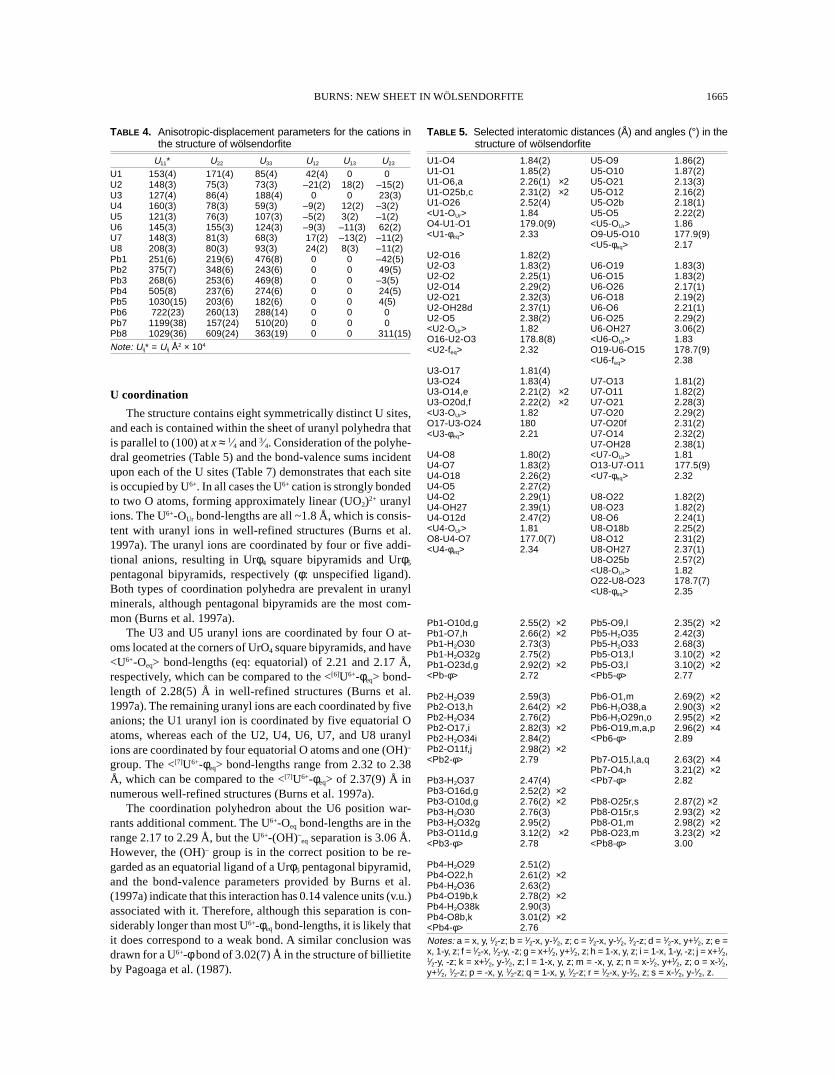
U coordination
The structure contains eight symmetrically distinct U sites,and each is contained within the sheet of uranyl polyhedra thatis parallel to (100) at x ≈ 1⁄4 and 3⁄4. Consideration of the polyhe-dral geometries (Table 5) and the bond-valence sums incidentupon each of the U sites (Table 7) demonstrates that each siteis occupied by U6+. In all cases the U6+ cation is strongly bondedto two O atoms, forming approximately linear (UO2)2+ uranylions. The U6+-OUr bond-lengths are all ~1.8 Å, which is consis-tent with uranyl ions in well-refined structures (Burns et al.1997a). The uranyl ions are coordinated by four or five addi-tional anions, resulting in Urφ4 square bipyramids and Urφ5
pentagonal bipyramids, respectively (φ: unspecified ligand).Both types of coordination polyhedra are prevalent in uranylminerals, although pentagonal bipyramids are the most com-mon (Burns et al. 1997a).
The U3 and U5 uranyl ions are coordinated by four O at-oms located at the corners of UrO4 square bipyramids, and have<U6+-Oeq> bond-lengths (eq: equatorial) of 2.21 and 2.17 Å,respectively, which can be compared to the <[6]U6+-φeq> bond-length of 2.28(5) Å in well-refined structures (Burns et al.1997a). The remaining uranyl ions are each coordinated by fiveanions; the U1 uranyl ion is coordinated by five equatorial Oatoms, whereas each of the U2, U4, U6, U7, and U8 uranylions are coordinated by four equatorial O atoms and one (OH)–
group. The <[7]U6+-φeq> bond-lengths range from 2.32 to 2.38Å, which can be compared to the <[7]U6+-φeq> of 2.37(9) Å innumerous well-refined structures (Burns et al. 1997a).
The coordination polyhedron about the U6 position war-rants additional comment. The U6+-Oeq bond-lengths are in therange 2.17 to 2.29 Å, but the U6+-(OH)–
eq separation is 3.06 Å.However, the (OH)– group is in the correct position to be re-garded as an equatorial ligand of a Urφ5 pentagonal bipyramid,and the bond-valence parameters provided by Burns et al.(1997a) indicate that this interaction has 0.14 valence units (v.u.)associated with it. Therefore, although this separation is con-siderably longer than most U6+-φeq bond-lengths, it is likely thatit does correspond to a weak bond. A similar conclusion wasdrawn for a U6+-φ bond of 3.02(7) Å in the structure of billietiteby Pagoaga et al. (1987).

BURNS: NEW SHEET IN WÖLSENDORFITE1666
FIGURE 2. Projection of the structure of wölsendorfite along [010]. The uranyl polyhedra are shaded with crosses, Pb2+ cations are shown ascross-hatched circles, and H2O groups are shown as open circles.
TABLE 7. Bond-valence sums* at cation and anion positions in the structure of wölsendorfite.
U1 5.86 Pb1 1.66 O1 1.87 O9 1.96 O17 1.88 O25 1.70 H2O33 0.22U2 6.00 Pb2 1.55 O2 2.05 O10 1.89 O18 2.08 O26 1.96 H2O34 0.31U3 6.27 Pb3 1.80 O3 1.59 O11 1.72 O19 1.79 OH27 1.18 H2O35 0.43U4 6.00 Pb4 1.73 O4 1.55 O12 1.83 O20 1.92 OH28 1.05 H2O36 0.25U5 6.27 Pb5 1.98 O5 1.87 O13 1.89 O21 2.06 H2O29 0.44 H2O37 0.38U6 6.06 Pb6 1.27 O6 2.06 O14 1.93 O22 1.81 H2O30 0.36 H2O38 0.28U7 6.09 Pb7 1.00 O7 1.75 O15 1.88 O23 1.72 H2O31 0.00 H2O39 0.27U8 5.94 Pb8 0.77 O8 1.71 O16 1.89 O24 1.52 H2O32 0.28 H2O40 0.00* Calculated using the parameters of Brese and O’Keeffe (1991) for Pb2+ and Burns et al. (1997a) for U6+.
Pb coordination
There are eight symmetrically distinct cation sites in theinterlayer (excluding H). Of these, the Pb1 through Pb5 sitescontain only Pb, and each is fully occupied according to site-scattering refinement. The Pb6 and Pb7 sites are partially oc-cupied, and the latter is disordered over two positions separatedby 0.56 Å. The occupancies of the Pb6 and Pb7 sites are 76 and96%, respectively, according to the results of site-scatteringrefinement. The Pb8 site contains both Pb and Ba, and all ofthe Ba indicated by the chemical analysis was assigned to thesite. Refinement of the Pb occupancy at the site indicates thatit contains ~36% Ba, ~30% Pb, and ~31% vacancy.
The polyhedral geometries (Table 5) and bond-valence sumsincident upon the sites (Table 7) are consistent with all Pbpresent as Pb2+, as is the case for all known Pb uranyl oxidehydrate minerals. The polyhedra are illustrated in Figure 3. Eachof the Pb sites is located on the m planes at x = 0, 1⁄2, and thePb6 and Pb7 sites are at the intersection of two m planes. ThePb1 through Pb7 cations are coordinated by OUr atoms of adja-cent sheets, as well as H2O groups located on the m planes inthe interlayer at x = 0, 1⁄2. In addition to OUr and interlayer H2Ogroups, the Pb8 site is coordinated by two O atoms that areequatorial ligands of uranyl polyhedra within the sheets. ThePb1 and Pb5 sites are coordinated by six OUr atoms and twoH2O groups, the Pb2, Pb3, and Pb4 polyhedra each contain six
OUr atoms and three H2O groups, and the Pb6 site is coordi-nated by six OUr atoms and four H2O groups. The Pb1 throughPb5 sites have <Pb-φ> bond-lengths in the range 2.72 to 2.79Å, and the <Pb-φ> bond-length of the tenfold-coordinated Pb6is 2.89 Å. The sixfold-coordinated Pb7 site involves shorterbonds, with a <Pb-φ> bond-length of 2.49 Å. The Pb8 site hasa <Pb-φ> bond-length of 3.00 Å, which is in accord with thesite containing substantial Ba as well as vacancy.
Polyhedra containing Pb2+ are often distorted owing to thepresence of a lone pair of electrons on the Pb2+ cation. Theanions are repelled away from the electron lone-pair, resultingin a one-sided coordination polyhedron. The Pb polyhedra inthe interlayer of wölsendorfite (Fig. 3) are fairly irregular, andsome are distinctly one-sided, suggesting that the Pb2+ cationsare lone-pair stereoactive.
Structural formula
Of the eight symmetrically distinct U6+ cations, only the U1site is on a special position, thus the unit cell contains 120 U6+
cations. The interlayer contains both Pb and Ba, and site-occu-pancy refinements have demonstrated that the Pb1 through Pb5sites are 100% occupied by Pb. The Pb6 and Pb7 sites are par-tially occupied by Pb, and the Pb8 site contains both Pb andBa. Each of the interlayer cations are on special positions, thus,according to site-scattering refinement, the unit cell contains

BURNS: NEW SHEET IN WÖLSENDORFITE 1667
FIGURE 3. The Pb2+ polyhedra that occur in the interlayer of wölsendorfite. Each polyhedron is shown projected along [100].
FIGURE 4. The sheet of uranyl polyhedra that occurs in the structure of wölsendorfite projected onto (100). (a) polyhedral representation ofthe sheet, (b) sheet anion-topology. The uranyl polyhedra are shaded with crosses.

BURNS: NEW SHEET IN WÖLSENDORFITE1668
49.3 Pb and 2.9 Ba. The bond-valence sums permit the identi-fication of (OH)- and H2O groups (Table 7), and the unit cellcontains 376 O atoms, 32 (OH)– groups, and 96 H2O groups.The bond-valence sums incident upon the O3, O4, and O24sites are ~1.5 v.u., a value that is intermediate between the ex-pected values for O and (OH)–. However, each of these atomsare part of uranyl ions; (OH)– groups are not known to occur inuranyl ions, leading to the conclusion that the O3, O4, and O24atoms are O, despite the low bond-valence sum incident uponthe sites. The formula for the wölsendorfite crystal studied canbe written as Pb6.16Ba0.36[(UO2)14O19(OH)4](H2O)12, with Z = 8.This formula has a net charge of –0.96, indicating that theremay be minor substitution of monovalent cations at theinterlayer sites, although none were detected by chemical analy-sis, or that the refined site occupancy factors are slightly inerror. The formula derived from the microprobe analysis, withH2O assumed on the basis of the structure determination, isPb6.10Ba0.36[(UO2)14O19(OH)4](H2O)12.
Sheets of uranyl polyhedra
The Urφ4 square bipyramids and Urφ5 pentagonal bipyramidsshare equatorial edges and corners to form complex sheets that
FIGURE 6. Chains of polygons that are required to construct sheetanion-topologies as chain-stacking sequences. After Miller et al. (1996).
FIGURE 5. The interlayer connectivity inthe structure of wölsendorfite projected onto(100). (a) ball-and-stick representation, (b)polyhedral representation. In (a) the Pb2+
cations are shown as circles shaded withparallel lines, anions that are bonded to thePb2+ cations are shown as small circles, andH2O groups that are held in the structure byH bonding only are shown as larger circles.The Pb2+ cations and H-bonded H2O groupsare numbered according to Table 3.

BURNS: NEW SHEET IN WÖLSENDORFITE 1669
are parallel to (100) (Fig. 4). Sheets of uranyl polyhedra aredominant structural features in uranium minerals, but thewölsendorfite sheet is more complex than any other known sheetof uranyl polyhedra, either in a mineral or a synthetic com-pound. The c unit-cell dimension of 55.969 Å is the longestprimitive repeat distance of any sheet of uranyl polyhedra. Theanion topology of the wölsendorfite sheet, derived using themethod of Burns et al. (1996), is shown in Figure 4b. Sheetanion-topologies are two-dimensional tessellations, and thewölsendorfite anion-topology contains triangles, squares andpentagons, as is often the case for the anion topologies of sheetsof uranyl polyhedra (Burns et al. 1996).
Interlayer connectivity
The interlayer at x = 0 is shown projected along [100] inFigure 5. The interlayer contains the eight symmetrically dis-tinct Pbφn polyhedra, as well as two symmetrically distinct H2Ogroups that are held in the structure by H bonding only. ThePbφn polyhedra are polymerized by the sharing of corners,edges, and faces, forming complex infinite heteropolyhedralchains that are parallel to [001] (Fig. 5).
COMPARISON TO RELATED STRUCTURES
The wölsendorfite sheet is an example of structural com-plexity that challenges our understanding of mineral structures.The vandendriesscheite sheet (Fig. 1i) is also very complex,but its primitive b unit-cell dimension of 41.378 Å (Burns 1997)is short compared to c = 55.969 Å in the wölsendorfite sheet.
FIGURE 7. The generation of sheet anion-topologies as chain-stacking sequences. (a) the α-U3O8 anion-topology, (b) the β-U3O8 anion-topology.
The sheets of uranyl polyhedra that occur in other lead uranyloxide hydrates (Fig. 1) are topologically simpler than eitherthe wölsendorfite or vandendriesscheite sheets. As is the casein the wölsendorfite sheet, both the sayrite (Fig. 1d) and curite(Fig. 1a) sheets contain both Urφ4 and Urφ5 polyhedra, whereasthe fourmarierite, richetite, and vandendriesscheite sheets con-tain only Urφ5 pentagonal bipyramids.
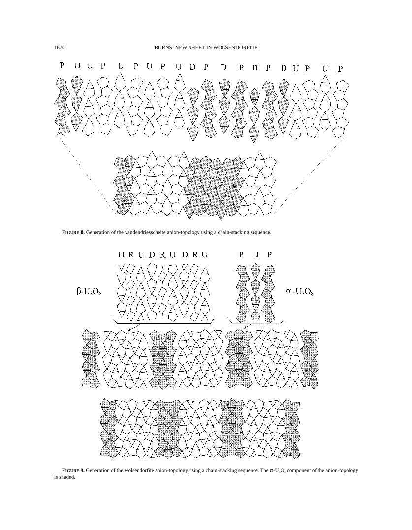
Miller et al. (1996) discussed the characterization of sheetanion-topologies as stacking sequences of chains, and this ap-proach was successfully applied to the vandendriesscheite an-ion-topology by Burns (1997). If consideration is restricted tothose sheets that contain only uranyl polyhedra that occur inminerals and synthetic phases (with the exception of the curiteanion-topology), only four chains of polygons (Fig. 6) are re-quired to construct all of the anion topologies. The P and Hchains involve edge-sharing pentagons and hexagons, respec-tively. The arrowhead chains involve the sharing of an edgebetween a pentagon and a triangle, with the other corner of thetriangle shared with the next pentagon in the chain. The arrow-head chain has a directional aspect, and may point either up(U) or down (D). The R chain consists of rhombs connected bylines. Various rules on how these chains fit together to formsheet anion-topologies were identified by Miller et al. (1996).
First consider the relatively simple α-U3O8 and β-U3O8 an-ion-topologies shown in Figure 7. The α-U3O8 anion-topology(also sometimes referred to as the protasite anion-topology) isthe basis for the sheets that occur in the structures of protasite,richetite, becquerelite, billietite, and compreignacite, as well
BURNS: NEW SHEET IN WÖLSENDORFITE1670
FIGURE 8. Generation of the vandendriesscheite anion-topology using a chain-stacking sequence.
FIGURE 9. Generation of the wölsendorfite anion-topology using a chain-stacking sequence. The α-U3O8 component of the anion-topologyis shaded.

BURNS: NEW SHEET IN WÖLSENDORFITE 1671
FIGURE 10. Various hypothetical sheet anion-topologies obtained by combining slabs of the α-U3O8 and β-U3O8 anion-topologies. The α-U3O8 component of the anion-topology is shaded. Only (e) is known from a mineral structure.

BURNS: NEW SHEET IN WÖLSENDORFITE1672
as in the mixed-valence α-U3O8 phase. The β-U3O8 anion-to-pology is less common; it is only known from the structure ofianthinite and the mixed-valence β-U3O8 phase. The genera-tion of each of these anion topologies using chain-stacking se-quences is illustrated in Figure 7. The α-U3O8 anion-topologycan be constructed using only the P and U or D chains, withthe repeat sequence PUPU… The β-U3O8 anion-topology re-quires the use of the P, D, and R chains, with the repeat se-quence DRUDRU…
The complex vandendriesscheite anion-topology can be derivedas a chain-stacking sequence (Fig. 8) using the P, D, and U chains,with the sequence PDUPUPUPUPUDPDPDPDUPUPU… (Burns1997). Note that the sequences UPUP… and DPDP… in thevandendriesscheite anion-topology are identical to the α-U3O8 an-ion-topology sequence, and the vandendriesscheite anion-topologycan be assembled using slabs of α-U3O8 anion-topology that alter-nate up and down.
The wölsendorfite anion-topology is generated using achain-stacking sequence with the R, D, P, and U chains, result-ing in a complex repeat sequence (Fig. 9). The repeat sequenceinvolves the strings DRUDRUDRU and PDP (Fig. 9), whichare slabs of the β-U3O8 and α-U3O8 anion-topologies, respec-tively. Thus, the wölsendorfite anion-topology may be as-sembled using slabs of the α-U3O8 and β-U3O8 anion-topologies,and it follows that the wölsendorfite sheet is structurally inter-mediate between the α-U3O8 and β-U3O8-type sheets.
As was the case with the sheets of uranyl polyhedra, theinterlayer contained in the structure of wölsendorfite is morecomplex than the interlayers in most uranyl minerals. Theinterlayers of lead uranyl oxide hydrate minerals display a rangeof polymerization. The Pbφ8 polyhedra in the interlayer ofsayrite are not polymerized, and only half of the Pbφn polyhe-dra in the interlayer of vandendriesscheite share polyhedral el-ements with other Pbφn polyhedra to form dimers. Thefourmarierite interlayer contains dimers of edge-sharing poly-hedra only. The richetite interlayer is very complex, with clus-ters of partially occupied Pbφn polyhedra that share faces andedges, as well as sharing polyhedral elements with M2+φ6 octa-hedra. The curite interlayer involves chains of face-sharing Pbφn
polyhedra that are two polyhedra wide, but they do not resemblethe chains found in the interlayer of wölsendorfite. Thus, thecomplex chains of polymerized polyhedra (Fig. 5) in thewölsendorfite interlayer are unusual in both their complexityand degree of polymerization.
DISCUSSION
It is well known that uranyl minerals possess fascinatinglydiverse structures, but a new level of structural complexity inuranyl minerals was revealed by the wölsendorfite sheet. Thesheet illustrates well the usefulness of the sheet anion-topol-ogy approach for the classification and comparison of the struc-tures of uranyl minerals. Using this approach, it is readilyapparent that the sheet contains slabs of simpler, previouslyknown sheets.
Why do the uranyl polyhedra in wölsendorfite adopt such acomplex configuration? Is it likely that similarly complex sheetsof uranyl polyhedra with different configurations exist in otherminerals? It is impossible to provide answers to either of these
questions at this time; considerable research into the energet-ics of these sheets, and into the structures of uranyl minerals, isessential to gain an understanding of the crystal-chemical prin-ciples that result in such complex sheets. However, it is appar-ent that there are many anion topologies and correspondingsheets that appear to be chemically feasible but that have notbeen observed in any structure. Consider, for example, sheetanion-topologies developed using slabs of various widths ofthe α-U3O8 and β-U3O8 anion topologies; a few of the possiblecombinations are shown in Figure 10. Of these, only one isknown from a structure (e), despite the fact that the topologiesare each complete tilings of two-dimensional space. We cur-rently do not have a means to discriminate which of these isenergetically favorable.
ACKNOWLEDGMENTS
The crystal used in this study was provided by the Canadian Museum ofNature. Robert A. Gault of the Canadian Museum of Nature carried out theelectron microprobe analysis of the crystals of wölsendorfite. This research wasfunded by the National Science Foundation (EAR98-04723).
REFERENCES CITED
Beddoe-Stephens, B. and Secher, K. (1982) Barian wölsendorfite from eastGreenland. Mineralogical Magazine, 46, 130–132.
Belov, L.N. and Fedorov, O.V. (1974) New data on wölsendorfite. ZapiskiVsesoyuznogo Mineralogicheskogo Obshchestva, 103, 718–719 (in Russian).
Brese, N.E. and O’Keeffe, M. (1991) Bond-valence parameters for solids. ActaCrystallographica, B47, 192–197.
Burns, P.C. (1997) A new uranyl oxide hydrate sheet in the structure ofvandendriesscheite: Implications for mineral paragenesis and the corrosion ofspent nuclear fuel. American Mineralogist, 82, 1176–1186.
———(1998a) The structure of richetite, a rare lead uranyl oxide hydrate. CanadianMineralogist, 36, 187–199.
———(1998b) Implications of solid-solution in the structure of boltwoodite. Cana-dian Mineralogist, 36, 1069–1075.
———(1998c) The structure of compreignacite, K2[(UO2)3O2(OH)3]2(H2O)7. Cana-dian Mineralogist, 36, 1061–1067.
———(1998d) CCD area detectors of X-rays applied to the analysis of mineral struc-tures. Canadian Mineralogist, 36, 847–853.
———(1999) Cs boltwoodite obtained by ion exchange from single crystals: Impli-cations for radionuclide release in a nuclear repository. Journal of Nuclear Ma-terials, 265, 218–223.
Burns, P.C. and Hill, F.C. (1999) Implications of the synthesis and structure of theSr analogue of curite. Canadian Mineralogist, in press.
Burns, P.C., Miller, M.L., and Ewing, R.C. (1996) U6+ minerals and inorganic phases:a comparison and hierarchy of crystal structures. Canadian Mineralogist, 34,845–880.
Burns, P.C., Ewing, R.C., and Hawthorne, F.C. (1997a) The crystal chemistry ofhexavalent uranium: Polyhedral geometries, bond-valence parameters, and poly-hedral polymerization. Canadian Mineralogist, 35, 1551–1570.
Burns, P.C., Ewing, R.C., and Miller, M.L. (1997b) Incorporation mechanisms ofactinide elements into the structures of U6+ phases formed during the oxidationof spent nuclear fuel. Journal of Nuclear Materials, 245, 1–9.
Burns, P.C., Finch, R.J., Hawthorne, F.C., Miller, M.L., and Ewing, R.C. (1997c)The crystal structure of ianthinite, [U4+
2 (UO2)4O6(OH)4(H2O)4](H2O)5: A pos-sible phase for Pu4+ incorporation during the oxidation of spent nuclear fuel.Journal of Nuclear Materials, 249, 199–206.
Evans, H.T., Jr. (1963) Uranyl ion coordination. Science, 141, 154–157.Finch, R.J. and Ewing, R.C. (1992) The corrosion of uraninite under oxidizing con-
ditions. Journal of Nuclear Materials, 190, 133–156.Finch, R.J., Miller, M.L., and Ewing, R.C. (1992) Weathering of natural uranyl
oxide hydrates: schoepite polytypes and dehydration effects. Radiochimica Acta,58/59, 433–443.
Finch, R.J., Cooper, M.A., Hawthorne, F.C., and Ewing, R.C. (1996) The crystalstructure of schoepite, [(UO2)8O2(OH)12](H2O)12. Canadian Mineralogist, 34,1071–1088.
Finch, R.J., Hawthorne, F.C., and Ewing, R.C. (1998) Structural relations amongschoepite, metaschoepite and “dehydrated schoepite”. Canadian Mineralogist,36, 831–845.
Finn, P.A., Hoh, J.C., Wolf, S.F., Slater, S.A., and Bates, J.K. (1996) The release ofuranium, plutonium, cesium, strontium, technetium and iodine from spent fuelunder unsaturated conditions. Radiochimica Acta, 74, 65–71.
Fleischer, M. and Mandarino, J.A. (1995) Glossary of mineral species. The Miner-

BURNS: NEW SHEET IN WÖLSENDORFITE 1673
alogical Record Inc., Tucson, Arizona.Frondel, C. (1958) Systematic mineralogy of uranium and thorium. U.S. Geologi-
cal Survey Bulletin 1064.Ibers, J.A. and Hamilton, W.C., eds. (1974) International Tables for X-ray Crystal-
lography, volume IV. The Kynoch Press, Birmingham, U.K.Kovba, L.M. (1972) Crystal structure of K2U7O22. Journal of Structural Chemistry,
13, 235–238.Miller, M.L., Finch, R.J., Burns, P.C., and Ewing, R.C. (1996) Description and clas-
sification of uranium oxide hydrate sheet topologies. Journal of Materials Re-search, 11, 3048–3056.
Murakami, T., Ohnuki, T., Isobe, H., and Sato, T. (1997) Mobility of uranium duringweathering. American Mineralogist, 82, 888–899.
Pagoaga, M.K., Appleman, D.E., and Stewart, J.M. (1987) Crystal structures andcrystal chemistry of the uranyl oxide hydrates becquerelite, billietite, andprotasite. American Mineralogist, 72, 1230–1238.
Piret, P. (1985) Structure cristalline de la fourmariérite, Pb(UO2)4O3(OH)4.4H2O.Bulletin de Minéralogie, 108, 659–665.
Piret, P., Deliens, M., Piret-Meunier, J., and Germain, G. (1983) La sayrite,Pb2[(UO2)5O6(OH)2]◊4H2O, nouveau minéral; propriétés et structure crystal-line. Bulletin de Minéralogie, 106, 299–304.
Protas, J. (1957) La wölsendorfite, nouvelle espèce uranifere. Compte Rendus, 244,2942
Sowder, A.G., Clark, S.B., and Fjeld, R.A. (1996) The effect of silica and phosphateon the transformation of schoepite to becquerelite and other uranyl phases.
Radiochimica Acta, 74, 45–49.Taylor, J.C., Stuart, W.L., and Mumme, I.A. (1981) The crystal structure of curite.
Journal of Inorganic and Nuclear Chemistry, 43, 2419–2423.Toussaint, J. (1961) Sur la structure de la wölsendorfite de Shinkolobwe. Annales
Sociéte Géologique de Belgique, 84, 365–373.Vochten, R. and van Haverbeke, L. (1990) Transformation of schoepite into the
uranyl oxide hydrates; becquerelite, billietite and wölsendorfite. Mineralogyand Petrology, 43, 65–72.
Vochten, R., van Haverbeke, L., Van Springel, K., and De Grave, E. (1995) Soddyite:Synthesis under elevated-temperature and pressure, and study of some physico-chemical characteristics. Neues Jahrbuch für Mineralogie. Monatshefte, 10, 470–480.
Vochten, R., Blaton, N., Peeters, O., Van Springel, K., and Van Haverbeke, L. (1997)A new method of synthesis of boltwoodite and of formation of sodiumboltwoodite, uranophane, sklodowskite and kasolite from boltwoodite. Cana-dian Mineralogist, 35, 735–741.
Wronkiewicz, D.J., Bates, J.K., Wolf, S.F., and Buck, E.C. (1996) Ten-year resultsfrom unsaturated drip tests with UO2 at 90°C: implications for the corrosion ofspent nuclear fuel. Journal of Nuclear Materials, 238, 78–95.
MANUSCRIPT RECEIVED DECEMBER 7, 1999MANUSCRIPT ACCEPTED JULY 6, 1999PAPER HANDLED BY RODNEY C. EWING