Embed Size (px)
Citation preview

Neuron, Vol. 47, 13–28, July 7, 2005, Copyright ©2005 by Elsevier Inc. DOI 10.1016/j.neuron.2005.05.019
ReviewThe Mauthner Cell Half a Century Later:A Neurobiological Model for Decision-Making?
Henri Korn1,* and Donald S. Faber2
1Laboratoire Recepteurs et CognitionCNRS, URA 2182Institut Pasteur25, rue du Docteur-Roux75724 Paris Cedex 15France2Department of NeuroscienceAlbert Einstein College of Medicine1300 Morris Park AvenueBronx, New York 10461
The Mauthner (M) cell is a critical element in a vitalescape “reflex” triggered by abrupt or threateningevents. Its properties at the molecular and synapticlevels, their various forms of plasticity, and the de-sign of its networks, are all well adapted for this sur-vival function. They guarantee that this behavior isappropriately unilateral, variable, and unpredictable.The M cell sets the behavioral threshold, and, actingin concert with other elements of the brainstem es-cape network, determines when, where, and how theescape is executed.
The Mauthner (M) cell is a critical element in a vital es-cape “reflex” that can be triggered by abrupt or threat-ening events, and this neuron determines whether ornot there will be a response.
The modern era of M cell studies began about 50years ago. The initial tone was set on electrophysiologi-cal (Furshpan and Furukawa, 1962) and behavioral (Wil-son, 1959) grounds. Since then, studies of this neuronand its networks have often opened new directions forwork in the more popular model systems of today. Thisprivileged role derives from its morphological and elec-trophysiological identifiability in fish and amphibia, par-ticularly teleosts, and from the fact that most of thisresearch has been carried out with in vivo preparations.
The many investigations dealing with this complexsystem made it possible to gradually reconstruct thewiring diagram of the underlying neuronal networks(Figure 1) and to appreciate their functional properties,including their remarkable plasticity and adaptability toa continuously varying environment. Thus, even thougha large number of studies were concerned with basicaspects of synaptic transmission and excitability, theynow converge on higher-order issues related to themechanisms and information processing, or decision-making operations involved in the choice of a behaviorand its subsequent execution.
Brief Historical ReminderSeveral structural features made the M cell ideal formorphological studies (Cajal, 1908; Bodian, 1937; see
*Correspondence: [email protected]
Zottoli, 1978). These include its large size, limited num-ber (two per individual), and a stereotyped gross mor-phology with two major dendrites, a large crossed axonthat descends in the spinal cord, and an initial segmentsurrounded by a particularly dense neuropil called theaxon cap. Authoritative descriptions of synaptic struc-ture were obtained at light and electron microscopiclevels, including the defining features of mixed electri-cal and chemical excitatory synapses and of varioustypes of inhibitory terminals and the soma-dendriticdistribution of their endings (Nakajima, 1974). The M cellhas also been a privileged model for developmentalinvestigations (see Kimmel and Model, 1978). Particularattention was paid to factors and cues influencingcellular determination (Detwiler, 1933; Stefanelli, 1951),guidance and orientation (Oppenheimer, 1942; Swisherand Hibbard, 1967), and neuronal differentiation (Le-ghissa, 1941). For some of these experiments, prospec-tive hindbrain regions were transplanted in the belly(Stefanelli, 1951) or midbrain (Model, 1978; Swisher andHibbard, 1967) of several embryonic species, thus serv-ing as some of the earliest examples of grafts in thenervous system.
Synaptic FunctionsStudy of the M cell system has contributed to funda-mental descriptions of the primary forms of communi-cation between neurons that are conserved throughoutmetazoan phylogeny, particularly the basic propertiesof electrical and chemical interactions. Its accessibilityfor a wide range of experimental approaches, includingsimultaneous recordings from the presynaptic andpostsynaptic sides of identified connections with intra-cellular staining, or from different regions of the M cell,stems from one striking feature. Specifically, when theM cell is activated, the extracellular currents associatedwith its action potential produce a negative field poten-tial that can be as large as 20–40 mV close to the axonhillock. This discovery by Furshpan and Furukawa(1962) signaled the beginning of the modern era of Mcell research. Remarkably, these currents also underliea class of neuronal interactions that still tend to beoverlooked in other preparations, despite their potentialfunctional relevance.Field EffectsNonsynaptically mediated electrical inhibition was dem-onstrated beautifully by Furukawa and Furshpan (1963).They found that activation of the M cell’s recurrent col-lateral network causes an immediate inhibition of thiscell. This early inhibition is correlated with an extracel-lular positive field, called the extrinsic hyperpolarizingpotential (EHP), in the central core of the axon cap. Itis due to outward currents generated by nearby axonsthat flow inward across the M cell axon hillock and hy-perpolarize it. The EHP and its effect on excitability aremonophasic because the presynaptic action potentialsfail to propagate actively to the axonal terminals. Thesepresynaptic cells constitute a defined population of in-hibitory interneurons with processes that terminate on

Neuron14
Figure 1. M Cell-Associated Circuits and Escape Reaction
(A) Horizontal view of the excitatory and inhibitory networks involved in the generation and control of the teleost escape reflex. One sensorysystem is shown from the eighth (VIII) nerve fibers, which are activated by hair cells in the ear and terminate on the lateral dendrite of the Mcell. Excitatory and inhibitory neurons are empty and filled, respectively. M cell output to supraspinal motoneurons (M) is relayed throughcranial relay neurons (CRN), which are excited by both M axons (thicker lines). At the spinal level there is a monosynaptic unilateral activationof primary motoneurons (PM). Several classes of inhibitory cells, the feed-forward commissural (COM I), the recurrent collateral (COL I), thecrossed (CI), and the descending (DI) interneurons are shown. Note that COM I and COL I also mediate electrical inhibition of the M cell.Alternative reticulospinal pathways are indicated by thick lines to the left of the figure. (Inset) Symbols for chemical and electrotonic synapses(modified from Faber et al., 1989, used with permission from the New York Academy of Sciences USA).(B) Sagittal view of the midbrain networks that manifest long-term changes (LTP) in synaptic strength at the ipsi- and contralateral M cellsfollowing tetanization (Stim) of an VIIIth nerve and control the unaffected collateral (Coll.) circuit. Symbols + and − refer to the correspondingsynaptic function, i.e., excitation and inhibition (modified from Korn et al., 1992).(C) (Upper left) Temporal relationship between activation of the M cell and the C-start. (Upper right) Superimposed silhouettes and anteriormidlines, monitored every 4 ms, with the initial stage 1, or C-start (CM, center of mass), and the subsequent propulsive stage 2. (Below).Time sequence of the same events as above (reprinted from Eaton et al., 2001, with permission from Elsevier).
the soma and proximal dendrites of the M cell (Faber csand Korn, 1973; Korn and Faber, 1975). Conversely, and
also a consequence of the high resistance of the axon hrcap that channels current intracellularly, when the M
cell fires, its field hyperpolarizes these interneurons, oTproducing a passive hyperpolarizing potential (PHP).
Discovery of the PHP was essential for a number of dssubsequent studies, because it allowed reliable identifi-
cation of axons presynaptic to the M cell. tmField effects, also known as ephaptic interactions
(Faber and Korn, 1989), represent a powerful mecha- tbnism for synchronizing neuronal populations, such as
cerebellar interneurons (Korn and Axelrad, 1980) and ehippocampal pyramidal cells in mammals (Dudek et al.,1998), including during seizures. p
fElectrical Connections via Gap JunctionsThroughout the first half of the twentieth century, a con- j
gtroversy raged over whether synaptic transmission inthe vertebrate central nervous system is electrical or h
hemical, as described in detail by Eccles (1964). Iteemed that the issue was resolved in favor of the latterypothesis with the advent of motoneuron intracellularecordings in the early 1950s. However, the questionf electrical transmission reappeared shortly thereafter.he issue was provoked by electron microscopic evi-ence in a few systems (Bennett et al., 1963; Robert-on, 1961) of what are now called gap junctions. A no-able example is the gap junction between a largeyelinated club ending of an eighth nerve afferent and
he lateral dendrite of the M cell, which was interpretedy Robertson et al. (1963) as being suggestive oflectrical transmission.Separate intracellular recordings from the pre- and
ostsynaptic elements demonstrated the electrotoniclow of current in both directions across M cell gapunctions (Furshpan, 1964). Furthermore, EM data sug-ested that these junctions are, in fact, mixed, i.e., theyave both electrical and chemical transmission (Naka-

Review15
jima, 1974). Since then, electrical coupling has beenfound in an increasing number of central structures,with morphologically mixed synapses in some (inferiorolive, cortex, lateral vestibular nucleus, retina, and hip-pocampus; see Pereda et al., 2003). However, the com-bined morphological and electrophysiological accessi-bility of the M cell, and of its auditory afferents, allowedin-depth study of their properties. For example, suchrecordings proved the hypothesis of dual transmissionat single terminals (Lin and Faber, 1988a) and demon-strated that electrical transmission is amplified by asubthreshold voltage-dependent sodium current in thepresynaptic endings, a mechanism that could be im-portant in cases of weak coupling between dendrites(Curti and Pereda, 2004).
Recently, connexin35, the fish ortholog of the neuron-specific human and mouse connexin36, was localizedto these junctions and to others in the goldfish brain,using a combination of confocal microscopy andfreeze-fracture replica immunogold labeling (Pereda etal., 2003). A subunit of the NMDA glutamate receptor isin postsynaptic densities quite close to the gap junc-tion plaques, providing a potential substrate for variousforms of activity-dependent synaptic plasticity at thesecontacts (see below).Chemically Mediated Inhibition and ExcitationExcitatory inputs diverge to the M cell and its inhibitoryinterneurons. Graded afferent stimulations revealedone of the operative rules postulated to link inhibitionto excitation: the disynaptic inhibition produced bythe bilateral feed-forward (or commissural) glycinergicpathway dominates for the weak strengths, and, onlywhen it is saturated, can excitation bring the cell tothreshold; (although the inhibition is disynaptic, it oc-curs without time lag relative to excitation, because allof the elements across the pathway have an electricalcomponent). This parallel inhibitory pathway controlsthe effectiveness of the M cell’s excitatory inputs. Theoutput circuit includes a powerful Renshaw-like (or col-lateral) feedback loop, and these interneurons can alsobe activated by sensory afferents. The same basic de-sign, i.e., convergence of feedforward and feedbackinhibitory connections onto common targets, pertainsto a number of central circuits in vertebrates, includingthose in the mammalian brain.
The M cell is also a prototype for understanding tar-geting and integration of inputs to specific local post-synaptic domains. Visual and statoacoustic inputs tothis neuron are segregated to separate dendrites. Fur-thermore, different components of the latter pathway(auditory and vestibular otoliths, lateral line) are local-ized to specific regions of the lateral dendrite (Faberand Korn, 1978). The chemical map of transmittersystems along this dendrite, determined with ionto-phoresis (Diamond, 1963; Diamond and Huxley, 1968),pharmacology (Wolszon et al., 1997), and immunocyto-chemistry (Sur et al., 1994), also indicated regional spe-cialization: AMPA, glycine, and GABA receptors aredistributed throughout, while NMDA receptors and do-paminergic inputs are restricted to the distal dendriticregion (with colocalization of somatostatin and GABAor glutamate in some terminals) (Sur et al., 1994). Sero-
tonergic inputs are excluded from the dendrite (Figure2, see also Korn et al., 1990).
The ability to record intradendritically from the M celland its small membrane time constant were essentialfor quantifying the strength of the two forms that syn-aptic inhibition can manifest (Fukami et al., 1965) de-pending upon the Cl− equilibrium potential (Furukawaand Furshpan, 1963, Furukawa et al., 1963). That is, itcan appear as a shunt of an excitatory input, as initiallydemonstrated in crayfish muscle (Fatt and Katz, 1953),and as a frank and prolonged change in membrane po-tential. These properties also made it possible to estab-lish the reality and specificity of dendritic, or “remote,”inhibition (Diamond and Huxley, 1968), as first pos-tulated by Frank (1959) and subsequently observed byLlinas and Terzuolo (1965). Thus, in some cases inhibi-tion can be purely shunting due to an increased con-ductance, and its effectiveness can be highly localizedand restricted to a distance of w50 �m along the largeprimary lateral dendrite. This conclusion was validatedwith predictions based on an equivalent circuit modelof the M cell implemented by Furukawa (Furukawa,1966) and by Huxley (Diamond and Huxley, 1968), withresults that matched experimental observations. Fur-thermore, these issues have resurfaced, as in vitropatch-clamp and imaging techniques allowed them tobe addressed in a wide range of mammalian neurons(Andersen, 1990, Fregnac et al., 2003).
The physical separation between adjacent synapticunits in the CNS might appear to preclude crosstalkbetween them. However, the postsynaptic conductancechanges evoked by two separate inhibitory inputs withadjacent terminals summate supralinearly when theyare co-activated. Kinetic modeling of quantal currents,based on biophysical and morphological parametersand patterned after pioneering models of the neuro-muscular junction (Land et al., 1980), indicated that thiseffect is due to lateral diffusion of transmitter (Faberand Korn, 1988). This type of facilitation depends onrapid diffusion of glycine from one synapse to the next(0.5–1.0 �m in 300 �sec) and on the requirement thatglycine receptors be at least doubly liganded to open.Previously described for the snake neuromuscularjunction after cholinesterase inhibition (Hartzell et al.,1975), but unrecognized in the CNS, this phenomenon,later referred to as “spillover” (Kullmann et al., 1999), isimportant in various forms of synaptic plasticity.
Analysis of the kinetics of M cell responses to ionto-phoretic application of glycine and GABA suggestedthe presence of distinct receptors for the two aminoacids, with their effects being primarily diffusion-limited(Diamond and Roper, 1973). When fluctuation analysis,first developed for isolated preparations (Anderson andStevens, 1973), was used in the M cell, the mean opentime of both glycine- and GABA-activated channelscorresponded to the decay time constant of unitaryIPSPs, suggesting that diffusion is quite rapid andchannel kinetics are rate-limiting in physiological condi-tions (Faber and Korn, 1980). Interestingly, Werman inMazliah (described in Faber and Korn, 1978) had ob-tained reliable steady-state dose-response curves forGABA and glycine receptor interactions at the M cell’slateral dendrite, as required theoretically (Werman, 1969).Furthermore, their results indicated that GABA can allo-
Neuron16
Figure 2. Schematic Representation and“Transmitter Network” of the M Cell
Diagram of the cell and of the distribution ofits afferent synaptic endings, some of whichare localized in discrete regions, particularlyaround the soma, the initial part of the axon,and the distal lateral dendrite. The cell issubdivided according to regions defined bythe dominance of a given transmitter, ratherthan by common morphological boundaries.For each area, the relative weights of the in-dicated substances, are proportional to thecharacter size (modified from Korn et al.,1990, used with permission from Elsevier).(Inset) Junctions recognized by electron mi-croscopy, which carry gap junctions (ar-rows), chemical, or mixed synapses. Theyare excitatory (LMCE, large myelinated clubendings; MCE, small myelinated club end-ings; LVB, large vesicle boutons) or inhibitory(UCE, unmyelinated club endings; SVB,small vesicle boutons). Terminals of the thinfibers that spiral around the M axon (SF) areexcitatory, according to Scott et al. (Scott etal., 1994) (modified from Nakajima, 1974,used with permission from Wiley-Liss, Inc., asubsidiary of John Wiley & Sons, Inc.).
sterically modify the glycine receptor, thereby increas- aeing the affinity of glycine to its binding site (see Faber
and Korn, 1978). The still unresolved problem of whether etthe M cell glycine receptor could be activated by GABA
acquired functional relevance when it emerged that thedtwo inhibitory transmitters are colocalized in the same
presynaptic terminals in the M cell system and in mam- iimals (Triller et al., 1987; Ottersen et al., 1988; Todd and
Sullivan, 1990). Furthermore, GABAergic terminals can Htbe apposed to postsynaptic GlyRs (Triller et al., 1987),
and, in rat spinal cord both transmitters can be core- mcleased from individual vesicles (Jonas et al., 1998). A
novel subunit of the glycine receptor, αZ1, with a high itdegree of homology with mammalian α subunits, was
cloned from zebrafish brain (David-Watine et al., 1999). scIt is present in the M cell (Imboden et al., 2001), and the
homomeric receptor that it forms can be activated by tpboth transmitters, albeit with different kinetics and
EC50s (Fucile et al., 1999), as is also the case for human smα1 and α2 homomeric receptors (De Saint Jan et al.,
2001). eThe Probabilistic Nature of Synaptic TransmissionQuantal Release. The discovery that the presynaptic in- i
oterneurons that can be identified by the presence of aPHP (see above) mediate glycinergic inhibition of the M 1
ccell (Korn and Faber, 1976) allowed paired recordingsfrom these connected neurons. This advance enabled 1
aa series of experiments designed to ask 1) if the quantalmodel of transmission, developed by Katz and his col- t
claborators (Katz, 1969) for the neuromuscular junction,is pertinent to central synapses, and 2) if structural cor-
mrelates could be determined for its statistical parame-ters. Such investigations were timely since both issues n
rwere being widely debated and there were alterna-tive and conflicting speculations about the nature of t
ltransmitter release in the CNS (see McLachlan, 1978;Redman, 1990). As expected, the postsynaptic re- a
sponses recorded in the soma fluctuated in size. Theirmplitude distributions, whether peaky or not, were ad-quately fit with a simple binomial release model (Kornt al., 1981) convolved with a statistical description ofhe background noise.
Normally the variable driving force for Cl−, which isetermined by intracellular injections of this ion, makes
t impossible to compare quantitative measurements ofnhibitory responses between different experiments.owever, a major advantage of the M cell system is
hat this limitation can be overcome by adopting a nor-alization procedure, based on the finding that the
onductance change underlying the collateral inhibitions approximately equivalent to the cell’s input conduc-ance (Faber and Korn, 1982). This was an importanttep because the collateral IPSP could be used to cal-ulate the IPSP driving force and convert quantal sizeo a conductance measure. This procedure made itossible to confirm subsequently the derived quantalize with that obtained using direct measurements ofiniature (m) IPSPs recorded in the soma in the pres-
nce of TTX (Korn et al., 1987).The quantal conductance was the basis for estimat-
ng the number of glycine-activated receptor channelspen at the peak of the quantum. Initial estimates of000 channels or more, based on a putative single-hannel conductance of 25 pS (Neher and Stevens,977) were subsequently revised downwards (Korn etl., 1994), after outside-out recordings in the M cell ofhe zebrafish embryo showed higher single-channelonductances (Legendre and Korn, 1994).In individual experiments, the finding that the bino-ial parameter n was equivalent to, or close to, the
umber of boutons, which, in turn, contain only oneelease site or active zone (Triller and Korn, 1982), ledo the hypothesis that each of them independently re-eases the contents of at most one vesicle, with anverage probability p (Korn et al., 1982; Korn and Faber,
1991). That is, n corresponds to the number of release

Review17
Figure 3. The One-Vesicle Hypothesis
(A, B1, and B2) Experimental verification of the quantal size predicted from the binomial analysis. (A) Equivalent circuit of the M cell membranewith two microelectrodes used to measure the membrane conductance Gm, the chloride driving force E, and the amplitude of the full-sizedinhibitory postsynaptic potential Vcoll. The equation for estimating the quantal conductance gq, derived from the statistical analysis offluctuating IPSPs, predicts a value of 35 nS (modified from Faber and Korn, 1982, used with permission from the American PhysiologicalSociety). (B1 and B2) Unimodal distribution of spontaneous miniature inhibitory currents. (B1) Examples of quanta (arrows) recorded in the Mcell in voltage clamp and in presence of TTX, with the smallest one almost obscured by the instrumental noise, σN. (B2) Amplitude histogramof a population of quanta, with a mean quantal size close to the predicted one and a variance larger than σN (modified from Korn et al., 1987).(C) Extension of the one-vesicle hypothesis, as formulated for the M cell soma (above), to the synchronized “multivesicular” release in thecase of more complex dendritic synapses (reprinted from Korn et al., 1994, with permission from Lippincott, Williams, & Wilkins).(D) Superimposed simulated quantal responses (n = 6) obtained with a cluster of 57 channels after the “release” of 10,000 molecules ofglycine. The fluctuations of the peak amplitude and of the decay phase are accounted for by the unpredictable behavior of the activatedchannel population (reprinted from Faber et al., 1992).
sites at each terminal, rather than to the number of syn-aptic vesicles, as initially proposed (del Castillo andKatz, 1954). Precedence for this concept came fromstatistical studies of transmission at peripheral junc-tions, suggesting that the calculated binomial n corres-ponded to the number of release sites presumably de-tected by an extracellular microelectrode (Wernig,1972, 1975; Zucker, 1973). According to Martin (1977),who endorsed this view, the “growing inclination to as-sign n to a finite number of release sites rather than toa number of available quanta as first assumed” wasnevertheless based more on “intuition, than on experi-mental evidence.” Furthermore, in contrast to thesoma, inhibitory terminals impinging on the M cell den-drite have from one to four active zones (Sur et al.,1995) and the size and shape of the receptor matricesincrease regularly from the soma to the tip of the den-drite (Triller et al., 1990). Histograms of mIPSP ampli-tudes recorded there after blocking evoked transmis-
sion have a similar number of classes with comparableproportions (Korn et al., 1994; Korn, 1998). This resultimplies that an ending that contains several releasesites might at the same time release multiple quantacorresponding, again, to the number of active zones(Figure 3).
Data compatible with the one-vesicle hypothesis,which has sparked a great deal of related studies, havebeen obtained in a number of systems, while in othersrelease may instead be multivesicular. A more cautiousinterpretation is that each active zone releases aquantum (Korn et al., 1994), which “most likely could beone vesicle, but perhaps a group” (Silver et al., 2003).
Related Synaptic Properties. During this work, im-munocytochemical labeling of the glycine receptorsshowed that in contrast with the neuromuscular junc-tion, central receptors are localized in discrete postsyn-aptic clusters facing the active zone (Triller et al., 1985).Thus, it was proposed that the active zone and its post-synaptic receptor counterpart, including some “extra-
synaptic” receptors found just beyond this region
Neuron18
(Faber et al., 1985), represent a distinct building block aaof the connections between nerve cells. It is charac-
terized both morphologically and as a functional entity bw(Korn et al., 1990), it corresponds to the synaptic com-
plex (Palay, 1958; Peters et al., 1991), and it is equiva- solent to the synaptic unit as first defined at the neuro-
muscular junction by anatomists and physiologists om(Zucker, 1973; Wernig, 1975). This definition avoids po-
tential confusion when the term “synapse” is indiscrimi- zSnately used in reference to a single contact or to a con-
nection having as many as 10,000 release sites, as inisquid (Heuser and Reese, 1977). In this way the “synap-
tic connection” that an afferent input establishes with neits target can be weighted quantitatively, according to
the number of its constituent units. tcA related issue, that of saturation of receptors by a
quantum and the apparent invariance of the postsynap- TDtic response (Jack et al., 1981; Edwards et al., 1990),
was refined by studies at these inhibitory units in adult dcgoldfish and embryonic zebrafish M cells, using whole-
cell patch-clamp recordings in the latter (Legendre and taKorn, 1994) and Monte Carlo simulations (Faber et al.,
1992; Kruk et al., 1997). A single quantum fluctuates inisize and time course due to intrinsic factors, particu-
larly the stochastic properties of receptors. As pre- uadicted, this variability is greatest at junctions with the
fewest number of receptors. LTThe simulations also showed that the peak quantal
amplitude is relatively insensitive to the amount of sstransmitter in a vesicle such that two exocytoses give
larger responses than one alone only if the ratio of aLmolecules released to the number of receptors is no
more than 3:1. An increase in quantal size, attributed to rdeither the release of more transmitter or to the presence
of more receptors, can also follow changes in mor- bmphology, e.g., an expansion of the contact zone be-
tween the pre- and postsynaptic membranes. This istructural modification can be enough to slow diffusionof transmitter out of the synaptic cleft and is one more e
spotential substrate for dynamic changes in synapticstrength. a
ccSynaptic Plasticity
Studies of short- and long-term plasticity at M cell syn- acapses have complemented work in other systems. The
novelty of activity-dependent changes, both in vivo and roat the first stages of sensory processing, is particularly
noteworthy. That is, not only do the inhibitory and excit- wfatory connections onto the M cell exhibit heterosynap-
tic facilitations induced by endogenous modulators, aenamely, serotonin and dopamine, respectively, but they
also undergo homosynaptic potentiations. tsEffects of Neuromodulators
A brief application of serotonin (5-HT) in the axon cap,oor of its uptake blockers, produced increases in inhibi-
tory currents evoked by activation of presynaptic net- sLworks that lasted tens of minutes, while the mean size
of the spontaneous mIPSCs remained unaffected (Mintz Aaet al., 1989). In the M cell, synaptic noise is predomi-
nantly inhibitory. Its quantal composition can be re- bcsolved, provided the isolated quanta can be distin-
guished, because they either are so numerous within i(synaptic noise or are isolated in the presence of TTX.
Therefore, synaptic noise was used to determine the a
locus of the 5-HT action. Evidence of a presynapticction was obtained (Mintz and Korn, 1991). First, themine is found in a profuse network of varicose fi-ers in the M cell’s synaptic bed (Gotow et al., 1990),ith invaginations within the inhibitory terminals them-elves. Second, 5-HT acts by increasing the probabilityf glycine release. The presumed mechanism, a blockf voltage-dependent K+ channels in the axon ter-inals, is similar to that partially responsible for sensiti-
ation of sensorimotor synapses in Aplysia (Kandel andchwartz, 1982).Dopamine is stored in thin varicosities that are found
n the synaptic bed of the lateral dendrite only and doot make contacts with postsynaptic elements (Peredat al., 1992). When applied locally, dopamine increaseshe amplitudes of both the electrical and glutamatergicomponents of the eighth nerve-evoked responses.his action is mediated by activation of a postsynaptic2 receptor coupled via a G protein to a cyclic AMP-ependent cascade, as demonstrated pharmacologi-ally, in part, by intracellular injections of compoundsargeted to specific steps in the pathway (Pereda etl., 1994).Opposing effects of these modulations on the behav-
oral threshold of the escape response are likely, partic-larly in light of the consequences of long-term potenti-tion (LTP) in the M cell system.TP of Chemical and Electrotonic Synapseseleosts, notably goldfish, are hearing specialists, andtrikingly, both the excitatory and inhibitory premotorynapses in the auditory pathway to the M cell undergon activity-dependent LTP (Figure 4). Similarly to theTP described first in hippocampal area CA1 (Colling-idge and Bliss, 1995), these potentiations can be in-uced by repetitive presynaptic bursts of spikes, evokedy stimulation of the eighth nerve at frequenciesatching their auditory sensitivity (w500Hz). They are
ndependent of the modulations described above.The discovery of LTP at gap junctions in the club-
nding synapses was quite surprising, as electricalynapses were not commonly considered to be modifi-ble. Yet, this LTP is due to a true increase in junctionalonductance rather than to a nonspecific change in Mell membrane properties (Yang et al., 1990). It presum-bly involves modifications of existing gap junctionhannels, rather than changes in rates of insertion oremoval, given that a small fraction of the channels arepen in normal conditions. This enhancement, alongith that of the associated chemical synapses, results
rom a postsynaptic increase of Ca2+, which leads toctivation of Ca2+/calmodulin-dependent kinase II (Per-da et al., 1998). The potentiation is highly specific, withhe Ca2+ action being restricted to the local domain ofingle contacts (Smith and Pereda, 2003).Finally, both the chemical and electrical components
f the mixed synapses can undergo long-term depres-ion (LTD) in specific conditions (Yang and Faber, 1991).TP of Inhibitory Junctionslthough the neuronal firing threshold is determined inll neurons by the balance between excitation and inhi-ition, studies of plasticity were almost exclusivelyoncerned with the former, until definitive evidence for
nhibitory LTP was provided with the M cell systemKorn et al., 1992; Oda et al., 1995). Several other ex-mples followed shortly in mammalian hippocampus,
cerebellum, and cortex (Marty and Llano, 1995).

Review19
Figure 4. Convergent Cellular Mechanisms inLTP and Dopamine-Evoked Potentiation ofMixed Synapses between the Club Endingsand the M Cell Dendrite
(A) Superimposed intracellular recordings ofmixed electrical (arrow) and chemical (arrow-head) excitatory responses to ipsilateraleighth nerve stimulations. (Left) Intracellularinjections of the protein kinase A inhibitor(PKI) block the subsequent attempt to evokea dopamine (dop)-mediated enhancement,but tetanus (tet) still produces LTP. (Middle)Conversely, chelation of Ca2+ with BAPTAblocks LTP, but not the dopamine action.(Right) LTP occludes the effects of dopa-mine. (modified from Figures 4C1, 5B1, and2B1 in Kumar and Faber, 1999, used withpermission of the Society for Neuroscience).(B) Schematic model illustrating the distinctintracellular postsynaptic cascades that initi-ate tetanus-induced LTP (Ca2+ entry throughthe NMDA-R activates CaM-KII) and the do-pamine-evoked potentiation (D1/D5 receptorbinding increases cAMP levels and activatesPKA), although they converge on commontargets, AMPA-Rs, and gap junction connex-ins (modified from Figure 7F in Pereda et al.,2004, used with permission from Elsevier).
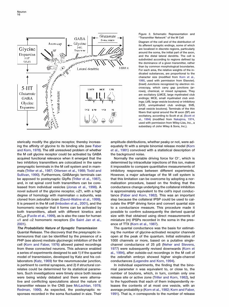
As noted already, the feed-forward glycinergic inhibi-tory pathway counters excitation of the two M cells byauditory afferents (see Figure 1). Stimuli with the samepatterns as those used to potentiate excitatory junc-tions, but weaker and below threshold for firing the Mcell, produce LTP of the inhibitory connections (Figure5). This phenomenon was confirmed with paired pre-and postsynaptic recordings. As at excitatory junc-tions, the induction is synapse specific and is blockedby postsynaptic Ca2+ chelation. However, quantal analy-sis indicated that the locus of LTP expression is presyn-aptic. Furthermore, the enhanced postsynaptic con-ductance is boosted by an additional LTP at the initialexcitatory relay between primary afferents and the in-terneurons themselves.
Repetitive sounds also induce inhibitory LTP and,remarkably, the same protocol has clear behavioral cor-relates in free-swimming fish. It is associated with amarked reduction in the probability of initiating an es-cape response due to a sudden stimulus, such asdropping a ball into the aquarium (Oda et al., 1998).This increased inhibition may thus be a mechanism fordesensitization or habituation (Kandel et al., 1983),expressed as a shift in the behavioral threshold (cf.below).Silent Connections: Substrate for Gain ControlA compound binomial model of transmitter release sug-gested that some of the terminal boutons in the con-nection between an Ia spinal afferent and an α-moto-neuron (Redman and Walmsley, 1983), or between agroup I afferent and a dorsal spinocerebellar tract neu-ron (Walmsley et al., 1988), could be ineffective. Thatis, although presynaptic conduction in an afferent is
normal, some terminals seem to exhibit a zero or near-zero release probability. Yet studies of M cell inputswere the first to show that, in fact, a significant fractionof a neuron’s afferent connections constitute a hiddenpopulation of silent chemical synaptic connections thatcan become functional in certain conditions. Sincethen, it has been recognized that chemical synapsescan be switched on and off, and this phenomenon isnow a mechanism commonly implicated in differentforms of LTP (Malinow and Malenka, 2002) and LTD. Acorollary of this notion of a labile fraction of cells is thatit allows a rapid reorganization of neural networks. Inpractice, this also means that the failure to obtain apostsynaptic response in paired recordings does notnecessarily imply the lack of anatomical contacts.
Approximately 80% of individual club ending con-nections with the M cell are chemically silent followinga presynaptic spike, and they become functional withsmall increases (w100 �s) in spike width produced byblocking voltage-dependent K+ channels with intra-axonal injections of Cs+ or 4-AP (Lin and Faber, 1988b;Faber et al., 1991a). Thus, in the case of these afferents,each of which has multiple release sites, transmissionmay be gated at the level of the full connections. More-over, the ratio of the chemical to the electrical EPSPsincreases as more afferents are stimulated, reflectingthe fact that the silent connections can be convertedinto functional ones when a significant population is co-activated (Pereda et al., 2004).
Impulses in at least 25% of the glycineric inhibitoryinterneurons produce no response in the M cell, al-though their pattern of connectivity is similar to that offunctional interneurons and may involve as many astens of release sites. Furthermore, intraaxonal injec-
tions of 4-AP or Ca2+ do not unmask transmission,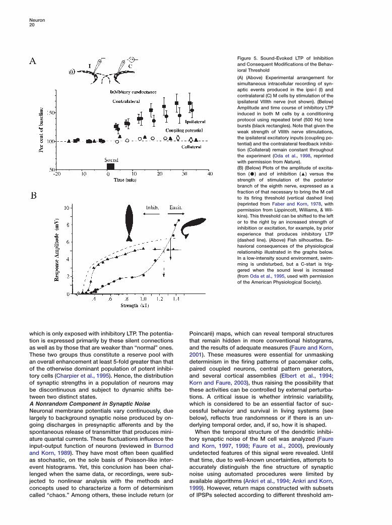
Neuron20
Figure 5. Sound-Evoked LTP of Inhibitionand Consequent Modifications of the Behav-ioral Threshold
(A) (Above) Experimental arrangement forsimultaneous intracellular recording of syn-aptic events produced in the ipsi-l (I) andcontralateral (C) M cells by stimulation of theipsilateral VIIIth nerve (not shown). (Below)Amplitude and time course of inhibitory LTPinduced in both M cells by a conditioningprotocol using repeated brief (500 Hz) tonebursts (black rectangles). Note that given theweak strength of VIIIth nerve stimulations,the ipsilateral excitatory inputs (coupling po-tential) and the contralateral feedback inhibi-tion (Collateral) remain constant throughoutthe experiment (Oda et al., 1998, reprintedwith permission from Nature).(B) (Below) Plots of the amplitude of excita-tion (C) and of inhibition (:) versus thestrength of stimulation of the posteriorbranch of the eighth nerve, expressed as afraction of that necessary to bring the M cellto its firing threshold (vertical dashed line)(reprinted from Faber and Korn, 1978, withpermission from Lippincott, Williams, & Wil-kins). This threshold can be shifted to the leftor to the right by an increased strength ofinhibition or excitation, for example, by priorexperience that produces inhibitory LTP(dashed line). (Above) Fish silhouettes. Be-havioral consequences of the physiologicalrelationship illustrated in the graphs below.In a low-intensity sound environment, swim-ming is undisturbed, but a C-start is trig-gered when the sound level is increased(from Oda et al., 1995, used with permissionof the American Physiological Society).
which is only exposed with inhibitory LTP. The potentia- Pttion is expressed primarily by these silent connections
as well as by those that are weaker than “normal” ones. a2These two groups thus constitute a reserve pool with
an overall enhancement at least 5-fold greater than that dpof the otherwise dominant population of potent inhibi-
tory cells (Charpier et al., 1995). Hence, the distribution aKof synaptic strengths in a population of neurons may
be discontinuous and subject to dynamic shifts be- tttween two distinct states.
A Nonrandom Component in Synaptic Noise wcNeuronal membrane potentials vary continuously, due
largely to background synaptic noise produced by on- bdgoing discharges in presynaptic afferents and by the
spontaneous release of transmitter that produces mini-tature quantal currents. These fluctuations influence the
input-output function of neurons (reviewed in Burnod auand Korn, 1989). They have most often been qualified
as stochastic, on the sole basis of Poisson-like inter- taevent histograms. Yet, this conclusion has been chal-
lenged when the same data, or recordings, were sub- najected to nonlinear analysis with the methods and
concepts used to characterize a form of determinism 1ocalled “chaos.” Among others, these include return (or
oincaré) maps, which can reveal temporal structureshat remain hidden in more conventional histograms,nd the results of adequate measures (Faure and Korn,001). These measures were essential for unmaskingeterminism in the firing patterns of pacemaker cells,aired coupled neurons, central pattern generators,nd several cortical assemblies (Elbert et al., 1994;orn and Faure, 2003), thus raising the possibility that
hese activities can be controlled by external perturba-ions. A critical issue is whether intrinsic variability,hich is considered to be an essential factor of suc-essful behavior and survival in living systems (seeelow), reflects true randomness or if there is an un-erlying temporal order, and, if so, how it is shaped.When the temporal structure of the dendritic inhibi-
ory synaptic noise of the M cell was analyzed (Faurend Korn, 1997, 1998; Faure et al., 2000), previouslyndetected features of this signal were revealed. Untilhat time, due to well-known uncertainties, attempts toccurately distinguish the fine structure of synapticoise using automated procedures were limited byvailable algorithms (Ankri et al., 1994; Ankri and Korn,999). However, return maps constructed with subsetsf IPSPs selected according to different threshold am-

Review21
plitudes as well as nonlinear measures disclosed sev-eral striking periodicities, centered around frequencieswithin the γ range that were observed in the brain activ-ity of higher vertebrates (Figure 6). Furthermore, mutualinteractions and the phase relationship between theIPSPs associated with the extracted frequencies wereconsistent with the notion that this signal is generatedby presynaptic interneurons behaving as weakly cou-pled oscillators, as opposed to independent ones. Inconfirmation, auditory-evoked LTP, which increasessome of the inhibitory synaptic strengths (Oda et al.,1998), permits complex presynaptic oscillating firing
Figure 6. Evidence for Dynamic Patterns Compatible with the No-tion of Chaos in Synaptic Noise
(A) Schematic representation of the presynaptic inhibitory networkafferent to the M cell and, below, a fragment of the synaptic noisegenerated by this network’s spontaneous activity. IPSPs are depo-larizing due to intracellular injections of Cl−.(B) Derivative of a segment of actual synaptic noise. IPSPS areselected according to a given size by a threshold θ. Intervals, in ms,between consecutive events are labeled In and I(n+1), respectively.(Reprinted from Faure et al., 2000, used with permission of theAmerican Physiological Society).(C) Return (Poincaré) map constructed with these intervals. Notethe striking signal-flag pattern, at the base of which the highestdensity of points reveals principal (πp) and secondary (πs) fre-quencies, with intervals equal to 13.3 and 14.4 ms, respectively(see text). Repeating this protocol after removing the related IPSPsand lowering the threshold would uncover a second, hidden trian-gle, with different underlying frequencies (not shown). (Reprintedfrom Faure et al., 2000, used with permission of the American Phys-iological Society).(D) Variations of the significance level of two measures of nonran-domness, the percentage of determinism (% det.) and the Kolmo-gorov-Sinai entropy (�(�)), as functions of θ. The horizontal line in-dicates the confidence level after comparison with surrogates(modified from Faure and Korn, 2003, used with permission fromthe MIT Press).
patterns to be transmitted more effectively to theirpostsynaptic target.
A model of the M cell system indicated that the stateof the pool of afferent interneurons is recapitulated inthe temporal structure of synaptic noise only if themean quantal content varies between the simulated“connections,” as observed experimentally (Korn et al.,1986). In this context, the probabilistic nature of neuro-transmission becomes a functional advantage ratherthan a limitation, since it allows the transmission of in-formation to be modulated by environmental factorswithout modifying the behavior of the synaptic net-works (Faure et al., 2000).
Sensory Motor Behavior and the M CellIs the M Cell a Command Neuron?The most common fast-start escape behavior is theC-start (Figure 1 C), so named on the basis of the shapeof the fish at the end of the first stage of this reflex,before forward propulsion (stage 2) begins (Weihs,1973; Webb, 1976; Eaton et al., 1977). Typically, in thegoldfish, one of the M cells fires a single spike about 3to 5 ms after the onset of an aversive acoustic stimulus(Zottoli, 1977; Eaton et al., 1988), and the first detectedmovement is a deviation of the head in the directionopposite to the activated M cell about 8 ms after thespike (Eaton et al., 1991). The C-start also occurs dur-ing prey capture and feeding (Canfield and Rose, 1993),probably during hatching (Eaton and Nissanov, 1985),and most likely in response to the disruption of anestablished social order (Fernald, 1975). Finally, the am-phibian M cells apparently can trigger a different typeof avoidance behavior, as they fire during the bilateralhindlimb contraction associated with the diving escapeof the frog (Will, 1991).
These features suggested that the M cell might bethe prototype of a “command neuron” (Kupfermannand Weiss, 1978), i.e., a neural decision-making cellwhose firing could be “necessary and sufficient” to trig-ger a complete behavioral act, such as the crayfish es-cape. This concept was challenged in a series of partic-ularly thoughtful experiments that showed that M cellablations (Eaton et al., 1982; Di Domenico et al., 1988)do not abolish the behavior, but rather delay its onsetby a few ms, and although M cell activation alone cantrigger stage 1, the resultant behavior is less variablethan is normal behavior (Nissanov et al., 1990; Eaton etal., 2001). Yet, the C-start almost never occurs withoutfiring of the M cell, and this neuron always fires first,before all other brainstem neurons (Casagrand et al.,1999; Eaton et al., 2001). Consequently, the M cell isa “command-like” neuron (Eaton et al., 2001), eventhough it participates in a parallel “brainstem escapenetwork” that finely regulates the escape trajectory.
The network comprises the M cell homologs MiD2cmand MiD3cm and other descending reticulospinal neu-rons (Figure 7). The homologs can fire bursts of actionpotentials, in contrast to the M cell (Nakayama andOda, 2004), and they also activate spinal circuits thatcause the trunk musculature to contract (Fetcho, 1991).These cells contribute to the normal behavior, but theyare activated with a longer latency by auditory inputsand they have a higher firing threshold than the M cell
Neuron22
Figure 7. Segmental Arrangement and Functional Relationships of Reticulospinal Neurons
(A) Reconstruction, from horizontal sections, of the seven rhombomeres RS1–RS7 of a goldfish. Note the large M cells’ somata and decussat-ing axons; neurons are not symmetrical due to incomplete labeling by HRP. n.mlf, nucleus of the medial longitudinal fasciculus.(B) Horizontal view of the segmentally homologous neurons. The M cell and the MiD2 and MiD3 cells occupy segments RS4, RS5, and RS6segments, respectively. VS, vestibular nucleus. Calibration bars = 200 �m in (A) and 50 �m in (B) (from Lee et al., 1993, reprinted withpermission of Wiley-Liss, Inc., a subsidiary of John Wiley & Sons, Inc.).(C) Effects of laser-induced ablations of the M cell array (above) and of the M cell alone (below) on the latency of the escape response. Asindicated, the left side of each panel shows the control mean latency of the response evoked by stimuli on the head or on the tail; the blackbars pertain to the side to be lesioned. Killing all three cells eliminated short-latency responses to both head- and tail-directed stimuli, whileeliminating the M cell alone affected only the tail-evoked activity (from Liu and Fetcho, 1999, reprinted with permission from Elsevier).
(Nakayama and Oda, 2004). Thus, there is a longer de- mslay when they compensate for the absence of the latter
(Eaton et al., 2001). ilSubsequently, these issues were elegantly addressed
using imaging techniques that allowed the activity of TIneurons in the brain and spinal cord to be visualized in
real time, in vivo, by taking advantage of the transpar- esency of larval zebrafish (Fetcho and O’Malley, 1995).
This approach confirmed predictions (Foreman and Ea- cston, 1993) that the serial Mauthner homologs are in-
volved in escapes and that the combination of neurons i1activated depends upon stimulus features (O’Malley et
al., 1996). In particular, the M cell alone fires when the tastimulus is to the tail, but the homologs are also active
when it is to the head, and causality was then demon- hstrated with photoablation of these neurons (Liu andFetcho, 1999). a
rSingle-cell imaging during behavior complementsand/or forces reconsideration of concepts drawn from s
telectrophysiology: (1) the brainstem escape network,including those neurons active during subsequent swim- t
ing, involves >80% of the neurons projecting to thepinal cord (Gahtan et al., 2001), and (2) spinal circuits
nvolved in swimming and escape behaviors are over-apping, but not identical (Ritter et al., 2001).o Escape: Yes or No?
n the case of an auditory stimulus, the speed of thescape reaction is attributed to the gap junctions atensory synapses, the small time constant of the Mell’s membrane, a rapidly conducting axon that mono-ynaptically excites primary motoneurons at the spike-
nitiating site, and the activation of fast muscles (Fetcho,991). On the other hand, the cellular properties andhe network design guarantee that a large number offferents are active before the M cell reaches its thres-old for spike initiation (Faber et al., 1991b).At the level of the neuron itself, these properties arelow input resistance, a short time constant, and a high
esting potential, which guarantee that only strong andynchronized inputs reach the firing level. In addition,he effects of the sensory-evoked feed-forward inhibi-ory network and of the spontaneous background in-

Review23
Figure 8. Diagram of the Spinal Circuits Shared by Different MotorBehaviors and Reconfiguration of Spinal Network Activity by Retic-ulospinal Commands as Escape Overrides Swimming
(Left) Fish silhouettes at indicated times, during swimming (above)and during the first stage of escape triggered by a sudden threat(below). The darkened areas represent the dominant muscular ac-tivity. (Right) Note that two spinal segments, represented by onebox on each side of the spinal cord, are sequentially activated dur-ing swimming (signified by a horizontal dashed line). Neurons andconnections in each box (not shown) are the same as detailed inFigure 1A. Escape priority is imparted by the M cell’s activation ofexcitatory synapses (empty arrows, + signs) and of crossed inhibi-tory interneurons (thick black arrows), which guarantee that onlyone side of the spinal cord will be activated in all segments (lowerdiagram). Shading designates the activated hemisegments. Notethat one M cell alone (shaded), or the cell with its homologs(hatched), is excited depending upon whether the stimulus is to thehead or tail, respectively (modified from Korn and Faber, 1996,used with permission from Elsevier).
hibitory noise (Hatta and Korn, 1999) are maximizednear threshold by the voltage-dependent open time ofthe Cl− channels (Faber and Korn, 1987; Legendre andKorn, 1995). The other property that maximizes inhibi-tion is the synergistic interaction between adjacentnonsaturated postsynaptic receptors that is due to lat-eral diffusion of the transmitter glycine (cf. above),which results in a greater inhibition than that expectedfrom the simple summation of their individual effects.Thus, subcellular and channel properties must be in-cluded among the factors that determine the likelihoodof an escape response.
Conversely, a number of nonlinearities can boost ex-
citation at the time that inhibition is saturated and canbe more easily overcome. For example, input resis-tance of the M cell is enhanced by depolarization(Faber and Korn, 1986).
At the network level, a bilateral feedback inhibitionprevents activation of both M cells (Furukawa andFurshpan, 1963; Hackett and Faber, 1983), which wouldcause bilateral muscle contractions and result in mini-mal displacements of the fish. Asymmetric tonic inhibi-tion of the two M cells may also bias the system in favorof activating one of them (Hatta and Korn, 1999). If,however, the two M cells are coactivated, a spinal in-hibitory pathway suppresses the excitatory effect of thetrailing action potential for intervals greater than 150 �s(Yasargil and Diamond, 1968). The midbrain inhibitorysystem, along with the role of a unique M cell dendro-toxin-I-voltage-gated potassium conductance (Naka-yama and Oda, 2004), avoids repetitive firing that wouldresult in multiple and ineffective body bends. Finally(Figure 8), there is a priority of the escape reaction overother motor behaviors (Svoboda and Fetcho, 1996)such as swimming, due to a high safety factor at allconnections downstream from the M cell (Fetcho,1991).To Escape: Where and How?During a typical acoustically triggered C-start, the ani-mal changes its orientation away from the startlingstimulus. The extent of this escape, as well as its varia-tions, is controlled by the parallel network, includinginteractions between the activated M cell and its homo-logs (Eaton et al., 1984; Di Domenico et al., 1988; Casa-grand et al., 1999).
Contrary to initial claims that the C-start trajectory isstereotypic, the stage 1 and stage 2 phases are highlyvariable in duration, angular displacement, and the dis-tance moved (Nissanov and Eaton, 1989). The variabil-ity of these phases is the consequence of differencesbetween the size of the initial agonist muscle con-traction and that of the later antagonistic contraction,as well as the timing between the two contractions(Eaton et al., 2001). The resulting unpredictability of theescape path is in marked contrast to the direction takenby a predator aiming at its prey, and it is an importantfeature of the M cell-triggered behavior that makes itdifficult for a predator to adapt, or to learn, a successfulstrategy for prey capture (Domenici, 2002).
This variability has been analyzed by defining escapetrajectories in circular coordinates, with the referencesbeing the stimulus orientations at rest, and by measur-ing the angles characterizing the fish’s orientation atthe end of phases 1 and 2 (Domenici and Blake, 1993,1997). Statistical measures indicate that variations intrajectories are not due to randomness, and they revealmultimodal adaptive patterns that remain hidden inclassical linear plots based on a fixed origin. The vari-ability of the C-start responses of solitary fish disap-pears in schooling, which produces a single trajectorypresumably to avoid collisions (Domenici and Batty,1997).
Despite these variations, the acoustically triggeredescape is appropriately directional when the fish are inopen field (Eaton and Emberley, 1991; Domenici andBlake, 1993), although this directionality can be re-versed without apparent reason or by a nearby physical

Neuron24
barrier or visual cues. Furthermore, in 10% to 20% of othe trials with an unobstructed path, the relationship pbetween the trajectory of the escape and the stimulus tangle is reversed and the fish turns toward the threat. nThis “tactical error” may be corrected by an aversive ssecond away movement that starts at the end of tstage1, which is the right timing for sensory feedbackto influence steering (Domenici and Blake, 1993), al- Fthough other authors (Di Domenico et al., 1988; Eaton Tet al., 1988) believe that it is preprogrammed. t
In addition to auditory inputs, several constituents of mthe octavolateralis system (lateral line, vestibular; re- cviewed in Faber and Korn, 1978) also activate the M pcells. It is likely that, as shown for the afferents from fthe swimbladder (Canfield and Eaton, 1990), they, along twith parallel inhibitory projections, influence the direc- ptionality of the escape by differentially biasing the excit- tability of the two M cells. In conjunction with visual in- aputs, they also provide the substrate for the directional Aoverride that occurs when a fish, close to a wall, turns caway from it (Eaton and Emberley, 1991; Preuss and sFaber, 2003). The long latency of visually evoked excit- catory potentials in the M cell suggests that vision can tmodify response direction before, rather than during, a Mpredator’s strike (Canfield, 2003). s“Decision” and the Escape Behavior 1Studies of the M cell, long ago termed a “miniature tbrain” by Steve Kuffler, illustrate some of the difficulties sencountered when trying to understand the relationship dbetween brain and behavior. Initially, the escape reac- ation was simply viewed as a classical sensory-to-motor h“reflex.” Consequently, it was believed that this beha- dvior could be explained by reducing it to the sum ofits smallest possible components, or building blocks. nHowever, it turns out that a rather complex system is gactually involved. a
The case of the M cell-triggered escape reactions il- alustrates several basic principles. First, even if the ana- ttomical design of a motor behavior seems well adapted tto a given function, other behaviors can utilize or build 1on that basic circuitry. It follows that “it is appropriate gto define the goal that an organism has to reach before rexamining the mechanism (and the hardware) in which Nit is embodied” (D. Marr, quoted by Glimcher, 2003). aThis guideline was at the core of investigations that re- lvealed the role of reticulospinal neurons other than the oM cell and their hierarchical organization within the ibrain stem (Figure 8). Second, the role of the nervous
rsystem is to maximize the chances of achieving a spe-
tcific aim, which in the case of the M cell-triggered reac-
wtion is most often equivalent to survival. This requiresothat the neural networks actively encode and decoderrecent and external ongoing events in order to deal with2the uncertainties of the outside world (Oda et al., 1998;mPreuss and Faber, 2003), while simultaneously intro-tducing sources of variability such as synaptic noise,
which act as internal randomizers (see above).cThe decision by the M cell to trigger a specific motordreaction can be viewed as a process that shifts the bal-fance between inhibition and excitation and favors the
latter. In other words, as suggested by Schall andAThompson (1999), there is a “decisional threshold” that
the rising activity has to cross in order for a movement Wto be produced. In this perspective, the decisional level
for the escape is equivalent to the firing threshold ofne neuron, and the likelihood that it is reached de-ends upon external conditions. Overall, the M cell sys-em, with its various forms of plasticity, exemplifies theotion that “the environmental problems animals facehape not only behavior but also the neural hardwarehat generates that behavior” (Glimcher, 2002).
uture Avenueshe M cell continues to be an invaluable model for in-erdisciplinary research. Recent investigations of theirorphological and electrophysiological properties indi-
ate that M cells and their networks have the sameroperties in the zebrafish as in the more familiar gold-
ish (Hatta and Korn, 1998; Hatta et al., 2001) and thathey share a common developmental and segmentationattern (Metcalfe et al., 1986; Lee et al., 1993). Thus,he wealth of information generated over decadesbout these systems can be extrapolated to each other.lso, the transparency of the hindbrain has made singleells accessible in whole-brain preparations at earlytages of development. Consequently, patch-clamp re-ordings at early stages at the M cell level have helpedo clarify single-channel and synaptic properties at the
cell inhibitory synapses and to correlate them withome of the behavioral features (Legendre and Korn,994; Legendre, 1997; Triller et al., 1997). The sameechniques should allow experimental manipulation ofpecific proteins in models of nervous system disor-ers, for example, mutating M cell glycine receptors, toddress issues related to severe forms of spasticity inumans called hyperekplexia, or startle disease (Rajen-ra and Schofield, 1995).Newly available powerful optical approaches such as
oninvasive calcium imaging, laser photoablation, andenetic manipulation using transgenic lines or mutantsre less difficult technically than electrophysiologicalpproaches. They should make it possible to elucidatehe causal links between neurons and behavior usinghe zebrafish as a vertebrate model (Fetcho and Liu,999). Hopes were raised when the Nusslein-Volhardroup began to screen for mutants defective for touchesponsiveness and locomotor behavior (Mullins andusslein-Volhard, 1993; Granato et al., 1996). However,number of the mutants are lethal. Another issue, re-
ated to the mechanisms that guarantee that there isnly one pair of M cells in the r4 segment of each fish,
s whether the same segmentation rules apply to theeticulospinal neurons of mammals. Recent investiga-ions indicate the involvement of the Notch-Delta path-ay in singling out the M cell and controlling its devel-pment (Haddon et al., 1998; Gray et al., 2001) and theole of Hox genes in specifying the cell (Hale et al.,004). Mutations of both pathways demonstrate a re-arkable plasticity and adaptability of the M cell sys-
em to duplication (Liu et al., 2003) and to misallocation.All of these results highlight the potential of the M
ell system to link molecular biology and genetics withevelopment and behavior, particularly since the zebra-
ish genome is known today.
cknowledgments
e would like to thank Robert Eaton, P. Faure, Joe Fetcho, Jim
Hudspeth, and N. Maubourguet for their helpful comments andsupport for this manuscript.
Review25
References
Andersen, P. (1990). Synaptic integration in hippocampal CA1 pyra-mids. Prog. Brain Res. 83, 215–222.
Anderson, C.R., and Stevens, C.F. (1973). Voltage clamp analysis ofacetylcholine produced end-plate current fluctuations at frog neu-romuscular junction. J. Physiol. 235, 655–691.
Ankri, N., and Korn, H. (1999). A statistical method for correctingdistortions of amplitude distribution histograms due to collisions ofsynaptic events. J. Neurosci. Methods 91, 83–99.
Ankri, N., Legendre, P., Faber, D.S., and Korn, H. (1994). Automaticdetection of spontaneous synaptic responses in central neurons.J. Neurosci. Methods 52, 87–100.
Bennett, M.V., Aljure, E., Nakajima, Y., and Pappas, G.D. (1963).Electrotonic junctions between teleost spinal neurons: electrophys-iology and ultrastructure. Science 19, 262–264.
Bodian, D. (1937). The structure of the vertebrate synapse. A studyof the axon endings on Mauthner’s cell and neighboring centers inthe goldfish. J. Comp. Neurol. 68, 117–159.
Burnod, Y., and Korn, H. (1989). Consequences of stochastic re-lease of neurotransmitters for network computation in the centralnervous system. Proc. Natl. Acad. Sci. USA 86, 352–356.
Cajal, S.R (1908). Sur un noyau spécial du nerf vestibulaire despoissons et des oiseaux. Trab. Lab. Invest. Biol. Univ. Madrid 6,1–20.
Canfield, J.G. (2003). Temporal constraints on visually directedC-Start responses: Behavioral and physiological correlates. BrainBehav. Evol. 61, 148–158.
Canfield, J.G., and Eaton, R.C. (1990). Swimbladder acoustic pres-sure transduction initiates Mauthner-mediated escape. Nature 347,760–762.
Canfield, J.G., and Rose, G.J. (1993). Activation of Mauthner neu-rons during prey capture. J. Comp. Physiol. [A] 172, 611–618.
Casagrand, J.J., Guzik, A.L., and Eaton, R.C. (1999). Mauthner andreticulospinal responses to the onset of acoustic pressure and ac-celeration stimuli. J. Neurophysiol. 82, 1422–1437.
Charpier, S., Behrends, J.C., Triller, A., Faber, D.S., and Korn, H.(1995). “Latent” inhibitory connections become functional duringactivity-dependent plasticity. Proc. Natl. Acad. Sci. USA 92, 117–120.
Collingridge, G.L., and Bliss, T.V. (1995). Memories of NMDA recep-tors and LTP. Trends Neurosci. 18, 54–56.
Curti, S., and Pereda, A. (2004). Voltage-dependent enhancementof electrical coupling by a subthreshold sodium current. J. Neu-rosci. 24, 3999–4010.
David-Watine, B., Goblet, C., de Saint Jan, D., Fucile, S., Devignot,V., Bregestovski, P., and Korn, H. (1999). Cloning, expression andelectrophysiological characterization of glycine receptor alpha sub-unit from zebrafish. Neuroscience 90, 303–317.
De Saint Jan, D., David-Watine, B., Korn, H., and Bregestovski, P.(2001). Activation of human α1 and α2 homomeric glycine recep-tors by taurine and GABA. J. Neurophysiol. 535, 741–755.
del Castillo, J., and Katz, B. (1954). Quantal components of endplate potential. J. Physiol. 124, 560–573.
Detwiler, S.R. (1933). Further experiments upon the extirpation ofMauthner’s neurones in amphibian embryos (Amblystoma mexica-num). J. Exp. Zool. 64, 415–431.
Diamond, J. (1963). Variation in the sensitivity to gamma-aminobu-tyric acid of different regions of the Mauthner neurone. Nature 199,773–775.
Diamond, J., and Huxley, J. (1968). The activation and distributionof GABA and L-glutamate receptors on goldfish Mauthner neu-rones: an analysis of dendritic remote inhibition. J. Physiol. 194,669–723.
Diamond, J., and Roper, S. (1973). Analysis of Mauthner cell re-sponses to iontophoretically delivered pulses of GABA, glycine andL-glutamate. J. Physiol. 232, 113–128.
Di Domenico, R., Nissanov, J., and Eaton, R.C. (1988). Lateraliza-
tion and adaptation of a continuously variable behavior followinglesions of a reticulospinal command neuron. Brain Res. 473, 15–28.Domenici, P. (2002). Escape trajectory, ecological. In Encyclopediaof Environmetrics, Volume 2, A.H. El-Shaarawi and W.W. Piegorsch,eds. (Chichester: J. Wiley and Sons, Ltd.), pp. 708–711.
Domenici, P., and Batty, R.S. (1997). The escape behavior of solitaryherring (Clupea harengus L.) and comparisons with schooling in-dividuals. Mar. Biol. 128, 29–38.
Domenici, P., and Blake, R.W. (1993). Escape trajectories in angel-fish (Pterophyllum Eeimekei.). J. Exp. Biol. 177, 253–272.
Domenici, P., and Blake, R.W. (1997). The kinematics and perfor-mance of fish fast-start swimming. J. Exp. Biol. 200, 1165–1178.
Dudek, F.E., Yasamura, T., and Rash, J.E. (1998). Non-synapticmechanisms in seizures and epileptogenesis. Cell Biol. Int. 22,793–805.
Eaton, R.C., and Emberley, D.S. (1991). How stimulus direction de-termines the trajectory of the Mauthner-initiated escape responsein a teleost fish. J. Exp. Biol. 161, 469–487.
Eaton, R.C., and Nissanov, J. (1985). A review of Mauthner-initiatedescape behavior and its possible role in hatching in the immaturezebrafish, Brachydanio rerio. Environ. Biol. Fishes 12, 265–279.
Eaton, R.C., Bombardieri, R.A., and Meyer, O.H. (1977). TheMauthner-initiated startle response in teleost fish. J. Exp. Biol. 66,65–81.
Eaton, R.C., Lavender, W.A., and Wieland, C.M. (1982). Alternativeneural pathways initiate fast-start responses following lesions ofthe Mauthner neuron in goldfish. J. Comp. Physiol. [A] 145, 485–496.
Eaton, R.C., Nissanov, J., and Wieland, C.M. (1984). Differential ac-tivation of Mauthner and non-Mauthner startle circuits in zebrafish:Implications for functional substitution. J. Comp. Physiol. [A] 155,813–820.
Eaton, R.C., Di Domenico, R., and Nissanov, J. (1988). Flexible bodydynamics of the goldfish C-start: implications for reticulospinalcommand mechanisms. J. Neurosci. 8, 2758–2768.
Eaton, R.C., Di Domenico, R., and Nissanov, J. (1991). Role of theMauthner cell in sensorimotor integration by the brain stem escapenetwork. Brain Behav. Evol. 37, 272–285.
Eaton, R.C., Lee, R.K., and Foreman, M.B. (2001). The Mauthnercell and other identified neurons of the brainstem escape networkof fish. Prog. Neurobiol. 63, 467–485.
Eccles, J.C. (1964). The Physiology of Synapses (Berlin-Göttingen-Heidelberg: Springer-Verlag).
Edwards, F.A., Konnerth, A., and Sakmann, B. (1990). Quantalanalysis of inhibitory synaptic transmission in the dentate gyrusof rat hippocampal slices: a patch-clamp study. J. Physiol. 430,213–249.
Elbert, T., Ray, W.J., Kowalik, Z.J., Skinner, J.E., Graf, K.E., andBirnbaumer, N (1994). Chaos and physiology: deterministic chaosin excitable cell assemblies. Physiol. Rev. 74, 1–47.
Faber, D.S., and Korn, H. (1973). A neuronal inhibition mediatedelectrically. Science 179, 577–578.
Faber, D.S., and Korn, H. (1978). Electrophysiology of the Mauthnercell: basic properties, synaptic mechanisms, and associated net-works. In Neurobiology of the Mauthner Cell, D.S. Faber and H.Korn, eds. (New York: Raven Press), pp. 47–131.
Faber, D.S., and Korn, H. (1980). Single-shot channel activation ac-counts for duration of inhibitory postsynaptic potentials in a centralneuron. Science 208, 612–615.
Faber, D.S., and Korn, H. (1982). Transmission at a central inhibitorysynapse. I. Magnitude of unitary postsynaptic conductance changeand kinetics of channel activation. J. Neurophysiol. 48, 654–678.
Faber, D.S., and Korn, H. (1986). Instantaneous inward rectificationin the Mauthner cell; a postsynaptic booster for excitatory inputs.Neuroscience 19, 1037–1043.
Faber, D.S., and Korn, H. (1987). Voltage-dependence of glycine-activated Cl− channels: a potentiometer for inhibition? J. Neurosci.7, 807–811.
Faber, D.S., and Korn, H. (1988). Synergism at central synapses
due to lateral diffusion of transmitter. Proc. Natl. Acad. Sci. USA85, 8708–8712.
Neuron26
Faber, D.S., and Korn, H. (1989). Electrical field effects: Their rele- Ftvance in central neural networks. Physiol. Rev. 69, 821–863.
Faber, D.S., Funch, P.G., and Korn, H. (1985). Evidence that recep- Gtors mediating central synaptic potentials extend beyond the post- Esynaptic density. Proc. Natl. Acad. Sci. USA 82, 3504–3508. b
Faber, D.S., Fetcho, J.R., and Korn, H. (1989). Neuronal networks Gunderlying the escape response in goldfish. Ann. N.Y. Acad. Sci. b563, 11–33. GFaber, D.S., Lin, J.-W., and Korn, H. (1991a). Silent synaptic con- enections and their modifiability. Ann. N Y Acad. Sci. 627, 151–164. GFaber, D.S., Korn, H., and Lin, J.-W. (1991b). Role of medullary snetworks and postsynaptic membrane properties in regulating rMauthner cell responsiveness to sensory excitation. Brain Behav. GEvol. 37, 286–297. FFaber, D.S., Young, W.S., Legendre, P., and Korn, H. (1992). Intrin- Jsic quantal variability due to stochastic properties of receptor- ttransmitter interactions. Science 258, 1494–1498. 3Fatt, P., and Katz, B. (1953). The effect of inhibitory nerve impulses Gon a crustacean muscle fibre. J. Physiol. 121, 374–389. (
2Faure, P., and Korn, H. (1997). A nonrandom dynamic componentin the synaptic noise of a central neuron. Proc. Natl. Acad. Sci. USA H94, 6506–6511. d
fFaure, P., and Korn, H. (1998). A new method to estimate the Kol-mogorov entropy on recurrence plots: its application to neuronal Hsignals. Physica D. 94, 6506–6511. r
bFaure, P., and Korn, H. (2001). Is there chaos in the brain? I. Con-3cepts of nonlinear dynamics and methods of investigation. C.R.
Acad. Sci. III 324, 773–793. HHFaure, P., and Korn, H. (2003). Synaptic noise and chaos in a verte-abrate neuron. In Handbook of Brain Theory and Neural Networks,3M. Arbib, ed. (Cambridge, MA: MIT Press), pp. 1130–1133.
HFaure, P., Kaplan, D., and Korn, H. (2000). Synaptic efficacy andpthe transmission of complex firing patterns between neurons. J.sNeurophysiol. 84, 3010–3025.
Fernald, D.R. (1975). Fast body turns in a cichlid fish. Nature 258, H228–229. M
4Fetcho, J.R. (1991). The spinal network of the Mauthner cell. BrainBehav. Evol. 37, 298–316. H
nFetcho, J.R., and Liu, K. (1999). Zebrafish as a model system forSstudying neuronal circuits and behavior. Ann. N Y Acad. Sci. 860,
333–345. HpFetcho, J.R., and O’Malley, D.M. (1995). Visualization of active neu-
ral circuitry in the spinal cord of intact zebrafish. J. Neurophysiol. H73, 399–406. H
MForeman, M.B., and Eaton, R.C. (1993). The direction change con-Acept for reticulospinal control of goldfish escape. J. Neurosci. 13,
4101–4113. IdFrank, K. (1959). Basic mechanisms of synaptic transmission in thevcentral nervous system. I.R.E. Trans. Med. Electron. ME-6, 85–88.JFregnac, Y., Monier, C., Chavane, F., Baudot, P., and Graham, L.s(2003). Shunting inhibition, a silent step in visual cortical computa-ition. J. Physiol. (Paris) 97, 441–451.
JFucile, S., De Saint Jan, D., David-Watine, B., Korn, H., and Breges-otovski, P. (1999). Comparison of glycine and GABA actions on the4zebrafish homomeric glycine receptor. J. Physiol. 517, 369–383.
KFukami, Y., Furukawa, T., and Asada, Y. (1965). Excitability changesiof the Mauthner cell during collateral inhibition. J. Gen. Physiol. 48,
581–600. KcFurshpan, E.J. (1964). “Electrical transmission” at an excitatoryssynapse in a vertebrate brain. Science 144, 878–880.fFurshpan, E.J., and Furukawa, T. (1962). Intracellular and extracel-Klular responses of several regions of the Mauthner cell of the gold-Sfish. J. Neurophysiol. 25, 732–771.
KFurukawa, T. (1966). Synaptic interaction at the Mauthner cell ofMgoldfish. Prog. Brain Res. 21, 44–70.aFurukawa, T., and Furshpan, E.J. (1963). Two inhibitory mecha-
nisms in the Mauthner neurons of goldfish. J. Neurophysiol. 26, Kv140–176.
urukawa, T., Fukami, Y., and Asada, Y. (1963). A third type of inhibi-ion in the Mauthner cell of goldfish. J. Neurophysiol. 26, 759–774.
ahtan, E., Nagarajan, S., Campos, J.B., and O’Malley, D.M. (2001).vidence for a widespread brainstem escape network in larval ze-rafish. J. Neurophysiol. 87, 608–614.
limcher, P.W. (2002). Decisions, decisions, decisions: Choosing aiological science of choice. Neuron 36, 323–332.
limcher, P.W. (2003). Decision, Uncertainty and the Brain: The Sci-nce of Neuroeconomics (Cambridge, MA: MIT Press).
otow, T., Triller, A., and Korn, H. (1990). Differential distribution oferotoninergic inputs on the goldfish Mauthner cell. J. Comp. Neu-ol. 292, 255–268.
ranato, M., Van Eeden, F.J.M., Schach, U., Trowe, T., Brand, M.,urutani-Seiki, M., Haffer, P., Hammerschmidt, M., Heisenberg, C.P.,iang, Y.J., et al. (1996). Genes controlling and mediating locomo-ion behavior in the zebrafish embryo and larva. Development 123,99–413.
ray, M., Moens, C.B., Amacher, S.L., Eisen, J.S., and Beattie, C.E.2001). Zebrafish deadly seven functions in neurogenesis. Dev. Biol.37, 306–323.
ackett, J.T., and Faber, D.S. (1983). Mauthner axon networks me-iating supraspinal components of the startle response in the gold-
ish. Neuroscience 8, 317–331.
addon, C., Smithers, L., Schneider-Maunoury, S., Coche, T., Hen-ique, D., and Lewis, J. (1998). Multiple delta genes and lateral inhi-ition in zebrafish primary neurogenesis. Development 125, 359–70.
ale, M.E., Kheirbek, M.A., Schreifer, J.E., and Prince, V.E. (2004).ox gene misexpression and cell-specific lesions reveal function-lity of homeotically transformed neurons. J. Neurosci. 12, 3070–076.
artzell, H.C., Kuffler, S.W., and Yoshikami, D. (1975). Post-synapticotentiation: interaction between quanta of acetylcholine at thekeletal neuromuscular synapse. J. Physiol. 251, 427–463.
atta, K., and Korn, H. (1998). Physiological properties of theauthner system in the adult zebrafish. J. Comp. Neurol. 395,
93–509.
atta, K., and Korn, H. (1999). Tonic inhibition alternates in pairedeurons that set direction of fish escape reaction. Proc. Natl. Acad.ci. USA 96, 12090–12095.
atta, K., Ankri, N., Faber, D.S., and Korn, H. (2001). Slow inhibitoryotentials in the teleost Mauthner cell. Neuroscience 103, 561–579.
euser, J.E., and Reese, T.S. (1977). Structure of the synapse. Inandbook of Physiology, Section I: The Nervous System, V.B.ountcastle, J.M. Brookhart, and E.R. Kandel, eds. (Bethesda, MD:mer. Physiol. Soc.), pp. 261–295.
mboden, M., Devignot, V., Korn, H., and Goblet, C. (2001). Regionalistribution of glycine receptor messenger RNA in the central ner-ous system of zebrafish. Neuroscience 103, 811–830.
ack, J.J., Redman, S.J., and Wong, K. (1981). The components ofynaptic potentials evoked in cat spinal motoneurones by impulses
n single group Ia afferents. J. Physiol. 321, 65–96.
onas, P., Bischofberger, J., and Sandkühler, J. (1998). Co-releasef two fast neurotransmitters at a central synapse. Science 281,19–424.
andel, E.R., and Schwartz, S.H. (1982). Molecular biology of learn-ng: modulation of transmitter release. Science 218, 433–443.
andel, E.R., Brunelli, M., Byrne, J., and Castelluci, V. (1983). Aommon presynaptic locus for the synaptic changes underlyinghort-term habituation and sensitization of the gill-withdrawal re-lexes in Aplysia. Cold Spring Harb. Symp. Quant. Biol. 48, 465–482.
atz, B. (1969). The Release of Neural Transmitter Substances. Theherrington Lectures X (Springfield, IL: Charles C. Thomas).
immel, C.B., and Model, P.G. (1978). Developmental studies of theauthner cell. In Neurobiology of the Mauthner Cell, D.S. Faber
nd H. Korn, eds. (New York: Raven Press), pp. 183–217.
orn, H. (1998). Correlation of morphology and function: the one-esicle hypothesis at Mauthner neuron inhibitory synapses. In

Review27
Central Synapses: Quantal Mechanisms and Plasticity, D.S. Faber,H. Korn, S.J. Redman, S.M. Thompson, and S. Altman, eds. (Stras-bourg: HFSP), pp. 37–47.
Korn, H., and Axelrad, H. (1980). Electrical inhibition of Purkinjecells in the cerebellum of the rat. Proc. Natl. Acad. Sci. USA 77,6244–6247.
Korn, H., and Faber, D.S. (1975). An electrically mediated inhibitionin goldfish medulla. J. Neurophysiol. 38, 452–471.
Korn, H., and Faber, D.S. (1976). Vertebrate central nervous system:same neurons mediate both electrical and chemical inhibitions.Science 194, 1166–1169.
Korn, H., and Faber, D.S. (1991). Quantal analysis and synaptic effi-cacy in the CNS. Trends Neurosci. 14, 439–445.
Korn, H., and Faber, D.S. (1996). Escape behavior. Brainstem, spi-nal cord circuitry and function. Curr. Opin. Neurobiol. 6, 826–832.
Korn, H., and Faure, P. (2003). Is there chaos in the brain? II. Experi-mental evidence and related models. C.R. Acad. Sci. III 326, 787–840.
Korn, H., Triller, A., Mallet, A., and Faber, D.S. (1981). Fluctuatingresponses at a central synapse: n of binomial fit predicts numberof stained presynaptic boutons. Science 213, 898–901.
Korn, H., Mallet, A., Triller, A., and Faber, D.S. (1982). Transmissionat a central inhibitory synapse. II. Quantal description of release,with a physical correlate for binomial n. J. Neurophysiol. 48, 679–707.
Korn, H., Faber, D.S., and Triller, A. (1986). Probabilistic determina-tion of synaptic strength. J. Neurophysiol. 55, 402–421.
Korn, H., Burnod, Y., and Faber, D.S. (1987). Spontaneous quantalcurrents in a central neuron match predictions from binomial analy-sis of evoked responses. Proc. Natl. Acad. Sci. USA 84, 5981–5985.
Korn, H., Faber, D.S., and Triller, A. (1990). Convergence of morpho-logical physiological, and immunocytochemical techniques for thestudy of single Mauthner cells. In Handbook of Chemical Neuro-anatomy, Volume 8, Analysis of Neuronal Microcircuits and Synap-tic Interactions, A. Björkland, T. Hökfelt, F.G. Wouterlood, and A.N.Van den Pol, eds. (Amsterdam: Elsevier), pp. 403–480.
Korn, H., Oda, Y., and Faber, D.S. (1992). Long-term potentiation ofinhibitory circuits and synapses in the central nervous system.Proc. Natl. Acad. Sci. USA 89, 440–443.
Korn, H., Sur, C., Charpier, S., Legendre, P., and Faber, D.S. (1994).The one-vesicle hypothesis and multivesicular release. In Ad-vances in Second Messenger Phosphoprotein Research, Volume29, Molecular and Cellular Mechanisms of Neurotransmitter Re-lease, L. Stjärne, P. Greengard, S.E. Grillner, T.G.M. Hökfelt, andD.R. Ottoson, eds. (New York: Raven Press), pp. 301–322.
Kruk, P.J., Korn, H., and Faber, D.S. (1997). The effects of geometri-cal parameters on synaptic transmission: A Monte Carlo simulationstudy. Biophys. J. 73, 2874–2890.
Kullmann, D.M., Min, M.Y., Asztely, F., and Rusakov, D.A. (1999).Extracellular glutamate diffusion determines the occupancy of glu-tamate receptors at CA1 synapses in the hippocampus. Philos.Trans. R. Soc. Lond. B Biol. Sci. 28, 395–402.
Kumar, S.S., and Faber, D.S. (1999). Plasticity of first-order sensorysynapses: interactions between homosynaptic long-term potentia-tion and heterosynaptically evoked dopaminergic potentiation. J.Neurosci. 19, 1620–1635.
Kupfermann, I., and Weiss, K.R. (1978). The command neuron con-cept. Behav. Brain Sci. 1, 3–39.
Land, B.R., Salpeter, E.E., and Salpeter, M.M. (1980). Acetylcholinereceptor site density affects the rising phase of miniature endplatecurrents. Proc. Natl. Acad. Sci. USA 77, 3736–3740.
Lee, R.K., Eaton, R.C., and Zottoli, S.J. (1993). Segmental arrange-ment of reticulospinal neurons in the goldfish hindbrain. J. Comp.Neurol. 329, 539–556.
Legendre, P. (1997). Pharmacological evidence for two types ofpostsynaptic glycinergic receptors on the Mauthner cell of 52-hour-old zebrafish larvae. J. Neurophysiol. 77, 2400–2415.
Legendre, P., and Korn, H. (1994). Glycinergic inhibitory synaptic
currents and related receptor channels in the zebrafish brain. Eur.J. Neurosci. 6, 1544–1557.
Legendre, P., and Korn, H. (1995). Voltage-dependence of conduc-tance changes evoked by glycine release in the zebrafish Brain. J.Neurophysiol. 73, 2404–2412.
Leghissa, S. (1941). Sviluppo dell’apparato del Mauthner in larve diAmbystoma mexicanum (Axolotl). Arch. Zool. Ital. 29, 213–253.
Lin, J.-W., and Faber, D.S. (1988a). Synaptic transmission mediatedby single club endings on the goldfish Mauthner cell. I. Characteris-tics of electrotonic and chemical postsynaptic potentials. J. Neu-rosci. 8, 1302–1312.
Lin, J.-W., and Faber, D.S. (1988b). Synaptic transmission mediatedby single club endings on the goldfish Mauthner cell. II. Plasticityof excitatory postsynaptic potentials. J. Neurosci. 8, 1313–1325.
Liu, K.S., and Fetcho, J.R. (1999). Laser ablations reveal functionalrelationships of segmental hindbrain neurons in zebrafish. Neuron23, 325–335.
Liu, K.S., Gray, M., Otto, S.J., Fetcho, J.R., and Beattie, C.E. (2003).Mutations in deadly seven/notch1a reveal developmental plasticityin the escape response circuit. J. Neurosci. 23, 8159–8166.
Llinas, R., and Terzuolo, C.A. (1965). Mechanisms of supraspinalactions upon spinal cord activities.Reticular inhibitory mechanismsupon flexor motoneurons. J. Neurophysiol. 28, 413–422.
Malinow, R., and Malenka, R.C. (2002). AMPA receptor traffickingand synaptic plasticity. Annu. Rev. Neurosci. 25, 103–126.
McLachlan, E.M. (1978). The statistics of transmitter release atchemical synapses. Int. Rev. Physiol. 17, 49–117.
Martin, A.R. (1977). Junctional transmission. II. Presynaptic mecha-nisms. In Handbook of Physiology, Volume 1, Part I, J.M. Brookhartand V.B. Mountcastle, Section eds., E.R. Kandel, Volume ed.(Bethesda, MD: Amer. Physiol. Soc.), pp. 329–388.
Marty, A., and Llano, I. (1995). Modulation of inhibitory synapses inthe mammalian brain. Curr. Opin. Neurobiol. 5, 335–341.
Metcalfe, W.K., Mendelson, B., and Kimmel, C.B. (1986). Segmentalhomologies among reticulospinal neurons in the hindbrain of thezebrafish larva. J. Comp. Neurol. 252, 147–159.
Mintz, I., and Korn, H. (1991). Serotonergic facilitation of quantalrelease at central inhibitory synapses. J. Neurosci. 11, 3359–3370.
Mintz, I., Gotow, T., Triller, A., and Korn, H. (1989). Effect of seroto-nergic afferents on quantal release at central inhibitory synapses.Science 245, 190–192.
Model, P.G. (1978). Regulation of the Mauthner cell following uni-lateral rotation of the prospective hindbrain in axolotl (Ambystomamexicanum) neurulae. Brain Res. 153, 135–143.
Mullins, M.C., and Nusslein-Volhard, C. (1993). Mutational ap-proaches to studying embryonic pattern formation in the zebrafish.Curr. Opin. Genet. Dev. 3, 648–654.
Nakajima, Y. (1974). Fine structure of the synaptic endings on theMauthner cell of the goldfish. J. Comp. Neurol. 156, 375–402.
Nakayama, H., and Oda, Y. (2004). Common sensory inputs anddifferential excitability of segmentally homologous reticulospinalneurons in the hindbrain. J. Neurosci. 24, 3199–3209.
Neher, E., and Stevens, C.F. (1977). Conductance fluctuations ofionic pores in membranes. Annu. Rev. Biophys. Bioeng. 6, 354–381.
Nissanov, J., and Eaton, R.C. (1989). Reticulospinal control of rapidescape turning maneuvers in fishes. Am. Zool. 29, 103–121.
Nissanov, J., Eaton, R.C., and Di Domenico, R. (1990). The motoroutput of the Mauthner cell, a reticulospinal command neuron.Brain Res. 517, 88–98.
Oda, Y., Charpier, S., Murayama, Y., Suma, C., and Korn, H. (1995).Long-term potentiation of glycinergic inhibitory synaptic transmis-sion. J. Neurophysiol. 74, 1056–1074.
Oda, Y., Kawasaki, K., Morita, M., Korn, H., and Matsui, H. (1998).Inhibitory long-term potentiation underlies auditory conditioning ofgoldfish escape behavior. Nature 394, 182–185.
O’Malley, D., Kao, Y.-H., and Fetcho, J. (1996). Imaging the func-tional organization of zebrafish hindbrain segments during escapebehaviors. Neuron 17, 1145–1155.
Neuron28
Oppenheimer, J.M. (1942). The decussation of ectopic Mauthner stfibers in fundulus embryos. J. Comp. Neurol. 77, 577–587.
SOttersen, O.P., Davanger, S., and Somogyi, P. (1988). Colocalizationnof glycine-like and GABA-like immunoreactivities in Golgi cell ter-1minals in the rat cerebellum: a postembedding light and electron
microscopic study. Brain Res. 450, 342–353. SoPalay, S.L. (1958). The morphology of synapses in the central ner-4vous system. Exp. Cell Res. Suppl. 5, 1634–1644.TPereda, A., Triller, A., Korn, H., and Faber, D.S. (1992). Dopaminetenhances both electrotonic coupling and chemical excitatory post-ssynaptic potentials at mixed synapses. Proc. Natl. Acad. Sci. USAT89, 2088–2092.sPereda, A., Nairn, A.C., Wolszon, L.R., and Faber, D.S. (1994). Post-tsynaptic modulation of synaptic efficacy at mixed synapses on theTMauthner cell. J. Neurosci. 14, 3704–3712.DPereda, A.E., Bell, T.D., Chang, B.H., Czernik, A.J., Nairn, A.C., Sod-merling, T.R., and Faber, D.S. (1998). Ca2+/calmodulin-dependentTkinase II mediates simultaneous enhancement of gap-junctionalcconductance and glutamatergic transmission. Proc. Natl. Acad.sSci. USA 95, 13272–13277.TPereda, A., O’Brien, J., Nagy, J.I., Bukauskas, F., Davidson, K.G.,aKamasawa, N., Yasumura, T., and Rash, J.E. (2003). Connexin 35smediates electrical transmission at mixed synapses on MauthnerTcells. J. Neurosci. 23, 7489–7503.fPereda, A.E., Rash, J.E., Nagy, J.I., and Bennett, M.V. (2004). Dy-cnamics of electrical transmission at club endings on the MauthnerWcells. Brain Res. Brain Res. Rev. 47, 227–244.rPeters, A., Palay, S.L., and de Webster, F. (1991). The Fine Structuremof the Nervous System: Neurons and Supporting cells, Third Edition9(Oxford: Oxford University Press).W
Preuss, T., and Faber, D.S. (2003). Central cellular mechanisms un-o
derlying temperature-dependent changes in the goldfish startle-r
escape behavior. J. Neurosci. 23, 5617–5626.W
Rajendra, S., and Schofield, P.R. (1995). Molecular mechanisms of Binherited startle syndromes. Trends Neurosci. 18, 80–82.
WRedman, S. (1990). Quantal analysis of synaptic potentials in neu- rrons of the central nervous system. Physiol. Rev. 70, 165–198.
WRedman, S., and Walmsley, B. (1983). Amplitude fluctuations in syn- taptic potentials evoked in cat spinal motoneurones at identified 7group Ia synapses. J. Physiol. 343, 135–145.
WRitter, D.A., Bhatt, D.H., and Fetcho, J.R. (2001). In vivo imaging of czebrafish reveals differences in the spinal networks for escape and 2swimming movements. J. Neurosci. 21, 8956–8965. WRobertson, J.D. (1961). Ultrastructure of excitable membranes and 3the crayfish median-giant synapse. Ann. N Y Acad. Sci. 6, 339–389. WRobertson, J.D., Bodenheimer, T.S., and Stage, D.E. (1963). The lultrastructure of Mauthner cell synapses and nodes in goldfish Wbrains. J. Cell Biol. 19, 159–199. p
pSchall, J.D., and Thompson, K.G. (1999). Neural selection and con-trol of visually guided eye movements. Annu. Rev. Neurosci. 22, Y241–259. e
sScott, J.W., Zottoli, S.J., Beatty, N.P., and Korn, H. (1994). Originand function of spiral fibers projecting to the goldfish Mauthner Ycell. J. Comp. Neurol. 339, 76–90. t
5Silver, R.A., Lübke, J., Sakmann, B., and Feldmayer, D. (2003). High-probability uniquantal transmission at excitatory synapses in barrel Ycortex. Science 302, 1981–1984. f
2Smith, M., and Pereda, A. (2003). Chemical synaptic activity modu-Zlates nearby electrical synapses. Proc. Natl. Acad. Sci. USA 8,t4849–4854.
ZStefanelli, A. (1951). The Mauthnerian apparatus in the Ichthyop-isida: Its nature and function and correlated problems of neurohis-Ftogenesis. Q. Rev. Biol. 26, 17–34.ZSur, C., Korn, H., and Triller, A. (1994). Colocalization of somato-dstatin with GABA or glutamate in distinct afferent terminals presyn-
aptic to the Mauthner cell. J. Neurosci. 14, 576–589.
Sur, C., Triller, A., and Korn, H. (1995). Morphology of the release
ite of inhibitory synapses on the soma and dendrite of an iden-ified neuron. J. Comp. Neurol. 351, 247–260.
voboda, K.R., and Fetcho, J.R. (1996). Interactions between theeural networks for escape and swimming in goldfish. J. Neurosci.6, 843–852.
wisher, J.E., and Hibbard, E. (1967). The course of Mauthner ax-ns in Janus-headed Xenopus embryos. J. Exp. Zool. 165, 433–40.
odd, A.J., and Sullivan, A.C. (1990). Light microscopic study ofhe coexistence of GABA and glycine-like immunoreactivities in thepinal cord of the rat. J. Comp. Neurol. 296, 496–505.
riller, A., and Korn, H. (1982). Transmission at a central inhibitoryynapse. III. Ultrastructure of physiologically identified and stainederminals. J. Neurophysiol. 48, 708–736.
riller, A., Cluzeaud, F., Pfeiffer, F., Betz, H., and Korn, H. (1985).istribution of glycine receptors at central synapses: An electronicroscopy study. J. Cell Biol. 101, 683–688.
riller, A., Cluzeaud, F., and Korn, H. (1987). Gamma-aminobutyric-ontaining terminals can be apposed to glycine receptors at centralynapses. J. Cell Biol. 104, 947–956.
riller, A., Seitanidou, T., Franksson, O., and Korn, H. (1990). Sizend shape of glycine receptor clusters in a central neuron exhibit aomato-dendritic gradient. New Biol. 2, 637–641.
riller, A., Rostaing, P., Korn, H., and Legendre, P. (1997). Morpho-unctional evidence for mature synaptic contacts on the Mauthnerell of 52-hour-old zebrafish larvae. Neuroscience 80, 133–145.
almsley, B., Edwards, F.R., and Tracey, D.J. (1988). Non-uniformelease probabilities underlie quantal synaptic transmission at aammalian excitatory central synapse. J. Neurophysiol. 60, 889–
08.
ebb, P.W. (1976). The effect of size on the fast-start performancef rainbow trout Salmo cairdneri, and a consideration of piscivo-ous predator-prey interactions. J. Exp. Biol. 65, 157–177.
eihs, D. (1973). The mechanism of rapid starting of slender fish.iorheology 10, 343–350.
erman, R. (1969). An electrophysiological approach to drug-eceptor mechanisms. Comp. Biochem. Physiol. 30, 997–1017.
ernig, A. (1972). Changes in statistical parameters during facilita-ion at the crayfish neuromuscular junction. J. Physiol. 226, 751–59.
ernig, A. (1975). Estimates of statistical release parameters fromrayfish and frog neuromuscular junctions. J. Physiol. 244, 207–21.
ill, U. (1991). Amphibian Mauthner cells. Brain Behav. Evol. 37,17–332.
ilson, D.M. (1959). Function of giant Mauthner’s neurons in theungfish. Science 129, 841–842.
olszon, L.R., Pereda, A.E., and Faber, D.S. (1997). A fast synapticotential mediated by NMDA and non-NMDA receptors. J. Neuro-hysiol. 78, 2693–2706.
ang, X.-D., and Faber, D.S. (1991). Initial synaptic efficacy influ-nces induction and expression of long-term changes in transmis-ion. Proc. Natl. Acad. Sci. USA 88, 4299–4303.
ang, X.-D., Korn, H., and Faber, D.S. (1990). Long-term potentia-ion of electrotonic coupling at mixed synapses. Nature 348, 542–45.
asargil, G.M., and Diamond, J. (1968). Startle-response in teleostish: an elementary circuit for neural discrimination. Nature 220,41–245.
ottoli, S.J. (1977). Correlation of the startle reflex and M cell audi-ory responses in unrestrained goldfish. J. Exp. Biol. 66, 243–254.
ottoli, S.J. (1978). Comparative morphology of the Mauthner celln fish and amphibians. In Neurobiology of the Mauthner Cell, D.S.aber and H. Korn, eds. (New York: Raven Press), pp. 13–45.
ucker, R.S. (1973). Changes in the statistics of transmitter releaseuring facilitation. J. Physiol. 229, 787–810.