Embed Size (px)
Citation preview

A Mouse Model for Human Norovirus
Stefan Taube,a* Abimbola O. Kolawole,a Marina Höhne,b John E. Wilkinson,c Scott A. Handley,d Jeffrey W. Perry,a
Larissa B. Thackray,d Ramesh Akkina,e Christiane E. Wobusa
Department of Microbiology and Immunology, University of Michigan, Ann Arbor, Michigan, USAa; Consultant Laboratory for Noroviruses, Robert Koch-Institute, Berlin,Germanyb; Unit for Laboratory Animal Medicine and Department of Pathology, University of Michigan, Ann Arbor, Michigan, USAc; Department of Pathology andImmunology, Washington University, St. Louis, Missouri, USAd; Department of Microbiology, Immunology and Pathology, Colorado State University, Fort Collins, Colorado,USAe
* Present address: Stefan Taube, Institute for Virology and Cell Biology, University of Lübeck, Lübeck, Germany.
S.T. and A.O.K. contributed equally to this work.
ABSTRACT Human noroviruses (HuNoVs) cause significant morbidity and mortality worldwide. However, despite substantialefforts, a small-animal model for HuNoV has not been described to date. Since “humanized” mice have been successfully used tostudy human-tropic pathogens in the past, we challenged BALB/c mice deficient in recombination activation gene (Rag) 1 or 2and common gamma chain (�c) (Rag-�c) engrafted with human CD34� hematopoietic stem cells, nonengrafted siblings, andimmunocompetent wild-type controls with pooled stool isolates from patients positive for HuNoV. Surprisingly, both human-ized and nonhumanized BALB/c Rag-�c-deficient mice supported replication of a GII.4 strain of HuNoV, as indicated by in-creased viral loads over input. In contrast, immunocompetent wild-type BALB/c mice were not infected. An intraperitonealroute of infection and the BALB/c genetic background were important for facilitating a subclinical HuNoV infection of Rag-�c-deficient mice. Expression of structural and nonstructural proteins was detected in cells with macrophage-like morphology inthe spleens and livers of BALB/c Rag-�c-deficient mice, confirming the ability of HuNoV to replicate in a mouse model. In sum-mary, HuNoV replication in BALB/c Rag-�c-deficient mice is dependent on the immune-deficient status of the host but not onthe presence of human immune cells and provides the first genetically manipulable small-animal model for studying HuNoV in-fection.
IMPORTANCE Human noroviruses are a significant cause of viral gastroenteritis worldwide, resulting in significant morbidity andmortality. Antivirals and vaccines are currently not available, in part due to the inability to study these viruses in a geneticallymanipulable, small-animal model. Herein, we report the first mouse model for human noroviruses. This model will accelerateour understanding of human norovirus biology and provide a useful resource for evaluating antiviral therapies.
Received 18 June 2013 Accepted 20 June 2013 Published 16 July 2013
Citation Taube S, Kolawole AO, Höhne M, Wilkinson JE, Handley SA, Perry JW, Thackray LB, Akkina R, Wobus CE. 2013. A mouse model for human norovirus. mBio 4(4):e00450-13. doi:10.1128/mBio.00450-13.
Editor Terence Dermody, Vanderbilt University School of Medicine
Copyright © 2013 Taube et al. This is an open-access article distributed under the terms of the Creative Commons Attribution-Noncommercial-ShareAlike 3.0 Unportedlicense, which permits unrestricted noncommercial use, distribution, and reproduction in any medium, provided the original author and source are credited.
Address correspondence to Christiane E. Wobus, [email protected], or Ramesh Akkina, [email protected].
Human noroviruses (HuNoVs) are the leading etiologic agentof viral epidemic gastroenteritis globally in people of all ages
(1), causing an estimated 200,000 deaths each year in childrenunder 5 years old in developing countries (2). In the United States,~21 million cases of gastroenteritis and ~800 deaths due toHuNoV occur annually, making HuNoV infections the leadingcause of death among individuals with viral gastroenteritis (3, 4).Virus transmission occurs by the fecal-oral route, with person-to-person and food- or waterborne spread being the most common(5). The economic impact of these infections is staggering, with aneconomic cost for norovirus (NoV)-associated food-borne out-breaks alone of $5.8 billion annually in the United States (6). Un-fortunately, no specific therapies are currently available to controlor prevent HuNoV infections.
HuNoV research has been stifled by the absence of a HuNoVcell culture system and a genetically manipulable small-animal
model for HuNoV. Some aspects of HuNoV biology have beenstudied in chimpanzees (7), gnotobiotic pigs, and calves (8, 9).However, these large-animal models are cumbersome, expensive,and not widely available.
HuNoVs are single-stranded positive-sense [(�)-sense] RNAviruses in the Caliciviridae family (10). Their ~7.5-kb genomeencodes three open reading frames (ORFs). ORF1 encodes thenonstructural proteins N-terminal protein or NS1/2, NTPase orNS3, 3A-like protein or NS4, VPg (viral protein, genome-linked)or NS5, protease or NS6, and RNA-dependent RNA polymerase(RdRp) or NS7, while ORF2 and -3 encode the major and minorcapsid proteins VP1 and VP2, respectively (10). HuNoVs are clas-sified based on sequence similarity into three genogroups (GI, GII,and GIV) and at least 25 genotypes (11). However, the majority ofHuNoV strains causing outbreaks today belong to the GII, geno-type 4 (GII.4) cluster (12). Over the past decade, GII.4 epidemics
RESEARCH ARTICLE
July/August 2013 Volume 4 Issue 4 e00450-13 ® mbio.asm.org 1
m
bio.asm.org
on May 17, 2018 - P
ublished by m
bio.asm.org
Dow
nloaded from

have occurred every 2 to 3 years, with new strains emerging bymutation and recombination (13), including the most recentGII.4 variant, Sydney 2012 (14).
To combat the problem of HuNoV morbidity and mortality, awidely accessible, genetically manipulable, small-animal platformand reproducible tissue culture system are urgently needed to in-vestigate molecular mechanisms regulating HuNoV infection andto develop effective antiviral strategies (15). Here we report over-coming one of these major barriers in the field through the devel-opment of the first mouse model for HuNoV.
RESULTSReconstitution of a humanized immune system is not requiredfor susceptibility of mice to HuNoV. HuNoV antigen is detectedin B lymphocytes and dendritic cells in the intestinal lamina pro-pria of HuNoV-infected chimpanzees (7), and a HuNoV GII.4strain binds to cells in the lamina propria of human duodenum inan in vitro whole-virus binding assay (16). This raised the possi-bility that HuNoV may replicate in human immune cells. To testwhether human immune cells are important for HuNoV infec-tion, we used “humanized” mice (i.e., mice engrafted with humanCD34� hematopoietic stem cells to reconstitute a functional hu-man immune system) (35). Recombination activation gene (Rag)1 or 2- and common �-chain (�c)-deficient (Rag�/� �c�/�)BALB/c mice were irradiated 2 to 3 days after birth and injected
with human CD34� hematopoietic stem cells, as previously de-scribed (17). Approximately 6 months later, when these mice hadreconstituted a human immune system, seven humanized mice,five nonhumanized Rag�/� �c�/� mice, and six BALB/c wild-typemice were challenged with pooled stool suspensions from patientscontaining either GI and GII (GI�II mix) or only GII (GII mix)HuNoV strains (see Table S1 in the supplemental material andMaterials and Methods). Mice were infected with 8.56 � 103 GIand 4.08 � 103 � 7.08 � 10e4 GII total genomes (for GI�II mix)or 7.45 � 103 GII total genomes (for GII mix) (see Table S2 in thesupplemental material).
Mice were initially challenged with pooled human stool sam-ples by both the intraperitoneal and oral routes to increase thechance of detecting HuNoV replication (Fig. 1; see also Table S2 inthe supplemental material). All tissue and fecal samples from the12 humanized and nonhumanized mice were analyzed using mul-tiplex quantitative reverse transcriptase PCR (qRT-PCR) as de-scribed elsewhere (18) to measure GI and GII genome loads. NoGI sequences were detected in murine feces or tissues in any of the7 mice infected with the GI�II mix for 24 or 72 h postinfection(hpi) (see Table S2 in the supplemental material). Therefore, onlypooled human stool suspensions containing GII viruses (GII mix)were used for subsequent infections, in which mice were infectedfor 48 or 72 hpi. None of the mice succumbed to infection, but onehumanized mouse with high titers in feces and tissue developed
FIG 1 HuNoV infects humanized and nonhumanized Rag�/� �c�/� mice. Mice were infected intraperitoneally and orally with HuNoV-containing stoolfiltrates. Symbols indicate the mouse background: open, Rag-�c; filled, Rag-�c-hu. Open triangles indicate wild-type (wt) mice. Inocula are colored blue (GI�IImix) or red (GII mix). (A) HuNoV genomes were measured by qRT-PCR in stomach (ST), jejunum/duodenum (JD), proximal ileum (PI), distal ileum (DI),cecum (CE), and colon (CO), mesenteric lymph nodes (MLN), liver (LI), spleen (SP), kidney (KI), heart (HT), lung (LU), bone marrow (BM), and brain (BR)from mice sacrificed 24 (top panel), 48 (middle panel), or 72 (bottom panel) hours postinfection (hpi). (B) HuNoV genomes were measured in total fecesexcreted 12 h before infection (pre) or between 0 to 24, 24 to 48, or 48 to 72 hpi. (C) Total genome titers recovered from humanized (Rag-�c-hu), nonhumanized(Rag-�c), and wild-type (WT) BALB/c mice at the time of harvest were compared to inoculum titers to determine the log fold increase in viral titer over inoculum.Data are from at least two independent experiments, and each symbol represents titers from an individual mouse.
Taube et al.
2 ® mbio.asm.org July/August 2013 Volume 4 Issue 4 e00450-13
m
bio.asm.org
on May 17, 2018 - P
ublished by m
bio.asm.org
Dow
nloaded from
watery diarrhea 24 hpi (mouse 3 in Table S2 in the supplementalmaterial; also data not shown).
Analysis of the tissue samples indicated that HuNoV GII ge-nomes were detectable in tissues from both humanized (5/7) andnonhumanized (4/5) mice (see Table S2 in the supplemental ma-terial). Tissue titers peaked at 24 to 48 hpi but declined by 72 hpi(Fig. 1A). HuNoV genomes were present in all regions of the in-testine and in many of the extraintestinal sites (Fig. 1A; see alsoTable S2 in the supplemental material). The tissue sites consis-tently negative for HuNoV genome were brain and spleen. Thesedata suggest that HuNoV does not cross the blood-brain barrier ofhumanized and nonhumanized mice.
Analysis of fecal samples indicated that GII genomes were de-tected in feces from 9 out of 12 mice, with similar proportions ofhumanized (5/7) and nonhumanized (4/5) mice (see Table S2 inthe supplemental material). No HuNoV genomes were detected inmurine feces prior to infection, but fecal titers peaked at 24 hpi(Fig. 1B). Of the 8 mice with detectable HuNoV genome titers inthe tissues, only one had no detectable genome in the feces (seeTable S2 in the supplemental material, mouse 8). In analogy toepidemiological analysis of HuNoV outbreaks (e.g., see reference19), we obtained sequence information for HuNoV strains pres-ent in murine feces. Genotyping of murine feces revealed the pres-ence of GII.4 genotype in feces from 8 out of 9 mice with detect-able fecal titers. A natural recombinant, GII.g/II.1, was the onlyother genotype isolated from mouse feces. This GII strain exhibitsclosest sequence homology in the polymerase region to GII.gstrains, while a region in the capsid aligned more closely withthose of GII.1 strains. Interestingly, no GII.g/II.1 genomes weredetected in the inoculum (see Table S1 in the supplemental mate-rial), suggesting that this genotype may have been present in hu-man stool in quantities below the limit of detection. A GII.6 ge-notype that was detected in one of the human stools present in theGII mix (see Table S1 in the supplemental material) was not re-covered from infected mice (see Table S2 in the supplementalmaterial). Thus, the GII.4 isolate was detected predominantly inmurine feces, possibly because it was present at higher titers thanthe other genotypes in the inoculum. Whether GI.3, GII.6, orother genotypes are infectious at higher doses or in general needsto be analyzed in the future to clarify the strain dependence in thismodel.
Since qRT-PCR for HuNoV does not distinguish between in-put and replication of HuNoV, genomes in tissues and murinefeces at the experimental endpoint were combined and comparedto the inoculum titer to determine whether HuNoV replicated inmice. Four of seven humanized and four of five nonhumanizedanimals showed 3- to 1,400-fold increases in genome titers overinput (Fig. 1C; see also Table S2 in the supplemental material).This analysis suggested that HuNoV replicated in 9 of 12 micefollowing a combined intraperitoneal and oral challenge. No sta-tistically significant difference using the Mann-Whitney U test (P� 1.000) was observed in the log fold increase between the hu-manized and nonhumanized mice challenged with HuNoV. SixBALB/c mice infected with the GII mix (1.47 � 105 GII genomecopies total/mouse) intraperitoneally and orally showed no in-creases in genome loads over input (Fig. 1C). Taken together,these data suggest that HuNoV infection of Rag�/� �c�/� micecan occur in the absence of human immune cells and show thatthe immunodeficiency conferred by the lack of Rag and/or �c isrequired for HuNoV replication in mice.
Minimal changes occurred in the fecal GII.4 HuNoV se-quence. Adaptation of human viruses is frequently required toestablish robust infection in a mouse model (e.g., see reference20). To determine whether HuNoV adapted to its murine host, wedetermined the complete sequence from an inoculating humanstool sample (sample 9 [see Table S1 in the supplemental mate-rial]) (norovirus Hu/GII.4/MI002/2011/USA) by 454 sequencingand compared it to the genome sequence collected from diarrheticmouse feces 48 hpi (mouse 3 [see Table S2]) (norovirus Hu/GII.4/MI001/2011/USA) obtained by direct sequencing of PCR contigs.Only two silent nucleotide exchanges in the genome (C1576G inNS3 [NTPase] and T3058C in NS6 [protease]) were identified(data not shown), suggesting that HuNoV present in the murinefeces did not undergo extensive adaptation. However, the meth-odologies used preclude us from determining whether the full-length genomic sequence determined from the feces of the diar-rhetic mouse represents input or replicated virus.
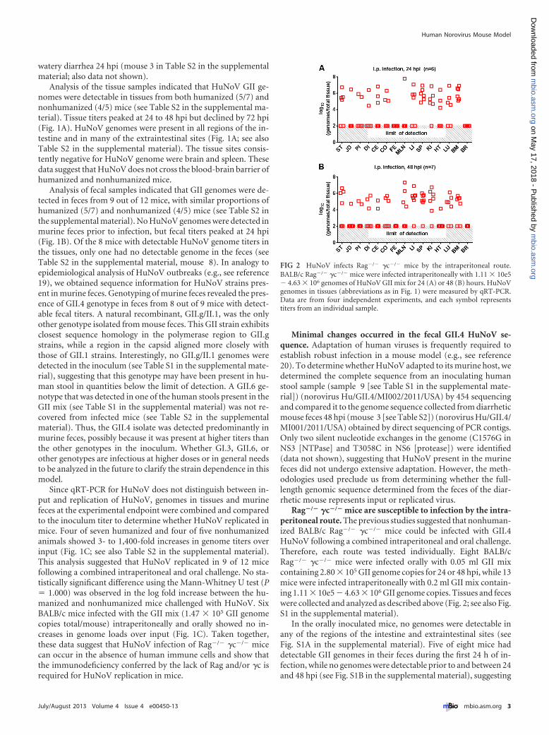
Rag�/� �c�/� mice are susceptible to infection by the intra-peritoneal route. The previous studies suggested that nonhuman-ized BALB/c Rag�/� �c�/� mice could be infected with GII.4HuNoV following a combined intraperitoneal and oral challenge.Therefore, each route was tested individually. Eight BALB/cRag�/� �c�/� mice were infected orally with 0.05 ml GII mixcontaining 2.80 � 105 GII genome copies for 24 or 48 hpi, while 13mice were infected intraperitoneally with 0.2 ml GII mix contain-ing 1.11 � 10e5 � 4.63 � 106 GII genome copies. Tissues and feceswere collected and analyzed as described above (Fig. 2; see also Fig.S1 in the supplemental material).
In the orally inoculated mice, no genomes were detectable inany of the regions of the intestine and extraintestinal sites (seeFig. S1A in the supplemental material). Five of eight mice haddetectable GII genomes in their feces during the first 24 h of in-fection, while no genomes were detectable prior to and between 24and 48 hpi (see Fig. S1B in the supplemental material), suggesting
FIG 2 HuNoV infects Rag�/� �c�/� mice by the intraperitoneal route.BALB/c Rag�/� �c�/� mice were infected intraperitoneally with 1.11 � 10e5� 4.63 � 106 genomes of HuNoV GII mix for 24 (A) or 48 (B) hours. HuNoVgenomes in tissues (abbreviations as in Fig. 1) were measured by qRT-PCR.Data are from four independent experiments, and each symbol representstiters from an individual sample.
Human Norovirus Mouse Model
July/August 2013 Volume 4 Issue 4 e00450-13 ® mbio.asm.org 3
m
bio.asm.org
on May 17, 2018 - P
ublished by m
bio.asm.org
Dow
nloaded from
that the detected genome was from input virus. No increases inviral loads over input were measured (data not shown). These dataindicate that nonhumanized BALB/c Rag�/� �c�/� mice were notinfected orally.
All 13 mice infected intraperitoneally had detectable genometiters in tissues at 24 or 48 hpi (Fig. 2). Viral genomes were de-tected in the intestine and extraintestinal sites, except in the brain.However, no virus was detected in the feces, suggesting thatBALB/c Rag�/� �c�/� mice do not shed virus following intraperi-toneal infection (Fig. 2). Overall, 12 of the 13 mice had 3- to60-fold increases in combined genome titers in the tissues com-pared to the inoculum titer (Fig. 3). As a control, two mice wereinfected intraperitoneally with 0.2 ml GII mix containing 1.32 �10e5 GII genome copies for 3 h, a time point prior to replication,and analyzed as before. No increases in genome titer over inputwere detected, suggesting that detection of increased genome ti-ters over input titers depends on the ability of HuNoV to replicate.These data suggest that the intraperitoneal route facilitates a sub-clinical GII.4 infection of BALB/c Rag�/� �c�/� mice.
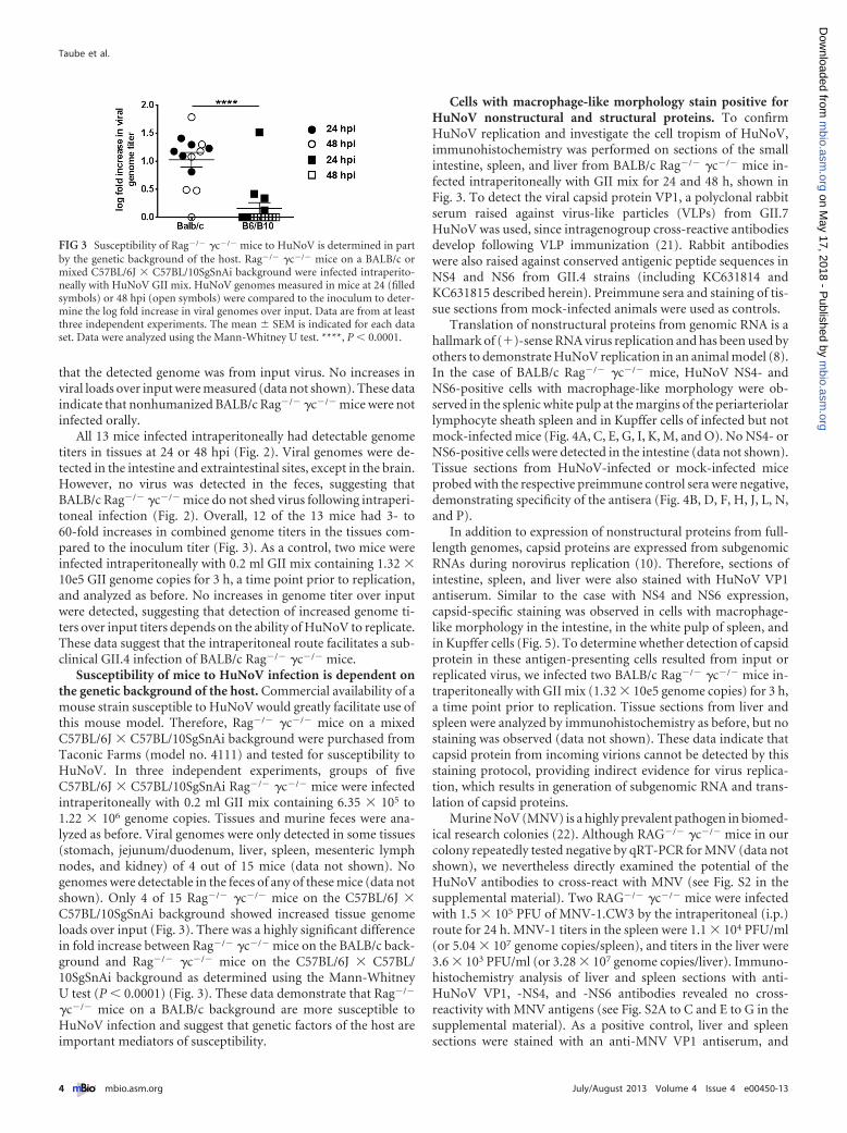
Susceptibility of mice to HuNoV infection is dependent onthe genetic background of the host. Commercial availability of amouse strain susceptible to HuNoV would greatly facilitate use ofthis mouse model. Therefore, Rag�/� �c�/� mice on a mixedC57BL/6J � C57BL/10SgSnAi background were purchased fromTaconic Farms (model no. 4111) and tested for susceptibility toHuNoV. In three independent experiments, groups of fiveC57BL/6J � C57BL/10SgSnAi Rag�/� �c�/� mice were infectedintraperitoneally with 0.2 ml GII mix containing 6.35 � 105 to1.22 � 106 genome copies. Tissues and murine feces were ana-lyzed as before. Viral genomes were only detected in some tissues(stomach, jejunum/duodenum, liver, spleen, mesenteric lymphnodes, and kidney) of 4 out of 15 mice (data not shown). Nogenomes were detectable in the feces of any of these mice (data notshown). Only 4 of 15 Rag�/� �c�/� mice on the C57BL/6J �C57BL/10SgSnAi background showed increased tissue genomeloads over input (Fig. 3). There was a highly significant differencein fold increase between Rag�/� �c�/� mice on the BALB/c back-ground and Rag�/� �c�/� mice on the C57BL/6J � C57BL/10SgSnAi background as determined using the Mann-WhitneyU test (P � 0.0001) (Fig. 3). These data demonstrate that Rag�/�
�c�/� mice on a BALB/c background are more susceptible toHuNoV infection and suggest that genetic factors of the host areimportant mediators of susceptibility.
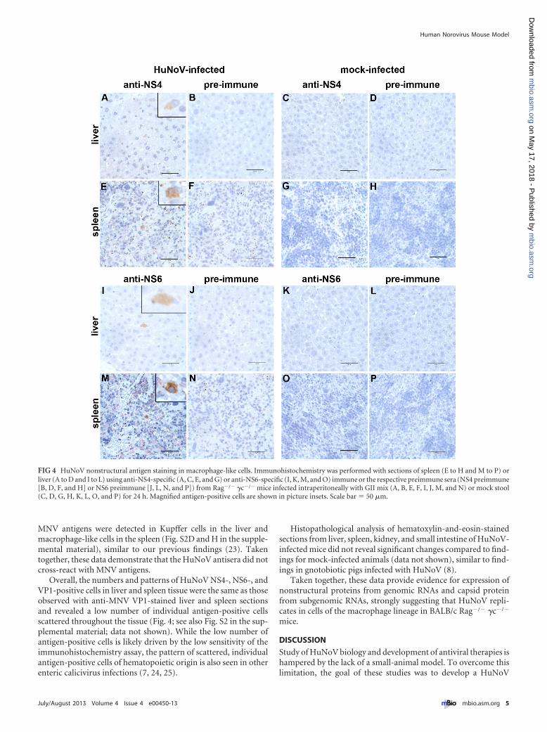
Cells with macrophage-like morphology stain positive forHuNoV nonstructural and structural proteins. To confirmHuNoV replication and investigate the cell tropism of HuNoV,immunohistochemistry was performed on sections of the smallintestine, spleen, and liver from BALB/c Rag�/� �c�/� mice in-fected intraperitoneally with GII mix for 24 and 48 h, shown inFig. 3. To detect the viral capsid protein VP1, a polyclonal rabbitserum raised against virus-like particles (VLPs) from GII.7HuNoV was used, since intragenogroup cross-reactive antibodiesdevelop following VLP immunization (21). Rabbit antibodieswere also raised against conserved antigenic peptide sequences inNS4 and NS6 from GII.4 strains (including KC631814 andKC631815 described herein). Preimmune sera and staining of tis-sue sections from mock-infected animals were used as controls.
Translation of nonstructural proteins from genomic RNA is ahallmark of (�)-sense RNA virus replication and has been used byothers to demonstrate HuNoV replication in an animal model (8).In the case of BALB/c Rag�/� �c�/� mice, HuNoV NS4- andNS6-positive cells with macrophage-like morphology were ob-served in the splenic white pulp at the margins of the periarteriolarlymphocyte sheath spleen and in Kupffer cells of infected but notmock-infected mice (Fig. 4A, C, E, G, I, K, M, and O). No NS4- orNS6-positive cells were detected in the intestine (data not shown).Tissue sections from HuNoV-infected or mock-infected miceprobed with the respective preimmune control sera were negative,demonstrating specificity of the antisera (Fig. 4B, D, F, H, J, L, N,and P).
In addition to expression of nonstructural proteins from full-length genomes, capsid proteins are expressed from subgenomicRNAs during norovirus replication (10). Therefore, sections ofintestine, spleen, and liver were also stained with HuNoV VP1antiserum. Similar to the case with NS4 and NS6 expression,capsid-specific staining was observed in cells with macrophage-like morphology in the intestine, in the white pulp of spleen, andin Kupffer cells (Fig. 5). To determine whether detection of capsidprotein in these antigen-presenting cells resulted from input orreplicated virus, we infected two BALB/c Rag�/� �c�/� mice in-traperitoneally with GII mix (1.32 � 10e5 genome copies) for 3 h,a time point prior to replication. Tissue sections from liver andspleen were analyzed by immunohistochemistry as before, but nostaining was observed (data not shown). These data indicate thatcapsid protein from incoming virions cannot be detected by thisstaining protocol, providing indirect evidence for virus replica-tion, which results in generation of subgenomic RNA and trans-lation of capsid proteins.
Murine NoV (MNV) is a highly prevalent pathogen in biomed-ical research colonies (22). Although RAG�/� �c�/� mice in ourcolony repeatedly tested negative by qRT-PCR for MNV (data notshown), we nevertheless directly examined the potential of theHuNoV antibodies to cross-react with MNV (see Fig. S2 in thesupplemental material). Two RAG�/� �c�/� mice were infectedwith 1.5 � 105 PFU of MNV-1.CW3 by the intraperitoneal (i.p.)route for 24 h. MNV-1 titers in the spleen were 1.1 � 104 PFU/ml(or 5.04 � 107 genome copies/spleen), and titers in the liver were3.6 � 103 PFU/ml (or 3.28 � 107 genome copies/liver). Immuno-histochemistry analysis of liver and spleen sections with anti-HuNoV VP1, -NS4, and -NS6 antibodies revealed no cross-reactivity with MNV antigens (see Fig. S2A to C and E to G in thesupplemental material). As a positive control, liver and spleensections were stained with an anti-MNV VP1 antiserum, and
FIG 3 Susceptibility of Rag�/� �c�/� mice to HuNoV is determined in partby the genetic background of the host. Rag�/� �c�/� mice on a BALB/c ormixed C57BL/6J � C57BL/10SgSnAi background were infected intraperito-neally with HuNoV GII mix. HuNoV genomes measured in mice at 24 (filledsymbols) or 48 hpi (open symbols) were compared to the inoculum to deter-mine the log fold increase in viral genomes over input. Data are from at leastthree independent experiments. The mean � SEM is indicated for each dataset. Data were analyzed using the Mann-Whitney U test. ****, P � 0.0001.
Taube et al.
4 ® mbio.asm.org July/August 2013 Volume 4 Issue 4 e00450-13
m
bio.asm.org
on May 17, 2018 - P
ublished by m
bio.asm.org
Dow
nloaded from
MNV antigens were detected in Kupffer cells in the liver andmacrophage-like cells in the spleen (Fig. S2D and H in the supple-mental material), similar to our previous findings (23). Takentogether, these data demonstrate that the HuNoV antisera did notcross-react with MNV antigens.
Overall, the numbers and patterns of HuNoV NS4-, NS6-, andVP1-positive cells in liver and spleen tissue were the same as thoseobserved with anti-MNV VP1-stained liver and spleen sectionsand revealed a low number of individual antigen-positive cellsscattered throughout the tissue (Fig. 4; see also Fig. S2 in the sup-plemental material; data not shown). While the low number ofantigen-positive cells is likely driven by the low sensitivity of theimmunohistochemistry assay, the pattern of scattered, individualantigen-positive cells of hematopoietic origin is also seen in otherenteric calicivirus infections (7, 24, 25).
Histopathological analysis of hematoxylin-and-eosin-stainedsections from liver, spleen, kidney, and small intestine of HuNoV-infected mice did not reveal significant changes compared to find-ings for mock-infected animals (data not shown), similar to find-ings in gnotobiotic pigs infected with HuNoV (8).
Taken together, these data provide evidence for expression ofnonstructural proteins from genomic RNAs and capsid proteinfrom subgenomic RNAs, strongly suggesting that HuNoV repli-cates in cells of the macrophage lineage in BALB/c Rag�/� �c�/�
mice.
DISCUSSION
Study of HuNoV biology and development of antiviral therapies ishampered by the lack of a small-animal model. To overcome thislimitation, the goal of these studies was to develop a HuNoV
FIG 4 HuNoV nonstructural antigen staining in macrophage-like cells. Immunohistochemistry was performed with sections of spleen (E to H and M to P) orliver (A to D and I to L) using anti-NS4-specific (A, C, E, and G) or anti-NS6-specific (I, K, M, and O) immune or the respective preimmune sera (NS4 preimmune[B, D, F, and H] or NS6 preimmune [J, L, N, and P]) from Rag�/� �c�/� mice infected intraperitoneally with GII mix (A, B, E, F, I, J, M, and N) or mock stool(C, D, G, H, K, L, O, and P) for 24 h. Magnified antigen-positive cells are shown in picture insets. Scale bar � 50 �m.
Human Norovirus Mouse Model
July/August 2013 Volume 4 Issue 4 e00450-13 ® mbio.asm.org 5
m
bio.asm.org
on May 17, 2018 - P
ublished by m
bio.asm.org
Dow
nloaded from

mouse model. Our data demonstrate that humanized and nonhu-manized Rag�/� �c�/� mice on a BALB/c background were sus-ceptible to a subclinical infection with HuNoV as indicated bydetection of nonstructural protein-positive cells and capsid-positive cells, as well as increases in genome titers over input.
First, expression of nonstructural proteins, which are ex-pressed only during infection (10), has been used in the case ofHuNoV infection of gnotobiotic pigs (8) as one line of evidence todemonstrate HuNoV replication. Similarly, our data show thattwo independent antibodies directed against two nonstructuralproteins detected antigen-positive cells in infected but not mock-infected mice (Fig. 4). Furthermore, the corresponding preim-mune sera did not react with any antigens, and no cross-reactivitywith the related MNV nonstructural proteins was observed.
Second, subgenomic RNAs are made during replication to ex-press the noroviral capsid protein (10). Thus, detection of capsidprotein at levels greater than those of incoming capsids is addi-tional evidence for HuNoV replication and was used in severalother HuNoV animal models (7, 8, 9). We detected capsid proteinexpression in livers and spleens of mice infected intraperitoneallywith HuNoV for 24 h but failed to detect capsid-positive cells inthe livers and spleens of mice infected for 3 h (Fig. 5 and data notshown). This demonstrated that capsid protein from input viruscannot be detected by immunohistochemistry and suggested thatthe capsid protein detected at 24 hpi was translated from newlysynthesized viral subgenomic RNA. The number and pattern ofHuNoV nonstructural protein-positive cells in liver and spleentissue was similar to that observed using an antibody against theHuNoV capsid protein (Fig. 4 and 5), suggesting that translationof nonstructural proteins, subgenomic replication, and transla-tion of the capsid protein occurred in a single cell.
Third, increases in overall genome titers over input were usedas another independent piece of evidence of replication. Up to
1,400-fold increases over input were detected in genome titers inmice infected by a combined oral/intraperitoneal administration,and up to 60-fold increases were detected in mice infected intra-peritoneally. The significant increases in genome titers over inputwere observed in two independent laboratories. Moreover, thelikelihood that merely input virus in tissue samples was detected islow because no increases over input virus were observed in mul-tiple experiments in which mice were inoculated with HuNoV,e.g., combined oral/intraperitoneal infection of wild-type BALB/cmice (Fig. 1C), oral infection of Rag�/� �c�/� mice (see Fig. S2in the supplemental material), and intraperitoneal infection ofRag�/� �c�/� mice for 3 h, a time point prior to replication (datanot shown).
Our data further demonstrated that Rag�/� �c�/� mice recon-stituted with CD34-positive human stem cells showed no statisti-cally significant difference from nonhumanized mice. Thus, sus-ceptibility was not linked to human cell reconstitution in thismouse model, and this suggested that HuNoV may replicate incells of murine origin. Immunohistochemistry of infected tissuesshowed HuNoV capsid-, NS4-, and NS6-positive Kupffer cellsand macrophage-like cells in the spleen (Fig. 4 and 5). This issimilar to murine norovirus (MNV), where antigen-positiveKupffer cells in the liver and splenic cells with macrophage-likemorphology were observed (23, 26) (see Fig. S2 in the supplemen-tal material). A tropism of HuNoV for macrophages, a cell typepresent in the intestinal lamina propria, is also consistent with theprevious finding that HuNoV GII.4 virus-like particles bind tolamina propria cells of human duodenum tissue sections (16).Thus, HuNoV and MNV may share a tropism for cells of thehematopoietic lineage. The identification of a cell type from themurine host, which supports HuNoV infection, may lead to thedevelopment of a HuNoV tissue culture system in the future.
We also demonstrated that Rag�/� �c�/� mice on a C57BL/6J
FIG 5 HuNoV capsid protein staining in macrophage-like cells. Immunohistochemistry was performed on sections of small intestine (A and D), spleen (B andE), and liver (C and F) using anti-HuNoV VP1-specific immune (A to C) and preimmune (D to F) sera from Rag�/� �c�/� mice infected intraperitoneally withGII mix for 24 (A, B, D, and E) or 48 (C and F) h. Magnified antigen-positive cells are shown in picture insets. Scale bar � 50 �m.
Taube et al.
6 ® mbio.asm.org July/August 2013 Volume 4 Issue 4 e00450-13
m
bio.asm.org
on May 17, 2018 - P
ublished by m
bio.asm.org
Dow
nloaded from

� C57BL/10SgSnAi background were less susceptible to HuNoVthan Rag�/� �c�/� mice on a BALB/c background. Therefore,susceptibility may depend on the genetic background and/or im-mune deficiencies. Differences in susceptibility due to geneticbackgrounds are also observed with other pathogens, and an in-creased susceptibility of BALB/c mice compared to that of C57BL6or C57BL10 mice due to differences in the host immune responsehas been described (e.g., see references 27 to 29). For example,production of type I interferons by peritoneal macrophages/monocytes was significantly increased in C57BL6 mice over that inBALB/c mice (27). HuNoV replication is sensitive to type I inter-ferons (30, 31). Thus, HuNoV injected intraperitoneally inC57BL/6J � C57BL/10SgSnAi mice potentially encounters higherlevels of type I interferons, which in turn could limit virus infec-tion. Further studies will be required to identify host factors de-termining HuNoV susceptibility in this mouse model.
Interestingly, our study suggests that Rag�/� �c�/� mice arenot susceptible to oral infection. This is in contrast to HuNoVtransmission in humans, which occurs via the fecal-oral route,and in gnotobiotic pigs, which are permissive to HuNoV follow-ing oral infection (31). This lack of oral infection in Rag�/� �c�/�
mice may be due to the genetic deficiency in the common cytokinereceptor gamma chain (�c) in these mice. Several enteric patho-gens use specialized microfold (M) cells present in the follicle-associated epithelium overlaying Peyer’s patches to gain access totheir host (for review, see reference 32). However, Peyer’s patcheswere absent from Rag�/� �c�/� mice following macroscopic andhistological observation (data not shown). This is not surprising,since signaling through the interleukin 7 (IL-7) receptor, which isa heterodimer comprised of the IL-7 alpha chain and commongamma chain, is critical for Peyer’s patch development (33, 34).We hypothesize that a lack of M cells may prevent oral HuNoVinfection in this mouse model, and studies are currently underway to test this hypothesis.
In addition, we did not observe HuNoV genome in the feces ofmice following intraperitoneal infection alone at any of the ana-lyzed time points. In contrast, viral genomes were detected in thefeces of mice infected both intraperitoneally and orally or orallyonly. HuNoV genome titers in murine feces were less than inputlevels following oral infection, suggesting that the viral genomesdetected represent input virus. Genome titers in murine fecesgreater than that of input virus were detected when mice wereinfected by combined intraperitoneal and oral inoculation. Futurestudies are needed to determine the relative amounts of replicatedvirus shed under these conditions. In addition, our finding raises abroader unanswered question in norovirus pathogenesis, which ishow noroviruses are shed from their host. Rag�/� �c�/� micemay provide an important tool to address this question in thefuture.
In summary, Rag�/� �c�/� mice on a BALB/c background aresusceptible to a subclinical infection by HuNoV, providing thefirst small-animal model for HuNoV infection. While this mousemodel does not recapitulate all aspects of HuNoV infection, suchas fecal-oral transmission, it will allow further mechanistic studiesof HuNoV biology, including host and viral factors determiningsusceptibility, which might enable improvement of the mousemodel. Furthermore, it is our hope that the availability of an easilymanipulable small-animal model for HuNoV infection will facil-itate not only basic but also translational HuNoV research, such asefficacy testing of compounds with anti-HuNoV activity, thereby
accelerating the development of urgently needed HuNoV thera-peutics.
MATERIALS AND METHODSDetailed methods can be found in Text S1 in the supplemental material.
HuNoV samples. Ten human stool samples from confirmed HuNoVoutbreaks (see Table S1 in the supplemental material) were processed asoutlined in Text S1.
Mice. All animal studies described herein were performed in accor-dance with local and federal guidelines. Please refer to Text S1 in thesupplemental material for details.
Infection of mice. Mice were housed individually in wire-bottomcages, and murine feces were collected over the indicated time frames.Mice were infected and harvested as outlined in Text S1.
Quantification/typing of HuNoV genomes by qRT-PCR. Quantita-tive RT-PCR of the HuNoV genome was performed with total RNA fromfecal and tissue samples as detailed in Text S1 in the supplemental mate-rial. The sensitivity and specificity of the assay are outlined in Text S1.Fecal samples were subjected to genotyping as detailed in Text S1.
Determination of fold increase. Inoculum titers were determined foreach experiment after back titration of the human stool suspensions byqRT-PCR. Total genome copies per mouse were calculated by addinggenome copies in all tissues and feces as outlined in Text S1 in the supple-mental material. The fold change was determined by dividing total ge-nome copies by inoculum genome copies.
Sequencing. Total nucleic acid was isolated from 0.2 ml of clarifiedhuman stool filtrate of patients 9 and 10 (see Table S1 in the supplementalmaterial), and 454 pyrosequencing was performed. A genome sequence ofHuNoV GII was obtained from mouse feces 48 h postinfection usingSanger sequencing of PCR amplicons (GenBank no. KC631815) (see TextS1 for additional details).
Generation of antibodies. Anti-VLP antibodies were made at Co-calico Biologicals, Inc., Reamstown, PA, in rabbits, following standardprotocols. Nonstructural antibodies against conserved antigenic NS4 andNS6 peptide sequences were generated and were affinity purified at Gen-Script USA Inc., Piscataway, NJ, in rabbits, following standard protocols.Preimmune sera were collected from each rabbit used to generate eachantibody (see Text S1 in the supplemental material for additional details).
Histopathology and immunohistochemistry. Mouse tissues werefixed and processed at the University of Michigan Pathology Core forAnimal Research, following standard histological procedures (see Text S1in the supplemental material for additional details).
Statistical analysis. The software program GraphPad Prism V5(GraphPad, La Jolla, CA) was used to perform statistical analyses. TheMann-Whitney U test was used to analyze differences. Results were con-sidered statistically significant when the P value was �0.05.
Nucleotide sequence accession numbers. The genomic sequences forthe HuNoV GII strain obtained from feces of the mouse with diarrhea(norovirus Hu/GII.4/MI001/2011/USA; accession number KC631814),the original inoculum (norovirus Hu/GII.4/MI002/2011/USA; accessionnumber KC631815), and the GII.7 HuNoV used for VLP production (ac-cession number KC832474) have been deposited in GenBank.
SUPPLEMENTAL MATERIALSupplemental material for this article may be found at http://mbio.asm.org/lookup/suppl/doi:10.1128/mBio.00450-13/-/DCSupplemental.
Text S1, DOCX file, 0.1 MB.Figure S1, TIF file, 1.1 MB.Figure S2, TIF file, 6.5 MB.Table S1, XLSX file, 0.1 MB.Table S2, XLSX file, 0.1 MB.
ACKNOWLEDGMENTS
This work was supported by NIH grants AI080611 to C.E.W. andAI073255 to R.A. S.A.H. and L.B.T. were supported by NIH grantsAI0544483, U54 AI057160, and AI084887.
Human Norovirus Mouse Model
July/August 2013 Volume 4 Issue 4 e00450-13 ® mbio.asm.org 7
m
bio.asm.org
on May 17, 2018 - P
ublished by m
bio.asm.org
Dow
nloaded from

We thank the University of Michigan Pathology Core for Animal Re-search for excellent technical assistance, I. Weissman (Stanford Univer-sity) for Rag�/� �c�/� mice, the Michigan Department of CommunityHealth for HuNoV-containing human stool samples, and M. J. Gonzalez-Hernandez (University of Michigan) for critical reading of the manu-script.
REFERENCES1. Widdowson MA, Monroe SS, Glass RI. 2005. Are noroviruses emerging?
Emerg. Infect. Dis. 11:735–737.2. Patel MM, Widdowson MA, Glass RI, Akazawa K, Vinjé J, Parashar
UD. 2008. Systematic literature review of role of noroviruses in sporadicgastroenteritis. Emerg. Infect. Dis. 14:1224 –1231.
3. Hall AJ, Curns AT, McDonald LC, Parashar UD, Lopman BA. 2012.The roles of Clostridium difficile and norovirus among gastroenteritis-associated deaths in the United States, 1999 –2007. Clin. Infect. Dis. 55:216 –223.
4. Scallan E, Hoekstra RM, Angulo FJ, Tauxe RV, Widdowson MA, RoySL, Jones JL, Griffin PM. 2011. Foodborne illness acquired in the UnitedStates—major pathogens. Emerg. Infect. Dis. 17:7–15.
5. Atmar RL, Estes MK. 2006. The epidemiologic and clinical importance ofnorovirus infection. Gastroenterol. Clin. North Am. 35:275–290.
6. Scharff RL. 2010. Health related costs from foodborne illness in theUnited States. Georgetown University, Washington, DC.
7. Bok K, Parra GI, Mitra T, Abente E, Shaver CK, Boon D, Engle R, YuC, Kapikian AZ, Sosnovtsev SV, Purcell RH, Green KY. 2011. Chim-panzees as an animal model for human norovirus infection and vaccinedevelopment. Proc. Natl. Acad. Sci. U. S. A. 108:325–330.
8. Cheetham S, Souza M, Meulia T, Grimes S, Han MG, Saif LJ. 2006.Pathogenesis of a genogroup II human norovirus in gnotobiotic pigs. J.Virol. 80:10372–10381.
9. Souza M, Azevedo MS, Jung K, Cheetham S, Saif LJ. 2008. Pathogenesisand immune responses in gnotobiotic calves after infection with the geno-group II.4-HS66 strain of human norovirus. J. Virol. 82:1777–1786.
10. Green KY. 2007. Caliciviridae, p 949 –980. In Knipe DM, Howley PM(ed), Fields Virology, vol 1, 5th ed. Lippincott Williams & Wilkins, Phila-delphia, PA.
11. Zheng DP, Ando T, Fankhauser RL, Beard RS, Glass RI, Monroe SS.2006. Norovirus classification and proposed strain nomenclature. Virol-ogy 346:312–323.
12. Bull RA, Eden JS, Rawlinson WD, White PA. 2010. Rapid evolution ofpandemic noroviruses of the GII.4 lineage. PLoS Pathog. 6:e1000831. http://dx.doi.org/doi:10.1371/journal.ppat.1000831.
13. Bull RA, White PA. 2011. Mechanisms of GII.4 norovirus evolution.Trends Microbiol. 19:233–240.
14. CDC. 2013. Notes from the field: emergence of new norovirus strain GII.4Sydney—United States, 2012. MMWR Morb. Mortal. Wkly. Rep. 62:55.
15. Vinjé J. 2010. A norovirus vaccine on the horizon? J. Infect. Dis. 202:1623–1625.
16. Chan MC, Ho WS, Sung JJ. 2011. In vitro whole-virus binding of anorovirus genogroup II genotype 4 strain to cells of the lamina propria andBrunner’s glands in the human duodenum. J. Virol. 85:8427– 8430.
17. Akkina R, Berges BK, Palmer BE, Remling L, Neff CP, Kuruvilla J,Connick E, Folkvord J, Gagliardi K, Kassu A, Akkina SR. 2011. Hu-manized Rag1-/-gammac-/- mice support multilineage hematopoiesisand are susceptible to HIV-1 infection via systemic and vaginal routes.PLoS One 6:e20169. doi: 10.1371/journal.pone.0020169.
18. Hoehne M, Schreier E. 2006. Detection of norovirus genogroup I and IIby multiplex real-time RT-PCR using a 3=-minor groove binder-DNAprobe. BMC Infect. Dis. 6:69. doi: 10.1186/1471-2334-6-69.
19. Matthews JE, Dickey BW, Miller RD, Felzer JR, Dawson BP, Lee AS,
Rocks JJ, Kiel J, Montes JS, Moe CL, Eisenberg JN, Leon JS. 2012. Theepidemiology of published norovirus outbreaks: a review of risk factorsassociated with attack rate and genogroup. Epidemiol. Infect. 140:1161–1172.
20. Zaini Z, Phuektes P, McMinn P. 2012. Mouse adaptation of a sub-genogroup B5 strain of human enterovirus 71 is associated with a novellysine to glutamic acid substitution at position 244 in protein VP1. VirusRes. 167:86 –96.
21. Parker TD, Kitamoto N, Tanaka T, Hutson AM, Estes MK. 2005.Identification of genogroup I and genogroup II broadly reactive epitopeson the norovirus capsid. J. Virol. 79:7402–7409.
22. Henderson KS. 2008. Murine norovirus, a recently discovered and highlyprevalent viral agent of mice. Lab. Anim. (NY) 37:314 –320.
23. Wobus CE, Karst SM, Thackray LB, Chang KO, Sosnovtsev SV, BelliotG, Krug A, Mackenzie JM, Green KY, Virgin HW. 2004. Replication ofnorovirus in cell culture reveals a tropism for dendritic cells and macro-phages. PLoS Biol. 2:e432. doi: 10.1371/journal.pbio.0020432.
24. Mumphrey SM, Changotra H, Moore TN, Heimann-Nichols ER, Wo-bus CE, Reilly MJ, Moghadamfalahi M, Shukla D, Karst SM. 2007.Murine norovirus 1 infection is associated with histopathological changesin immunocompetent hosts, but clinical disease is prevented by STAT1-dependent interferon responses. J. Virol. 81:3251–3263.
25. Sestak K, Feely S, Fey B, Dufour J, Hargitt E, Alvarez X, Pahar B,Gregoricus N, Vinjé J, Farkas T. 2012. Experimental inoculation ofjuvenile rhesus macaques with primate enteric caliciviruses. PLoS One7:e37973. doi: 10.1371/journal.pone.0037973.
26. Ward JM, Wobus CE, Thackray LB, Erexson CR, Faucette LJ, Belliot G,Barron EL, Sosnovtsev SV, Green KY. 2006. Pathology of immunodefi-cient mice with naturally occurring murine norovirus infection. Toxicol.Pathol. 34:708 –715.
27. Ellermann-Eriksen S, Liberto MC, Iannello D, Mogensen SC. 1986.X-linkage of the early in vitro alpha/beta interferon response of mouseperitoneal macrophages to herpes simplex virus type 2. J. Gen. Virol. 67:1025–1033.
28. Geist LJ, Hinde SL. 2001. Susceptibility to cytomegalovirus infection maybe dependent on the cytokine response to the virus. J. Investig. Med.49:434 – 441.
29. Güler ML, Gorham JD, Hsieh CS, Mackey AJ, Steen RG, Dietrich WF,Murphy KM. 1996. Genetic susceptibility to Leishmania: IL-12 respon-siveness in TH1 cell development. Science 271:984 –987.
30. Chang KO, George DW. 2007. Interferons and ribavirin effectively in-hibit Norwalk virus replication in replicon-bearing cells. J. Virol. 81:12111–12118.
31. Jung K, Wang Q, Kim Y, Scheuer K, Zhang Z, Shen Q, Chang KO, SaifLJ. 2012. The effects of simvastatin or interferon-alpha on infectivity ofhuman norovirus using a gnotobiotic pig model for the study of antivirals.PLoS One 7:e41619. doi: 10.1371/journal.pone.0041619.
32. Miller H, Zhang J, Kuolee R, Patel GB, Chen W. 2007. Intestinal M cells:the fallible sentinels? World J. Gastroenterol. 13:1477–1486.
33. Honda K, Nakano H, Yoshida H, Nishikawa S, Rennert P, Ikuta K,Tamechika M, Yamaguchi K, Fukumoto T, Chiba T, Nishikawa SI.2001. Molecular basis for hematopoietic/mesenchymal interaction duringinitiation of Peyer’s patch organogenesis. J. Exp. Med. 193:621– 630.
34. Luther SA, Ansel KM, Cyster JG. 2003. Overlapping roles of CXCL13,interleukin 7 receptor alpha, and CCR7 ligands in lymph node develop-ment. J. Exp. Med. 197:1191–1198.
35. Berges BK, Wheat WH, Palmer BE, Connick E, Akkina R. 2006. HIV-1infection and CD4 T cell depletion in the humanized Rag2-/-gamma c-/-(RAG-hu) mouse model. Retrovirology 3:76. doi: 10.1186/1742-4690-3-S1-S76.
Taube et al.
8 ® mbio.asm.org July/August 2013 Volume 4 Issue 4 e00450-13
m
bio.asm.org
on May 17, 2018 - P
ublished by m
bio.asm.org
Dow
nloaded from