Embed Size (px)
Citation preview

Citation: Palangasinghe DR, Kodithuwakku G, Dahanayaka N, Dissanayake A and Yahathugoda C. A Mixed Malaria and Mycoplasma Co-Infection in a Sri Lankan Patient: A Case Report. J Bacteriol Mycol. 2018; 5(1): 1062.
J Bacteriol Mycol - Volume 5 Issue 1 - 2018ISSN : 2471-0172 | www.austinpublishinggroup.com Palangasinghe. © All rights are reserved
Journal of Bacteriology and MycologyOpen Access
BackgroundMalaria was once a common infection in Sri Lanka where the
country had to suffer several out brakes that had claimed many lives in the 19th century [1,2]. In last decade malaria was in a steady decline and last indigenous case was reported in 2012 [1]. World health organization has declared the country as malaria eradicated since 2016 [1]. But still several imported cases of malaria were reported in Sri Lanka from travellers returning from countries like India, Pakistan, Haiti and Africa. Out of them several patients developed severe malaria due to delayed diagnosis [1]. Early identification of imported cases of malaria is of paramount importance to avoid re introduction of the infection to Sri Lankan community. Severe malaria has a high morbidity and mortality which could be minimized by early identification and treatment.
Presence of other infections with malaria may alter the clinical presentation and poses a challenge to diagnosis as well as treatment [7,8,9]. Mycoplasma is a common respiratory pathogen that can
Case Report
A Mixed Malaria and Mycoplasma Co-Infection in a Sri Lankan Patient: A Case ReportPalangasinghe DR*1, Kodithuwakku G2, Dahanayaka N3, Dissanayake A3 and Yahathugoda C4
1Senior Registrar in Medicine, University Medical Unit, Teaching Hospital Karapitiya, Sri Lanka2Registrar in medicine, University Medical Unit, Teaching Hospital Karapitiya, Galle, Sri Lanka3Department of Medicine, Senior Lecturer in Medicine, Faculty of Medicine, University of Ruhuna, Sri Lanka4Department of Parasitology, Professor in Parasitology, University of Ruhuna, Sri Lanka
*Corresponding author: Palangasinghe DR, Senior Registrar in Medicine, University Medical Unit, Teaching Hospital Karapitiya, Sri Lanka
Received: February 06, 2018; Accepted: February 21, 2018; Published: February 28, 2018
cause a wide spectrum of disease manifestations; the most common being upper respiratory tract infections [12]. Mycoplasma is well known to contaminate cell cultures of plasmodium species, posing a great challenge to researchers and microbiologists [3,4]. Co-existence and clinical implications of malaria and mycoplasma infections are yet to be evaluated. We encountered one previous reported case of falciparum malaria and mycoplasma co-infection in the literature [5].
We report the first case a case of malaria infection caused by 3 Plasmodium species (P. falciparum, P. ovale and P. vivax) with a Mycoplasma pneumoniae co-infection. Even though there were case reports of imported malaria cases to Sri Lanka since 2012, this is the 1st triple infection identified.
Case PresentationA 45 year old gem businessman from Galle district (Southern
Province, Sri Lanka) presented on 01.09.2017 with fever and dry cough for 16 days. The fever was associated with chills and rigors.
Abstract
Background: Although last indigenous case of malaria in 2012 and achievement of malaria free status since 2016; there were few imported cases throughout last five years. Therefore it is important to suspect malaria in a patient with a febrile illness with recent history of travel to malarial areas. Delay in diagnosing can end up in severe malaria which has a high morbidity and mortality. Complex inter-relationship between mycoplasma and plasmodium species were reported in vitro. We report the first case of malaria mixed species infection with mycoplasma pneumoniae co-infection.
Case Presentation: A 45 year old gem businessman from Southern Sri Lanka came with 16 days of high fever with chills and rigors, dry cough and dyspnea after returning recently from India. Patient did not receive malaria chemoprophylaxis before or during a one month stay in India. On examination he was ill and had hepatomegaly. With dry cough and dyspnea he had alveolar nodular shadows in the right middle zone of chest radiograph with a significantly high mycoplasma pneumonia antibody titre for which he was commenced on oral clarithromycin treatment. With persistent fever with chills and rigors while on clarithromycin treatment for 2 days, carefully examined blood films revealed Plasmodium falciparum, vivax and ovale parasites. Antigen tests and Polymerase Chain Reaction (PCR) tests confirmed the mixed Plasmodium species infection. Hewas continued on oral Clarythromycin, together with Artemether-Lumefantrine combination therapy followed by primaquine. He became fever free from day 2 of treatment with artemether and lumafantrine combination while plasmodium parasitaemia and antigenemia disappeared from blood after 2 days of antimalarial treatment. He was apparently well at 2 weeks review without fever, cough or dyspnea and repeat chest radiograph showed resolution of alveolar nodular shadows on right middle zone.
Conclusions: Even in a malaria eradicated country it is important suspect malaria in a traveller returning from an endemic area for prompt diagnosis and treatment. Mycoplasma pneumonia could be a co infection which might alter the clinical presentation in a patient with malaria.
Keywords: Malaria Mixed Infection; Mycoplasma; Co Infection; Sri Lanka
J Bacteriol Mycol 5(1): id1062 (2018) - Page - 02
Palangasinghe DR Austin Publishing Group
Submit your Manuscript | www.austinpublishinggroup.com
He had type 2 diabetes mellitus with a satisfactory glycaemic control while on Gliclazide 80mg twice daily. He developed fever, 2 days after returning from India where he went on a business tour for one month. He stayed in Kolkata during that month (from 14.07.2017 to 15.08.2017) and he came back to Sri Lanka through Madras where he stayed one night. During the stay in India he had numerous mosquito bites and he had not taken chemoprophylaxis for malaria. The last time he visited India was 10 years back where he did not have any febrile illnesses. During the current febrile illness, he was seen by several doctors on 3 outpatient visits where he was treated with oral antibiotics (with amoxicillin on one occasion and with amoxicillin and clavulinic acid combination on another occasion), antipyeretics and bronchodilators without improvement of his symptoms. From the 10th day of illness he was developed shortness of breath but he did not have orthropnea or paroxysmal nocturnal dyspnea.
He had fever daily during this period with fever spikes appearing around 7pm associating with chills and rigors. The temperature was recorded in the range of 103-104 F. On examination he was pale and was not icteric. He was looking ill during height of fever with tachypnea and tachycardia. The throat was inflamed with no pustules and there were no palpably enlarged lymph nodes or rashes. He was alert with no signs of meningeal irritation. There was hepatomegaly, with a non tender diffusely enlarged liver which was palpable 6cm below the right costal margin and there was no surface nodularity or bruits over it. There was no splenomegaly. No free fluid or ballotable masses were noted. His cardiovascular, respiratory and nervous system examinations were unremarkable.
Investigations on admission (day 17 of illness) revealed Hemoglobin of 10g/dL with a hematocrit of 30, Mean Corpuscular Volume (MCV) was 72fL, White Cell Count (WBC) of 11110/uL with neutrophils and lymphocytes being59% and 28% respectively. He was thrombocytopenic with a platelet count of 67000/uL. He had a high C-reactive protein (CRP) value of 75mg/dL and Erythrocyte Sedimentation Rate (ESR) was 60mm in 1st hour. Urine analysis was normal and there was no bacterial growth in urine culture. Liver functions revealed an Aspartate Transaminase (AST) 28IU/L, Alanine Transaminase (ALT) 48IU/L, Alkaline Phosphatase (ALP) 337IU/L and Gammaglutamyltransferase (GGT) 314IU/L. His total Bilirubin level was17umol/L and direct bilirubin level was 10umol/L. He had a serum total protein concentration of 53g/dL and serum albumin level of 35g/dL. His renal functions were normal throughout with a Serum creatinine of 75umol/L and blood urea of 25mg/dL on admission. Ultrasound scan (USS) of the abdomen revealed hepatomegaly, but no splenomegaly or bile duct dilatation. Chest radiograph revealed right middle zone alveolar nodular shadows. He was commenced on oral clarithromycin 500mg twice daily on day 1 of admission (day 17 of illness) after taking blood for culture and mycoplasma antibodies which became positive at a titre of 1: 320. A blood film was analyzed on 2nd day of admission was reported as neutrophil leukocytosis with features of coexisting iron deficiency anemia (hypochromic microcytic cells and pencil shaped cells). Although cough has improved he continued to have high fever with chills and rigors despite 3 days of treatment with clarithromycin with persistent shortness of breath. Arterial blood gas analysis performed on day 3 of admission showed following. PH 7.44, PaO2 69mmHg, PCO2 24mmHg and HCO3- 17mmol/L, Lactate level was 2.4mmol/L.
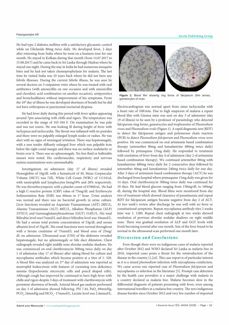
Electrocardiogram was normal apart from sinus tachycardia with a heart rate of 108/min. Due to high suspicion of malaria a repeat blood film with Giemsa stain was sent on day 3 of admission (day 19 of illness) to be seen by a professor of parasitology who detected falciparum ring forms, gametocytes and trophozoites of Plasmodium vivax and Plasmodium ovale (Figure 1). A rapid diagnostic test (RDT) to detect the falciparum antigen and polymerase chain reaction (PCR) to detect Plasmodium falciparum and Plasmodium vivax were positive. He was commenced on oral artimisnin based combination therapy (artemether 80mg and lumafantrine 480mg twice daily) followed by primaquine 15mg daily. He responded to treatment with resolution of fever from day 4 of admission (day 2 of artimisnin based combination therapy). We continued artemether 80mg with lumafantrine 480mg twice daily for 2 consecutive days followed by artemether 40mg and lumafantrine 240mg twice daily for one day. After 3 days of artimisnin based combination therapy (ACT) he was discharged from hospital where primaquine 15mg daily was given for 14 days. Oral clarithromycin 500mg twice daily was continued for 10 days. He had blood glucose ranging from 130mg/dL to 160mg/dL during the hospital stay. Blood films were monitored from day two of treatment which showed clearance of parasitaemia and repeat RDT for falciparum antigen became negative from day 2 of ACT. At two week’s review after discharge he was well with no fever or constitutional symptoms. Repeat mycoplasma antibody titre 2 weeks later was 1: 1280. Repeat chest radiograph at two weeks showed resolution of previous alveolar nodular shadows on right middle zone. There was gradual resolution of ALP and GGT levels with levels becoming normal after one month. Size of the liver found to be normal in the ultrasound scan performed one month later.
Discussion and ConclusionsEven though there were no indigenous cases of malaria reported
after October 2012 and WHO declared Sri Lanka as malaria free in 2016, imported cases poses a threat for the reintroduction of the disease in the country [1,2,6]. This case report is of particular interest as it is a mixed plasmodium infection with mycoplasma coinfection. We came across one reported case of Plasmodium falciparum and mycoplasma co infection in the literature [5]. Prompt case detection by the health care providers is a major challenge with malaria in a country declared as malaria free. Malaria becomes slow in the differential diagnosis of patients presenting with fever, even among international travellers in a malaria free country. The zero indigenous disease burden since October 2012 and very few number of imported
Figure 1: Blood film showing ring forms of falciparum (thin arrow), gametocytes of vivax.

J Bacteriol Mycol 5(1): id1062 (2018) - Page - 03
Palangasinghe DR Austin Publishing Group
Submit your Manuscript | www.austinpublishinggroup.com
cases reported might contribute to lack of clinical as well as laboratory expertise in the diagnosis and treatment of malaria. This is further complicated by the epidemic of dengue in the country which is the priority in a differential diagnosis when a person has fever and thrombocytopenia. There had been occasions where the diagnosis of malaria was delayed sometimes exceeding 30 days since the onset of fever [6]. As a result there is a risk of delay in treatment which could not only increase the disease transmission potential but also increases the risk of person developing the severe disease.
Our patient had a febrile illness with a dry cough, shortness of breath, inflamed pharynx, alveolar nodular shadows in the right middle zone of the chest radiograph and a significantly positive mycoplasma antibody titer compatible with a diagnosis of mycoplasma infection. Certain features in our patient made us to think beyond the mycoplasma infection; which included fever with chills and rigors; which continued despite treatment with clarithromycin and a recent travel to a malaria endemic area. A blood film seen on day 18 of the illness in the routine laboratory could not detect the malarial parasites despite the clinical suspicion. It could have been due to lack of laboratory expertise with very low number of malaria cases during the last decade. Since we had a high index of suspicion about malaria in our patient a second blood film was sent to an expert (a professor of parasitology) together with the antigen test (RDT for malaria) followed by initiation of ACT as for falciparum malaria. Subsequently blood film, RDT and PCR testing was compatible with a mixed Plasmodium species infection. Our patient was initiated on ACT on day 3 of hospital admission (day 19 of the illness) which resulted in resolution of fever from the next day. The presence of thrombocytopenia and type 1 respiratory failure in our patient could have been due to either malaria or mycoplasma. With malaria both thrombocytopenia and hypoxia are features of severe infection [15]. Liver is an important organ in the life cycle of plasmodium parasites. In all Plasmodium species sporozoites develop into merozoites in the hepatocytes whereas in Plasmodium vivax and Plasmodium ovale there is a dormant phase (hypnozoite stage) in hepatocytes which allows recrudescences to occur [13]. Fatty changes, portal tract inflammation and kupffer cells hyperplasia had been recognized as different patterns of liver injury in severe malaria. The extent of hepatocellular dysfunction varies from common mild abnormalities in liver function tests to rare cases of hepatic failure [13,14]. Hyperbillirubinaemia seen in malaria is mostly an unconjugated hyperbillirubinaemia due to haemolysis of red cells than liver dysfunction. Commonest liver enzyme abnormality noted in malaria was rise in transaminases to three times the upper limit of normal. There are several reports of liver involvement with mycoplasma pneumoniae infection including acute hepatitis and cholestatic hepatitis [10,11]. The pattern of liver involvement in our patient with hepatomegaly, elevated ALP and GGT with normal transaminases, prothrombin time, billirubin had been unusual for both mycoplasma and malaria infections. We presume that had been a manifestation of both or either malaria or mycoplasma infection due to resolution of hepatomegaly with normalization of ALP and GGT one month later. Non immune status for malaria with a recent visit to malarial area is another factor that made our patient a susceptible to develop severe malaria. Although our patient stayed in Kalkata which is in the boarder of a hyperendemic area for malaria in India he was
neither aware nor consume any chemoprophylaxis for malaria. There were no indigenous cases of malaria reported in Sri Lanka after 2012 together with recent travel made malaria infection very likely to be an imported infection in our patient. Co infections associated with malaria may contribute to increase the morbidity and mortality [7,8]. The presence of co infections might alter the clinical presentation making the diagnosis more challenging in malaria. The most frequent pathogens isolated from patients with severe falciparum malaria infection were non typhoid Salmonella species and other gram negative bacteria [7,8,9]. The extent of causal relationship between Plasmodium species and these bacteria are currently unknown. But concurrent bacteraemia has resulted in high morbidity and mortality among patients with malaria [8,9]. Mycoplasma is known to coexist with malaria in vitro with yet undefined clinical implications. Accelerated growth of contaminant Mycoplasma species in cultures of P. falciparum had resulted in subsequent reduction in Plasmodium parasitaemia [3,4]. This is the first report of mixed malaria species and Mycoplasmapneumoniae co-infection in a human host. We treated our patient for both malaria mixed species infection and mycoplasma.
With increasing global travel it is a challenge to maintain the malaria free status in countries like Sri Lanka. Delay in diagnosis could result from lack of clinical suspicion as well as lack of laboratory expertise. Resultant delay in treatment in a patient with malaria could not only lead to severe malaria but also increases the risk of transmission of the disease. Among 144 imported cases of malaria in 2013 and 2014, fifteen persons develop severe disease as a result of delay in diagnosis [6]. Therefore sustaining the malaria free status which is a remarkable public health achievement and zero mortality due to disease both will become challenges to Sri Lanka. This emphasizes the importance of suspecting and actively looking for malaria in any patient with fever returning from an endemic area.
In our patient fever had been there for 19 days (3rd day of hospital admission) before the identification of illness. Blood film sent on the 2nd day of admission suspecting malaria was reported as having features of a bacterial infection with evidence of iron deficiency anemia (neutrophil leukocytosis with hypochromic microcytic red cells and pencil shaped cells. Diagnosis of mixed malaria speicies infection was made since we got an experienced person (a professor of parasitology) to review the blood films on the third day of admission which helped us to treat the patient successfully. This shows the importance of getting an experienced person to see the blood film for malarial parasites when the clinical suspicion is high; since it could be missed otherwise especially with the rare occurrence of infection in a country where the infection is already eradicated.
Early detection and initiation of treatment had been the success in our patient which resulted in recovery as well as the reduction of transmission. The change in clinical presentation, forgetfulness of malaria as a cause of febrile illness and deterioration of the competence of microscopists are all likely to contribute to a delay in the diagnosis especially in a country where malaria is eradicated. Even in a malaria free country the high degree of suspicion on possible imported cases is of importance to diagnose and treat cases promptly to maintain the malaria free status.
References1. Nadira DK, Gawrie NL Galappaththy and Dyann FW. On the road to eliminate
J Bacteriol Mycol 5(1): id1062 (2018) - Page - 04
Palangasinghe DR Austin Publishing Group
Submit your Manuscript | www.austinpublishinggroup.com
malaria in Sri Lanka: Lessons from history, challenges, gaps in knowledge and research needs. Malaria Journal. 2014; 13: 59
2. Premaratna R, Galappaththy G, Chandrasena N, Fernando R, Nawasiwatte T, Nilanthi R de Silva, et al. What clinicians who practice in countries reaching malaria elimination should be aware of lessons learnt from recent experience in Sri Lanka. Malaria Journal. 2011; 10: 302.
3. Agarwal P, Srivastava K, Rajeev, Puri SK, Srivastava K. Management of mycoplasma contamination in vitro culture of Plasmodium falciparum without antibiotic treatment- A preliminary report. Research in microbiology. 2012; 164: 211-215.
4. Singh S, Puri SK, Srivastava K. Treatment and control of Mycoplasma contamination in Plasmodium falciparum culture. Parasitol Res. 2008; 104: 181-184.
5. Praveen W, Gowri R, Ahalya S, Panduka K, Ariaranee G and Thashi C. Plasmodium falciparum and Mycoplasma pneumoniae co-infection presenting with cerebral malaria manifesting orofacial dyskinesia and haemophagocytic lymphohistiocytosis. 2016.
6. Priyani D, Risintha GP, WM Kumudunayana T de AW Gunasekera, Mihirini H, Kamini M and Deepika F. Characterization of imported malaria, the largest threat to sustained malaria elimination from Sri Lanka. Malaria Journal. 2015; 14: 177.
7. Berkley J, Mwarumba S, Bramham K, Lowe B, Marsh K. Bacteremia complicating severe malaria in children. Trans. R. Soc. Trop. Med. Hyg. 1999; 93: 283-286.
8. Were T, Davenport GC, Hittner JB, Ouma C, Vulule JM, Ong’echa JM, Perkins DJ. Bacteremia in Kenyan children presenting with malaria. J. Clin. Microbiol. 2011; 49: 671-676.
9. Johanna S, Pontus N, Saduddin D, Akhar S, Sara E, Marika H, Lillemor K, et al. Bacterial Co infections in Travelers with Malaria. Rationale for Antibiotic Therapy. 2013; 51: 15-21.
10. Grullich C, Baumert TF, Blum HE. Acute Mycoplasma pneumoniae infection presenting as cholestatic hepatitis. J Clin Microbiol. 2003; 41: 514-515.
11. Romero-Gomez M, Otero MA, Sanchez Munoz D, Ramirez-Arcos M, Larraona JL, Suarez Garcia E, et al. Acute hepatitis due to Mycoplasma pneumoniae infection without lung involvement in adult patients. J Hepatol. 2006; 44: 827-828.
12. Surender K and Malay S. Mycoplasma pneumonia: Clinical features and management. Lung India. 2010; 27: 75-85.
13. Hollingdale MR. Malaria and the liver. Hepatology. 1985; 5: 327-335.
14. Parnpen V, Vasant K and Chuchard P. Liver changes in severe Plasmodium falciparum malaria: Histopathology, apoptosis and nuclear factor kappa B expression. Malaria Journal. 2014; 13: 106.
15. Severe malaria: Tropical Medicine and International Health 19, Supplement 1 (November 2014). WHO. 2014.
Citation: Palangasinghe DR, Kodithuwakku G, Dahanayaka N, Dissanayake A and Yahathugoda C. A Mixed Malaria and Mycoplasma Co-Infection in a Sri Lankan Patient: A Case Report. J Bacteriol Mycol. 2018; 5(1): 1062.
J Bacteriol Mycol - Volume 5 Issue 1 - 2018ISSN : 2471-0172 | www.austinpublishinggroup.com Palangasinghe. © All rights are reserved


![THE WITHIN-HOST DYNAMICS OF MALARIA INFECTION WITH …ruan/MyPapers/... · THE WITHIN-HOST DYNAMICS OF MALARIA INFECTION 1001 approximately the same times (Rouzine and Mckenzie [34])](https://img.dokumen.tips/doc/110x75/6039d2a5dafed858e1329708/the-within-host-dynamics-of-malaria-infection-with-ruanmypapers-the-within-host.jpg)