Embed Size (px)
Citation preview

LETTERSPUBLISHED ONLINE: 7 OCTOBER 2012 | DOI: 10.1038/NMAT3430
A magnetic switch for the control of cell deathsignalling in in vitro and in vivo systemsMi Hyeon Cho1†, Eun Jung Lee1,2†, Mina Son2†, Jae-Hyun Lee1, Dongwon Yoo1, Ji-wook Kim1,Seung Woo Park3, Jeon-Soo Shin2,4* and Jinwoo Cheon1,2*The regulation of cellular activities in a controlled manner isone of the most challenging issues in fields ranging from cellbiology to biomedicine1,2. Nanoparticles have the potential ofbecoming useful tools for controlling cell signalling pathwaysin a space and time selective fashion3,4. Here, we havedeveloped magnetic nanoparticles that turn on apoptosis cellsignalling by using a magnetic field in a remote and non-invasive manner. The magnetic switch consists of zinc-dopediron oxide magnetic nanoparticles5 (Zn0.4Fe2.6O4), conjugatedwith a targeting antibody for death receptor 4 (DR4) of DLD-1colon cancer cells. The magnetic switch, in its On mode when amagnetic field is applied to aggregate magnetic nanoparticle-bound DR4s, promotes apoptosis signalling pathways. Wehave also demonstrated that the magnetic switch is operableat the micrometre scale and that it can be applied in an in vivosystem where apoptotic morphological changes of zebrafishare successfully induced.
Cell signalling is an important process in biological systemsfor exchanging information through networks of various signalmolecules to control cellular activities, such as differentiation,growth, metabolism and death6. Owing to their newly developedhigh precision and accuracy, physical stimuli using optical,electrical and magnetic methods have been devised to regulatecell signalling1–4. Among these, magnetic techniques are uniquelyadvantageous because magnetic fields can penetrate deeply withnegligible attenuation into biological tissues7,8. Consequently,it has distinctive benefits for in vivo applications. Moreover,when coupled with magnetic nanoparticles, magnetic fields canbe transformed into other forms of energy, such as heat andmechanical force9–16. The magnetic heat induction has been usedfor gating of the thermosensitive ion channel9 as well as forhyperthermia therapy10. Although relatively large mechanical force(in the piconewton range) has been used in in vitro and invivo systems for direct stretching of ion channels and cytoskeletalstimulation11–13, two recent in vitro studies have revealed thatthe induction of calcium influx15 and tubulogenesis16 usingreceptor clustering is also possible by using nanoparticles. Magneticnanoparticles can exert a gentle force (in the femtonewton range) onmembrane receptors to induce their clustering without disturbingthe rheological and cytoskeletal properties. Furthermore, thenanoscale dimensions of nanoparticles conjugated with targetingmolecules make them beneficial for probing cellular sensorystructures and functions at the molecular level and for inducingspecific cellular activation processes7,8. Nonetheless, the nanoscalemagnetic switching technique for receptor clustering is still at too
1Department of Chemistry, Yonsei University, Seoul 120-749, Korea, 2Graduate Program for Nanomedical Science, Yonsei University, Seoul 120-749, Korea,3Department of Internal Medicine, Institute of Gastroenterology, College of Medicine, Yonsei University, Seoul 120-752, Korea, 4Department ofMicrobiology, Severance Biomedical Science Institute, Institute for Immunology and Immunological Diseases, College of Medicine, Yonsei University, Seoul120-752, Korea. †These authors contributed equally to this work. *e-mail: [email protected]; [email protected].
early a stage of development to guarantee that it will be generallyapplicable to the control of cell signalling in other biologicallyimportant systems. In addition, it is not known whether it will beeffective in in vivo systems.
Apoptosis, programmed cell death, is known to be amajor factorin maintaining homeostasis and removing undesired cells17–19.Recently, an extrinsic apoptosis signalling pathway that is initiatedby death receptors has emerged as one of themain targets for cancertherapy20–22. Extrinsic apoptosis signalling is usually activated byclustering of death receptors through docking of biochemicalligands, such as the TNF-related apoptosis inducing ligand (TRAIL)that is a potent inducer of apoptosis23,24. However, direct use of suchligands in clinical applications is limited by the short plasmahalf-lifeand the ease of degradation25,26.
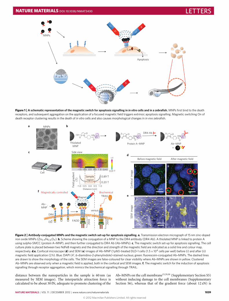
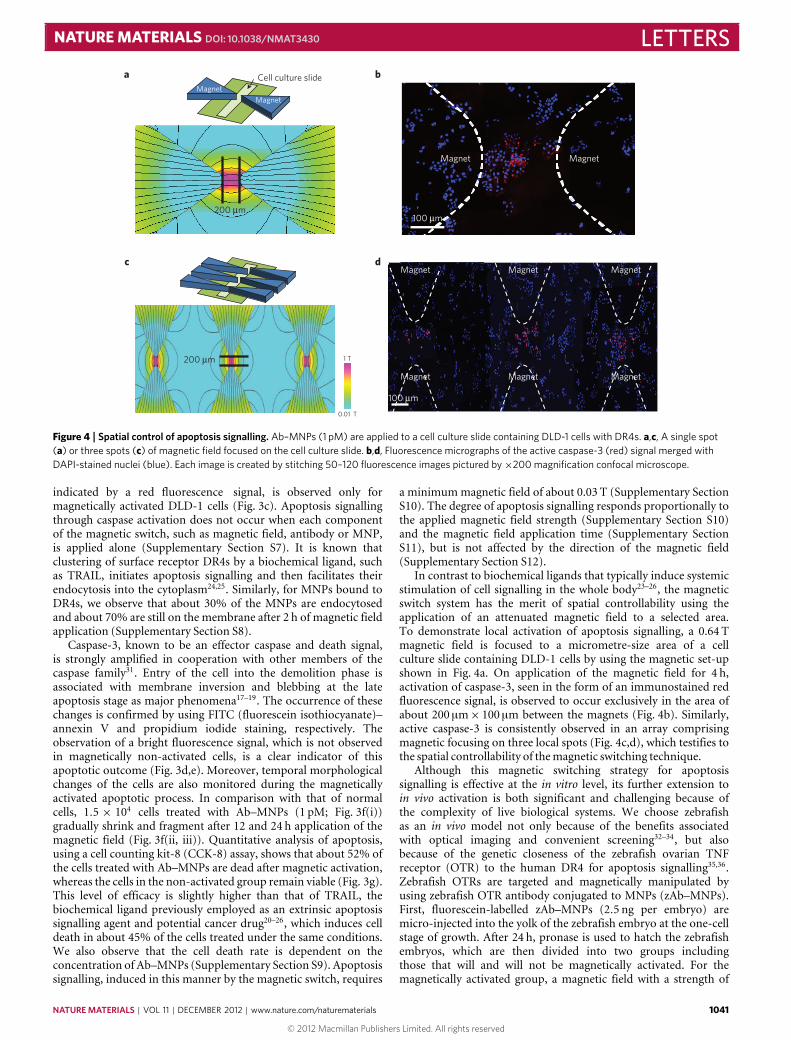
In the study described below, we have developed a magneticswitch for apoptosis signalling, and demonstrate its in vivo feasi-bility through the receptor clustering process (Fig. 1). The mag-netic switch for cell signalling consists of DR4 monoclonal anti-body conjugated to magnetic nanoparticles (Ab–MNPs). DR4s arehighly expressed on tumour cells20–27 and magnetic nanoparticlesare designed to bind DR4s through a specific monoclonal anti-body interaction. Zinc-doped iron oxide magnetic nanoparticles(Zn0.4Fe2.6O4 MNPs, 15 nm) are chosen (Fig. 2a) owing to their highsaturation magnetization value (161 e.m.u. g−1), which is essentialfor effective utilization of magnetic force5 (Supplementary SectionS1). For preparation of the Ab–MNPs, protein A is conjugated tothiolated MNPs through a sulpho-SMCC (sulphosuccinimidyl-4-[N -maleimidomethyl]cyclohexane-1-carboxylate) crosslinker. TheDR4 antibody is then conjugated to protein A on MNPs in a DR4antibody/MNP stoichiometric ratio of 1:1 (Fig. 2b and Supple-mentary Section S1).
For in vitro magnetic switching On of apoptosis, Ab–MNPs (1 pM) are applied to DLD-1 colon cancer cells(1.5×10⁴ cells per well), expressing DR4s (Supplementary SectionS2), and the cells are placed in between two NdFeB mag-nets (Fig. 2c). In the absence of magnetic field, Ab–MNPs areobserved as an evenly dispersed weak green fluorescence signal(Fig. 2d(i)) and remain dispersed over time (Supplementary SectionS3). However, large aggregated spots exhibiting a strong greenfluorescence signal are observed after the application of a magneticfield for 2 h (Fig. 2d(ii) red circles and Supplementary SectionS4). Scanning electronmicroscope (SEM) images consistently showthat the initial evenly distributed Ab–MNPs change to denselypopulated aggregates (Fig. 2e and Supplementary Section S4). Here,the simulatedmagnetic field is 0.20 T at the centre28 and the average
1038 NATURE MATERIALS | VOL 11 | DECEMBER 2012 | www.nature.com/naturematerials
© 2012 Macmillan Publishers Limited. All rights reserved
NATURE MATERIALS DOI: 10.1038/NMAT3430 LETTERS
MNPs
Apoptosis
S
S
N
N
S
N S
N
Figure 1 | A schematic representation of the magnetic switch for apoptosis signalling in in vitro cells and in a zebrafish. MNPs first bind to the deathreceptors, and subsequent aggregation on the application of a focused magnetic field triggers extrinsic apoptosis signalling. Magnetic switching On ofdeath receptor clustering results in the death of in vitro cells and also causes morphological changes in in vivo zebrafish.
Side view
Magnet Magnet
0.0 1 .0¬1.0 ¬0.5 0.5
0.0Distance (cm)
1 .0¬1.0 ¬0.5 0.5
(cm)
1 T
0.01 T
0.20 T
0.89 TSample region
Fiel
d gr
adie
nt (
T)
Ab¬ MNP
DR4
DD FADD
f
DISC
DISC
versus
TRAILBiochemical
SEM
im
age
Con
foca
l im
age
i ii
5 µm 5 µm
100 nm 100 nm
Magnet
Magnetically controlled
20 nm
MNPsa
MagnetMagnet
Cell
cd
e
b
ThiolatedMNP
–SH +O
NO
O
HNS
O
O
O
N HN
DR4 Ab S
O
O
O
N HN
=
S
SN
N
Ab¬MNPProtein A¬MNP
Before magnetic field After magnetic field
Figure 2 | Antibody-conjugated MNPs and the magnetic switch set-up for apoptosis signalling. a, Transmission electron micrograph of 15 nm zinc-dopediron oxide MNPs (Zn0.4Fe2.6O4). b, Scheme showing the conjugation of a MNP to the DR4 antibody (DR4 Ab). A thiolated MNP is linked to protein Ausing sulpho-SMCC (protein A–MNP), and then further conjugated to DR4 Ab (Ab–MNPs). c, The magnetic switch set-up for apoptosis signalling. The cellculture plate is placed between two NdFeB magnets and the direction and strength of the magnetic field are indicated as a solid line and colour map,respectively. d,e, Confocal microscope (d) and SEM (e) images of Ab–MNP (1 pM)-treated DLD-1 cells (1.5× 10⁴ cells per well) before (i) and after (ii)magnetic field application (2 h). Blue: DAPI (4′,6-diamidino-2-phenylindole)-stained nucleus; green: fluorescein-conjugated Ab–MNPs. The dashed linesare drawn to show the morphology of the cells. The SEM images are false-coloured for clear visibility where Ab–MNPs are shown in yellow. ClusteredAb–MNPs are observed only when a magnetic field is applied, both in the confocal and SEM images. f, The magnetic switch for the induction of apoptosissignalling through receptor aggregation, which mimics the biochemical signalling through TRAIL.
distance between the nanoparticles in the sample is 60 nm (asmeasured by SEM images). The interparticle attraction force iscalculated to be about 30 fN, adequate to promote clustering of the
Ab–MNPs on the cell membrane15,29,30 (Supplementary Section S5)without inducing damage to the cell membranes (SupplementarySection S6), whereas that of the gradient force (about 12 zN) is
NATURE MATERIALS | VOL 11 | DECEMBER 2012 | www.nature.com/naturematerials 1039
© 2012 Macmillan Publishers Limited. All rights reserved

LETTERS NATURE MATERIALS DOI: 10.1038/NMAT3430
DR4
a
DIS
C FADD
Procaspase-8
Procaspase-3
Caspase-8
Non
-act
ivat
ed
Mag
netic
ally
ac
tivat
ed
Caspase-3 Membrane inversion
Late apoptosis
i
ii
i
ii
i
ii
i
ii
b c d e
Ab¬MNP
MF
Apoptotic body
f
i ii iii
g
0
20
40
60
80
100
Cel
l dea
th (
%)
Cellsalone
Non-activated
TRAIL Magneticallyactivated
******
**
Figure 3 | In vitro apoptosis induction in the DLD-1 colon cancer cell line. a, Cascade of extrinsic apoptosis signalling pathways. Assembly of DR4s leads torecruitment of DISC, composed of FADD and procaspase-8. Procaspase-8 is cleaved to form active caspase-8 and leads to subsequent caspase-3activation. b,c, Confocal microscope images of fluorescently stained active caspase-8 (b) and active caspase-3 (c) in DLD-1 cells for non-activated (i) andmagnetically activated (ii) groups. Active caspase-8 and caspase-3 are immunostained with Alexa-594-labelled secondary antibodies (red) and nuclei arestained with DAPI (blue). d,e, Confocal micrographs of membrane inversion (d) and blebbing at the late apoptosis stage (e), stained with FITC–annexin V(green) and propidium iodide (red), respectively. f, Differential interference contrast micrographs of DLD-1 cell morphology before (i), 12 h (ii) and 24 h (iii)after magnetic field application. Scale bars, 10 µm. g, Cell death measured by CCK-8 assay. Cells treated with Ab–MNPs (1 pM) and magnetic field arecompared with the cells alone, Ab–MNPs without magnetic field and TRAIL treatment. Error bars represent standard deviation. (∗∗P< 0.01, ∗∗∗P< 0.001.)
orders of magnitude weaker and negligible in this set-up wherethe mid-point of the sample area is 1 cm away from the magnet15(Supplementary Section S5). The Ab–MNPs induce clustering oftheDR4s in a similarmanner to the biochemical ligand, TRAIL, andthey have the unique advantage of being magnetically switched Onto activate cell signalling remotely and non-invasively in a spatiallyand temporally controlled fashion (Fig. 2f).
To examine the extrinsic apoptosis signalling process withconcurrent assembly of DR4s promoted by using the magneticswitch, biologically important intermediate species of the signalling
cascades are monitored. It is known that clustering of DR4s formsthe death-inducing signalling complex (DISC) containing theFas-associated death domain (FADD) and procaspase-8 (refs 20–22). In the DISC, procaspase-8 is cleaved to active caspase-8(initiator caspase), which leads to further activation of caspase-3(Fig. 3a). Treatment of Ab–MNPs (1 pM) on DLD-1 cells for 2 hwith a 0.20 T magnetic field results in a strong red fluorescencesignal arising from the active caspase-8 in the cytoplasm, but nofluorescence is observed in the control group not exposed to amagnetic field (Fig. 3b). Subsequent generation of active caspase-3,
1040 NATURE MATERIALS | VOL 11 | DECEMBER 2012 | www.nature.com/naturematerials
© 2012 Macmillan Publishers Limited. All rights reserved
NATURE MATERIALS DOI: 10.1038/NMAT3430 LETTERS
MagnetMagnet
Magnet Magnet Magnet
Magnet Magnet Magnet
100 µm
100 µm
MagnetMagnet
200 µm
200 µm
1 T
0.01 T
Cell culture slidea b
c d
Figure 4 | Spatial control of apoptosis signalling. Ab–MNPs (1 pM) are applied to a cell culture slide containing DLD-1 cells with DR4s. a,c, A single spot(a) or three spots (c) of magnetic field focused on the cell culture slide. b,d, Fluorescence micrographs of the active caspase-3 (red) signal merged withDAPI-stained nuclei (blue). Each image is created by stitching 50–120 fluorescence images pictured by×200 magnification confocal microscope.
indicated by a red fluorescence signal, is observed only formagnetically activated DLD-1 cells (Fig. 3c). Apoptosis signallingthrough caspase activation does not occur when each componentof the magnetic switch, such as magnetic field, antibody or MNP,is applied alone (Supplementary Section S7). It is known thatclustering of surface receptor DR4s by a biochemical ligand, suchas TRAIL, initiates apoptosis signalling and then facilitates theirendocytosis into the cytoplasm24,25. Similarly, for MNPs bound toDR4s, we observe that about 30% of the MNPs are endocytosedand about 70% are still on the membrane after 2 h of magnetic fieldapplication (Supplementary Section S8).
Caspase-3, known to be an effector caspase and death signal,is strongly amplified in cooperation with other members of thecaspase family31. Entry of the cell into the demolition phase isassociated with membrane inversion and blebbing at the lateapoptosis stage as major phenomena17–19. The occurrence of thesechanges is confirmed by using FITC (fluorescein isothiocyanate)–annexin V and propidium iodide staining, respectively. Theobservation of a bright fluorescence signal, which is not observedin magnetically non-activated cells, is a clear indicator of thisapoptotic outcome (Fig. 3d,e). Moreover, temporal morphologicalchanges of the cells are also monitored during the magneticallyactivated apoptotic process. In comparison with that of normalcells, 1.5 × 10⁴ cells treated with Ab–MNPs (1 pM; Fig. 3f(i))gradually shrink and fragment after 12 and 24 h application of themagnetic field (Fig. 3f(ii, iii)). Quantitative analysis of apoptosis,using a cell counting kit-8 (CCK-8) assay, shows that about 52% ofthe cells treated with Ab–MNPs are dead after magnetic activation,whereas the cells in the non-activated group remain viable (Fig. 3g).This level of efficacy is slightly higher than that of TRAIL, thebiochemical ligand previously employed as an extrinsic apoptosissignalling agent and potential cancer drug20–26, which induces celldeath in about 45% of the cells treated under the same conditions.We also observe that the cell death rate is dependent on theconcentration of Ab–MNPs (Supplementary Section S9). Apoptosissignalling, induced in this manner by the magnetic switch, requires
a minimummagnetic field of about 0.03 T (Supplementary SectionS10). The degree of apoptosis signalling responds proportionally tothe applied magnetic field strength (Supplementary Section S10)and the magnetic field application time (Supplementary SectionS11), but is not affected by the direction of the magnetic field(Supplementary Section S12).
In contrast to biochemical ligands that typically induce systemicstimulation of cell signalling in the whole body23–26, the magneticswitch system has the merit of spatial controllability using theapplication of an attenuated magnetic field to a selected area.To demonstrate local activation of apoptosis signalling, a 0.64 Tmagnetic field is focused to a micrometre-size area of a cellculture slide containing DLD-1 cells by using the magnetic set-upshown in Fig. 4a. On application of the magnetic field for 4 h,activation of caspase-3, seen in the form of an immunostained redfluorescence signal, is observed to occur exclusively in the area ofabout 200 µm× 100 µm between the magnets (Fig. 4b). Similarly,active caspase-3 is consistently observed in an array comprisingmagnetic focusing on three local spots (Fig. 4c,d), which testifies tothe spatial controllability of themagnetic switching technique.
Although this magnetic switching strategy for apoptosissignalling is effective at the in vitro level, its further extension toin vivo activation is both significant and challenging because ofthe complexity of live biological systems. We choose zebrafishas an in vivo model not only because of the benefits associatedwith optical imaging and convenient screening32–34, but alsobecause of the genetic closeness of the zebrafish ovarian TNFreceptor (OTR) to the human DR4 for apoptosis signalling35,36.Zebrafish OTRs are targeted and magnetically manipulated byusing zebrafish OTR antibody conjugated to MNPs (zAb–MNPs).First, fluorescein-labelled zAb–MNPs (2.5 ng per embryo) aremicro-injected into the yolk of the zebrafish embryo at the one-cellstage of growth. After 24 h, pronase is used to hatch the zebrafishembryos, which are then divided into two groups includingthose that will and will not be magnetically activated. For themagnetically activated group, a magnetic field with a strength of
NATURE MATERIALS | VOL 11 | DECEMBER 2012 | www.nature.com/naturematerials 1041
© 2012 Macmillan Publishers Limited. All rights reserved

LETTERS NATURE MATERIALS DOI: 10.1038/NMAT3430
Zebrafish embryo1-cell stage 24 h.p.f.
Fluorescein- conjugatedzAb-MNP
No Magnetic field Yes
48 h.p.f.
zAb¬MNP (+)zAb¬MNP (¬)
Brig
ht-f
ield
imag
eM
NP
b
a
d
e
f
iii iii
iii iii
iii iii
iii iii
1 mm 1 mm 1 mm
Magneticallyactivated Non-activatedControl
100 μm 100 μm 100 μm
Cas
pase
-3
ZebrafishOTR
Magnetic field
TT
PR Yolk
c
0
5
10
15
20
25
30
Ang
le o
f TT
() θ
g
Num
ber
of c
ells
pos
itive
fo
r ac
tive
casp
ase-
3 si
gnal
0
20
40
60
80
100
120
140
Control Non-activated
Magneticallyactivated
Control Non-activated
Magneticallyactivated
******
θ
Figure 5 | In vivo magnetic apoptosis signalling for zebrafish. a, The apoptosis experimental scheme of zebrafish. Fluorescein-labelled zAb–MNPs(2.5 ng embryo−1) are micro-injected into yolk of embryo at one-cell stage to label extrinsic apoptosis receptor (OTR). At 24 h post-fertilization (h.p.f.), themagnetic field is applied to zebrafish. b, Bright-field microscope images of three different groups of zebrafish, control (i), non-activated (ii) andmagnetically activated (iii). The magnetically activated group of zebrafish shows morphological alterations in the tail region compared with other groups.c, Quantitative analysis on tail bending by measuring the angle between the line on the pronephros (PR) and the line of tail tip (TT) for each group.d–f, Fluorescence images of zebrafish in which zAb–MNPs are green and active caspase-3 is immunostained as red. Green and red fluorescence are onlyobserved in the tail region of the magnetically activated group (d(iii),e(iii)). Insets in d and e are magnified images of tail region. f, Magnified fluorescencemicrographs of active caspase-3 in zebrafish tail region. g, A graph of caspase-3 activity measured by using fluorescence intensity of zebrafish. Error barsare standard deviation. (∗∗∗P< 0.001).
0.50 T is applied to a zebrafish chamber for 24 h (Fig. 5a). Afterapplication of the magnetic field, characteristic features associatedwith apoptosis, such as morphological deformation of embryos andcaspase-3 activation35,36, are examined at the 48 h post-fertilization(h.p.f.) stage. Inspection of bright-field microscope images showsthat both the control and non-activated groups exhibit normalontogenic zebrafish embryo development (Fig. 5b(i,ii)). In contrast,morphological alterations in the tail region are clearly observedin images of the magnetically activated group (Fig. 5b(iii)). Thismorphological change is visualized by determining the angle oftail tip bending. For this purpose, a straight line is drawn alongthe pronephros and the angles between this line and tail tip lineare measured in three different groups, as shown in Fig. 5c. Themagnetically activated group shows an approximately 3.5-foldlarger angle (about 22◦) than that of the control and non-activatedgroups (about 6◦). This morphological change is proposed to be aconsequence of apoptosis signalling35. Besides, zebrafish embryos
without zAb–MNPs injection are not affected by magnetic fieldapplication alone (Supplementary Section S7).
To gain further evidence for the occurrence of apoptosis, thelocation of zAb–MNPs and the apoptosis signalling products areexamined using an optical method. zAb–MNPs are fluorescentlylabelled and the strong green fluorescence signal is observed in theyolk for both the magnetically activated and non-activated groupsowing to residual zAb–MNPs after the injection. Interestingly,clumps of strong green fluorescence signal are seen in zebrafishtail regions that are magnetically activated, whereas this regionof non-activated zebrafish shows only a dispersed and weakgreen fluorescence signal (Fig. 5d(ii,iii)). These observations arecaused by the combination of higher OTR expression in the tailregion of zebrafish in a manner that is consistent with previousobservations35,36, and subsequentmagnetic clustering of theOTRs.
The activation of caspase-3 at the tail region is confirmedby using immunostaining. Both the control and non-activated
1042 NATURE MATERIALS | VOL 11 | DECEMBER 2012 | www.nature.com/naturematerials
© 2012 Macmillan Publishers Limited. All rights reserved

NATURE MATERIALS DOI: 10.1038/NMAT3430 LETTERSgroups of zebrafish embryos show no apparent signals associatedwith active caspase-3 (Fig. 5e(i,ii)). In contrast, strong red spotsassociated with active caspase-3 are clearly observed in the tailregion for the magnetically activated group (Fig. 5e(iii),f(iii)),which match closely with the region of green fluorescence signalof clustered zAb–MNPs. Note, the faint red signals seen for thecontrol and non-activated groups in this immunostaining processare due to the naturally occurring mild apoptosis process ofzebrafish (Fig. 5f(i,ii)). The results of quantitative measurementsof active caspase-3 show that the magnetically activated grouphas an approximately 6-fold higher fluorescence intensity levelthan those of the control and non-activated groups (Fig. 5g). Ithas been documented that a bent tail is regarded as one of themost representative traits of apoptosis35,36. Consistent with this, thehighly localized presence of OTR receptors, clustered MNPs andactivated caspase-3 in the lower part of the tail stem is likely to causethe tail to bend up through localized apoptotic cell death in the tail.The combined results demonstrate that the magnetic switch can beeffectively applied to in vivo live vertebrates.
In the study described above, we have demonstrated thatapoptosis signalling can be turned On in vitro and in a zebrafishin vivomodel by using a magnetic switch. Our magnetic switch maybe broadly applicable to any type of surface membrane receptorsthat exhibit cellular functions on clustering. With apoptosis beingone of the main cancer research targets, the development of anextrinsic apoptosis agonist that can avoid p53 mutation-induceddrug resistance is important and here ourmagnetic switch can serveas a selective inducer19,27. Likewise, the applications can be extendedto other clinically useful membrane receptors, such as the vascularendothelial growth factor receptor for regenerative medicine37 andthe Toll-like receptor for immune potentiation38. Although at anearly stage of development, the spatially and temporally controlledmagnetic switch system has the potential to be a useful tool for theactivation of various cell signals at the target region.
Received 20March 2012; accepted 23 August 2012;published online 7 October 2012
References1. Hoffman, B. D., Grashoff, C. & Schwartz, M. A. Dynamic molecular processes
mediate cellular mechanotransduction. Nature 475, 316–323 (2011).2. Gorostiza, P. & Isacoff, E. Y. Optical switches for remote and noninvasive
control of cell signaling. Science 322, 395–399 (2008).3. Lee, S. E., Liu, G. L., Kim, F. & Lee, L. P. Remote optical switch for localized
and selective control of gene interference. Nano Lett. 9, 562–570 (2009).4. Chung, I. et al. Spatial control of EGF receptor activation by reversible
dimerization on living cells. Nature 464, 783–787 (2010).5. Jang, J-t. et al. Critical enhancements of MRI contrast and hyperthermic
effects by dopant-controlled magnetic nanoparticles. Angew. Chem. Int. Ed. 48,1234–1238 (2009).
6. Jordan, J. D., Landau, E. M. & Iyengar, R. Signaling networks: The origins ofcellular multitasking. Cell 103, 193–200 (2000).
7. Pankhurst, Q. A., Connolly, J., Jones, S. K. & Dobson, J. Application ofmagnetic nanoparticles in biomedicine. J. Phys. D 36, R167–R181 (2003).
8. Gupta, A. K. & Gupta, M. Synthesis and surface engineering of iron oxidenanoparticles for biomedical applications. Biomaterials 26, 3995–4021 (2005).
9. Huang, H., Delikanli, S., Zeng, H., Ferkey, D. M. & Pralle, A. Remote controlof ion channels and neurons through magnetic-field heating of nanoparticles.Nature Nanotech. 5, 602–606 (2010).
10. Creixell, M. et al. EGFR-targeted magnetic nanoparticle heaters kill cancer cellswithout a perceptible temperature rise. ACS Nano 5, 7124–7129 (2011).
11. Wang, N., Butler, J. P. & Ingber, D. E. Mechanotransduction across the cellsurface and through the cytoskeleton. Science 260, 1124–1127 (1993).
12. Kanczler, J. M. et al. Controlled differentiation of human bone marrowstromal cells using magnetic nanoparticle technology. Tissue Eng. 16,3241–3250 (2010).
13. Hughes, S., Haj, A. J. E. & Dobson, J. Magnetic micro- and nanoparticlemediated activation of mechanosensitive ion channels. Med. Eng. Phys. 27,754–762 (2005).
14. Dobson, J. Remote control of cellular behavior with magnetic nanoparticles.Nature Nanotech. 3, 139–143 (2008).
15. Mannix, R. J. et al. Nanomagnetic actuation of receptor-mediated signaltransduction. Nature Nanotech. 3, 36–40 (2008).
16. Lee, J-H. et al. Artificial control of cell signaling and growth by magneticnanoparticles. Angew. Chem. Int. Ed. 49, 5698–5702 (2010).
17. Danial, N. N. & Korsmeyer, S. J. Cell death: Critical control points. Cell 116,205–219 (2004).
18. Williams, G. T. Programmed cell death: Apoptosis and oncogenesis. Cell 65,1097–1098 (1991).
19. Storey, S. Targeting apoptosis: Selected anticancer strategies. Nature Rev. 7,971–972 (2008).
20. Fulda, S. & Debatin, K-M. Extrinsic versus intrinsic apoptosis pathways inanticancer chemotherapy. Oncogene 25, 4798–4811 (2006).
21. Ashkenazi, A. Targeting death and decoy receptors of the tumour-necrosisfactor superfamily. Nature Rev. Cancer 2, 420–430 (2002).
22. Sayers, T. J. Targeting the extrinsic apoptosis signaling pathway for cancertherapy. Cancer Immunol. Immunother. 60, 1173–1180 (2011).
23. Mahalingam, D., Szegezdi, E., Keane, M., Jong, S. de & Samali, A. TRAILreceptor signaling and modulation: Are we on the right TRAIL? Cancer Treat.Rev. 35, 280–288 (2009).
24. Vondálová Blanárová, O. et al. Cisplatin and a potent platinum(IV)complex-mediated enhancement of TRAIL-induced cancer cells killing isassociated with modulation of upstream events in the extrinsic apoptoticpathway. Carcinogenesis 32, 42–51 (2011).
25. Schneider-Brachert, W. et al. Compartmentalization of TNF receptor 1signaling: Internalized TNF receptosomes as death signaling vesicles. Immunity21, 415–428 (2004).
26. Kelley, S. K. & Ashkenazi, A. Targeting death receptors in cancer withApo2L/TRAIL. Curr. Opin. Pharmacol. 4, 333–339 (2004).
27. Ashkenazi, A. & Herbst, R. S. To kill a tumor cell: The potential of proapoptoticreceptor agonists. J. Clin. Invest. 118, 1979–1990 (2008).
28. Meeker, D. C. Finite element method magnetics.http://www.femm.info (accessed Feb 9, 2011).
29. Türkcan, S. et al. Observing the confinement potential of bacterial pore-formingtoxin receptors inside rafts with nonblinking Eu3+-doped oxide nanoparticles.Biophys. J. 102, 2299–2308 (2012).
30. Pralle, A., Keller, P., Florin, E-L., Simons, K. & Hörber, J. K. H.Sphingolipid-cholesterol rafts diffuse as small entities in the plasmamembrane of mammalian cells. J. Cell Biol. 148, 997–1007 (2000).
31. Porter, A. G. & Jänicke, R. U. Emerging roles of caspase-3 in apoptosis.Cell Death Differ. 6, 99–104 (1999).
32. Pyati, U. J., Look, A. T. & Hammershmidt, M. Zebrafish as a powerfulvertebrate model system for in vivo studies of cell death. Semin. Cancer Biol. 17,154–165 (2007).
33. Eimon, P. M. & Ashkenazi, A. The zebrafish as a model organism for the studyof apoptosis. Apoptosis 15, 331–349 (2010).
34. Yamashita, M. Apoptosis in zebrafish development. Comp. Biochem. Phys. B136, 731–742 (2003).
35. Eimon, P. M. et al. Delineation of the cell-extrinsic apoptosis pathway in thezebrafish. Cell Death Differ. 13, 1619–1630 (2006).
36. Bobe, J. & Goetz, F. W. Molecular cloning and expression of a TNF receptorand two TNF ligands in the fish ovary. Comp. Biochem. Phys. B 129,475–481 (2001).
37. Matsuda, H. The current trends and future prospects of regenerative medicinein cardiovascular diseases. Asian Cardiovasc. Thorac. Ann. 13, 101–102 (2005).
38. Oblak, A. & Jerala, R. Toll-like receptor 4 activation in cancer progression andtherapy. Clin. Dev. Immunol. 475, 1–12 (2011).
AcknowledgementsThis work was financially supported by grants from the Creative Research Initiative(2010-0018286), WCU Program (R32-10217), National Research Foundation of Korea(2011-0017611) and the second stage BK21 for Chemistry and Medical Sciences ofYonsei University. S.W.P. was supported by the National Research Foundation,Mid-career Researcher Program (72011-0043). M.H.C. was supported by a Hi SeoulScience/Humanities Fellowship from the Seoul Scholarship Foundation.
Author contributionsJ.C. and J-S.S. conceived and designed the experiments. M.H.C., E.J.L., M.S. and J-w.K.performed the experiments. S.W.P. provided advice on the in vivo zebrafish experiments.M.H.C., E.J.L., J-H.L., D.Y., J-S.S. and J.C. wrote the manuscript.
Additional informationSupplementary information is available in the online version of the paper. Reprints andpermissions information is available online at www.nature.com/reprints.Correspondence and requests for materials should be addressed to J-S.S. or J.C.
Competing financial interestsThe authors declare no competing financial interests.
NATURE MATERIALS | VOL 11 | DECEMBER 2012 | www.nature.com/naturematerials 1043
© 2012 Macmillan Publishers Limited. All rights reserved