Embed Size (px)
Citation preview

THE HEART IN THYROID DISEASE. I. THE EFFECT OF THYROIDECTONIYONTHE ORTHODIAGRAM1
By ALEXANDERMARGOLIES, EDWARDROSEAND FRANCIS C. WOOD
(From the Robinette Fouindationt and the Thyroid Clinic, Hospital of the University of Pcinnsylvania, Philadelphia)
(Received for publication March 28, 1935)
The observations which we wish to report havebeen made with two purposes in mind: to deter-mine (1) what changes, if any, occur in the sizeand shape of the heart, as determined by ortho-diagraphy, in patients with toxic and non-toxicgoiter, and (2) the effect of partial or subtotalthyroidectomy, in the same subjects, upon theorthodiagram. Our material consisted of 102 pa-tients with toxic goiters, and 35 with non-toxicgoiters. The non-toxic group consisted of thenodular variety except for one case of carcinoma;several patients had diffuse enlargements of thegland in association with the non-toxic nodules.In a few instances the presence of hyperthyroid-ism was not easy to determine. Such cases wereclassified only after consideration and correlationof the history, physical findings, basal metabolismreadings, and histologic appearance of the ex-cised thyroid tissue. All patients were on theThyroid Surgical Service of Dr. C. H. Frazier atthe Hospital of the University of Pennsylvaniaand were studied through his courtesy. The op-eration consisted of subtotal thyroidectomy in 84of the toxic patients (performed in one stage in51 cases and in two stages or more in 33) andunilateral lobectomy in the remaining 18 (16nodular and 2 diffuse). In the non-toxic groupunilateral lobectomy was done in 23, and sub-total thyroidectomy in 12. The anesthesia in al-most all instances was a combination of nitrousoxide and local, often with pre-anesthetic narco-sis obtained by avertin.
The plan of study included-in addition to theusual history, physical examination, and routineblood and urine studies-electrocardiograms, or-thodiagrams, and basal metabolism determinationsmade as soon as possible after admission to theWard.2 The latter three studies were repeated
1 Read in Abstract before the Section on GeneralMedicine, College of Physicians of Philadelphia, April23, 1934.
2A number of patients were examined in the Outpa-tient Clinic before admission to the Ward, and thesestudies were included in the analysis.
within 7 days after operation and at intervals ofabout 3, 7, and 12 months thereafter. All threestudies were usually made on the same day; whenthis was not possible, the interval between themdid not exceed 3 days. In those patients of thetoxic group who had multiple stage operations,follow-up intervals were dated from the last op-eration. Additional clinical studies were madewhen indicated. Thirty patients were followedfor less than one year. Four toxic patients havebeen followed for over 2 years. Observations onthe general physical condition with particular re-spect to the cardiovascular system and thyroidwere made at each follow-up examination. Allorthodiagrams were made by the same individual(A. M.).
HEART SIZE
The literature concerning heart size in patientswith goiter presents certain difficulties when at-tempts at comparison are made because of (1)varying classifications of thyroid disease; (2)different methods employed in measuring theheart and determining significant variations fromnormal in size; and (3) the inclusion of patientswith associated conditions which might, per se,affect the size and shape of the heart.
Table I presents a review of the literature onheart size in goiter before and after thyroid-ectomy. Considering only those authors whoclassify their cases as toxic and non-toxic, itwill be seen that the preoperative percentage ofcases with cardiac enlargement varies from 26 to83 per cent in the toxic cases and from 10 to 59per cent in the non-toxic cases. Exclusion ofgroups with associated cardiovascular disease re-duces the percentages in the toxic group to therange 26 to 32 per cent and in the non-toxic groupto the range 1.0 to 14 per cent. The analysis ofpostoperative changes in heart size in the fewsatisfactory reports indicates that there is a tend-ency for hearts enlarged before operation to be-come smaller or remain unchanged in size,whether associated with toxic or non-toxic goiter.
483

484 ALEXANDERMARGOLIES, EDWARDROSE AND FRANCIS C. WOOD
TABLE I
Sutiimmary of literature on heart size in patients with goiter before antd after thyroidectonmy
Goiter type and percentage Heart size-postoperativeof cases enlarged
Enlarged Normal Small Per-c4o.. before before before centage_ c opera- opera- opera- cases
tion tion tion enlargedAuthor ____________>__ _ ____ ____
0 a.C
C~ a
-e a~~~~~~- u -~~~ 4
Steiner (4) 300 No 28 __ _ 8o55 P +O 16 12 __ ____ __
Kerr and Hensel (7) 181 7 _ _ _ _ _ _ 33 74 P _ __ __ a_
Meyer and Sulger (8) 125 __ 33 _73 3s9 78 _ __ T 64 24 to 36 4 7 27 10 0 16_____ _
Meyer-Borstel (9) 156 ? 83 59 ? ? ? some 0
Hurxthal, Menard, Bogen (10) 200 oes 1 46 a41 11T
Parkinson and Cookson(()) 130 No 45 O+T 11 1 to 3 3 3 5
Menard and Hurxthal (12) 115 Yes ? T 115 1 to 24 very little change in uncom-Cr() 30 _ 18 to 50 POplicated hyperthyroidism
Lerman and Means (14) 399 No 32 10 P
Jones, Seabrook, M enne (15) 827 Yes 33 410
Burnett and Durbin (16) 148 Yes 30 P+T
61T 12 + 0 7 8 10 429 30 0Margolies, Rose, Wood 137 No 26 14 0 -6_T1.
25NT 12+ 0 1 3 8 0 9 2 0 2 11 16
* Method used to determine heart size, P, percussion; T, teleoroentgenograms; 0, orthodiagrams.
Postmortemi studies on thyrotoxic patients con-firm the presence of cardiac hypertrophy in 50 to75 per cent of cases (5, 11, 17). (Many of thesepatients, however, had associated cardiovasculardisease.) Furthermiiore, thyroid feeding in dogscauses cardiac hypertrophy, which involves allchambers of the heart with slightly greater pro-portional increase in the left ventricle (18).
METHOD
We employed the orthodiagraphic method torecord the contours of the heart. In our opinionthis method, when carefully performed, is morereliable and accurate than percussion and teleo-roentgenography. The patients were examinedin the erect posture, in the four standard posi-
tions.3 The diaphragmi and heart borders weredrawn at the end of normual inspiration. Cardiacsize was determined by measuring the frontalarea, transverse diameter and the anteroposteriordiameter. To obtain the frontal area it is neces-sary to comiiplete arbitrarily the upper and lowerborders of the heart by continuing the curves ofthe right and left borders toward the midline(Figure 1). No attempt was miiade to obtain thesvstolic and diastolic size of the heart for tworeasons: (1) the difficulty of doing this in arapidly beating heart, and (2) the difference infrontal area in the two phases of the heart beat
3 Anteroposterior position, right anterior oblique posi-tion at 45 degrees, left anterior oblique position at 45degrees and the left lateral position.

EFFECT OF THYROIDECTOMYON ORTHODIAGRAM
FIG. 1. TYPICAL ORTHODIAGRAMIN HYPERTHYROID-isM, SHOWINGINCREASED PROMINENCEOF PULMONARYARTERY (P. A.).
The broken line indicates the position of the borderof the pulmonary artery after relief of hyperthyroidism.The curved broken lines show the method of arbitrarilycompleting the upper and lower borders of the heart toobtain the frontal area. (See text).
would fall within the limits of error of themethod and within the range of normal variation.
Relative heart size
Simple empiric observation of the relative sizesof the heart and thoracic cage has been supersededto a considerable extent by more exact measure-
ments (19). Diagonal measurements are unre-
liable, in our experience, because of their vari-ability with cardiac position and their wide nor-
mal range. Determination of cardio-thoracicratio in a series of normals has convinced us thatthe range of normal variation is too wide to be ofvalue in the determination of cardiac enlargementin the individual case. The position of the heartin the chest and the cross-sectional shape of thechest are so variable normally that attempts to re-
late heart size to chest measurements are subjectto error. This criticism applies also to attemptsto measure differential enlargements of the ven-
tricles by the median right and median left diam-eters. Many normal hearts occupy a central or
a left lateral position without respect to a more
or less transverse position. No reliable methodhas yet been devised to determine roentgeno-
graphically the relative contributions of the rightand left ventricles in an enlarged heart (20).Alterations in shape may, however, suggest pre-ponderance of one or the other ventricle. De-termination of the size and shape of the heartshadow does not, of course, give any informationas to whether increase in size is due to truehypertrophy, dilatation of the chambers, or toboth.
It seems more reasonable to relate heart size tobody size in the individual. Theoretically, theideal would be the heart-volume, body-size ratio,but in the absence of criteria for that correlationwe placed greatest reliance on the frontal areaand transvterse diameter in relation to height andweight, and made use of the tables of Hodges andEyster (21, 22). The heart size was consideredwithin normal limits when the area was not morethan 15 per cent above or below the predicted nor-mal for height and weight,4 and the transversediameter less than 1.5 cm. from the prediction.All graphs and tables, however, refer to areaalone. The anteroposterior diameter of the heartwas used to determine the normality of heartdepth. We believe that if a sufficiently widerange of normal variation is allowed, cardiac areamay be accepted as a reliable index of heart sizein patients whose anteroposterior cardiac diameterfalls within the average normal range (23).Two patients were excluded from our analysisbecause their hearts presented a definitely en-larged frontal area, but with anteroposterior di-ameters much below the average normal rangedue to compression in a shallow chest.
The heart size was considered to have changedsignificantly when the variation in area was 10 percent or more of the measurement with which itwas compared. This standard was adopted so asto include the maximum possible technical errorand the change in predicted heart size due tochange in weight.5
Our analysis of changes in the orthodiagramas shown in the accompanying figures has includedonly those patients without associated conditions
4 Normal hearts are rarely found to be more than 15per cent above the predicted area.
5 From the tables of Hodges and Eyster (21, 22) itwas computed that the cardiac area should vary 3 per centfor each 20 pounds change in weight, height remainingthe same.
485

ALEXANDER'MARGOLIES, EDWARDROSE AND FRANCIS C. WOOD
a)a)n. a-0
26 4-< 1
n t
aa a)Y :
4-)0.
3 . ,a .Zc ;a)
_Za) 0 t
Ca -
3- 0- 3-
a.) t
. ot 4 t
CZ 0
tt w b~~~~~~~~~~~Z C)n
- a) ~
a)3- iN 1?rK V
v(\j :~- q u-00 -
Ca*_ _ Car fn :< g < x::a)
,n )X; n'a- it ._
486
1IL3V
WnALV

EFFECT OF THYROIDECTOMYON ORTHODIAGRAM
(particularly cardiovascular lesions) which mightaffect the size and shape of the heart. This hasresulted in the exclusion of 21 of the toxic groupand 7 of the non-toxic group. Two patients withpersistent auricular fibrillation, but without signsof failure, were included (Figure 5, A. F.).
Figure 2A shows the relation between theactual and predicted cardiac areas in a group of60 females and 20 miiales without cardiovascularor thyroid disease. The ages of the femalesranged from 14 to 61 years, with an average of34.8 years; the ages of the males ranged from 14to 60 years, with an average of 38.9 years.
Figure 2B shows the samiie relation betweenactual and predicted area in a group of 60 femalesand 20 males with thyrotoxicosis. The femalesvaried in age from 14 to 61 years, with an aver-age of 36.7 years; the males varied in age from13 to 59, with an average of 35.6 years. Twenty-six per cent of the thyrotoxic group had en-larged hearts, and 5 per cent had small hearts.In the control group (Figure 2A) cardiac en-largement was present in none, and 12.5 per centhad smiiall hearts.
Fourteen per cent of 28 patients with non-toxicgoiter showed enlarged hearts, and 18 per centsmnall hearts (Figure 2C). Twenty-seven ofthis group were females, ranging in age from 14to 61 years, with an average age of 39. The onemale was 44 years old.
Figure 3 shows individual variations in cardiacarea throughout the period of observation in the28 patients with non-toxic goiters. Twenty-fiveof these patients were followed for one year ormore after operation. Examination of this fig-ure shows that cardiac enlargement was presentbefore operation in 4 cases (14 per cent) ; 3 ofthese showed no significant change in size afteroperation; one became smaller. Increase incardiac area after operation occurred in 11 (39per cent) ; 8 were normal in size and 3 weresmall before operation. Sixteen (57 per cent)slhowed no significant ultimate change. Of the 5hearts classified as smi-all before operation, 3 in-creased in size postoperatively and 2 were un-changed.
Figures 4 and 5 represent similar individualvariations in 80 thyrotoxic patients (one case isnot recorded because of insufficient follow-updata). Sixty-one of these patients were followed
--40N---00
FIG. 3. INDIVIDUAL VARIATIONS IN CARDIAC AREATHROUGHOUTTHE PERIOD OF OBSERVATION IN 28 PA-TIFNTS WITH NON-TOXIC GOITER.
Thyroidectomy was done in all cases between the firstand second measurements. Abscissa represents weeksof observation. The percentage relation to predictedarea is expressed by the figures in parentheses in thecolumn on the left; other figures represent actual cardiacarea before operation and at the last examination afteroperation. G patient growing throughout the periodof observation. The scale (in sq. cm.) may be usedto determine variations in area between any two measure-ments on the same curve. Cases in Figures 3, 4 and 5are grouped according to heart size before operation.
487

ALEXANDERMARGOLIES, EDWARDROSE AND FRANCIS C. WOOD
'?76
NORMAL
so
45
46
21
20
Is
go
FIG. 4.See Figure 5 for legend.
488
(-.8)6' -- 16
(-22)6 s ,6
(-20)65 ' / SMALLL(-23)62 6
. ,,,,1,,.
-
-
EFFECT OF THYROIDECTOMYON ORTHODIAGRAM
FIG. 5.
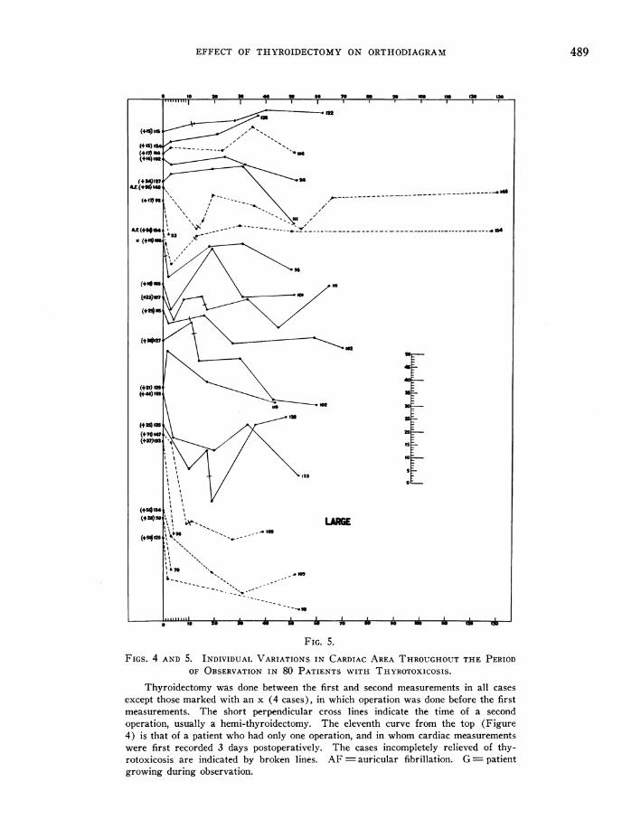
FIGS. 4 AND 5. INDIVIDUAL VARIATIONS IN CARDIAC AREA THROUGHOUTTHE PERIODOF OBSERVATION IN 80 PATIENTS WITH THYROTOXICOSIS.
Thyroidectomy was done between the first and second measurements in all cases
except those marked with an x (4 cases), in which operation was done before the firstmeasurements. The short perpendicular cross lines indicate the time of a secondoperation, usually a hemi-thyroidectomy. The eleventh curve from the top (Figure4) is that of a patient who had only one operation, and in whom cardiac measurementswere first recorded 3 days postoperatively. The cases incompletely relieved of thy-rotoxicosis are indicated by broken lines. AF auricular fibrillation. G=patientgrowing during observation.
489
c
D_
ALEXANDER'MARGOLIES, EDWNARDROSE AND FRANCIS C. WN OOD
for one year or more after operation: of these,52 were completely relieved of their thyrotoxico-sis at the time of the last examiinlation, and 9showed evidence of incomiiplete relief. Preop-erative cardiac enlargemenit was almiiost twice as
frequient (21 cases or 26 per cenit) as in the non-
toxic group. Significant ultimlate clhanges in sizeoccurred in 31 (39 per cent). -None of the en-
lafged hearts showed further inicrease in size, and11 slhowed reduction, including 6 in patients wlhowere incompletely relieved of their thyrotoxicosis.All of the 4 smzall lhearts l)ecame larger after op-
eration. Of the 55 patielnts witlh hearts of nor-
mal size before operationi, 12 increased signifi-cantly in size after operation, 39 remained Uin-changed, and 4 1)ecame smiialler. Cases presentedin Figures 3, 4 andI 5 whichl were followed forone year or more are incluided in Table I.
The frequency witlh whliclh cardiac enlargemenitoccurred in our cases of uncomplicated tlhyro-toxicosis seems to be significant. The nutmlber ofcases of non-toxic goiter studied was not largeenough to warrant any definite conclusions re-
garding the incidence of cardiac enlargement, butits frequency was greater tlhan normal. Theaverage degree of enlarogemiient (expressed interms of percentage increase albove the predictedarea) in the toxic group (21 cases) was 32 per
cent, and in the non-toxic group (4 cases) 21 per
cent. Of the 21 thvrotoxic, patients with en-
larged lhearts, 19 lhad difftuse goiters, and 2nodular goiters.
Of 53 patients in the thyrotoxic group, elevenper cent showed enlargemlenit one year or more
after suiccessful thyroidectonuw ; the remainderwere witlhin the normal zone (Figture 6A; cf.Figure 2B).
Of 12 thyrotoxic patients who Were inlcom11-
pletely relieved, nine wvere followed for one -earor more, and one each for 20, 30 and 40 weeksrespectively. Six showe(d chanige in actual area
of less than 10 per cent, 4 had decreased 10 per
cent or more, aind only 2 showed increase in size
(Figure 6B).Figure 6C shows the relation betweeni actual
and p)redicted areas in 25 patients with lnon-toxicgoiter one year or miiore after operation. Comi-parison of the relative proportion of large, smiialland niormlal hearts in 25 patients with non-toxicgoiter before and onie year after operation shows
no significant shift (Figure 2C, cf. Figure 6C).WVe were unable to demiionstrate any significant
relation between the duration of thyrotoxicosisor of non-toxic goiter, as dletermiiined roughlyfromii the history, and the inicid(ence of cardiac en-
largement. In the thyrotoxic patients witlhoutheart failure somiie of the mllost miiarked cardiac en-
largemiients occurredI in cases of apparently shortdluration. Because of the miiarked tendency to
spontaneous fluctuation in the intensity of thyro-toxicosis, we mlade no attempt to correlate thisfactor with abnormalities of heart size.
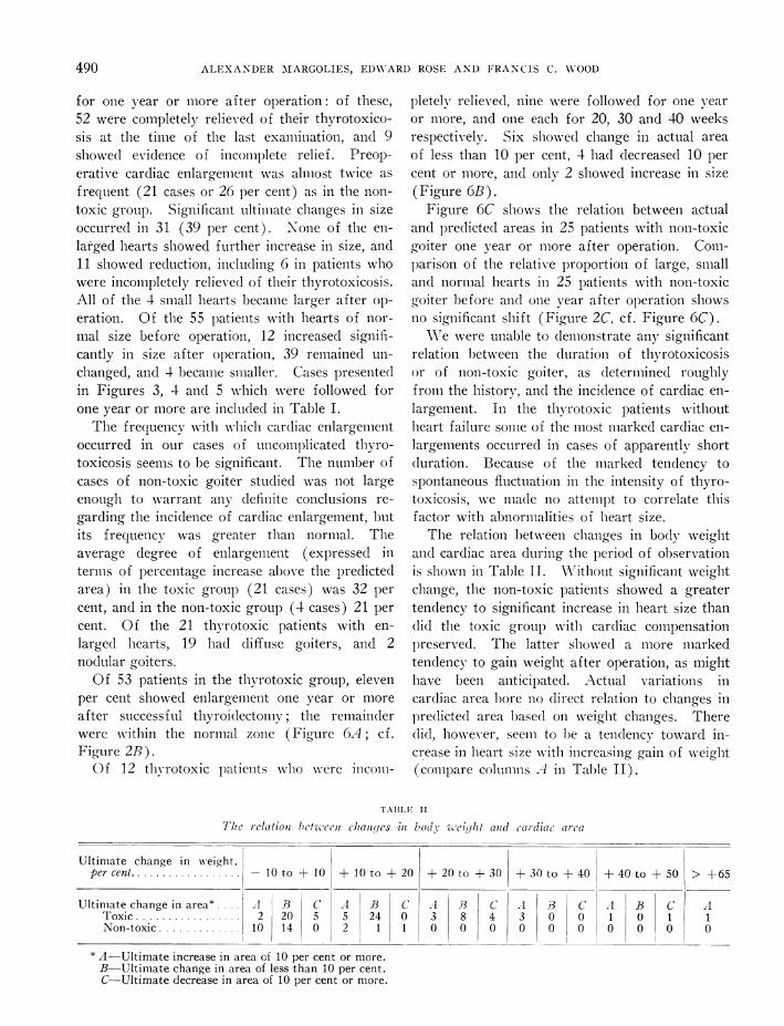
The relationi between chaniges in body weightand cardiac area during the period of observationis shown in Table II. Without significant weightchanige, the non-toxic patients showed a greatertendency to significant inicrease in heart size than(lid the toxic group with cardiac compensationp)reserved. The latter showe(d a mlore markedtendency to gain weight after operation, as mighthave been anticipated. Actual variations incar(liac area 1)ore no direct relation to changes in
predictedl area based on weight changes. There(lid, however, seemii to be a tendency toward in-crease in heart size with inicreasing gain of weigllt(comiipare colutmilns A-1 in Table II).
TABLE. II
7TlIc r-elaltionl betweent clhaneJCs in body z ei'iJt (a11(d cardiac area
Ultimate change in w-eight,per cent .................. - o+10 + to + 20 + 20 to + 30 + 30 to +40 + 40 to + 50 > +65
Ultimate change in area* .... A1 B8 C A B8 C A B C A B C A4 B C 1Toxic.2 20 5 5 24 0 3 8 4 3 0 0 1 0 1 1Non-toxic. 10 14 0 2 1 1 0 0 0 0 0 0 0 0 0 0
* A-Ultimate increase in area cf 10 per cent or more.B-Ultimate change in area of less than 10 per cent.C-Ultimate decrease in area of 10 per cent or more.
490

EFFECT OF THYROIDECTOMYON ORTHODIAGRAM
oC
0 C)
._ r
~0
I ._
) 0
CZ C
Q 0
fr 0d
01..CZ )-
I _ A
X~ 01-,
C)-. cd
CZ° Fbd CT
1..
01.. C)
Cd
0Cl C)
o 0
1..
CE- 1.o
C)C) E° C)
* 0.
*
11 0 *-* )r)
1- 1- C)
cY ce 'Q t
1Q .
s ;r Y Y1.1-00 0lC0Y.
CbCt-.Y
C)C). C
C)C 1..
C)Q C)C)
.0
C) C)- C
: W~0
491
l"n-W
ALEXANDERMARGOLIES, EDWARDROSE AND FRANCIS C. WOOD
Nine toxic patients showed either a reductionof the basal metabolic rate to - 15 per cent orbelow, or exhibited other evidence of hypothy-roidism after operation. In 5 these changes werenot associated with any significant variation incardiac area. In 4 there was an increase in heartsize varying fromii 9 to 18 per cent up to the timeof appearance of the thyreoprival signs. In 3 ofthese patients there was coincidental weight gain.In 3 non-toxic patients the basal metabolic ratedropped after operation to - 15 per cent or be-low, and in 2 others mild thyreoprival symiiptomiisappeared. In the latter two no change in cardiacarea occurred. Of the former, 2 showed increasein cardiac area of 11 and 12 per cent respectivelywithout significant weight gain. It appears thatpostoperative hypothyroidismii may be partly re-sponsible for increase in cardiac area occurringafter thyroidectomy (13).
Sex did not seem to influence the incidence ofcardiac enlargement in the toxic group. All butonle of the non-toxic patients were females.
Table III shows the age (listribution with refer-ence to the incidence of cardiac enlargement. The
TABLE III
The relation between age anzd tf/e incidencc of cardiacenlargement (15 per cenlt or mitore above predictiont)
Toxic Non-toxic
DecadeTotal Ntimber Total Number
number with large nuimber with largeof cases hiearts of cases lhearts
2 5 0 3 03 18 4 3 14 23 6 11 15 ... 25 8 7 06 8 5 3 17 1 1
frequency of enlargement al)pears to increase di-rectly with age in the toxic patients, but there isno apparent relationship in the non-toxic group.
Fifteen (19 per cent) of 81 toxic patienitsshowed tem1tporary variations of 10 per cent ormore in cardiac area during the perio(d of ob-servation. In these cases there were no signifi-cant ultimate changes in area from the first to thelast measurements. lThese variations did not bearany relation to changes in heart rate or blood pres-sure. It is not certaini that thvrotoxicosis influ-
enced these variations in size as no a(lequate con-trol observations have been made in normals.
Eight toxic patients witlh preserved compensa-tion received digitalis before or after ol)eration inmoderate dosage for short perio(ds without appar-ent effect on their cardiac measurements.
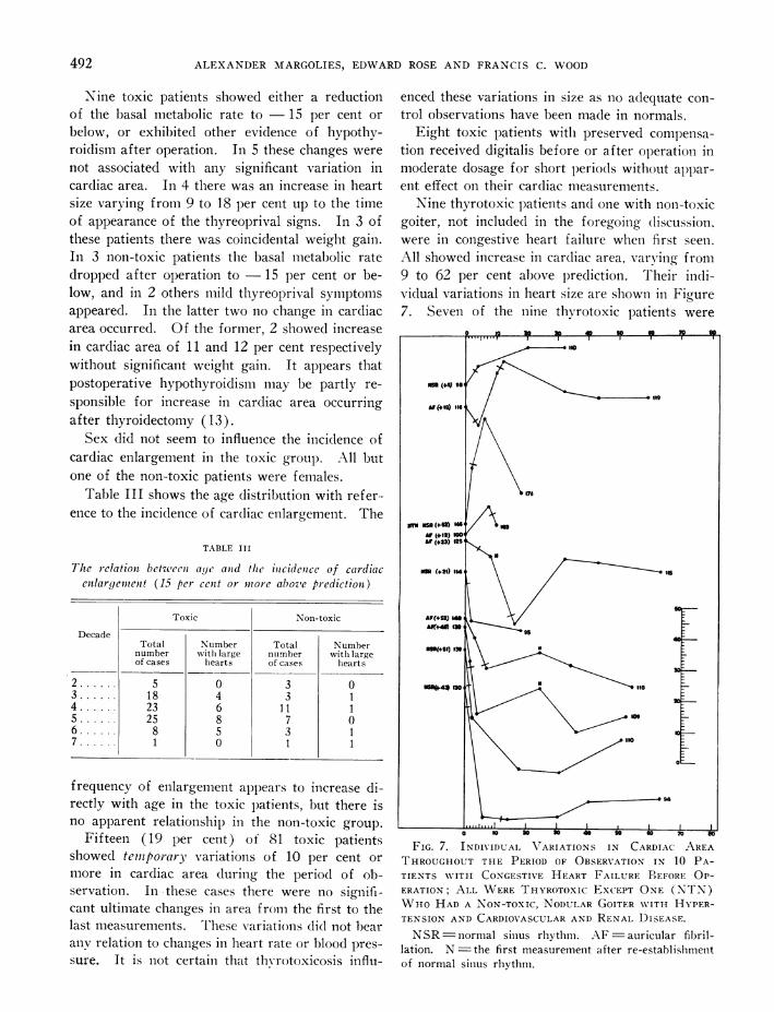
Nine thyrotoxic patients and oine with non-toxicgoiter, not includedI in the foregoing tliscussion.were in congestive heart failure wheni first seeni.All showed increase in cardiac area, varying from9 to 62 per cent above lprediction. Their indi-vidual variations in heart size are showni in Figure7. Seven of the nine thvrotoxic patients were
FIG. 7. INDIVIDUAI VARIATIONS IN CARDIAC AREATHROUGHOUTTHE PERIOD OF OBSERVATION IN 10 PA-TIENTS WNITH CONGESTIVE HEART FAILURE BEFORE OP-ERATION; ALL WERETHlYROTOXIc ExCEPT ONE (NTN)WHOHAD A NON-TOXIC, NODULARGOITER W'ITH HYPER-TENSION AND CARDIOVASCULARAND RENAL DISEASE.
NSR normal sinus rhythm. AF auricular fibril-lation. N =-the first measurement after re-establishmentof normal sinlus rhythiml.
492

EFFECT OF THYROIDECTOMYON ORTHODIAGRAM
relieved of their thyrotoxicosis after operation.Five were in auricular fibrillation; in 3 normalrlhythm returned postoperatively. In 6 the car-diac area diminished after operation, despite gainof weight in 5. One patient was followed foronly a few days after operation, and her fate isunknown. The one patient known not to havebeen completely relieved of thyrotoxicosis showeda slight increase in cardiac area 11 months afteroperation. Eight of the nine toxic patients wererelieved of their decompensation (including theone who remained thyrotoxic). The one non-toxic patient (a male) had cardiovascular-renaldisease with hypertension and heart failure; hewas unimproved four and a half months afteroperation.
Traclheal stentosis
The consensus in the literature is that trachealstenosis from thyroid pressure has no demonstra-ble effect on the size of the heart. Parkinson andCookson (11), in postmortem examinations of43 thyrotoxic patients, found cardiac hypertrophyin 22, but only 2 of these showed tracheal stenosis.WVe made no systematic attempt to analyze theincidence or degree of severity of tracheal stenosis.\Ve have, however, observed a number of patientswith substernal goiter causing pressure symptoms,in whom there was no cardiac enlargement.
Pulnionary artery
Bauer and Helm (24) first called attention, inpatients with thyroid disease, to the prominenceand increased pulsation of the pulmonary arteryand pointed out that increased prominence andincreased pulsation are not always combined.Parkinson and Cookson (11) found a definiteprominence of this arc in 42 (32 per cent) of 130thvrotoxic patients. The heart was normal in sizein over one third (16) of the cases with definiteprominence. They confirmed the presence ofdilatation of the pulmonary artery by postmortemexamination and pointed out that it may be ob-scured during life by the position of the heart.They attributed the dilatation to the widened pulsepressure and increased heart rate in thyrotoxicosis.Menard and Hurxthal (12) found a prominent(straight or convex) pulmonary arc in 52 (45 percent) of their 115 toxic patients. They further
report the same finding in 30 per cent of theirpatients with non-toxic goiter and in 32 per centof non-goitrous individuals but think the prom-inence is greater in thyrotoxicosis. They observeda definte reduction after thyroidectomy or afterdisappearance of heart failure in 19 (37 per cent)of their toxic patients.
In our series of 81 thyrotoxic patients, 44 (54per cent) showed prominence and/or increasedpulsation of the pulmonary artery (Figure 1).Of these, 35 showed prominence and increasedpulsation combined. Four showed prominencealone, and 5 increased pulsation alone. Of the28 patients with non-toxic goiter, 4 (14 per cent)showed one or the other of these pulmonary ar-tery changes. This group included one patientwith slight prominence of the artery, in whom adiagnosis of chronic thyroiditis was made. An-other patient showed slight dilatation of the ptil-monary artery in the right anterior oblique posi-tion but not anteroposteriorly. Two patientsshowed increased pulsation without prominenceof the artery.
In 34 of the 44 toxic patients, the abnormalityof the pulmonary artery disappeared in from 1 to7 months after operation (average 2.3 months).In this group, 16 hearts became smaller, 12 be-came larger, and 6 did not change in size. In the10 patients in whomthe changes persisted, 8 wereclassed as successful results, one was unsuccessful,and one was not followed long enough to beclassified. Of these 9 patients followed, the car-diac area decreased in 2 and was unchanged in 7.In 3 of the 4 non-toxic patients, the changes in thepulmonary artery disappeared in 1, 4 and 12months respectively without significant change inheart size. In the fourth case there was nochange one year after thyroidectomy.
Seven patients with other complicating condi-tions showed abnormalities of the pulmonary ar-tery before operation. Three were in decompen-sation; the others had aortic insufficiency, mitralinsufficiency, mitral stenosis and hypertension, re-spectively. The abnormality disappeared afteroperation in all but the two patients with mitrallesions.
Wewere unable to demonstrate any relationshipbetween the occurrence of changes in the pul-monary artery and (a.) changes in heart size, (b)systemic blood pressure, (c) preoperative heart
493

ALEXANDERMARGOLIES, EDWARDROSE AND FRANCIS C. WOOD
rate, or (d) basal metabolism, in either the toxicor non-toxic group. The relative importance ofalterations in pressure within the pulmonary cir-culation in this connection cannot be determinedat present.
Cardiac pulsationKraus (1) first called attention to the marked
change in heart size during contraction in hyper-thyroidism. Bauer and Helm (24) noted the in-creased pulsation of the whole left border. R6s-ler (25) believes that the pulsation is character-istic in that the systolic contraction is quicker thanin a normal heart beating at the same rate; thatthe contraction is not wave-like as in normals, butthe whole ventricle seems to contract at the sametime. Our own fluoroscopic observations sup-ported by roentgen-kymographic studies (27, 28)of the pulmonary artery and left ventricular pulsa-tions (Figure 8), confirm the presence of thecharacteristic pulsation in hyperthyroidism. Thischaracteristic pulsation disappears after relief ofhyperthyroidism.
Cardiac shapeTIhe following characteristics of the cardiac con-
figuration in hyperthyroidism have been described:prominence of the middle left border (pulmonaryartery) (9, 11, 24, 25, 26); prominence and in-creased width of the superior vena cava (11, 25);high aortic knob (24); rounded apex (24); amitral configuration (9); a ham-like contour(11); cardiac shape and pulsation may, at times,first draw the attention of the clinician to the ex-istence of thyrotoxicosis (9, 25). The lung fieldsmay be unusually clear (25, 26).
We found that, at times, the anteroposteriorcardiac silhouette partially simulates the " mitralconfiguration," because of the filling out of thenormally concave middle left border by the dilatedpulmonary artery; the clouded lung fields and pos-terior enlargement of the left auricle seen in mitralstenosis are not present in hyperthyroidism.Furthermore, the aortic knob and its pulsationare not usually as obscure as in the case of mitralstenosis. The height of the aortic knob was notsignificantly different from that seen in a groupof non-goitrous patients of corresponding agedistribution (25). We did not find increasedwidth of the superior vena cava (12) (middle
right border), but the extreme upper right vas-cular border was frequently somewhat oblique,probably due to right lateral displacement of theright innominate vein by the enlarged thyroidgland. We found nothing characteristic in theshape of the heart itself, exclusive of the vascularpedicle. When the heart was enlarged, the con-figuration suggested bilateral increase with prob-ably slight left ventricular preponderance in mostcases.
SUMMARY
Orthodiagraphic studies have been made in 102thvrotoxic patients and 35 patients with non-toxicgoiter. These studies were made before partialor subtotal thyroidectomy and at successive inter-vals thereafter up to one year in most instances.Our findings have been analyzed with respect tothe following: (1) the incidence of abnormalitiesof cardiac area before operation; (2) changes incardiac area following complete and incompletesurgical relief; (3) the relation between changesin cardiac area and (a) duration of goiter or thy-rotoxicosis, (b) postoperative weight changes, (c)postoperative hvpothyroidism, (d) age, (e) sex,(f) heart failure; (4) the incidence of changesin the appearance of the pulmonary artery, andthe effect of thyroidectomy on these changes; and(5) the shape and character of pulsation of theheart in hyperthyroidism.
CONCLUSIONS
1. Significant increase in cardiac area occursfrequently in uncomplicated hyperthyroidism (26per cent of our cases). The incidence of suchincrease in patients with uncomplicated non-toxicgoiter is also probably abnormal (14 per cent inour series of 28 cases).
2. Following thyroidectomy in uncomplicatedhyperthyroidism, there is a tendency for heartsof abnormal size, whether large or small, to returntoward normal and for hearts within the normalzone to remain so. This tendency is not materi-ally affected by failure to control completely thehyperthyroidism.
3. Significant changes in cardiac area do not oc-cur following thyroidectomy for uncomplicatednon-toxic goiter.
4. Postoperative hypothyroidism may occasion-ally be a factor in increasing cardiac area.
494

FIG. 8. ROENTGEN-KYMOGRAMSOF THE BORDERSOF THE LEFT VENTRICLEAND PULMONARYARTERY.
The lung fields show as the lighter zones; the lheart and pulmonary artery respectively produce the black zones;the serrated edges represent the cardiac and vascular pulsations. The ventricular tracings (A, B and C) should beread from above downward; systole is represented by the deflection of the heart border to the left, diastole by thedeflection to the right. The arterial tracings (D, E and F) should be read from left to right; systole is representedby the upward deflection of the vascular shadow, as in the usual pulse tracing. The letter P identifies the positionof the beginning of the P wave in the simultaneously recorded electrocardiogram; Q, the beginning of the QRScom-plex; T, the end of the T wave (27, 28).
A. Pulsation of the left ventricular border in a normal heart, rate 110.B. Pulsation of the left ventricular border in a normal heart, rate 140.C. Pulsation of the left ventricular border in a thyrotoxic heart, rate 136. The systolic contraction is quicker
and greater in extent than in the normal heart beating at approximately the same rate.D. Pulsation of the pulmonary artery in a normal patient, heart rate 115.E. Pulsation of the pulmonary artery in a thyrotoxic patient, heart rate 150 (faster moving casette).F. Pulsation of the pulmonary artery in a thyro.oxic patient, heart rate 110. The amplitude of the arterial pulsa-
tion is greater in the thyrotoxic patients.

ALEXANDERMARGOLIES, EDWARDROSE AND FRANCIS C. WOOD
5. Temporary variations in cardiac area, with-out significant ultimate changes, occur after thy-roidectomy for uncomplicated hyperthyroidism insome cases (19 per cent in our series). Thecause of these variations is not known.
6. Congestive heart failure in hyperthyroidismis almost always accompanied by enlargement ofthe cardiac area. This tends to decrease withpostoperative restoration of compensation, pro-vided the thyrotoxicosis is also relieved.
7. Increased prominence and/or pulsation ofthe pulmonary artery is frequent in hyperthyroid-istn (54 per cent of our cases). It occurs lessfrequently in association with non-toxic goiter(14 per cent of our cases). The cause of thesechanges is unknown. Wewere unable to demon-strate any relation between them and (a) changesin heart size, (b) systemic blood pressure, (c)preoperative heart rate, or (d) basal metabolism,in patients with toxic or non-toxic goiter. Inabout three-fourths of our cases, the abnormalityof the pulmonary artery disappeared within 1 to7 months after thyroidectomy.
8. Cardiac pulsation, as observed fluoroscopic-ally, is usually characteristically altered in hyper-thyroidism.
9. The heart (exclusive of the vascular pedicle)does not assume a characteristic shape in personswith toxic or non-toxic goiter.
BIBLIOGRAPHY1. Kraus, F., Ueber das Kropfherz. Wien. klin.
Wchnschr., 1899, 12, 416.2. Kraus, F., Ueber Kropfherz. Deutsche med.
Wchnschr., 1906, 32, 1889.3. Blauel, Muller, O., and Schlayer, Ueber das Verhalten
des Herzens bei Struma. Beitr. z. klin. Chir., 1909,62, 119.
4. Steiner, O., Beziehungen zwischen Kropf und Herz,Ihr Verhalten nach der Strumektomie. Mitt. a. d.Grenzgeb. d. Med. u. Chir., 1922, 35, 39.
5. Willius, F. A., and Boothby, W. M., The heart inexophthalmic goiter and adenoma with hyperthy-roidism. M. Clin. North America, 1923, 7, 189.
6. Coller, F. A., The morbidity of endemic goiter. J. A.M. A., 1924, 82, 1745.
7. Kerr, W. J., and Hensel, G. C., Observations of thecardiovascular system in thyroid disease. Arch.Int. Med., 1923, 31, 398.
8. Meyer, A. W., and Sulger, E., Das Kropfherz vorund nach der Operation. Med. Klin., 1926, 22,838.
9. Meyer-Borstel, H., Vber Form- und Gr6ssenverander-ungen des Herzens bei Struma. Fortschr. a. d.Geb. d. R6ntgenstrahlen, 1930, 41, 695.
10. Hurxthal, L. M., Menard, 0. J., and Bogen, M. E.,The size of the heart in goiter. A teleoroentgeno-graphic study, A. J. M. Sc., 1930, 180, 772.
11. Parkinson, J., and Cookson, H., The size and shapeof the heart in goiter. Quart. J. Med., 1931, 24,499.
12. Menard, 0. J., and Hurxthal, L. M., Changes observedin the heart shadow in toxic goiter before and aftertreatment. Ann. Int. Med., 1933, 6, 1634.
13. Hurxthal, L. M., Discussion in Transactions Amer.Heart Assoc., Am. Heart J., 1932, 8, 152.
14. Lerman, J., and Means, J. H., Cardiovascular symp-tomatology in exophthalmic goiter. Am. Heart J.,1932, 8, 55.
15. Jones, N. W., Seabrook, D. B., and Menne, F. R., Aclinical study of goiter in the Pacific Northwest,with special reference to the state of the heart.Am. Heart J., 1932, 8, 41.
16. Burnett, C. T., and Durbin, E., The signs and symp-toms of heart changes in toxic goiter. Am. HeartJ., 1932, 8, 29.
17. Kepler, E. J., Hypertrophy of the heart and con-gestive heart failure in hyperthyroidism: A clin-ical and pathologic study of 178 fatal cases. Proc.Staff Meet., Mayo Clin., 1932, 7, 397.
18. Simonds, J. P., and Brandes, W. W., The size of theheart in experimental hyperthyroidism. Arch.Int. Med., 1930, 45, 503.
19. Hodges, F. J., Heart; x-ray examination. WisconsinM. J., 1929, 28, 46.
20. Margolies, A., X-ray of the heart and great vessels.Cyclopedia of Medicine, F. A. Davis Co., Phila-delphia, 1932, 3, 424.
21. Hodges, P. C., and Eyster, J. A. E., Estimation ofcardiac area in man. Am. J. Roentgenol., 1924,12, 252.
22. Hodges, F. J., and Eyster, J. A. E., Estimation oftransverse cardiac diameter in man. Arch. Int.Med., 1926, 37, 707.
23. Roesler, H., The relation of the shape of the heartto the shape of the chest. Am. J. Roentgenol.,1934, 32, 464.
24. Bauer, J., and Helm, F., Vber Rontgenbefunde beiKropfherzen. Deutsches Arch. f. klin. Med., 1912,109, 73.
25. Rosler, H., Das Rontgenbild des Herzen beim HIyper-thyreoidismus. Wien. Arch. f. inn. Med., 1928, 15,539.
26. Pollitzer, H., Ueber Volumen pulmonis diminutumals Symptom des Morbus Basedowii. Wien klin.Wchnschr., 1924, 37, 735.
27. Margolies, A., and Wolferth, C. C., The opening snap(Claquement d'ouverture de la mitrale) in mitralstenosis, its characteristics, mechanism of pro-duction and diagnostic importance. Am. Heart J..1932, 7, 443.
28. Wolferth, C. C., and Margolies, A., Asynchronism incontraction of the ventricles in the so-called com-mon type of bundle branch block. Am. Heart J.,1935, 10, 425.
496


![[XLS]reports.mca.gov.inreports.mca.gov.in/Reports/MasterDataExcels/company... · Web view500000 500000 100000 100000 100000 100000 12/3/2015 100000 100000 3/3/2015 4/3/2015 100000](https://img.dokumen.tips/doc/110x75/5a9ec0867f8b9a0d158bcbbb/xls-view500000-500000-100000-100000-100000-100000-1232015-100000-100000-332015.jpg)


![[XLS] · Web view2/1/2016 100000 100000 0 2/5/2016 1000000 100000 0 2/5/2016 100000 100000 0 2/17/2016 2000000 900000 0 2/19/2016 2000000 100000 0 2/19/2016 100000 100000 0 …](https://img.dokumen.tips/doc/110x75/5b4c4a3a7f8b9a481a8b82c4/xls-web-view212016-100000-100000-0-252016-1000000-100000-0-252016-100000.jpg)


![[XLS]reports.mca.gov.inreports.mca.gov.in/Reports/MasterDataExcels/company... · Web view100000 100000 100000 100000 10000000 430000 100000 100000 100000 100000 10000000 425000 100000](https://img.dokumen.tips/doc/110x75/5aa857b27f8b9a86188b6f26/xls-view100000-100000-100000-100000-10000000-430000-100000-100000-100000-100000.jpg)