Embed Size (px)
Citation preview

A Historical Look at Bioterrorism
Christine ChungAaron Little
Angela SerranoLaurie Wallis

Introduction
• Bioterrorism is a growing concern in social, political, and scientific communities globally
• Historically, Category A Select Agents, as identified by the CDC, have been used or acquired with intent to disseminate
• Category A Select Agents are those with high morbidity or mortality combined with high transmission rates including Anthrax, Botulism, Plague, Smallpox. Also included is Category B Agent, Salmonella because of the relative ease of acquisition and historical use.
• Note: Follow hyperlinked buttons in the timeline to view Select Agent information.

Biological Weapons Timeline
• 1346: Plague victims are catapulted over city walls during the Tarter siege of Kaffa.
• 1746: British army distributes smallpox infected blankets to Native Americans during the “French and Indian War”
• 1925: The Geneva Protocol is signed, banning the use of BW in warfare
• 1932 – 1945: Japan uses BW against China and POWs• 1942: British test weaponized anthrax on sheep on
Gruinard Island, Scotland leaving the island under quarantine for 48 years.

Biological Weapons Timeline
• 1969: US President Richard Nixon announces that the US will never use BW under any circumstances
• 1972: The Biological Weapons Convention is signed. There are currently 163 signatories to the BWC.
• 1975: US signs the Geneva Protocol• 1979: Anthrax is accidentally released from BW
production facility in Sverdlovsk, USSR. • 1984: The Rajneeshees deliberately contaminate salad
bars in Oregon with salmonella bacteria. • 1992: Russian President Boris Yeltsin reaffirms Russia’s
commitment to the BWC after disclosing the existence of Biopreparat, a major clandestine BW program

Biological Weapons Timeline
• 1995: Larry Wayne Harris obtains vials of plague from ATCC.
• 1995: Aum Shinrikyo develops and attempts to disseminated botulinum toxin and anthrax.
• 1998: All US Armed Service personnel must be vaccinated against anthrax.
• 2001: Genetic manipulation significantly increases virulence of mousepox virus (similar to smallpox)
• 2001: Letters containing weaponized anthrax are sent through the US postal system.
• 2002: Polio virus is artificially synthesized within laboratory• 2009 and beyond: Advances in biotechnology create
countless benefits, but introduce new proliferation threats…

Bacillus anthracis Toxin• Disease Caused:
− Anthrax
• Produced by Bacillus anthracis− Related to B. Cereus and B. thuringiensis
which do not produce capsules and not infectious to humans
• Large gram-positive rod
• Capable of endosporulation, particularly in high CO2 (>5%) concentration− Spores can survive in soil and harsh conditions for decades
• Zoonotic disease primarily infects cattle, horses, goats, sheep− Natural transmission is extremely rare− No human to human transmission

Bacillus anthracis Toxin• 89 known strains, including
– Ames strain of 2001 attacks– Vollum strain of 1935 WWII Gruinard
bioweapon trials– Sterne strain for vaccines
• Pathogenicity is via poly-D-glutamyl capsule and 3 factors – Edema Factor (EF)– Protective Antigen (PA)– Lethal Factor (LF)
• LD50: varies greatly within species– Rat: 1,590 colony-forming units/kg– Monkey 7.5 million units/kg– Human estimated 8,000 units/kg

Anthrax• Pathogenic B. anthracis requires a capsule to mediate
the invasive stage and a multicomponent toxin to mediate the toxigenic stage– poly-D-glutamate polypeptite coating nontoxic, protects
against complement, and phagocytosis and bactericidal components of macrophages
– construction requires pX02 plasmid, obtained from conjugation– Protective Antigen (PA) acts as the binding (B) domain– Edema Factor (EF) acts as an active (A) domain, homologous
to the alpha domain of adenylate cyclase– Lethal Factor (LF) acts as an active (A) domain, a Zn++
dependent protease and member of the MAPKK family

Anthrax
• Mechanism– PA, EF, and LF combine to form an A-B enzymatic binding
structure– cause edema, attracting leucocytes to the area– impair macrophage and neutrophil phagocytosis
• PA+EF elevates cAMP levels, reducing permeability• also depletes ATP, required for engulfment process
– PA+LF act to disrupt cell signaling pathway; not entirely understood
– septicemia causes death from oxygen depletion, secondary shock, increased vascular permeability, respiratory failure, cardiac failure; sudden and unexpected after 3-10 days

Anthrax• Routes of infection
– Cutaneous: boil, then eschar, then necrotic ulcer; painless; minor lethality– Gastrointestinal: severe gastrointestinal irritability; highly lethal route– Pulmonary: induces flu-like symptoms; most lethal route
• Treatment– Vaccination for potential contact, given yearly and at least 4 weeks prior to
exposure– Antibiotics (eg penicillin) for inhalation victims, given within 24 hours– Cutaneous inoculation has minor lethality, usually none with antibiotics
• Cleanup– Spores are hardy, resistant to dessication, heat, extreme chemicals, and
natural decay– CDC and BW protocol recommend steam sterilization or burning for at least
30 minutes– other approved chemicals may not destroy them all

Anthrax in Bioterrorism• Location: More than 60 sites in the US • Perpetrator: Bruce Ivins, suspect• Objective: Unknown• Organism:
– Bacillus anthracis spores• Dissemination
– 4-7 letters sent through postal system– 22 confirmed cases of anthrax
• 11 Cutaneous• 11 Inhalational (5 Deaths)
• Outcome: FBI Named Bruce Ivins of USAMRIID as suspect. Ivins committed suicide before he could be tried.
“Amerithrax”
Back to Timeline

Plague
• Organism:– Yersinia pestis (formerly known as
Pasteurella pestis)
• Location: Africa, former Soviet Union, the Americas, Asia, and the Middle East
• Types of Plague:– Sylvatic plague: in wild rodent populations
– Urban plague: involves rats and is the major source for human endemics
• The WHO (World Health Organization) reports 1,000-3,000 cases of plague worldwide each year, with an average of 5-15 in the western U.S. – probably an underestimate– Highest incidence in Africa (>90% of cases worldwide)
– 90% of U.S. cases in New Mexico, Arizona, Colorado, and California

Urban Plague• Bubonic Plague
– Infection of lymphatic system– Occurs within a week of infected flea bite– Known as “Black Death” as multiplication of bacteria produces “buboes” (swollen, painful
lymph nodes)– 75% mortality rate– Up to 15% of bubonic plague victims develop secondary pneumonic plague
• Pneumonic Plague– Infection of respiratory system– Occurs in crowded conditions when contaminated respiratory droplets are expelled by
infected humans and directly inhaled by others– Characterized by shorter incubation period and greater mortality (90%)
• Septicemic Plague– “blood-poisoning” form– Can result from bubonic and pneumonic plague when bacteria enters the bloodstream from
the lymphatic and respiratory systems– Least common form of plague, characterized by high fevers, purple skin patches and
vomiting– Can cause DIC (disseminated intravascular coagulation)– Almost always fatal (near 100%)

Yersinia pestis• Gram-negative coccobacillus (an Enterobacteriaceae), non-motile
• Facultative anaerobe
• “Safety pin” appearance in bipolar staining
• Colonial Morphology: grey-white, “fried-egg”, irregularity, hemolytic, grows faster and larger at 28°C
• Produces a thick anti-phagocytic slime layer
• Produces two antiphagocytic components necessary for virulence:– F1 antigen and VW antigens– Both produced at 37°, not lower, thus not virulent in fleas that have body temp. of 25°)
• Expresses vadBC gene, allowing for adherence and invasion of epithelium
• Type III secretion system: allows bacteria to inject six different substances into macrophages and immune cells for cytolysis, apoptosis, platelet aggregation, actin microfilament disruption (limits phagocytosis by targeting actin)
– Necessary for virulence

Yersinia pestis (cont.)
• Bacteria can survive for weeks outside of a host– Viable in blood for 100 days– Dried blood for three weeks– In flea feces for five weeks– In infected human bodies for up to
270 days– Can survive in soil for some time
• Fleas act as vectors• Mainly rodents are the reservoirs, but hundreds of
animals can be potential hosts, including cats, dogs, rabbits, and squirrels (most prevalent vector in the U.S.)

Mechanisms of Action
• (Pneumonic plague) Can spread between humans through sneezing, coughing, and/or direct contact of infected tissue– Estimated that only 50kg of Y. pestis released as an aersol in a city of 5
million would infect 150,000, of which 36,000 would die
• Most carnivores, except cats, are resistant to plague infection• Epidemiology
– Occurs in urban and/or wild rodent populations
– Humans acquire primarily via infected fleas
– Y. pestis multiplies in flea intestinal tract
• Known mechanisms– Sites of Entry and Exit
– Incubation period: 1-3 days (pneumonic); 2-6 days (bubonic)
– Infection spreads from lymph nodes near the bite site where swelling occurs, then spreads to other organs such as the spleen, liver, lungs, skin, mucous membranes, and the brain (but usually not the kidney)

Current Researchon Mechanisms, etc.
• The CDC considers an average of one flea per rodent as the maximum threshold to reduce the risk of Y. pestis transmission to humans.
– A reduction of rodent and/or flea populations is optimal– Chitin synthesis inhibitors used to reduce flea populations effectively on certain
species of rodents
• Type III secretion of Y. pestis targeted for new therapeutics
• Interleukin-10-deficient mice are resistant to Y. pestis– Heterozygotes also able to survive high doses of IV infections– Two substrains of 129 mice resistant to high-dose KIM5– Resistance is not recessive– 129-derived genomic DNA near IL-10 confers resistant to Y. pestis KIM5
• Ail (Attachment and Invasion locus) protein found to be crucial in binding and cytotoxic Yop protein delivery into the host cell (type III secretion)
– Single deletion in ail locus severely hindered Yop delivery– Mice with KIM5 ∆ail mutant:
• >3,000-fold increased LD50
• 1,000-fold less bacteria in spleens, livers, and lungs

Effects
• Symptoms– General malaise; pain or tenderness at regional lymph nodes;
septicemia; DIC; convulsions; shock; headache; prostration; bacteria in blood
• “Signs”– High fever (hyperpyrexia); diffuse, hemorraghic changes in the skin;
dark skin at extremities that led to the name “black death”; coughing and sneezing in the case of pneumonic plague
• Secondary Illnesses– Complications: DIC, pneumonia, meningitis
– Bubonic plague victims may develop pneumonic plague, which is contagious through coughing and considered the most severe form of the disease

Plague in Bioterrorism• Location: Ningbo, China and Changde, China• Perpetrator: Japanese secret biological
warfare research facility (Unit 731)• Objective: Infect civilian populations• Organism:
– Yersinia pestis• Dissemination
– Ceramic bombs full of bubonic plague-carrying fleas dropped over Ningbo by the Imperial Japanese Army Air Force
– 80% of fleas survived the bombing to infect civilians– Changde: plague-contaminated foods were distributed to
civilians and water supplies contaminated• Outcome: Largely ineffective in comparison to
distribution as aerosols; caused epidemic plague outbreaks; 400,000 Chinese killed in Ningbo
Back to Timeline

Plague Events(as Biological weapon)
• Earliest reference to bubonic plague (approx. 1320 BC) in book of I Samuel• 6th Century: Plague of Justinian (First Pandemic) through Byzantine Empire, greatly weakening
the Roman empire by reducing the population by one-third• 1346: Mongol warriors of the Golden Horde threw infected corpses over the walls of the besieged
Crimean city of Kaffa (in present day Ukraine)• 1347-1351: “Black Death” (Second Pandemic), possibly originated in Gobi Desert• 1710: Russian forces attacked the Swedes by fling plague-infected corpses over the city walls of
Reval in Estonia (Tallinn)• 1855-1950s: The Third Pandemic, originated in China, spread worldwide via ships• WWI: the German Army allegedly spread plague in St. Petersburg, Russia• 1940: Imperial Japanese Army Air Force bomb Ningbo with plague-carrying flea ceramic bombs• 1941: Unit 731 air-drop plague-carrying fleas on Changde• 1944: the Japanese planned on dropping porcelain plague-flea bombs on invading Gis to defend
their airstrip on Saipan, but failed when carrier submarine sank before reaching the island• Biological warfare generally not used after WWII, but challenged by China and North Korea, who
accuse the U.S. of using disease-carrying insects against them during the Korean War.• 1953: U.S. initiated disease vector weaponization efforts with focus on plague-fleas, etc.• Vietnam War: plague was endemic among natives; U.S. soldiers well-protected with vaccines• 1995: Discovery of multi-drug resistant (MDR) strain of plague in Madagascar• 1996: an Ohio man attempted to obtain bubonic plague cultures through the mail

Pathogenesis
• Y. pestis primarily a rodent pathogen; humans are accidental hosts when bitten by infected rat fleas
• Y. pestis grows and multiplies in flea’s intestinal tract, blocking the flea’s proventriculus, and loses its capsular layer. Several proteins including hemin storage (Hms) system and Yersinia murine toxin (Ymt) contribute to maintenance of bacteria in flea’s digestive tract.
• Hms genetic loci aggregate in esophagus and proventriculus of flea, which ruptures blood cells, which inhibits feeding causing the flea to feel hungry.
• Ingested blood is pumped into the esophagus, dislodging the bacteria cultivating there and is regurgitated and transferred into a new host (i.e. humans) when fleas feed
• Most get phagocytosed and killed by leukocytes in human.
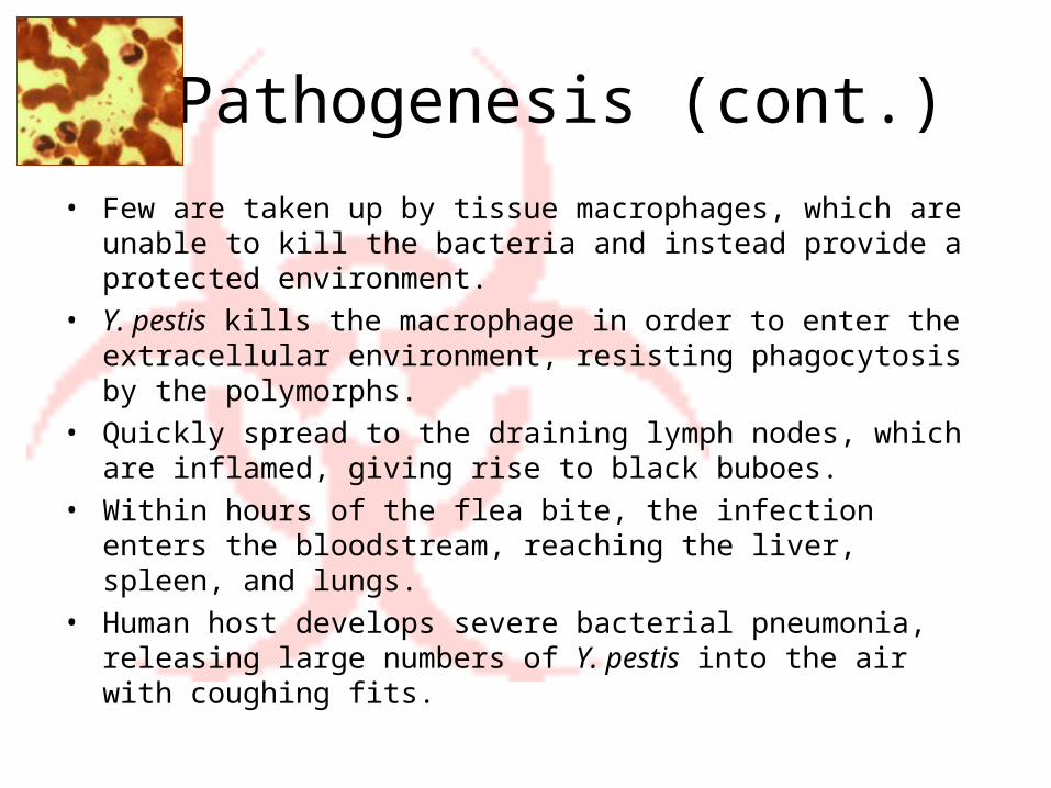
Pathogenesis (cont.)
• Few are taken up by tissue macrophages, which are unable to kill the bacteria and instead provide a protected environment.
• Y. pestis kills the macrophage in order to enter the extracellular environment, resisting phagocytosis by the polymorphs.
• Quickly spread to the draining lymph nodes, which are inflamed, giving rise to black buboes.
• Within hours of the flea bite, the infection enters the bloodstream, reaching the liver, spleen, and lungs.
• Human host develops severe bacterial pneumonia, releasing large numbers of Y. pestis into the air with coughing fits.

Present uses and/orPossible future uses
• Present treatments/therapy– Rapid diagnosis essential
• Rapid disease progression• High mortality rate
– Bubonic plague: 75% mortality in few days– Pneumonic plague: >90% mortality within 24 hours
– Without treatment, fatality rates increase up to 90% for bubonic plague, 100% for septicemic or pneumonic plague
– With treatment, fatality rate = 5-20%• Rapid treatment crucial to survival rates
– Antibiotics: Streptomycin, chloramphenicol, tetracycline, fluoroquinolones, sulfonamides, ciprofloxacin, and potentially doxycycline or gentamicin, etc.
– Multi-drug resistant strains have been isolated
– Best laboratory diagnosis is made by PCR, etc.

Present uses and/orPossible future uses
• Weaponized plague development and Other Current Events– On the list of potential terrorist agents
– Transmission by aerosols is potentially deadly and can spread from person to person
– Institute of Ultra Pure Biochemical Preparations, Leningrad: a weaponized plague center
– Al Qaeda• Roughly 40 al-Qaeda terrorists reportedly died from bubonic plague in their
Algerian training camp• Late 1990s: Osama bin Laden set up 19 chemical and biological weapons
laboratories in Afghanistan, which were stocked with anthrax, plague, and botulinum toxins.
• Possible threats: could spread in public by a lone suicidal bioterrorist (i.e. in subways), or could contaminate self with the plague first to conduct a bioterrorist attack
– Could take up to a week for symptoms to appear
– 2002: two NYC residents acquired plague from New Mexico

Present uses and/orPossible future uses
• Present research being conducted in the maintenance of plague– Experiments with genetic engineering of vaccines based on F1 and V
antigens are underway• Bacteria lacking F1 antigen are still virulent• These vaccines may not fully protect potential hosts
– Research suggests that descendants of medieval European plague survivors are less likely to catch plague
– Recent research indicates that ongoing outbreaks of plague can be caused by viral hemorrhagic disease, similar to Ebola
– A handful of Western laboratories are actively conducting research on MDR Y. pestis (resistant to at least eight drugs traditionally used to treat plague)
• Should conduct additional research to effectively fight MDR strain

Present uses and/orPossible future uses
• Prevention and vaccination– By law, pneumonic plague patients must be isolated
– Sanitation measures• Control of rat populations and elimination of fleas; etc.
– Formalin-inactivated vaccine for adults (age 18-61) at high risk, and continuous booster shots
• Not very effective• May lead to severe inflammation
– As of the mid-1990s, the vaccine is no longer available in the U.S.
– Research being conducted currently for more effective vaccines
– Scientists hypothesize that a mutation in the CCR5 gene, which gives rise to a natural immunity to the HIV virus, may also confer immunity to Y. pestis
– Currently, hospitals are poorly equipped/prepared to deal with patients in the case bioterrorism may occur.
Back to Timeline

Salmonella
• Disease Caused: − Salmonellosis/Gastroenteritis/Enteric Fevers
(including Typhoid Fever)
• Gram-negative Bacterium• Characterized by O, H, and Vi antigens• Ingested in contaminated food and water• Zoonotic-worldwide human and animal
disease• Resilient and capable of survival for several
years• Sensitive to moist and dry heat and many
disinfectants

Salmonella• Mechanism
– Bacteria pass through gastric acid barrier and invades mucosa of small and large intestines and produce toxins
– Irritation of the small and large intestines
• Results of Exposure– Profuse vomiting and diarrhea – Leads to dehydration– Can result in death with severe dehydration.
• Treatment– Antibiotics– Hydration

Salmonella in Bioterrorism• Location: The Dalles, Oregon• Perpetrator: Rajneesh Cult• Objective: Gain control of the Wasco County
Court by affecting the election• Organism: Salmonella typhimurium
– Purchased from commercial supplier• Dissemination
– Restaurant salad bars– 751 illnesses
• Early investigation by CDC suggested the event was a naturally occurring outbreak
• Cult member arrested on unrelated charge confessed involvement with the event
Bhagwan Shree
Rajneesh
Back to Timeline

Smallpox
• Disease caused by Variola major virus.• Humans are the only natural reservoir for variola virus
− Originated in Egypt or India over 3000 years ago− Eradicated in nature by vaccination programs in 1970-1980s.
• Only known stocks of virus at CDC in Atlanta, and a Russian repository.
• Mortality rate of 30%− Up to 90% mortality for flat and hemorrhagic forms of virus.

Smallpox
• Mechanism− Entry through the respiratory mucosa − virus migrates rapidly to regional lymph nodes, then to
spleen, bone marrow, kidneys, and liver − virus localizes in small blood vessels of the dermis and
oropharyngeal mucosa, and evolves into skin lesions.
• Results of Exposure− Incubation period 7-14 days− Flu-like symptoms− Development of pustules
• Treatment− None

Smallpox in Bioterrorism
• Location: North America during French and Indian War • Perpetrator: British Forces in North America• Objective: Infect Native Americans with Smallpox disease• Organism
− Variola Major• Dissemination:
− Blankets used by smallpox patients given to Native Americans as “gifts” during war
− 50% Native American mortality rate• Outcome: Smallpox outbreak claimed lives of many Native
Americans, British leader Jeffrey Amherst claimed parts of Canada and the United States in war victory
Back to Timeline

Botulinum Toxin
• Disease caused: Botulism• Produced by Clostridium botulinum, C. baratii, and
C. butyricum– Multiple types of toxin: A, B, C, D, E, F, G– Only A, B, E, and F produce human disease
• Spore forming bacteria is highly stable• Toxic protein is degraded by heat and
humidity – relatively stable for a protein• LD50: 0.001µg/kg
– The lowest known LD50 of all toxins
• Found in soil, water, and contaminated food• Therapeutic use as a paralyzing agent when highly
diluted

Botulinum Toxin
• Mechanism– Neurotoxin degrades the SNARE complex in the synaptic
bulb.– Permanently interferes with the release of acetylcholine,
preventing nerve stimulation for muscle contraction.
• Results of Exposure– Paralysis– Respiratory failure
• Treatment– Antitoxin (limited supplies)– Supportive care
• Ventilator machine• Extensive rehab

Botulinum Toxin in Bioterrorism• Location: Tokyo, Japan• Perpetrator: Aum Shinrikyo Cult• Objective: Over throw government via targeted
assassination and pubic dissemination.• Organisms:
– Bacillus anthracis • Vaccine strain
– Clostridium botulinum• Environmental isolate• Avirulent strain
– Ebola virus• Attempted to acquire from Zaire outbreak under guise of
an “Humanitarian mission”• Dissemination
– Aerosolization in Tokyo• B. anthracis • Botulinum toxin
• Outcome:– Use of non-virulent strains and ineffective dissemination
methods resulted in no casualties from biological weapons– Successful sarin nerve gas attack in subway– Leader Asahara was convicted of criminal activity
Aerosolization of Bacillus anthracis and botulinum toxin by
Aum Shinrikyo
Back to Timeline

Summary
• The future threat of bioterrorism is increasing with the advances in biotechnology.– Increasing ease of acquisition and production
• Sequencing technology advances and publications• Small-scale operations sufficient to incite fear
– Historical failures decreasing• many past attempts failed because of bad science• Terrorists gaining required knowledge and skills
– Political treaties / federal restrictions reducing ability of research
– Biodefense difficulties• Vaccines are expensive, and preventative only • antibiotics are best line of defense, but select agents increasingly resistant• Slow spreading awareness of complex topics
Back to Timeline

References• Aceto, D., Astuto-Gribble, L., and Gaudioso, J. 2007. The Acquisition of Dangerous Biological
Materials: Technical Facts Sheets to Assist Risk Assessments of 46 Potential BW Agents. Sandia National Laboratories Report.
• Ackerman, G., and Tamsett, J., Editors. 2009. Jihadists and Weapons of Mass Destruction. Chapter Six “Jihadists and Biological and Toxin Weapons” by Loeb, C. Pg. 153-171. CRC Press.
• Ajayi, T. 2002. Smallpox and Bioterrorism. Stanford Journal of International Relations. [Internet] Vol. 3 Issue 2. [cited 2009 April 4] Available from: http://www.stanford.edu/group/sjir/3.2.02_ajayi.html
• Biological Warfare. In: Wikipedia the Free Encyclopedia. [Internet]: Wikipedia; [cited 2009 Mar 1]. Available from: http://en.wikipedia.org/wiki/Biological_warfare
• Black Death. In: Wikipedia the Free Encyclopedia. [Internet]: Wikipedia; [cited 2009 Mar 28]. Available from: http://en.wikipedia.org/wiki/Black_Death
• Bubonic Plague. In: Wikipedia the Free Encyclopedia. [Internet]: Wikipedia; [cited 2009 Mar 1]. Available from: http://en.wikipedia.org/wiki/Bubonic_plague
• Center for Infectious Disease Research and Policy [Internet]. Smallpox page. [cited 2009 April 4] Available from: http://www.cidrap.umn.edu/cidrap/content/bt/smallpox/index.html
• Chamberlain, Neal R. [Internet]. [updated 2004 Aug 3]. Kirksville (MO): Kirksville College of Osteophathic Medicine, A.T. Still University; [cited 2009 Mar 3]. Available from: http://www.kcom.edu/faculty/chamberlain/website/lectures/lecture/plague.htm

References• Cunha CB, Cunha BA. 2006. Impact of Plague on Human History. Infect Dis Clin N Am 20(2006):
253-272.
• Davis RM, Cleugh E, Smith RT, Fritz CL. 2008. Use of a chitin synthesis inhibitor to control fleas on wiild rodents important in the maintenance of plague, Yersinia pestis, in California. Journal of Vector Ecology 33(2):278-284.
• Deadly Diseases: Plague. In: RX for Survival, a Global Health Challenge. [Internet]: PBS; [cited 2009 Apr 10]. Available from: http://www.pbs.org/wgbh/rxforsurvival/series/diseases/plague.html
• Dire DJ. 2005. Biological Warefare. In: eMedicineHealth Practical Guide to Health. [Internet]: eMedicineHealth; [cited 2009 Mar 13]. Available from: http://www.emedicinehealth.com/biological_warfare/article_em.htm
• Drysdale M, Heninger S, Hutt J, Chen Y, Lyons CR, et al. (2005) Capsule synthesis by Bacillus anthracis is required for dissemination in murine inhalation anthrax. Embo J 24: 221–227.
• Felek S, Krukonis ES. 2009. The Yersinia pestis Ail Protein Mediates Binding and Yop Delivery to Host Cells Required for Plague Virulence. Infection and Immunity 77(2):825-836.
• Fix, Douglas F. [Internet]. Carbondale (IL): Southern Illinois University Carbondale; [cited 2009 Mar 3]. Available from: http://www.cehs.siu.edu/fix/medmicro/yersi.htm
• Inglesby TV, Dennis DT, et. al. 2000. Plague as a Biological Weapon: Medical and Public Health Management. JAMA 283(17):2281-2290.
• Kare, John. 2002. Plague and Anthrax: Ancient Diseases, Modern Warfare. Top Emerg Med 24(3):77-87.

References• List of Historical Plagues. In: Wikipedia the Free Encyclopedia. [Internet]: Wikipedia; [cited 2009
Mar 1]. Available from: http://en.wikipedia.org/wiki/List_of_historical_plagues
• McGovern TW, Friedlander, AM. 2007. Chapter 23: Plague. Medial Aspects of Chemical and Biological Warfare. [Internet]: Borden Institute the Textbooks of Military Medicine; [cited 2009 Apr 14]. Available from: http://www.bordeninstitute.army.mil/published_volumes/chemBio/Ch23.pdf
• Mangold T, Goldberg J. 2001. Plague Wars: The Terrifying Reality of Biological Warfare. New York: St. Martin’s Griffin.
• Maginnis, Robert. 2009. Al-Qaeda and The Plague. In: Human Events. [Internet]: Human Events; [cited 2009 Mar 24]. Available from: http://www.humanevents.com/article.php?id=30382
• Marik PE, Bowles S. 2002. Management of Patients Exposed to Biological and Chemical Warfare Agents. Journal of Intensive Care Medicine 17(4):147-161.
• Pan NJ, Brady MJ, Leong JM, Goguen JD. 2009. Targeting Type III Secretion in Yersinia pestis. Antimicrobial Agents and Chemotherapy 53(2):385-392.
• Plague Attributes – Biological Weapons. In: GlobalSecurity.org. [Internet]: GlobalSecurity; [cited 2009 Mar 13]. Available from: http://www.globalsecurity.org/wmd/intro/bio_plague-att.htm
• Relman, D. A. 2006. Bioterrorism — Preparing to Fight the Next War. The New England Journal of Medicine. Vol. 354: 113-115.
• Septicemic plague. In: Wikipedia the Free Encyclopedia. [Internet]: Wikipedia; [cited 2009 Apr 3]. Available from: http://en.wikipedia.org/wiki/Septicemic_plague

References• Todar, K. 2009. [Internet] "Bacillus anthracis and anthrax." Todar's Online Textbook of
Bacteriology. University of Wisconsin-Madison Department of Bacteriology. [cited 2009 April 12] Available from: www.textbookofbacteriology.net/Anthrax.html
• Turner JK, Xu JL, Tapping RI. 2009. Substrains of 129 Mice Are Resistant to Yersinia pestis KIM5: Implications for Interleukin-10-Deficient Mice. Infection and Immunity 77(1):367-373.
• University of Texas Medical Branch. [Internet] Epidemiology Corner: Infectious Disease Page: Salmonella. [cited 2009 April 4] Available from: http://www.utmb.edu/mchd/Information/Epidemiology/InfectiousDiseases/Salmonella
• Weyant, R. S., J. W. Ezzelland, Jr., and T. Popovic. 2001. [Internet] Basic laboratory protocols for the presumptive identification of Bacillus anthracis. Centers for Diseases Control and Prevention, Atlanta, Ga. [cited 2009 April 12] Available from: http://www.bt.cdc.gov/Agent/Anthrax/Anthracis20010417.pdf
• West, Alex. 2009. Al-Qaeda terrorists killed by Black Death after the killer bug also known as the plague sweeps. In: The Sun. [Internet]: The Sun; [cited 2009 Mar 24]. Available from: http://www.thesun.co.uk/sol/homepage/news/article2146286.ece
• Willey JM, Sherwood LM, Woolverton CJ. 2008. The Epidemiology of Infectious Disease. Prescott, Harley, and Klein’s Microbiology. 7th ed. Boston: McGraw-Hill. p. 885-912.
• Yersinia pestis. In: Wikipedia the Free Encyclopedia. [Internet]: Wikipedia; [cited 2009 Mar 10]. Available from: http://en.wikipedia.org/wiki/Yersinia_pestis