Embed Size (px)
Citation preview
*For correspondence:
(DJM); [email protected].
harvard.edu (GMC); fields@uw.
edu (SF); [email protected] (DB)
†These authors contributed
equally to this work
Competing interest: See
page 20
Funding: See page 20
Received: 06 August 2015
Accepted: 17 December 2015
Published: 29 December 2015
Reviewing editor: Jeffery W
Kelly, Scripps Research Institute,
United States
Copyright Feng et al. This
article is distributed under the
terms of the Creative Commons
Attribution License, which
permits unrestricted use and
redistribution provided that the
original author and source are
credited.
A general strategy to construct smallmolecule biosensors in eukaryotesJustin Feng1,2†, Benjamin W Jester3,4†, Christine E Tinberg5†, Daniel J Mandell2,6*†,
Mauricio S Antunes7, Raj Chari2, Kevin J Morey7, Xavier Rios2, June I Medford7,George M Church2,6*, Stanley Fields3,4,8*, David Baker4,5*
1Program in Biological and Biomedical Sciences, Harvard Medical School, Boston,United States; 2Department of Genetics, Harvard Medical School, Boston, UnitedStates; 3Department of Genome Sciences, University of Washington, Seattle, UnitedStates; 4Howard Hughes Medical Institute, University of Washington, Seattle,United States; 5Department of Biochemistry, University of Washington, Seattle,United States; 6Wyss Institute for Biologically Inspired Engineering, HarvardUniversity, Boston, United States; 7Department of Biology, Colorado StateUniversity, Fort Collins, United States; 8Department of Medicine, University ofWashington, Seattle, United States
Abstract Biosensors for small molecules can be used in applications that range from metabolic
engineering to orthogonal control of transcription. Here, we produce biosensors based on a ligand-
binding domain (LBD) by using a method that, in principle, can be applied to any target molecule.
The LBD is fused to either a fluorescent protein or a transcriptional activator and is destabilized by
mutation such that the fusion accumulates only in cells containing the target ligand. We illustrate
the power of this method by developing biosensors for digoxin and progesterone. Addition of
ligand to yeast, mammalian, or plant cells expressing a biosensor activates transcription with a
dynamic range of up to ~100-fold. We use the biosensors to improve the biotransformation of
pregnenolone to progesterone in yeast and to regulate CRISPR activity in mammalian cells. This
work provides a general methodology to develop biosensors for a broad range of molecules in
eukaryotes.
DOI: 10.7554/eLife.10606.001
IntroductionBiosensors capable of sensing and responding to small molecules in vivo have wide-ranging applica-
tions in biological research and biotechnology, including metabolic pathway
regulation (Zhang et al., 2012), biosynthetic pathway optimization (Raman et al., 2014; Tang and
Cirino, 2011), metabolite concentration measurement and imaging (Paige et al., 2012), environ-
mental toxin detection (Gil et al., 2000), and small molecule-triggered therapeutic
response (Ye et al., 2013). Despite such broad utility, no single strategy for the construction of bio-
sensors has proven sufficiently generalizable to gain widespread use. Current methods typically cou-
ple binding of a small molecule to a single output signal, and use a limited repertoire of natural
protein- (Tang et al., 2013) or nucleic acid aptamer-binding (Yang et al., 2013) domains, which nar-
rows the scope of small molecules that can be detected. A general solution to small molecule bio-
sensing should be adaptable to a range of small molecules and responses.
A promising approach to biosensor design in eukaryotes uses conditionally stable ligand-binding
domains (LBDs) (Banaszynski et al., 2006; Tucker and Fields, 2001). In the absence of a cognate
ligand, these proteins are degraded by the ubiquitin proteasome system (Egeler et al., 2011). Bind-
ing of the ligand stabilizes the LBD and prevents its degradation. Fusing the destabilized LBD to a
Feng et al. eLife 2015;4:e10606. DOI: 10.7554/eLife.10606 1 of 23
RESEARCH ARTICLE
suitable reporter protein, such as an enzyme, fluorescent protein, or transcription factor, renders the
fusion conditionally stable and generates sensor response (Figure 1a). Naturally-occurring LBDs can
be engineered to be conditionally stable (Banaszynski et al., 2006; Miyazaki et al., 2012;
Iwamoto et al., 2010), making it possible in principle to convert them into biosensors for target
ligands. Designed LBDs can also be used, especially in cases for which natural binding proteins do
not exist or lack sufficient specificity or bioorthogonality.
Here, we convert a single designed LBD scaffold into multiple highly specific biosensors for the
clinically relevant steroids digoxin and progesterone. We engineer LBDs fused with fluorescent
reporters to be conditionally stable in the budding yeast Saccharomyces cerevisiae. Attaching these
conditionally-stabilized LBDs to transcription factors (TFs) yields biosensors that respond to their tar-
get ligands with greater signal induction than observed with fusions to fluorescent proteins. We use
TF-biosensors to improve the biosynthetic yield of progesterone in yeast. The biosensors retain func-
tion when ported directly into mammalian cells, with up to 100-fold activation over background,
allowing us to develop a method for tight control of CRISPR/Cas9 genome editing. The biosensors
also show up to 50-fold activation by ligand in Arabidopsis thaliana. The method presented here
enables the rapid development of eukaryotic biosensors from natural and designed binding
domains.
Results
Fluorescent biosensors built from engineered LBDsLBDs intended for biosensor development should recognize their targets with high affinity and speci-
ficity. We began with the computationally-designed binding domain DIG10.3 (Tinberg et al., 2013),
hereafter DIG0, which binds the plant steroid glycoside digoxin and its aglycone digoxigenin with
picomolar affinities. Introduction of three rationally-designed binding site mutations into DIG0
resulted in a progesterone binder (PRO0) with nanomolar affinity (Tinberg et al., 2013). We con-
structed genetic fusions of DIG0 and PRO0 to a yeast-enhanced GFP (yEGFP, LBD-biosensors DIG0-
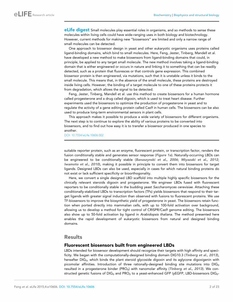
eLife digest Small molecules play essential roles in organisms, and so methods to sense these
molecules within living cells could have wide-ranging uses in both biology and biotechnology.
However, current methods for making new “biosensors” are limited and only a narrow range of
small molecules can be detected.
One approach to biosensor design in yeast and other eukaryotic organisms uses proteins called
ligand-binding domains, which bind to small molecules. Here, Feng, Jester, Tinberg, Mandell et al.
have developed a new method to make biosensors from ligand-binding domains that could, in
principle, be applied to any target small molecule. The new method involves taking a ligand-binding
domain that is either engineered or occurs in nature and linking it to something that can be readily
detected, such as a protein that fluoresces or that controls gene expression. This combined
biosensor protein is then engineered, via mutations, such that it is unstable unless it binds to the
small molecule. This means that, in the absence of the small molecule, these proteins are destroyed
inside living cells. However, the binding of a target molecule to one of these proteins protects it
from degradation, which allows the signal to be detected.
Feng, Jester, Tinberg, Mandell et al. use this method to create biosensors for a human hormone
called progesterone and a drug called digoxin, which is used to treat heart disease. Further
experiments used the biosensors to optimize the production of progesterone in yeast and to
regulate the activity of a gene editing protein called Cas9 in human cells. The biosensors can be also
used to produce long-term environmental sensors in plant cells.
This approach makes it possible to produce a wide variety of biosensors for different organisms.
The next step is to continue to explore the ability of various proteins to be converted into
biosensors, and to find out how easy it is to transfer a biosensor produced in one species to
another.
DOI: 10.7554/eLife.10606.002
Feng et al. eLife 2015;4:e10606. DOI: 10.7554/eLife.10606 2 of 23
Research article Biochemistry Biophysics and structural biology

GFP and PRO0-GFP) and constitutively expressed them in S. cerevisiae (Supplementary file 1). The
fusions showed little change in fluorescence in response to digoxin or progesterone, respectively
(Figure 1b,c and Figure 1—figure supplement 1). Work by Wandless and co-workers has shown
that mutagenesis of LBDs can be used to identify variants that are stable only in the presence of a
target ligand (Banaszynski et al., 2006). We randomly mutagenized the LBDs of DIG0-GFP and
PRO0-GFP by error-prone PCR and subjected libraries of 105 (Gil et al., 2000) integrants to multiple
rounds of FACS, sorting alternately for high fluorescence in the presence of the ligand and low fluo-
rescence in its absence. We isolated LBD variants having greater than 5-fold activation by cognate
ligand (Figure 1b,c and Figure 1—figure supplement 1). By making additional variants that contain
only one of the up to four mutations found in the progesterone biosensors, we showed that some
mutations are additive, while others predominately contribute to sensitivity (Figure 1—figure sup-
plement 2a). Many of the conditionally-destabilizing mutations identified in DIG0 involve residues
that participate in key dimer interface interactions (Figure 1d). The conditionally-destabilizing muta-
tions of PRO0 are located throughout the protein (Figure 1—figure supplement 2b–d); the DIG0
interface mutations also rendered PRO0-GFP conditionally stable on binding progesterone (Fig-
ure 1—figure supplement 2e).
TF-biosensors amplify ligand-dependent responsesTo improve the dynamic range and utility of the biosensors, we built conditionally-stable LBD-tran-
scription factor fusions (TF-biosensors) by placing an LBD between an N-terminal DNA-binding
domain (DBD) and a C-terminal transcriptional activation domain (TAD, Figure 2a). The use of TFs
serves to amplify biosensor response and allows for ligand-dependent control of gene
expression (Shoulders et al., 2013; Beerli et al., 2000; Louvion et al., 1993). Our initial constructs
used the DBD of Gal4, the destabilized LBD mutant DIG1 (E83V), and either VP16 or VP64 as a TAD
to drive the expression of yEGFP under the control of a GAL1 promoter. The dynamic range of TF-
biosensor activity was maximal when the biosensor was expressed using a weak promoter and weak
activation domain, because of lower yEGFP expression in the absence of ligand (Figure 2—figure
supplement 1a,b).
We chose Gal4-DIG1-VP16 (hereafter G-DIG1-V) for further TF-biosensor development because it
has both a large dynamic range and maximal activation by ligand. A FACS-based screen of an error-
prone PCR library of G-DIG0-V, G-DIG1-V, and G-DIG2-V variants identified mutations L77F and
R60S in the Gal4 dimer interface (hereafter GL77F, GR60S) that further increased TF-biosensor
response by lowering background activity in the absence of ligand (Figure 2b and Figure 2—figure
supplement 1c). Although these Gal4 mutations were identified by screening the libraries of
digoxin-dependent TF-biosensors, they also increased progesterone-dependent activation of the G-
PRO-V series of biosensors, indicating a shared mechanism of conditional stability in both systems
(Figure 2—figure supplement 1d). Combining mutations in Gal4 and DIG0 or PRO0 led to activa-
tions of up to 60-fold by cognate ligand, a ten-fold improvement over the most responsive LBD-bio-
sensors (Figure 2c,d and Figure 2—figure supplement 2a) and a dynamic range that has been
challenging to achieve with stability-based biosensors in yeast (Rakhit et al., 2011). The TF-biosen-
sors were also rapidly activated, showing a five-fold increase in signal after 1 hr of incubation with
ligand and full activation after ~14 hr (Figure 2e,f and Figure 2—figure supplement 2b). In contrast
to the LBD-biosensors, the TF-biosensors exhibited a broad range of fluorescence levels across sin-
gle cells, as well as a population of nonfluorescent cells in the presence of ligand (Figure 2—figure
supplement 2). We used FACS to isolate cells from the nonfluorescent population and found those
cells to be inviable, possibly indicating plasmid loss or toxicity from biosensor activation.
Upon withdrawal of ligand, strains expressing TF-biosensors rapidly exhibited reduction in signal,
reaching half of their maximum yEGFP fluorescence after approximately 5 hr and nearly undetect-
able fluorescence after 10–15 hr (Figure 2g,h). The response of the TF-biosensors to the withdrawal
of ligand is likely much faster than observed by fluorescence, as the reduction in fluorescence signal
is dependent on both the degradation of the TF-biosensors as well as the degradation and dilution
of previously expressed yEGFP.
Feng et al. eLife 2015;4:e10606. DOI: 10.7554/eLife.10606 3 of 23
Research article Biochemistry Biophysics and structural biology

Figure 1. A general method for construction of biosensors for small molecules. (a) Modular biosensor construction from a conditionally destabilized
LBD and a genetically fused reporter. The reporter is degraded in the absence but not in the presence of the target small molecule. (b) yEGFP
fluorescence of digoxin LBD-GFP biosensors upon addition of 250 mM digoxin or DMSO vehicle. (c) yEGFP fluorescence of progesterone LBD-GFP
biosensors upon addition of 50 mM progesterone or DMSO vehicle. (d) Positions of conditionally destabilizing mutations of DIG0 mapped to the crystal
structure of the digoxin LBD (PDB ID 4J9A). Residues are shown as colored spheres and key interactions highlighted in insets. In b-c, fold activation is
shown above brackets, (-) indicates cells lacking biosensor constructs, and error bars represent the standard error of the mean (s.e.m.) of three
biological replicates.
DOI: 10.7554/eLife.10606.003
The following figure supplements are available for figure 1:
Figure supplement 1. Population responses to cognate ligand for cells bearing LBD-biosensors.
DOI: 10.7554/eLife.10606.004
Figure supplement 2. Characterization of mutations conferring progesterone-dependent stability.
DOI: 10.7554/eLife.10606.005
Feng et al. eLife 2015;4:e10606. DOI: 10.7554/eLife.10606 4 of 23
Research article Biochemistry Biophysics and structural biology
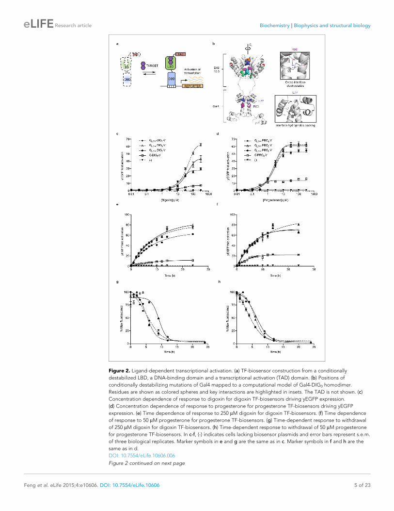
Figure 2. Ligand-dependent transcriptional activation. (a) TF-biosensor construction from a conditionally
destabilized LBD, a DNA-binding domain and a transcriptional activation (TAD) domain. (b) Positions of
conditionally destabilizing mutations of Gal4 mapped to a computational model of Gal4-DIG0 homodimer.
Residues are shown as colored spheres and key interactions are highlighted in insets. The TAD is not shown. (c)
Concentration dependence of response to digoxin for digoxin TF-biosensors driving yEGFP expression.
(d) Concentration dependence of response to progesterone for progesterone TF-biosensors driving yEGFP
expression. (e) Time dependence of response to 250 mM digoxin for digoxin TF-biosensors. (f) Time dependence
of response to 50 mM progesterone for progesterone TF-biosensors. (g) Time-dependent response to withdrawal
of 250 mM digoxin for digoxin TF-biosensors. (h) Time-dependent response to withdrawal of 50 mM progesterone
for progesterone TF-biosensors. In c-f, (-) indicates cells lacking biosensor plasmids and error bars represent s.e.m.
of three biological replicates. Marker symbols in e and g are the same as in c. Marker symbols in f and h are the
same as in d.
DOI: 10.7554/eLife.10606.006
Figure 2 continued on next page
Feng et al. eLife 2015;4:e10606. DOI: 10.7554/eLife.10606 5 of 23
Research article Biochemistry Biophysics and structural biology

TF-biosensors are tunable and modularAn attractive feature of the TF-biosensors is that the constituent parts – the DBD/promoter pair, the
LBD, the TAD, the reporter, and the yeast strain – are modular, such that the system can be modi-
fied for additional applications. To demonstrate tunability, we replaced the DBD of G-DIG1-V with
the bacterial repressor LexA and replaced the Gal4 DNA-binding sites in the GAL1 promoter with
those for LexA. LexA-based TF-biosensors with DIG1 and a weak TAD (B42) showed a strong
response to digoxin (nearly 40-fold) only when the promoter-driving reporter expression contained
LexA-binding sites (Figure 3a). These results demonstrate that the biosensors can function with dif-
ferent combinations of DBDs and TADs, which could produce diverse behaviors and permit their use
in eukaryotes requiring different promoters. Furthermore, the reporter gene can be swapped with
an auxotrophic marker gene to enable growth selections. The TF-biosensors drove the expression of
the HIS3 reporter most effectively when steroid was added to the growth media, as assessed by
the growth of a histidine auxotrophic strain in media lacking histidine (Figure 3b,d). Fusion of the
Mata2 degron to the biosensor improved dynamic range by reducing the growth of yeast in the
absence of ligand. Finally, the yeast strain could be modified to improve biosensor sensitivity toward
target ligands by the deletion of the gene for a multidrug efflux pump (Ernst et al., 2005), thereby
increasing ligand retention (Figure 3c–d).
TF-biosensors enable a selection in yeast to improve the bioproductionof a small moleculeImproving bioproduction requires the ability to detect how modifications to the regulation and com-
position of production pathways affect product titers. Current product detection methods such as
mass spectrometry or colorimetric assays are low-throughput and are not scalable or generalizable.
LBD- and TF-biosensors could be coupled with fluorescent reporters to enable high throughput
library screening or to selectable genes to permit rapid evolution of biosynthetic
pathways (Tang and Cirino, 2011; Dietrich et al., 2010; Chou and Keasling, 2013). Yeast-based
platforms have been developed for the biosynthesis of pharmaceutically relevant steroids, such as
progesterone and hydrocortisone (Duport et al., 1998; Szczebara et al., 2003). A key step in the
production of both steroids is the conversion of pregnenolone to progesterone by the enzyme 3b-
hydroxysteroid dehydrogenase (3b-HSD). We aimed to use a progesterone biosensor to detect and
improve this transformation. An important feature of biosensors intended for pathway engineering is
their ability to detect a product with minimal activation by substrate or other related chemicals. TF-
biosensors built from PRO1 showed the greatest dynamic range and selectivity for progesterone
over pregnenolone when driving yEGFP expression or when coupled with a HIS3 reporter assay
(Figure 4a,b and Figure 4—figure supplement 1a). We investigated whether this sensor could be
used to detect the in vivo conversion of pregnenolone to progesterone by episomally-expressed 3b-
HSD (Figure 4c). Using GL77F-PRO1-V driving a yEGFP reporter, we could detect progesterone pro-
duction, with biosensor response greatest when 3b-HSD was expressed from a high copy number
plasmid and from a strong promoter (Figure 4d).
We then sought to use the biosensor to improve this enzymatic transformation. To select for
improved progesterone production, we required a growth assay in which wild-type 3b-HSD could no
longer complement histidine auxotrophy when the yeast were grown on plates supplemented with
pregnenolone. To this end, the selection stringency was tuned by adding the His3 inhibitor 3-amino-
triazole (Figure 4—figure supplement 1e). We mutagenized the 3b-HSD coding sequence using
error-prone PCR and screened colonies that survived the HIS3 selection for their yEGFP activation
by pregnenolone. By transforming evolved 3b-HSD mutations into a fresh host background, we
showed that the mutations in the enzyme, and not off-target plasmid or host escape mutations,
Figure 2 continued
The following figure supplements are available for figure 2:
Figure supplement 1. Improvements to TF-biosensor response.
DOI: 10.7554/eLife.10606.007
Figure supplement 2. Population responses to cognate ligand for cells bearing TF-biosensors.
DOI: 10.7554/eLife.10606.008
Feng et al. eLife 2015;4:e10606. DOI: 10.7554/eLife.10606 6 of 23
Research article Biochemistry Biophysics and structural biology

Figure 3. Tuning TF-biosensors for different contexts. (a) The TAD and DBD of the TF-biosensor and the
corresponding binding site for the DBD in the reporter promoter can be swapped for a different application.
Expression of a plasmid-borne luciferase reporter was driven by TF-biosensors containing either a LexA or Gal4
DBD and either a VP16 or B42 TAD. Promoters for the reporter contained DNA-binding sites for either Gal4 or
LexA. (b) TF-biosensors were transformed into the yeast strain PJ69-4a and tested for growth on this minimal
media containing 1 mM 3-aminotriazole (3-AT) and the indicated steroid. To determine the effect of including an
additional destabilization domain, the degron from Mata2 was cloned into one of four positions. (c) G-DIG1-V
biosensor response to digoxigenin in yEGFP reporter strain PyE1 either with or without a deletion to the ORF of
PDR5. (d) Ligand and TF-biosensor-dependent growth on this media in yeast strains containing deleted ORFs for
efflux-related transcription factors (PDR1 and PDR3) or ABC transporter proteins (YOR1, PDR5, SNQ2). In a and c,
error bars represent s.e.m. of three biological replicates.
DOI: 10.7554/eLife.10606.009
Feng et al. eLife 2015;4:e10606. DOI: 10.7554/eLife.10606 7 of 23
Research article Biochemistry Biophysics and structural biology

Figure 4. Application of biosensors to metabolic engineering in yeast. (a) Fold activation of GL77F-PRO1-V by a panel of steroids in yEGFP reporter
strain PyE1. Data are represented as mean ± SEM. (b) Growth of degron-G-PRO1-V in HIS3 reporter strain PJ69-4a is stimulated by progesterone but
not pregnenolone. (c) Schematic for directed evolution of 3b-HSD using TF-biosensors for the conversion of pregnenolone to progesterone. (d) Fold
activation of GL77F-PRO1-V by a panel of plasmids expressing wild-type 3b-HSD under varying promoter strengths in yEGFP reporter strain PyE1 when
incubated in 50 mM pregnenolone. Data for plasmids containing CEN/ARS and 2 m (2 micron) origins are shown. (e) Fold activation of GL77F-PRO1-V by
a panel of evolved 3b-HSD mutants expressed under the TDH3 promoter on a CEN/ARS plasmid and incubated in 50 mM pregnenolone. (f)
Progesterone titer in 1 OD of cells produced by strains expressing 3b-HSD mutants. Progesterone became toxic at levels of 100 mM and above, leading
to substantial cell death. b-estradiol and hydrocortisone were not soluble in yeast growth media at levels above 25 mM. In a and d-f, data are presented
as mean ± s.e.m. of three biological replicates. In d and e, (-) indicates cells lacking 3b-HSD. *indicates significance with a threshold of p < 0.05 using 2-
tailed Student’s t-test.
DOI: 10.7554/eLife.10606.010
The following figure supplement is available for figure 4:
Figure supplement 1. Specificity of PRO biosensors enables selection for auxotrophy complementation.
DOI: 10.7554/eLife.10606.011
Feng et al. eLife 2015;4:e10606. DOI: 10.7554/eLife.10606 8 of 23
Research article Biochemistry Biophysics and structural biology

were responsible for increased biosensor response (Figure 4e). Two of the mutants, 3b-HSD N139D
and 3b-HSD F67Y, were assayed for progesterone production using gas chromatography and mass
spectrometry and were found to produce two-fold more progesterone per OD than cells bearing
the wild-type enzyme (Figure 4f).
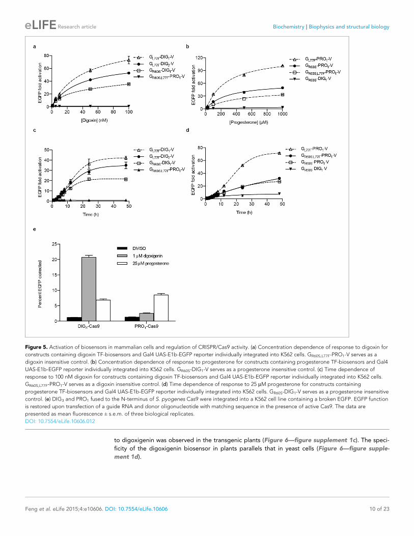
Yeast-derived biosensors port directly to mammalian cells and can beused to tightly regulate CRISPR/Cas9 genome editingYeast is an attractive platform for engineering in vivo biosensors because of its rapid doubling time
and tractable genetics. If yeast-derived biosensors function in more complex eukaryotes, the design-
build-test cycle in those organisms could be rapidly accelerated. We first assessed the portability of
yeast TF-biosensors to mammalian cells. Single constructs containing digoxin and progesterone TF-
biosensors with the greatest dynamic ranges (without codon optimization) were stably integrated
into human K562 cells using PiggyBac transposition. We characterized the dynamics of the TF-bio-
sensors in human cells by dose response and time course assays similar to the yeast experiments
(Figure 5a–d). As with yeast, the human cells demonstrated greater sensitivity to digoxin, with fluo-
rescence activation increasing up to 100 nM of cognate ligand for digoxin biosensors and 1 mM for
progesterone biosensors. We observed >100-fold activation for the most sensitive progesterone
biosensor GL77F-PRO1-V. The increase in mammalian dynamic range over yeast may arise from more
aggressive degradation of destabilized biosensors or greater accumulation of target-stabilized bio-
sensors or reporters resulting from larger cell sizes and slower doubling times. The time course data
show that fluorescence increased four-fold within 4 hr of target introduction and rose logarithmically
for 24–48 hr.
We next assessed whether these biosensors could drive more complex mammalian phenotypes.
The CRISPR/Cas9 system has proved to be an invaluable tool for genome editing (Mali et al., 2013;
DiCarlo et al., 2013; Gratz et al., 2013; Hwang, 2013). Despite the high programmability and
specificity of Cas9-mediated gene editing achieved to date, unchecked Cas9 activity can lead to off-
target mutations and cytotoxicity (Fu et al., 2013; Mali et al., 2013; Pattanayak et al., 2013). Fur-
ther, it may be desirable to tightly regulate Cas9 activity such that gene editing occurs only under
defined conditions. To facilitate inducible gene editing, we fused human codon-optimized versions
of the DIG3 and PRO1 LBDs to the N-terminus of Cas9 from S. pyogenes. We integrated this con-
struct into a reporter cell line containing an EGFP variant with a premature stop codon that renders
it non-functional. Upon separate stable integration of the DIG-Cas9 and PRO-Cas9 fusions, we trans-
fected a guide RNA targeting the premature stop codon as well as a donor oligonucleotide contain-
ing the sequence to restore EGFP activity via homologous recombination. After a 48-hr incubation
period, we observed an ~18-fold increase in GFP positive cells with digoxigenin relative to the mock
control (Figure 5e).
Environmental detection in the plant Arabidopsis thalianaTo assess generalizability of these sensors to multicellular organisms, we engineered G-DIG1-V to
function as an environmental biosensor in plants. The DIG1 sequence was codon optimized for
expression in Arabidopsis thaliana. We tested biosensor fusions to two different degrons, Mata2
from yeast and DREB2a from Arabidopsis (Sakuma et al., 2006), and we used the VP16 and VP64
variants as the TAD. We initially tested the G-DIG1-TAD variants with a transient expression assay
using Arabidopsis protoplasts and a reporter gene consisting of firefly luciferase under the control
of a Gal4-activated plant promoter (pUAS::Luc). The biosensor containing the Mata2 degron and
VP16 TAD showed the highest fold activation of luciferase in the presence of digoxigenin (Figure 6—
figure supplement 1a). We next inserted the genes encoding G-DIG1-V-Mata2 and the Gal4-acti-
vated pUAS::Luc into a plant transformation vector and stably transformed them into Arabidopsis
plants. Primary transgenic plants were screened in vivo for digoxigenin-dependent luciferase produc-
tion, and responsive plants were allowed to set seed for further testing. Second generation trans-
genic plants (T1, heterozygous) were tested for digoxin- or digoxigenin-dependent induction of
luciferase expression. After 42 hr, we observed 30-50-fold induction of luciferase activity in digoxin-
treated plants compared to the uninduced control (Figure 6). Both digoxin and digoxigenin were
capable of inducing the biosensor. Digoxigenin-dependent luciferase induction was observed in mul-
tiple independent transgenic T1 lines (Figure 6—figure supplement 1b), and a rising dose response
Feng et al. eLife 2015;4:e10606. DOI: 10.7554/eLife.10606 9 of 23
Research article Biochemistry Biophysics and structural biology
to digoxigenin was observed in the transgenic plants (Figure 6—figure supplement 1c). The speci-
ficity of the digoxigenin biosensor in plants parallels that in yeast cells (Figure 6—figure supple-
ment 1d).
Figure 5. Activation of biosensors in mammalian cells and regulation of CRISPR/Cas9 activity. (a) Concentration dependence of response to digoxin for
constructs containing digoxin TF-biosensors and Gal4 UAS-E1b-EGFP reporter individually integrated into K562 cells. GR60S,L77F-PRO1-V serves as a
digoxin insensitive control. (b) Concentration dependence of response to progesterone for constructs containing progesterone TF-biosensors and Gal4
UAS-E1b-EGFP reporter individually integrated into K562 cells. GR60S-DIG1-V serves as a progesterone insensitive control. (c) Time dependence of
response to 100 nM digoxin for constructs containing digoxin TF-biosensors and Gal4 UAS-E1b-EGFP reporter individually integrated into K562 cells.
GR60S,L77F-PRO1-V serves as a digoxin insensitive control. (d) Time dependence of response to 25 mM progesterone for constructs containing
progesterone TF-biosensors and Gal4 UAS-E1b-EGFP reporter individually integrated into K562 cells. GR60S-DIG1-V serves as a progesterone insensitive
control. (e) DIG3 and PRO1 fused to the N-terminus of S. pyogenes Cas9 were integrated into a K562 cell line containing a broken EGFP. EGFP function
is restored upon transfection of a guide RNA and donor oligonucleotide with matching sequence in the presence of active Cas9. The data are
presented as mean fluorescence ± s.e.m. of three biological replicates.
DOI: 10.7554/eLife.10606.012
Feng et al. eLife 2015;4:e10606. DOI: 10.7554/eLife.10606 10 of 23
Research article Biochemistry Biophysics and structural biology

DiscussionIn vivo biosensors for small molecules enable the regulation and detection of cellular responses to
endogenous metabolites and exogenous chemicals. Here, we show that LBDs can be conditionally
destabilized to create biosensors that function in yeast, mammalian cells, and plants, and we demon-
strate the use of these biosensors in metabolic engineering and genome editing applications. While
this method requires a high-affinity ligand-binding domain as a starting point, nearly all small mole-
cules of interest have a natural protein interactor. Furthermore, the use of de novo-designed binders
opens the possibility of generating biosensors for ligands with unsuitable or unknown binding pro-
teins. By incorporating standard mutagenesis and screening, our method constitutes a simple plat-
form for sensor development that can be applied to many areas of biotechnology. These sensors act
either at the level of post-translational control over protein function or at the level of transcription
(Figure 7a), and they can be tuned by altering any of their components (Figure 7b) or by modifying
efflux of the target ligand in the host organism. These tunable features should make the biosensors
useful in many different cellular and environmental contexts.
Our results suggest a general mechanism of conditional stabilization for LBDs, allowing the ratio-
nal development of biosensors for other targets. Furthermore, the portability of the mutations we
identified suggests a structural basis for conferring conditional stability to the DIG0 LBD scaffold.
Both the DIG0 LBD and Gal4 are homodimers, and the majority of the conditionally-stabilizing muta-
tions are located at the dimer interfaces. A computational model of the Gal4-DIG0 complex indicates
that the orientation of the two domains allows a homodimeric fusion to form (Figure 2b). These
results suggest an allosteric interplay between ligand binding and dimer formation: weakening of
the dimer interface, in either the DIG0 or the Gal4 domain, is compensated by ligand binding. This
LBD scaffold is derived from a member of the nuclear transport factor 2 family, a fold class that typi-
cally has a large dimer interface (~1200 A2) that facilitates the large and open ligand-binding site
Figure 6. Application of biosensors in plants. (a) Activation of luciferase expression in transgenic Arabidopsis plants containing the G-DIG1-V biosensor
in the absence (left) or presence (right) of 100 mM digoxin. Luciferase expression levels are false colored according to scale to the right. Relative
luciferase units corresponding to 1 min of image pixel integration (to avoid saturating pixels) are shown above each individual plant. (b) Brightfield
image of plants shown in a.
DOI: 10.7554/eLife.10606.013
The following figure supplement is available for figure 6:
Figure supplement 1. Characterization of DIG biosensor in plants.
DOI: 10.7554/eLife.10606.014
Feng et al. eLife 2015;4:e10606. DOI: 10.7554/eLife.10606 11 of 23
Research article Biochemistry Biophysics and structural biology

(~600 A2). These protein folds are well suited for de novo design of other LBDs because of their
large binding pocket and natural substrate diversity (Todd et al., 2002). Exploiting dimer interfaces
to modulate stability without impairing ligand binding may be a general mechanism to confer condi-
tional stability on LBDs. This possibility is supported by the observation that interface mutations in
DIG0 and Gal4 conferring digoxin-dependent stability lead to progesterone-dependent stability in a
progesterone biosensor (Figure 1—figure supplement 2a).
A long-standing challenge in metabolic engineering is to rapidly detect and control how changes
to the regulation and composition of biosynthetic pathways affect product titers. Transcriptional
control by a product or intermediate (Zhang et al., 2012; Raman et al., 2014; Tang and Cirino,
2011) and directed evolution of constituent pathway elements (Agresti et al., 2010; Alper et al.,
2005; Dietrich et al., 2013) have emerged as promising strategies towards this goal. These
approaches require high selectivity against intermediates (Zhang and Keasling, 2011), a feature
demonstrated here that can be explicitly considered during the computational design and screening
process. Our method allows biosensors to be generated that are highly selective for a small mole-
cule, facilitating a simple directed evolution strategy without requiring prior structural or bioinfor-
matic knowledge about the targeted enzyme(s) or pathway(s). Because the biosensors are TF-based,
sophisticated systems of optimizing metabolic output, such as dynamic control of gene
expression (Zhang et al., 2012) and feedback-regulated genome evolution (Chou and Keasling,
2013), are possible.
A reliance on the general principles of protein stability and ligand binding allows the develop-
ment of biosensors that function in any organism with similar protein quality control machinery.
Here, we engineered biosensors based on a designed scaffold derived from a bacterial protein.
These biosensors required only minimal modifications to retain high levels of sensitivity when devel-
oped in yeast and deployed across mammalian and plant species, demonstrating unprecedented
portability for biosensors. In some cases, these biosensors showed a greater dynamic range in mam-
malian cells relative to yeast, possibly due to larger cell volume and variations in protein degradation
machinery. Further work using biosensors based on multiple different LBD scaffolds and introduced
into diverse organisms will allow us to better understand the principles by which biosensor variability
across hosts arises.
Small molecule biosensors with the modularity incorporated here enable diverse cellular
responses to a variety of exogenous and endogenous signals (Banaszynski et al., 2008). Gene edit-
ing is an area that requires particularly tight coupling of cell response to activation signals. The
CRISPR/Cas9 system provides a facile and robust genome-editing platform, but it can result in off-
target genetic changes (Fu et al., 2013; Mali et al., 2013; Pattanayak et al., 2013). Proposed solu-
tions include optimizing guide RNA sequences (Fu et al., 2014; Cho et al., 2014), building chimeric
Figure 7. Schematic of biosensor platform. (a) Biosensors for small molecules are modularly constructed by replacing the LBD with proteins possessing
altered substrate preferences. (b) Activity of the biosensor can be tuned by 1) introducing destabilizing mutations (red Xs), 2) adding a degron, 3)
altering the strength of the TAD or DNA binding affinity of the TF, 4) changes in the number of TF-binding sites or sequences, and 5) titrating 3-
aminotriazole, an inhibitor of His3. (c) Yeast provide a genetically tractable chassis for biosensor development before implementation in more complex
eukaryotes, such as mammalian cells and plants.
DOI: 10.7554/eLife.10606.015
Feng et al. eLife 2015;4:e10606. DOI: 10.7554/eLife.10606 12 of 23
Research article Biochemistry Biophysics and structural biology

Cas9 fusions requiring the presence of two Cas9 molecules in close proximity (Mali et al., 2013;
Tsai et al., 2014; Guilinger et al., 2014; Ran et al., 2013), and regulating Cas9 activity by chemical
or light-based inducers (Dow et al., 2015; Zetsche et al., 2015; Polstein and Gersbach, 2015).
While small molecule inducers including doxycycline and rapamycin have been used, these molecules
may confer leaky expression and cytotoxicity (Xie et al., 2008). Thus, an expanded chemical reper-
toire is needed for tightly regulated gene editing and gene therapy applications. By exploiting the
low background of the LBD-biosensors, we produced biosensor-Cas9 fusions with tightly controlled
activation (Figure 5e). This switch-like control over CRISPR/Cas9 activity could reduce background
activity and off-target editing, a critical feature for safer gene therapies (Mandal, et al.,
2014; Wu et al., 2013; Schwank et al., 2013).
Our biosensor design approach should have numerous applications in agriculture. For example,
biosensors could be developed to enable plants to monitor the environment for pollutants, toxins or
dangerous compounds. Coupling biosensors with a phytoremediation trait could enable plants to
both sense a contaminant and activate a bioremediation gene circuit. When paired with an agro-
nomic or biofuel trait, such biosensors could serve as triggers for bioproduction. In the transgenic
Arabidopsis plants, we observed ligand-dependent activation in all cells, tissues and organs exam-
ined (Figure 6).
The technology introduced here operates at either the transcriptional or post-translational level.
These biosensors can be developed in yeast and readily transferred with minimal modification to
other eukaryotic cell types, where they retain a high level of sensitivity (Figure 7c). The generality of
our approach arises from the universality of the transcriptional activation and protein degradation
machinery across eukaryotes, together with the modularity and tunability of the constituent parts.
These biosensors should find broad application, including improving metabolically engineered path-
way flux and product titers, exerting ligand-dependent control over genome editing, and detecting
exogenous small molecules or endogenous metabolites.
Materials and methods
Culture and growth conditionsBiological replicates are defined as samples inoculated from distinct colonies. Growth media con-
sisted of YPAD (10 g/L yeast extract, 20 g/L peptone, 40 mg/L adenine sulfate, 20 g/L glucose) and
SD media (1.7 g/L yeast nitrogen base without amino acids, 5 g/L ammonium sulfate, 20 g/L glucose
and the appropriate amount of dropout base with amino acids [Clontech, Mountain View, CA]). The
following selective agents were used when indicated: G418 (285 mg/L), pen/strep (100 U/mL penicil-
lin and 100 mg/mL streptomycin).
LBD-yEGFP library constructionThe DIG10.3 sequence (Tinberg et al., 2013) was cloned by Gibson assembly (Gibson et al., 2009)
into a pUC19 plasmid containing yeast enhanced GFP (yEGFP, UniProt ID B6UPG7) and a KanMX6
cassette flanked by 1000 and 500 bp upstream and downstream homology to the HO locus. The
DIG10.3 sequence was randomized by error-prone PCR using a Genemorph II kit from Agilent Tech-
nologies. An aliquot containing 100 ng of target DNA (423 bp out of a 7.4 kb plasmid) was mixed
with 5 mL of 10X Mutazyme buffer, 1 mL of 40 mM dNTPS, 1.5 mL of 20 mM forward and reverse
primer containing 90 bp overlap with the pUC19 plasmid (oJF70 and oJF71), and 1 mL of Mutazyme
polymerase in 50 mL. The reaction mixture was subject to 30 cycles with Tm of 60˚C and extension
time of 1 min. Vector backbone was amplified using Q5 polymerase (NEB, Ipswich, MA) with oJF76
and oJF77 primers with Tm of 65˚C and extension time of 350 s. Both PCR products were isolated
by 1.5% agarose gel electrophoresis and the randomized target was inserted as a genetic fusion to
yEGFP by Gibson assembly (Gibson et al., 2009). Assemblies were pooled, washed by ethanol pre-
cipitation, and resuspended in 50 mL of dH2O, which was drop dialyzed (EMD Millipore, Billerica,
MA) and electroporated into E. cloni supreme cells (Lucigen, Middleton, WI). Sanger sequencing of
16 colonies showed a mutation rate of 0–7 mutations/kb. The library was expanded in culture and
maxiprepped (Qiagen, Valencia, CA) to 500 mg/ml aliquots. 16 mg of library was drop dialyzed and
electrotransformed into yeast strain Y7092 for homologous recombination into the HO locus.
Feng et al. eLife 2015;4:e10606. DOI: 10.7554/eLife.10606 13 of 23
Research article Biochemistry Biophysics and structural biology
Integrants were selected by growth on YPAD solid media containing G418 followed by outgrowth in
YPAD liquid media containing G418.
LBD-yEGFP library selectionsLibraries of DIG0-yEGFP and PRO0-yEGFP integrated into yeast strain Y7092 were subject to three
rounds of fluorescence activated sorting in a BD FACSAria IIu. For the first round, cells were grown
overnight to an OD600 of ~1.0 in YPAD containing steroid (500 mM digoxigenin or 50 mM progester-
one), and cells showing the top 5% of fluorescence activation were collected and expanded over-
night to an OD600 of ~1.0 in YPAD lacking steroid. In the second sort, cells displaying the lowest
~3% fluorescence activation were collected. Cells passing the second round were passaged over-
night in YPAD containing steroid to an OD600 of ~1.0 and sorted once more for the upper 5% of
fluorescence activation. The sorted libraries were expanded in YPAD liquid culture and plated on
solid YPAD media. Ninety-six colonies from each library were clonally isolated and grown overnight
in deep well plates containing 500 mL of YPAD. Candidates were diluted 1:50 into two deep well
plates with SD-complete media: one plate supplemented with steroid and the other with DMSO
vehicle. Cells were grown for another 4 h, and then diluted 1:3 into microtitre plates of 250 mL of the
same media. Candidates were screened by analytical flow cytometry on a BD LSRFortessa cell ana-
lyzer. The forward scatter, side scatter, and yEGFP fluorescence (530 nm band pass filter) were
recorded for a minimum of 20,000 events. FlowJo X software was used to analyze the flow cytometry
data. The fold activation was calculated by normalizing mean yEGFP fluorescence activation for each
steroid to the mean yEGFP fluorescence in the DMSO only control. Highest induction candidates
were subject to Sanger sequencing with primers flanking the LBD sequence.
TF-biosensor reporter plasmid construction and integrationReporter genes were cloned into the integrative plasmid pUG6 or the CEN plasmid pRS414 using
the Gibson method (Gibson et al., 2009). Each reporter (either yEGFP or firefly luciferase) was
cloned to include a 5’ GAL1 promoter (S. cerevisiae GAL1 ORF bases (-455)-(-5)) and a 3’ CYC1 ter-
minator. For integration, linearized PCR cassettes containing both the reporter and an adjacent
KanMX antibiotic resistance cassette were generated using primers containing 50 bp flanking
sequences of homology to the URA3 locus. Integrative PCR product was transformed into the yeast
strain PJ69-4a using the Gietz method (Gietz and Schiestl, 2007) to generate integrated reporter
strains.
G-DIG/PRO-V plasmid constructionG-DIG/PRO-V fusion constructs were prepared using the Gibson method (Gibson et al., 2009). Con-
structs were cloned into the plasmid p416CYC (p16C). Gal4 (residues 1–93, UniProt ID P04386),
DIG10.3 (Tinberg et al., 2013), and VP16 (residues 363-490, UniProt ID P06492) PCR products for
were amplified from their respective templates using Phusion high-fidelity polymerase (NEB, Wal-
tham, MA) and standard PCR conditions (98˚C 10 s, 60˚C 20 s, 72˚C 30 s; 30 cycles). The 8-residue
linker sequence GGSGGSGG was used between Gal4 and DIG10.3. PCR primers were purchased
from Integrated DNA technologies and contained 24–30 5’ bases of homology to either neighboring
fragments or plasmid. Clones containing an N-terminal degron were similarly cloned fusing residues
1–67 of Mata2 (UniProt ID P0CY08) to the 5’- end of G-DIG-V. Plasmids were transformed into yeast
using the Gietz method (Gietz and Schiestl, 2007), with transformants being plated on synthetic
complete media lacking uracil (SD -ura).
G-DIG-V mutant constructionMutations were introduced into DIG10.3/pETCON (Tinberg et al., 2013) or the appropriate G-DIG/
PRO-V construct using Kunkel mutagenesis (Kunkel, 1985). Oligos were ordered from Integrated
DNA Technologies, Inc. For mutants constructed in pETCON/DIG10.3, the mutagenized DIG10.3
gene was amplified by 30 cycles of PCR (98˚C 10 s, 61˚C 30 s, 72˚C 15 s) using Phusion high-fidelity
polymerase (NEB) and 5’- and 3’- primers having homologous overlap with the DIG10.3-flanking
regions in p16C-G-DIG-VP64 (Gal4_DIG10.3_VP64_hr_fwd and Gal4_DIG10.3_VP64_hr_rev_rc).
Genes were inserted into p16C-Gal4-(HE)-VP16 by Gibson assembly (Gibson et al., 2009) using vec-
tor digested with HindIII and EcoRI-HF.
Feng et al. eLife 2015;4:e10606. DOI: 10.7554/eLife.10606 14 of 23
Research article Biochemistry Biophysics and structural biology

G-PRO-V mutant constructionThe gene for DIG10.3 Y34F/Y99F/Y101F was amplified from the appropriate DIG10.3/
pETCON (Tinberg et al., 2013) construct by 30 cycles of PCR (98˚C 10 s, 59˚C 30 s, 72˚C 15 s) using
Phusion high-fidelity polymerase (NEB) and 5’- and 3’- primers having homologous overlap with the
DIG10.3-flanking regions in p16C-G-DIG-VP64 (DIG_fwd and DIG_rev). Genes were inserted into
p16C-GDVP16 by Gibson assembly (Gibson et al., 2009) using p16C-Gal4-(HE)-VP16 vector
digested with HindIII and EcoRI-HF.
G-DIG-V error-prone library constructionA randomized G-DIG-V library was constructed by error-prone PCR using a Genemorph II kit from
Agilent Technologies (Santa Clara, CA). An aliquot containing 20 ng p16C GDVP16, 20 ng p16C
GDVP16 E83V, and 20 ng p16C Y36H was mixed with 5 mL of 10X Mutazyme buffer, 1 mL of 40 mM
dNTPS, 1.5 mL of 20 mM forward and reverse primer containing 37- and 42-bp overlap with the p16C
vector for homologous recombination, respectively (GDV_ePCR_fwd and GDV_ePRC_rev), and 1 mL
of Mutazyme polymerase in 50 mL. The reaction mixture was subjected to 30 cycles of PCR (95˚C 30
s, 61˚C 30 s, 72˚C 80 s). Template plasmid was digested by adding 1 mL of DpnI to the reaction mix-
ture and incubating for 3 hr at 37˚C. Resulting PCR product was purified using a Quiagen PCR
cleanup kit, and a second round of PCR was used to amplify enough DNA for transformation. Gene
product was amplified by combining 100 ng of mutated template DNA with 2.5 mL of 10 mM primers
(GDV_ePCR_fwd and GDV_ePRC_rev), 10 mL of 5X Phusion buffer HF, 1.5 mL of DMSO, and 1 mL of
Phusion high-fidelity polymerase (NEB, Waltham, MA) in 50 mL. Product was assembled by 30 cycles
of PCR (98˚C 10 s, 65˚C 30 s, 72˚C 35 s). Following confirmation of a single band at the correct
molecular weight by 1% agarose gel electrophoresis, the PCR product was purified using a Quaigen
PCR cleanup kit and eluted in ddH2O. Yeast strain PyE1 DPDR5 was transformed with 9 mg of ampli-
fied PCR library and 3 mg of p16C Gal4-(HE)-VP16 triply digested with SalI-HF, BamHI-HR, and
EcoRI-HF using the method of Benatuil (Benatuil et al., 2010), yielding ~106 transformants. Follow-
ing transformation, cells were grown in 150 mL of SD -ura media. Sanger sequencing of 12 individual
colonies revealed an error rate of ~1–6 mutations per gene.
G-DIG-V library selectionsAn error-prone library of G-DIG0/DIG1/DIG2/-V transformed into yeast strain PyE1 DPDR5 was sub-
jected to three rounds of cell sorting using a Cytopeia (BD Influx) fluorescence activated cell sorter.
For the first round, cells displaying high fluorescence in the presence of digoxin (on-state) were col-
lected. Transformed cells were pelleted by centrifugation (4 min, 4000 rpm) and resuspended to a
final OD600 of 0.1 in 50 mL of SD -ura media, pen/step antibiotics, and 5 mM digoxin prepared as a
100 mM solution in DMSO. The library was incubated at 30˚C for 9 hr and then sorted. Cells display-
ing the highest fluorescent values in the GFP channel were collected (1,747,058 cells collected of
32,067,013 analyzed; 5.5%), grown up at 30˚C in SD -ura, and passaged twice before the next sort.
For the second round of sorting, cells displaying low fluorescence in the absence of digoxin (off-
state) were collected. Cells were pelleted by centrifugation (4 min, 4000 rpm) and resuspended to a
final OD600 of 0.1 in 50 mL of SD -ura media supplemented with pen/strep antibiotics. The library
was incubated at 30˚C for 8 hr and then sorted. Cells displaying low fluorescent values in the GFP
channel were collected (1,849,137 cells collected of 22,290,327 analyzed; 11.1%), grown up at 30˚Cin SD -ura, and passaged twice before the next sort. For the last sorting round, cells displaying high
fluorescence in the presence of digoxin (on-state) were collected. Cells were prepared as for the first
sort. Cells displaying the highest fluorescent values in the GFP channel were collected (359,485 cells
collected of 31,615,121 analyzed; 1.1%). After the third sort, a portion of cells were plated and
grown at 30˚C. Plasmids from 12 individual colonies were harvested using a Zymoprep Yeast mini-
prep II kit (Zymo Research Corporation, Irvine, CA) and the gene was amplified by 30 cycles of PCR
(98˚C 10 s, 52˚C 30 s, 72˚C 40 s) using Phusion high-fidelity polymerase (NEB) with the T3 and T7 pri-
mers. Sanger sequencing (Genewiz, Inc., South Plainfield, NJ) was used to sequence each clone in
the forward (T3) and reverse (T7) directions.
Feng et al. eLife 2015;4:e10606. DOI: 10.7554/eLife.10606 15 of 23
Research article Biochemistry Biophysics and structural biology

G-DIG-V error-prone library mutation screensOf 12 sequenced clones from the library sorts, two showed significantly improved (>2-fold) response
to DIG over the input clones (clone 3 and clone 6). Clone 3 contains the following mutations:
Gal4_T44T (silent), Gal4_L77F, DIG10.3_E5D, DIG10.3_E83V, DIG10.3_R108R (silent),
DIG10.3_L128P, DIG10.3_I137N, DIG10.3_S143G, and VP16_A44T. Clone 6 contains the following
mutations: Gal4_R60S, Gal4_L84L (silent), VP16_G17G (silent), VP16_L48V, and VP16_H98H (silent).
To identify which mutations led to the observed changes in DIG response, variants of these clones
with no silent mutations and each individual point mutant were constructed using Kunkel
mutagenesis (Kunkel, 1985). Oligos were ordered from Integrated DNA Technologies, Inc.
Sequence-confirmed plasmids were transformed into PyE1 DPDR5f and plated onto selective SD -
ura media. Individual colonies were inoculated into liquid media, grown at 30˚C, and passaged once.
Cells were pelleted by centrifugation (4 min, 1700 � g) and resuspended to a final OD660 of 0.1 in 1
mL of SD -ura media supplemented 50 mM DIG prepared as a 100 mM solution in DMSO. Following
a 6 hr incubation at 30˚C, cells were pelleted, resuspended in 200 mL of PBS, and cellular fluores-
cence was measured on an Accuri C6 flow cytometer using a 488 nm laser for excitation and a 575
nm band pass filter for emission. FlowJo software version 7.6 was used to analyze the flow cytometry
data. The data are given as the mean yEGFP fluorescence of the single yeast population in the
absence of DIG (off-state) and the mean yEGFP fluorescence of the higher fluorescing yeast popula-
tion in the presence of DIG (on-state).
Computational model of Gal4-DIGA model of the Gal4-DIG10.3 fusion was built using Rosetta Remodel (Huang et al., 2011) to assess
whether the linker between Gal4 and the DIG LBD, which are both dimers, would allow for the for-
mation of a dimer in the fusion construct. In the simulation, the Gal4 dimer was held fixed while the
relative orientation of the DIG LBD monomers were sampled symmetrically using fragment insertion
in the linker region. Constraints were added across the DIG LBD dimer interface to facilitate sam-
pling. The lowest energy model satisfied the dimer constraints, indicating that a homodimer configu-
ration of the fusion is possible.
TF-biosensor titration assays in yeastYeast strain PyE1 transformed with p16C plasmids containing G-LBD-V variants were inoculated
from colonies into SD –ura media supplemented and grown at 30˚C overnight (16 h). 10 mL of the
culture was resuspended into 490 mL of separately prepared media each containing a steroid of
interest (SD –ura media supplemented the steroid of interest and DMSO to a final concentration of
1% DMSO). Resuspended cultures were then incubated at 30˚C for 8 hr. 125 mL of incubated culture
was resuspended into 150 mL of fresh SD –ura media supplemented with the steroid of interest and
DMSO to a final concentration of 1%. These cultures were then assayed by analytical flow cytometry
on a BD LSRFortessa using a 488 nm laser for excitation. The forward scatter, side scatter, and
yEGFP fluorescence (530 nm band pass filter) were recorded for a minimum of 20,000 events.
FlowJo X software was used to analyze the flow cytometry data. The fold activation was calculated
by normalizing mean yEGFP fluorescence activation for each steroid to the mean yEGFP fluores-
cence in the DMSO only control. G-PRO0-V was assayed on a separate day from the other TF biosen-
sors under identical conditions.
TF-biosensor kinetic assays in yeastYeast strain PyE1 was transformed with p16C plasmids containing G-LBD-V variants were inoculated
from colonies into SD –ura media and grown at 30˚C overnight (16 hr). 5 mL of each strain was
diluted into 490 mL of SD –ura media in 2.2 mL plates. Cells were incubated at 30˚C for 8 hr. 5 mL of
steroid was then added for a final concentration of 250 mM digoxin or 50 mM progesterone. For
each time point, strains were diluted 1:3 into microtitre plates of 250 mL of the same media. Strains
were screened by analytical flow cytometry on a BD LSRFortessa cell analyzer. The forward scatter,
side scatter, and yEGFP fluorescence (530 nm band pass filter) were recorded for a minimum of
20,000 events. FlowJo X software was used to analyze the flow cytometry data. The fold activation
was calculated by normalizing mean yEGFP fluorescence activation for each time point to the mean
yEGFP fluorescence at T = 0 hr.
Feng et al. eLife 2015;4:e10606. DOI: 10.7554/eLife.10606 16 of 23
Research article Biochemistry Biophysics and structural biology

Luciferase reporter assayYeast strains containing either a plasmid-borne or integrated luciferase reporter were transformed
with p16C plasmids encoding TF-biosensors. Transformants were grown in triplicate overnight at
30˚C in SD –ura media containing 2% glucose in sterile glass test tubes on a roller drum. After ~16
hr of growth, OD600 of each sample was measured and cultures were back diluted to OD600 = 0.2 in
fresh SD –ura media containing steroid dissolved in DMSO or a DMSO control (1% DMSO final). Cul-
tures were grown at 30˚C on roller drum for 8 hr prior to taking readings. Measurement of luciferase
activity was adapted from a previously reported protocol(Leskinen et al., 2003). 100 mL of each cul-
ture was transferred to a 96-well white NUNC plate. 100 mL of 2 mM D-luciferin in 0.1 M sodium cit-
rate (pH 4.5) was added to each well of the plate and luminescence was measured on a Victor 3V
after 5 min.
Yeast deletion strain creationGenomic deletions were introduced into the yeast strains PJ69-4a and PyE1 using the 50:50
method (Horecka and Davis, 2014). Briefly, forward and reverse primers were used to amplify an
URA3 cassette by PCR. These primers generated a product containing two 50 bp sequences homol-
ogous to the 5’ and 3’ ends of the ORF at one end and a single 50 bp sequence homologous to the
middle of the ORF at the other end. PCR products were transformed into yeast using the Gietz
method (Gietz and Schiestl, 2007) and integrants were selected on SD –ura plates. After integration
at the correct locus was confirmed by a PCR screen, single integrants were grown for 2 days in YEP
containing 2.5% ethanol and 2% glycerol. Each culture was plated on synthetic complete plates con-
taining 5-fluoroorotic acid. Colonies were screened for deletion of the ORF and elimination of the
Ura3 cassette by PCR and confirmed by Sanger sequencing.
TF-biosensor specificity assaysYeast strains expressing the TF-biosensors and yEGFP reporter (either genetically fused or able to
be transcriptionally activated by the TAD) were grown overnight at 30˚C in SD –ura media for 12 hr.
Following overnight growth, cells were pelleted by centrifugation (5 min, 5250 rpm) and resus-
pended into 500 mL of SD –ura. 10 mL of the washed culture was resuspended into 490 mL of sepa-
rately prepared media each containing a steroid of interest (SD –ura media supplemented with the
steroid of interest and DMSO to a final concentration of 1% DMSO). Steroids were tested at a con-
centration of 100 mM digoxin, 50 mM progesterone, 250 mM pregnenolone, 100 mM digitoxigenin,
100 mM beta-estradiol, and 100 mM hydrocortisone. Stock solutions of steroids were prepared as a
50 mM solution in DMSO.
Resuspended cultures were then incubated at 30˚C for 8 hr. 125 mL of incubated culture was
resuspended into 150 mL of fresh SD –ura media supplemented the steroid of interest, and DMSO to
a final concentration of 1%. These cultures were then assayed by analytical flow cytometry on a BD
LSRFortessa using a 488 nm laser for excitation. The forward scatter, side scatter, and yEGFP fluores-
cence (530 nm band pass filter) were recorded for a minimum of 20,000 events. FlowJo X software
was used to analyze the flow cytometry data. The fold induction was calculated by normalizing mean
yEGFP fluorescence activation for each steroid to the mean yEGFP fluorescence in the DMSO only
control.
Yeast spotting assaysYeast strain PJ69-4a transformed with p16C plasmids containing degron-G-DIG-V variants were first
inoculated from colonies into SD -ura media and grown at 30˚C overnight (16 hr). 1 mL of each cul-
ture was pelleted by centrifugation (3000 rcf, 2 min), resuspended in 1 mL of fresh SD -ura and the
OD660 was measured. Each culture was then diluted in SD -ura media to an OD660 = 0.2 and incu-
bated at 30˚C for 4–6 hr. 1 mL of each culture was pelleted and resuspended in sterile, distilled
water and the OD660 measured again. Each transformant was then diluted to an OD660 = 0.1. Four
1/10 serial dilutions of each culture were prepared in sterile water (for a total of 5 solutions). 10 mL
of each dilution was spotted in series onto several SD –ura –his agar plates containing 1 mM 3-ami-
notriazole and the indicated steroid. Steroid solutions were added to agar from 200x steroid solu-
tions in DMSO (0.5% DMSO final in plates).
Feng et al. eLife 2015;4:e10606. DOI: 10.7554/eLife.10606 17 of 23
Research article Biochemistry Biophysics and structural biology

3b-HSD plasmid and library constructionThe 3b-HSD ORF was synthesized as double-stranded DNA (Integrated DNA Technologies,
Inc., Coralville, IA) and amplified using primers oJF325 and oJF326 using KAPA HiFi under standard
PCR conditions and digested with BsmBI to create plasmid pJF57. 3b-HSD expression plasmids
(pJF76 through pJF87) were generated by digesting plasmid pJF57 along with corresponding plas-
mids from the Yeast Cloning Toolkit (Lee et al., 2015) with BsaI and assembled using the Golden
Gate Assembly method (Engler et al., 2008). The 3b-HSD sequence was randomized by error-prone
PCR using a Genemorph II kit from Agilent Technologies. An aliquot containing 100 ng of target
DNA was mixed with 5 mL of 10X Mutazyme buffer, 1 mL of 40 mM dNTPS, 1.5 mL of 20 mM forward
and reverse primer containing 90-bp overlap with the 3b-HSD expression plasmids and 1 mL of Muta-
zyme polymerase in 50 mL. The reaction mixture was subject to 30 cycles with Tm of 60˚C and exten-
sion time of 1 min. Vector backbone was amplified using KAPA HiFi polymerase with oJF387 and
oJF389 (pPAB1) or oJF387 and oJF389 (pPOP6) with Tm of 65˚C and extension time of 350 s. PCR
products were isolated by 1.5% agarose gel electrophoresis and assembled using the Gibson
method (Gibson et al., 2009). Assemblies were pooled, washed by ethanol precipitation, and resus-
pended in 50 mL of dH2O, which was drop dialyzed (Millipore) and electroporated into E. cloni
supreme cells (Lucigen). Sanger sequencing of 16 colonies showed a mutation rate of 0–4 muta-
tions/kb. The library was expanded in culture and maxiprepped (Qiagen) to 500 mg/mL aliquots. 16
mg of library was drop dialyzed and electrotransformed into yeast strain PyE1.
3b-HSD progesterone selectionsPyE1 transformed with libraries of 3b-HSD were seeded into 5 mL of SD –ura –leu media supple-
mented and grown at 30˚C overnight (24 hr). Cultures were measured for OD600, diluted to an
OD600 of 0.0032, and 100 mL was plated onto SD –ura –leu –his plates supplemented 35 mM 3-AT
and either 50 mM pregnenolone or 0.5% DMSO.
Progesterone bioproduction and GC/MS analysisProduction strains were inoculated from colonies into 5 mL SD –ura media and grown at 30˚C over-
night (16 hr). 1 mL of each culture was washed and resuspended into 50 mL of SD –ura with 250 mM
of pregnenolone and grown at 30˚C for 76 hr. OD600 measurements were recorded for each culture
before pelleting by centrifugation. Cells were lysed by glass bead disruption, and lysates and growth
media were extracted separately with heptane. Extractions were analyzed by GC/MS.
TF-biosensor EGFP assays in mammalian cellsThe K562 cell line was obtained from the ATCC. The cell line was not authenticated and not tested
for mycoplasma contamination. For each TF-biosensor, 1 mg of the PiggyBac construct along with
400 ng of transposase were nucleofected into K562 cells using the Lonza Nucleofection system as
per manufacturer settings. Two days post-transfection, cells underwent puromycin selection (2 mg/
mL) for at least eight additional days to allow for unintegrated plasmid to dilute out and ensure that
all cells contained the integrated construct. An aliquot of 100,000 cells of each integrated population
were then cultured with 25 mM of progesterone, 1 mM of digoxigenin, or no small molecule. Forty-
eight hours after small molecule addition, cells were analyzed by flow cytometry using a BD Bioscien-
ces Fortessa system. Mean EGFP fluorescence of the populations was compared.
Construction of K562 cell linesThe PiggyBac transposase system was employed to integrate biosensor constructs into K562 cells.
Vector PB713B-1 (Systems Biosciences, Mountain View, CA) was used a backbone. Briefly, this back-
bone was digested with NotI and HpaI and G-LBD-V, Gal4BS-E1b-EGFP (EGFP; enhanced GFP ref
or UniProt ID A0A076FL24), and sEF1-Puromycin were cloned in. Gal4BS represents four copies of
the binding sequence. For hCas9, the PiggyBac system was also employed, but the biosensors were
directly fused on the N-terminus of Cas9 and were under the control of the CAGGS promoter. Cas9
from S. pyogenes was used.
Feng et al. eLife 2015;4:e10606. DOI: 10.7554/eLife.10606 18 of 23
Research article Biochemistry Biophysics and structural biology

TF-biosensor-Cas9 assaysConstruct integration was carried out as for the Cas9 experiments for EGFP assays, except that the
constructs were integrated into K562 containing a broken EGFP reporter construct. Introduction of
an engineered nuclease along with a donor oligonucleotide can correct the EGFP and produce fluo-
rescent cells. Upon successful integration (~10 days after initial transfection), 500,000 cells were
nucleofected with 500 ng of guide RNA (sgRNA) and 2 mg of donor oligonucleotide. Nucleofected
cells were then collected with 200 mL of media and 50 mL aliquots were added to wells containing
950 mL of media. Each nucleofection was split into four separate wells containing 1 mM of digoxige-
nin, 25 mM of progesterone, or no small molecule. Forty-eight hours later, cells were analyzed using
flow cytometry and the percentage of EGFP positive cells was determined.
TF-biosensor assays in protoplastsDigoxin transcriptional activators were initially tested in a transient expression assay using Arabidop-
sis protoplasts according previously described methods(Yoo et al., 2007), with some modifications.
Briefly, protoplasts were prepared from 6-week old Arabidopsis leaves excised from plants grown in
short days. Cellulase Onozuka R-10 and Macerozyme R-10 (Yakult Honsha, Inc., Japan) in buffered
solution were used to remove the cell wall. After two washes in W5 solution, protoplasts were re-sus-
pended in MMg solution at 2 � 105 cells/mL for transformation. Approximately 104 protoplasts were
mixed with 5 mg of plasmid DNA and PEG4000 at a final concentration of 20%, and allowed to incu-
bate at room temperature for 30 min. The transformation reaction was stopped by the addition of
two volumes of W5 solution, and after centrifugation, protoplasts were re-suspended in 200 mL of
WI solution (at 5 � 105/mL) and plated in a 96-well plate. Digoxigenin (Sigma-Aldrich, St. Louis, MO)
was added to the wells, and protoplasts were incubated overnight at room temperature in the dark,
with slight shaking (40 rpm). For luciferase imaging, protoplasts were lysed using Passive Lysis Buffer
(Promega, Madison, WI) and mixed with LARII substrate (Dual-Luciferase Reporter Assay System,
Promega, Madison, WI). Luciferase luminescence was collected by a Stanford Photonics XR/MEGA-
10 ICCD Camera and quantified using Piper Control (v.2.6.17) software.
Plant plasmid constructionG-DIG1-V was recoded to function as a ligand-dependent transcriptional activator in plants. Specifi-
cally, an Arabidopsis thaliana codon optimized protein degradation sequence from the yeast
MATa2 gene was fused in frame in between the Gal4 DBD and the DIG1 LBD. The resulting gene
sequence was codon-optimized for optimal expression in Arabidopsis thaliana plants and cloned
downstream of a plant-functional CaMV35S promoter to drive constitutive expression in plants, and
upstream of the octopine synthase (ocs) transcriptional terminator sequence. To quantify the tran-
scriptional activation function of DIG10.3, the luciferase gene from Photinus pyralis (firefly) was
placed downstream of a synthetic plant promoter consisting of five tandem copies of a Gal4
Upstream Activating Sequence (UAS) fused to the minimal (-46) CaMV35S promoter sequence. Tran-
scription of luciferase is terminated by the E9 terminator sequence. These sequences were cloned
into a pJ204 plasmid and used for transient expression assays in Arabidopsis protoplasts.
Construction of transgenic Arabidopsis plantsAfter confirmation of function in transient tests, the digoxin biosensor genetic circuit was transferred
to pCAMBIA 2300 and was stably transformed into Arabidopsis thaliana ecotype Columbia plants
using a standard Agrobacterium floral dip method (Clough and Bent, 1998). Transgenic plants were
selected in MS media (Murashige and Skoog, 1962) containing 100 mg/L kanamycin.
TF-biosensor assays in transgenic plantsTransgenic plants expressing the digoxin biosensor genetic circuit were tested for digoxigenin-
induced luciferase expression by placing 14–16 day-old plants in liquid MS (- sucrose) media supple-
mented with 0.1 mM digoxigenin in 24-well plates, and incubated in a growth chamber at 24˚C, 100mE.m2.s-1 light. Luciferase expression was measured by imaging plants with a Stanford Photonics XR/
MEGA-10 ICCD Camera, after spraying luciferin and dark adapting plants for 30 min. Luciferase
expression was quantified using Piper Control (v.2.6.17) software. Plants from line KJM58-10 were
used to test for the specificity of induction by incubating plants, as described above, in 0.1 mM
Feng et al. eLife 2015;4:e10606. DOI: 10.7554/eLife.10606 19 of 23
Research article Biochemistry Biophysics and structural biology

digoxigenin, 0.1 mM digitoxigenin, and 0.02 mM b-estradiol. All chemicals were obtained from
Sigma-Aldrich (St. Louis, MO).
AcknowledgementsWe thank James DiCarlo and Caleb Bashor for advice on construct development and strain engi-
neering. This work was supported by grants from the Defense Threat Reduction Agency (DTRA) to
DB and JIM and NIH grant P41 GM103533 to the UW Yeast Resource Center supporting DB and SF
GMC received support from the Department of Energy Grant DE-FG02-02ER63445 and from the
Synthetic Biology Engineering Research Center (SynBERC) through awards MCB-134189 and EEC-
0540879 from the National Science Foundation. JF is a National Science Foundation Graduate
Research Fellow (DGE1144152) and DJM is a Howard Hughes Medical Institute Fellow of the Life
Sciences Research Foundation. DB and SF are investigators of the Howard Hughes Medical Institute.
Additional information
Competing interestsJF, BWJ, CET, DJM, RC, XR, GMC, SF, DB: Harvard University has filed a provisional patent on this
work. The other authors declare that no competing interests exist.
Funding
Funder Grant reference number Author
National Science Foundation Graduate ResearchFellowship DGE1144152
Justin Feng
Howard Hughes MedicalInstitute
Life Sciences ResearchFoundation Fellowship
Daniel J Mandell
Defense Threat ReductionAgency
June I MedfordDavid Baker
U.S. Department of Energy DE-FG02- 02ER63445 George M Church
National Science Foundation Synthetic BiologyEngineering ResearchCenter (SynBERC) throughawards MCB-134189 andEEC-0540879
George M Church
National Institutes of Health P41 GM103533 Stanley FieldsDavid Baker
Howard Hughes MedicalInstitute
Stanley FieldsDavid Baker
The funders had no role in study design, data collection and interpretation, or the decision tosubmit the work for publication.
Author contributions
JF, BWJ, CET, DJM, Conception and design, Acquisition of data, Analysis and interpretation of
data, Drafting or revising the article; MSA, RC, KJM, Acquisition of data, Analysis and interpretation
of data, Drafting or revising the article; XR, Established the Cas9 GFP correction K562 cell line, Con-
tributed unpublished essential data or reagents; JIM, GMC, SF, DB, Conception and design, Analysis
and interpretation of data, Drafting or revising the article
Additional files
Supplementary files. Supplementary file 1. Oligonucleotides used in the construction of biosensors are shown.
DOI: 10.7554/eLife.10606.016
Feng et al. eLife 2015;4:e10606. DOI: 10.7554/eLife.10606 20 of 23
Research article Biochemistry Biophysics and structural biology

ReferencesAgresti JJ, Antipov E, Abate AR, Ahn K, Rowat AC, Baret J-C, Marquez M, Klibanov AM, Griffiths AD, Weitz DA.2010. Ultrahigh-throughput screening in drop-based microfluidics for directed evolution. Proceedings of theNational Academy of Sciences of the United States of America 107:4004–4009. doi: 10.1073/pnas.0910781107
Alper H, Miyaoku K, Stephanopoulos G. 2005. Construction of lycopene-overproducing e. coli strains bycombining systematic and combinatorial gene knockout targets. Nature Biotechnology 23:612–616. doi: 10.1038/nbt1083
Banaszynski LA, Chen Ling-chun, Maynard-Smith LA, Ooi AGL, Wandless TJ. 2006. A rapid, reversible, andtunable method to regulate protein function in living cells using synthetic small molecules. Cell 126:995–1004.doi: 10.1016/j.cell.2006.07.025
Banaszynski LA, Sellmyer MA, Contag CH, Wandless TJ, Thorne SH. 2008. Chemical control of protein stabilityand function in living mice. Nature Medicine 14:1123–1127. doi: 10.1038/nm.1754
Beerli RR, Schopfer U, Dreier B, Barbas CF. 2000. Chemically regulated zinc finger transcription factors. Journalof Biological Chemistry 275:32617–32627. doi: 10.1074/jbc.M005108200
Benatuil L, Perez JM, Belk J, Hsieh C-M. 2010. An improved yeast transformation method for the generation ofvery large human antibody libraries. Protein Engineering Design and Selection 23:155–159. doi: 10.1093/protein/gzq002
Cho SW, Kim S, Kim Y, Kweon J, Kim HS, Bae S, Kim J-S. 2014. Analysis of off-target effects of CRISPR/Cas-derived RNA-guided endonucleases and nickases. Genome Research 24:132–141. doi: 10.1101/gr.162339.113
Chou HH, Keasling JD. 2013. Programming adaptive control to evolve increased metabolite production. NatureCommunications 4. doi: 10.1038/ncomms3595
Clough SJ, Bent AF. 1998. Floral dip: a simplified method forAgrobacterium-mediated transformationofArabidopsis thaliana. The Plant Journal 16:735–743. doi: 10.1046/j.1365-313x.1998.00343.x
DiCarlo JE, Norville JE, Mali P, Rios X, Aach J, Church GM. 2013. Genome engineering in saccharomycescerevisiae using CRISPR-cas systems. Nucleic Acids Research 41:4336–4343. doi: 10.1093/nar/gkt135
Dietrich JA, McKee AE, Keasling JD. 2010. High-throughput metabolic engineering: advances in small-moleculescreening and selection. Annual Review of Biochemistry 79:563–590. doi: 10.1146/annurev-biochem-062608-095938
Dietrich JA, Shis DL, Alikhani A, Keasling JD. 2013. Transcription factor-based screens and synthetic selectionsfor microbial small-molecule biosynthesis. ACS Synthetic Biology 2:47–58. doi: 10.1021/sb300091d
Dow LE, Fisher J, O’Rourke KP, Muley A, Kastenhuber ER, Livshits G, Tschaharganeh DF, Socci ND, Lowe SW.2015. Inducible in vivo genome editing with CRISPR-Cas9. Nature Biotechnology 33:390–394. doi: 10.1038/nbt.3155
Duport C, Spagnoli R, Degryse E, Pompon D. 1998. Self-sufficient biosynthesis of pregnenolone andprogesterone in engineered yeast. Nature Biotechnology 16:186–189. doi: 10.1038/nbt0298-186
Egeler EL, Urner LM, Rakhit R, Liu CW, Wandless TJ. 2011. Ligand-switchable substrates for a ubiquitin-proteasome system. Journal of Biological Chemistry 286:31328–31336. doi: 10.1074/jbc.M111.264101
Engler C, Kandzia R, Marillonnet S, El-Shemy HA. 2008. A one pot, one step, precision cloning method with highthroughput capability. PLoS ONE 3:e3647. doi: 10.1371/journal.pone.0003647
Ernst R, Klemm R, Schmitt L, Kuchler K. 2005. Yeast ATP-binding cassette transporters: cellular cleaning pumps.Methods Enzymol 400:460–484. doi: 10.1016/S0076-6879(05)00026-1
Fu Y, Foden JA, Khayter C, Maeder ML, Reyon D, Joung JK, Sander JD. 2013. High-frequency off-targetmutagenesis induced by CRISPR-cas nucleases in human cells. Nature Biotechnology 31:822–826. doi: 10.1038/nbt.2623
Fu Y, Sander JD, Reyon D, Cascio VM, Joung JK. 2014. Improving CRISPR-cas nuclease specificity usingtruncated guide RNAs. Nature Biotechnology 32:279–284. doi: 10.1038/nbt.2808
Gibson DG, Young L, Chuang R-Y, Venter JC, Hutchison CA, Smith HO. 2009. Enzymatic assembly of DNAmolecules up to several hundred kilobases. Nature Methods 6:343–345. doi: 10.1038/nmeth.1318
Gietz RD, Schiestl RH. 2007. High-efficiency yeast transformation using the LiAc/SS carrier DNA/PEG method.Nature Protocols 2:31–34. doi: 10.1038/nprot.2007.13
Gil GC, Mitchell RJ, Chang ST, Gu MB. 2000. A biosensor for the detection of gas toxicity using a recombinantbioluminescent bacterium. Biosensors & Bioelectronics 15:23–30.
Gratz SJ, Cummings AM, Nguyen JN, Hamm DC, Donohue LK, Harrison MM, Wildonger J, O’Connor-Giles KM.2013. Genome engineering of drosophila with the CRISPR RNA-guided Cas9 nuclease. Genetics 194:1029–1035. doi: 10.1534/genetics.113.152710
Guilinger JP, Thompson DB, Liu DR. 2014. Fusion of catalytically inactive Cas9 to FokI nuclease improves thespecificity of genome modification. Nature Biotechnology 32:577–582. doi: 10.1038/nbt.2909
Horecka J, Davis RW. 2014. The 50:50 method for PCR-based seamless genome editing in yeast. Yeast 31:103–112. doi: 10.1002/yea.2992
Huang P-S, Ban Y-EA, Richter F, Andre I, Vernon R, Schief WR, Baker D, Uversky VN. 2011. RosettaRemodel: ageneralized framework for flexible backbone protein design. PLoS ONE 6:e24109. doi: 10.1371/journal.pone.0024109
Hwang W. 2013. Failure to launch. Nature Biotechnology 31:1–3. doi: 10.1038/nbt.2482
Feng et al. eLife 2015;4:e10606. DOI: 10.7554/eLife.10606 21 of 23
Research article Biochemistry Biophysics and structural biology

Iwamoto M, Bjorklund T, Lundberg C, Kirik D, Wandless TJ. 2010. A general chemical method to regulateprotein stability in the mammalian central nervous system. Chemistry & Biology 17:981–988. doi: 10.1016/j.chembiol.2010.07.009
Kunkel TA. 1985. Rapid and efficient site-specific mutagenesis without phenotypic selection. Proceedings of theNational Academy of Sciences of the United States of America 82:488–492. doi: 10.1073/pnas.82.2.488
Lee ME, DeLoache WC, Cervantes B, Dueber JE. 2015. A highly characterized yeast toolkit for modular,multipart assembly. ACS Synthetic Biology 4:975–986. doi: 10.1021/sb500366v
Leskinen P, Virta M, Karp M. 2003. One-step measurement of firefly luciferase activity in yeast. Yeast 20:1109–1113. doi: 10.1002/yea.1024
Louvion J-F, Havaux-Copf B, Picard D. 1993. Fusion of GAL4-VP16 to a steroid-binding domain provides a toolfor gratuitous induction of galactose-responsive genes in yeast. Gene 131:129–134. doi: 10.1016/0378-1119(93)90681-R
Mali P, Aach J, Stranges PB, Esvelt KM, Moosburner M, Kosuri S, Yang L, Church GM. 2013. CAS9 transcriptionalactivators for target specificity screening and paired nickases for cooperative genome engineering. NatureBiotechnology 31:833–838. doi: 10.1038/nbt.2675
Mali P, Yang L, Esvelt KM, Aach J, Guell M, DiCarlo JE, Norville JE, Church GM. 2013. RNA-guided humangenome engineering via Cas9. Science 339:823–826. doi: 10.1126/science.1232033
Mandal PK, Ferreira LM, Collins R, Meissner TB, Boutwell CL, Friesen M, Vrbanac V, Garrison BS, Stortchevoi A,Bryder D, Musunuru K, Brand H, Tager AM, Allen TM, Talkowski ME, Rossi DJ, Cowan CA. 2014. Efficientablation of genes in human hematopoietic stem and effector cells using CRISPR/Cas9. Cell Stem Cell 15:643–652. doi: 10.1016/j.stem.2014.10.004
Miyazaki Y, Imoto H, Chen Ling-chun, Wandless TJ. 2012. Destabilizing domains derived from the humanestrogen receptor. Journal of the American Chemical Society 134:3942–3945. doi: 10.1021/ja209933r
Murashige T, Skoog F. 1962. A revised medium for rapid growth and bio assays with tobacco tissue cultures.Physiologia Plantarum 15:473–497. doi: 10.1111/j.1399-3054.1962.tb08052.x
Paige JS, Nguyen-Duc T, Song W, Jaffrey SR. 2012. Fluorescence imaging of cellular metabolites with RNA.Science 335:1194. doi: 10.1126/science.1218298
Pattanayak V, Lin S, Guilinger JP, Ma E, Doudna JA, Liu DR. 2013. High-throughput profiling of off-target DNAcleavage reveals RNA-programmed Cas9 nuclease specificity. Nature Biotechnology 31:839–843. doi: 10.1038/nbt.2673
Polstein LR, Gersbach CA. 2015. A light-inducible CRISPR-Cas9 system for control of endogenous geneactivation. Nature Chemical Biology 11:198–200. doi: 10.1038/nchembio.1753
Rakhit R, Edwards SR, Iwamoto M, Wandless TJ. 2011. Evaluation of FKBP and DHFR based destabilizingdomains in saccharomyces cerevisiae. Bioorganic & Medicinal Chemistry Letters 21:4965–4968. doi: 10.1016/j.bmcl.2011.06.006
Raman S, Rogers JK, Taylor ND, Church GM. 2014. Evolution-guided optimization of biosynthetic pathways.Proceedings of the National Academy of Sciences of the United States of America 111:17803–17808. doi: 10.1073/pnas.1409523111
Ran FA, Hsu PD, Lin C-Y, Gootenberg JS, Konermann S, Trevino AE, Scott DA, Inoue A, Matoba S, Zhang Y,Zhang F. 2013. Double nicking by RNA-guided CRISPR Cas9 for enhanced genome editing specificity. Cell 154:1380–1389. doi: 10.1016/j.cell.2013.08.021
Sakuma Y, Maruyama K, Qin F, Osakabe Y, Shinozaki K, Yamaguchi-Shinozaki K. 2006. Dual function of anarabidopsis transcription factor DREB2A in water-stress-responsive and heat-stress-responsive gene expression.Proceedings of the National Academy of Sciences of the United States of America 103:18822–18827. doi: 10.1073/pnas.0605639103
Schwank G, Koo B-K, Sasselli V, Dekkers JF, Heo I, Demircan T, Sasaki N, Boymans S, Cuppen E, van der Ent CK,Nieuwenhuis EES, Beekman JM, Clevers H. 2013. Functional repair of CFTR by CRISPR/Cas9 in intestinal stemcell organoids of cystic fibrosis patients. Cell Stem Cell 13:653–658. doi: 10.1016/j.stem.2013.11.002
Shoulders MD, Ryno LM, Cooley CB, Kelly JW, Wiseman RL. 2013. Broadly applicable methodology for the rapidand dosable small molecule-mediated regulation of transcription factors in human cells. Journal of theAmerican Chemical Society 135:8129–8132. doi: 10.1021/ja402756p
Szczebara FM, Chandelier C, Villeret C, Masurel A, Bourot S, Duport C, Blanchard S, Groisillier A, Testet E,Costaglioli P, Cauet G, Degryse E, Balbuena D, Winter J, Achstetter T, Spagnoli R, Pompon D, Dumas B. 2003.Total biosynthesis of hydrocortisone from a simple carbon source in yeast. Nature Biotechnology 21:143–149.doi: 10.1038/nbt775
Tang S-Y, Cirino PC. 2011. Design and application of a mevalonate-responsive regulatory protein. AngewandteChemie International Edition 50:1084–1086. doi: 10.1002/anie.201006083
Tang S-Y, Qian S, Akinterinwa O, Frei CS, Gredell JA, Cirino PC. 2013. Screening for enhanced triacetic acidlactone production by recombinant escherichia coli expressing a designed triacetic acid lactone reporter.Journal of the American Chemical Society 135:10099–10103. doi: 10.1021/ja402654z
Tinberg CE, Khare SD, Dou J, Doyle L, Nelson JW, Schena A, Jankowski W, Kalodimos CG, Johnsson K,Stoddard BL, Baker D. 2013. Computational design of ligand-binding proteins with high affinity and selectivity.Nature 501:212–216. doi: 10.1038/nature12443
Todd AE, Orengo CA, Thornton JM. 2002. Sequence and structural differences between enzyme and nonenzymehomologs. Structure 10:1435–1451. doi: 10.1016/S0969-2126(02)00861-4
Feng et al. eLife 2015;4:e10606. DOI: 10.7554/eLife.10606 22 of 23
Research article Biochemistry Biophysics and structural biology

Tsai SQ, Wyvekens N, Khayter C, Foden JA, Thapar V, Reyon D, Goodwin MJ, Aryee MJ, Joung JK. 2014.Dimeric CRISPR RNA-guided FokI nucleases for highly specific genome editing. Nature Biotechnology 32:569–576. doi: 10.1038/nbt.2908
Tucker CL, Fields S. 2001. A yeast sensor of ligand binding. Nature Biotechnology 19:1042–1046. doi: 10.1038/nbt1101-1042
Wu Y, Liang D, Wang Y, Bai M, Tang W, Bao S, Yan Z, Li D, Li J. 2013. Correction of a genetic disease in mousevia use of CRISPR-Cas9. Cell Stem Cell 13:659–662. doi: 10.1016/j.stem.2013.10.016
Xie J, Nair A, Hermiston TW. 2008. A comparative study examining the cytotoxicity of inducible gene expressionsystem ligands in different cell types. Toxicology in Vitro 22:261–266. doi: 10.1016/j.tiv.2007.08.019
Yang J, Seo SW, Jang S, Shin S-I, Lim CH, Roh T-Y, Jung GY. 2013. Synthetic RNA devices to expedite theevolution of metabolite-producing microbes. Nature Communications 4:1413. doi: 10.1038/ncomms2404
Ye H, Charpin-El Hamri G, Zwicky K, Christen M, Folcher M, Fussenegger M. 2013. Pharmaceutically controlleddesigner circuit for the treatment of the metabolic syndrome. Proceedings of the National Academy ofSciences of the United States of America 110:141–146. doi: 10.1073/pnas.1216801110
Yoo S-D, Cho Y-H, Sheen J. 2007. Arabidopsis mesophyll protoplasts: a versatile cell system for transient geneexpression analysis. Nature Protocols 2:1565–1572. doi: 10.1038/nprot.2007.199
Zetsche B, Volz SE, Zhang F. 2015. A split-Cas9 architecture for inducible genome editing and transcriptionmodulation. Nature Biotechnology 33:139–142. doi: 10.1038/nbt.3149
Zhang F, Carothers JM, Keasling JD. 2012. Design of a dynamic sensor-regulator system for production ofchemicals and fuels derived from fatty acids. Nature Biotechnology 30:354–359. doi: 10.1038/nbt.2149
Zhang F, Keasling J. 2011. Biosensors and their applications in microbial metabolic engineering. Trends inMicrobiology 19:323–329. doi: 10.1016/j.tim.2011.05.003
Feng et al. eLife 2015;4:e10606. DOI: 10.7554/eLife.10606 23 of 23
Research article Biochemistry Biophysics and structural biology