Embed Size (px)
Citation preview

BioMed CentralBMC Genomics
ss
Open AcceResearch articleA functional analysis of the CREB signaling pathway using HaloCHIP-chip and high throughput reporter assaysDanette D Hartzell*1, Nathan D Trinklein*2, Jacqui Mendez1, Nancy Murphy†1, Shelley F Aldred†2, Keith Wood1 and Marjeta Urh1Address: 1Promega Corporation, 2800 Woods Hollow Road, Madison, WI 53711, USA and 2SwitchGear Genomics 1455 Adams Drive, Suite 1317, Menlo Park, CA 94025, USA
Email: Danette D Hartzell* - [email protected]; Nathan D Trinklein* - [email protected]; Jacqui Mendez - [email protected]; Nancy Murphy - [email protected]; Shelley F Aldred - [email protected]; Keith Wood - [email protected]; Marjeta Urh - [email protected]
* Corresponding authors †Equal contributors
AbstractBackground: Regulation of gene expression is essential for normal development and cellulargrowth. Transcriptional events are tightly controlled both spatially and temporally by specific DNA-protein interactions. In this study we finely map the genome-wide targets of the CREB proteinacross all known and predicted human promoters, and characterize the functional consequencesof a subset of these binding events using high-throughput reporter assays. To measure CREBbinding, we used HaloCHIP, an antibody-free alternative to the ChIP method that utilizes theHaloTag fusion protein, and also high-throughput promoter-luciferase reporter assays, whichprovide rapid and quantitative screening of promoters for transcriptional activation or repressionin living cells.
Results: In analysis of CREB genome-wide binding events using a comprehensive DNA microarrayof human promoters, we observe for the first time that CREB has a strong preference for bindingat bidirectional promoters and unlike unidirectional promoters, these binding events often occurdownstream of transcription start sites. Comparison between HaloCHIP-chip and ChIP-chip datareveal this to be true for both methodologies, indicating it is not a bias of the technology chosen.Transcriptional data obtained from promoter-luciferase reporter arrays also show anunprecedented, high level of activation of CREB-bound promoters in the presence of the co-activator protein TORC1.
Conclusion: These data suggest for the first time that TORC1 provides directional informationwhen CREB is bound at bidirectional promoters and possible pausing of the CREB protein afterinitial transcriptional activation. Also, this combined approach demonstrates the ability to morebroadly characterize CREB protein-DNA interactions wherein not only DNA binding sites arediscovered, but also the potential of the promoter sequence to respond to CREB is evaluated.
Published: 27 October 2009
BMC Genomics 2009, 10:497 doi:10.1186/1471-2164-10-497
Received: 11 June 2009Accepted: 27 October 2009
This article is available from: http://www.biomedcentral.com/1471-2164/10/497
© 2009 Hartzell et al; licensee BioMed Central Ltd. This is an Open Access article distributed under the terms of the Creative Commons Attribution License (http://creativecommons.org/licenses/by/2.0), which permits unrestricted use, distribution, and reproduction in any medium, provided the original work is properly cited.
Page 1 of 15(page number not for citation purposes)

BMC Genomics 2009, 10:497 http://www.biomedcentral.com/1471-2164/10/497
BackgroundControl of gene expression and transcription in mamma-lian cells is typically achieved through a multi-layered net-work of protein signaling pathways containing multiplecheckpoints to ensure specificity or correct transmissionof external stimuli. Regulation of transcriptional activa-tion or repression is crucial for proper development, cellgrowth, and routine progression through the cell cycle.There is a rapidly growing body of data describing DNA-protein interactions on a genome-wide scale, aided byavailability of complete mammalian genome sequencesand also the coupling of chromatin immunoprecipitation(ChIP) experiments [1-3] with DNA microarrays analysis(ChIP-chip) [4-10] or ultra high-throughput sequencing(ChIP-Seq) [11-16]. While genome-wide maps of DNA-protein interactions are crucial to understanding globaltranscriptional networks, understanding the functionalconsequences of these binding events is equally impor-tant. To expand existing approaches to study DNA-proteininteractions in living cells, we present two complementarytechnologies: HaloCHIP, an antibody-free alternativeapproach to ChIP, for mapping protein binding sites onDNA, and high-throughput reporter assays to measure thepromoter activity associated with binding events.
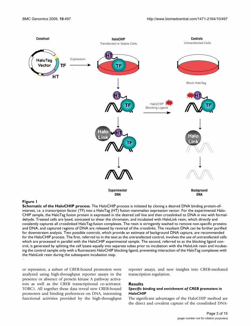
The success of ChIP relies heavily on the success of theimmunoprecipitation step in the process, creating a needfor alternative approaches when the antibody against theDNA binding protein is either not functional or availablefor the ChIP assay [17-21]. Such alternative approachesare derived from the standard ChIP method and includethe initial formaldehyde crosslinking of protein:DNAcomplexes, yet typically differ in the use of protein fusiontags, which allow for complexes to be isolated using eitheran antibody against the tag [17,21] or direct capture on aresin that interacts with the fusion tag [18-20]. The latteris the basis for the HaloCHIP method, which utilizes theHaloTag protein [19,20], a 33 kDa protein fusion tag, thatcan be cloned N- or C-terminally to a DNA binding pro-tein of interest[19,20] (Figure 1). In the HaloCHIPmethod, the HaloTag fusion protein is expressed eithertransiently or stably in mammalian cells and crosslinkedcomplexes can be directly captured from a cellular lysatevia covalent binding to a HaloTag-specific resin, termedHaloLink [19,20] (Figure 1). The complete covalent link-age established at this point allows for extensive washingto remove non-specific protein and DNA, followed bystandard reversal of the crosslinks to release the DNA frag-ments which were bound to the DNA binding protein(Figure 1). Several controls for the HaloCHIP method arepossible to show that capture is specific in this process andprovide an excellent estimate of background (Figure 1).
Both the HaloCHIP and ChIP method yield informationabout the location and timing of binding events on DNA,but do not provide information as to the cellular response
or consequence of the given binding event. Currently,mRNA and protein levels are measured to determinewhether or not a gene has been activated or repressed, buta more direct measure of the transcription potential orfunction of bound DNA sequences would be ideal. Tocomplement these approaches and also increase sensitiv-ity, high-throughput reporter assays can be used (Cooperet al. 2006). High-throughput reporter assays utilize a384-well format that enable the functional measure ofthousands of endogenous human promoters. Each indi-vidual promoter is fused to a luciferase reporter gene andtransiently delivered to living cells. Upon protein bindingto the promoter region, the luciferase reporter gene is acti-vated and the degree of this activation can be quantita-tively determined before and after a stimulus bymeasuring the light output. This allows real-time moni-toring of transcriptional activation or repression from thepromoter-reporter construct after stimulus of a pathwayor response to other cellular conditions.
To demonstrate the use of these approaches to furtherunderstanding of DNA-protein interactions in living cells,we chose to study the CREB transcription factor [22-25].The model system of the CREB signaling pathway hasbeen elegantly studied and its binding targets have beendescribed previously at the level of individual promotersas well as a genome-wide scale [12,22,26-29]. CREBbelongs to a family of transcription factors including acti-vating transcription factor 1 (ATF1) and the cAMPresponse element modulator (CREM), which regulategene expression in response to changes in cAMP and othercellular signals [23,24]. Upon activation of the proteinkinase A pathway or stimulation of other kinases, CREB isdirectly phosphorylated on several critical serines [22,30],though phosphorylation is not required for binding toDNA [31]. The phosphorylation events instead allow sub-sequent recruitment and binding of transcriptional co-activators CREB binding protein (CBP)/p300 as well astransducers of regulated CREB (TORCs) to the promoterregion [32-35]. Previous studies have shown that CREBco-factors are often necessary for transcription activationand that CREB binding to DNA, even in its phosphor-ylated form, is not usually sufficient to activate transcrip-tion [34-39].
In this paper, CREB binding is mapped at a much higherresolution than previous studies and covers all known andpredicted human promoters using the HaloCHIP methodin conjunction with DNA microarrays, ("HaloCHIP-chip"). As this is a new approach for studying genome-wide protein:DNA interactions, these data were comparedto the standard CREB ChIP-chip process using an anti-body against the endogenous CREB protein, revealing ahigh degree of overlap between the methods and also topreviously published data [26,29]. To further correlateDNA binding events to potential transcription activation
Page 2 of 15(page number not for citation purposes)
BMC Genomics 2009, 10:497 http://www.biomedcentral.com/1471-2164/10/497
or repression, a subset of CREB-bound promoters wereanalyzed using high-throughput reporter assays in thepresence or absence of protein kinase A pathway activa-tors as well as the CREB transcriptional co-activator,TORC1. All together these data reveal new CREB-boundpromoters and binding preferences on DNA, interestingfunctional activities provided by the high-throughput
reporter assays, and new insights into CREB-mediatedtranscription regulation.
ResultsSpecific binding and enrichment of CREB promoters in HaloCHIPThe significant advantages of the HaloCHIP method arethe direct and covalent capture of the crosslinked DNA-
Schematic of the HaloCHIP processFigure 1Schematic of the HaloCHIP process. The HaloCHIP process is initiated by cloning a desired DNA binding protein-of-interest, i.e. a transcription factor (TF) into a HaloTag (HT) fusion mammalian expression vector. For the experimental Halo-CHIP sample, the HaloTag fusion protein is expressed in the desired cell line and then crosslinked to DNA in vivo with formal-dehyde. Treated cells are lysed, sonicated to shear the chromatin, and incubated with HaloLink resin, which directly and covalently captures all crosslinked HaloTag-fusion complexes. The resin is stringently washed to remove non-specific proteins and DNA, and captured regions of DNA are released by reversal of the crosslinks. The resultant DNA can be further purified for downstream analysis. Two possible controls, which provide an estimate of background DNA capture, are recommended for the HaloCHIP process. The first, referred to in the text as the untransfected control, involves the use of untransfected cells which are processed in parallel with the HaloCHIP experimental sample. The second, referred to as the blocking ligand con-trol, is generated by splitting the cell lysate equally into separate tubes prior to incubation with the HaloLink resin and incubat-ing the control sample only with a fluorescent HaloCHIP blocking ligand, preventing interaction of the HaloTag complexes with the HaloLink resin during the subsequent incubation step.
Expression
ExperimentalDNA
BackgroundDNA
HaloCHIP ControlsConstructUntransfected Cells
HaloCHIPBlocking Ligand
Transfected or Stable Cells
Block HaloTag
� �
� � � �
� �
� � � � � �
� � � � � �
� �
� �
� � � �
� �
� �
� �
� �
� �
� �
� �
� �
Page 3 of 15(page number not for citation purposes)

BMC Genomics 2009, 10:497 http://www.biomedcentral.com/1471-2164/10/497
protein complexes on the HaloLink resin, eliminating theneed of an antibody and preventing loss or diffusion ofcomplexes after capture (Figure 1). As with the use of anyfusion tag, it is important to show the tagged proteinbehaves similar to the endogenous protein. Previous stud-ies with HaloTag fusion proteins have demonstratedproper physiology, including DNA binding and localiza-tion [19,40-45]. In order to demonstrate specific DNAbinding of the HaloTag-CREB fusion protein, HaloCHIPassays were performed in triplicates using transientlyexpressed HaloTag-CREB as the experimental sample anduntransfected HeLa cells as a control. The relative abun-dance of three known CREB-specific promoters, Fos, Jun,and p27, [24,28] and three negative control sequences,called C1, C2, and C3, which are non-genic regions fromthe human genome that lack CRE sites, were then ana-lyzed using Plexor quantitative PCR (Figure 2). The CREB-specific promoters show an average enrichment of 12.5fold, while the untransfected control showed an enrich-ment of 1.8 fold (Figure 2). The negative controlsequences are not enriched in either sample (Figure 2)Similar results were obtained using the HaloCHIP block-ing ligand (Figure 1) as a control (data not shown). Afterthis initial validation of specific binding, the HaloCHIPDNA was then prepared for hybridization to a humanpromoter microarray to ascertain binding on a genome-wide scale.
HaloCHIP-chip DNA oligo array designTo measure the genome-wide binding sites of CREB andto assess specificity of binding on a global scale, CREBHaloCHIP DNA was hybridized to a custom DNA oligomicroarray. In the HaloCHIP-chip strategy, two samples,the experimental and untransfected control, were eachtreated with or without forskolin (FSK) and processedthrough HaloCHIP (Figure 3). Similar to ChIP, the Halo-CHIP derived DNA from each sample, typically 10-100ng/reaction, required subsequent amplification to obtainamounts sufficient (1-10 μg) for microarray analysis. Thewhole genome amplification (WGA) method was used foramplification [46], and the experimental and controlsamples were labelled with Cy5 and Cy3, respectively(Figure 3). A custom DNA oligo microarray was designedbased upon promoter regions defined by SwitchGearGenomics genome-wide set of predicted transcriptionstart sites (Figure 3) [47-50]. An average of 14 probes,each 50-mers, was designed to span a 1.8 kb region ofeach known promoter region. The probes were chosen tosequences primarily upstream of a transcription start site,with an average of 131 bp spacing between probes. Intotal, approximately 385,000 probes were used to cover27,661 promoter regions, including 33,255 transcriptionstart sites (TSS) (Figure 3).
CREB HaloCHIP-chip array data analysis and cut-offsA total of 7 independent CREB HaloCHIP-chip experi-ments were conducted; 4 were treated with forskolin and3 without. All showed significant enrichment, howeververy few differences were detected between the untreatedand FSK treated samples, as was seen in previously pub-lished data [29]. The log2 ratios obtained for each pro-moter were averaged across the 7 independent arrayexperiments to obtain a single value for each promoterregion. To calculate the positive and negative predictivevalues (PPV and NPV respectively) of the array data at dif-ferent percentile thresholds, the promoters were sortedbased on their average log2 ratio for enrichment and vali-dated by qPCR. For the PPV determination, 12 sequenceseach were chosen from the Top 1%, 5%, and 10% of thesorted list and for the NPV determination, 36 sequenceswere randomly chosen from the bottom 50%. The Top1%, 5%, and 10% categories showed PPVs of 100%, 92%,and 50%, respectively (Table 1), while the NPV from the
Specific DNA binding of HaloTag-CREB in vivoFigure 2Specific DNA binding of HaloTag-CREB in vivo. Halo-CHIP experiments were performed in triplicates, on HeLa cells transiently expressing HaloTag-CREB or untransfected as a control. Resulting DNA from both the HaloTag-CREB and Untransfected control sample was amplified and analyzed using Plexor quantitative PCR. Total amounts of DNA for both samples were calculated for three promoters which CREB is known to bind [28], Fos, Jun, and p27, as well as three control sequences, C1, C2, and C3, which do not con-tain CRE consensus binding sites. Depicted in dark blue is the fold enrichment of each CREB-specific promoter over the average amount of the three control promoters for the HaloTag-CREB HaloCHIP experimental sample. In light blue is the identical calculation for the Untransfected HaloCHIP control sample.
Fold
Enr
ichm
ent
0
2
4
6
8
10
12
14
16
18
HaloTag-CREB
Untransfected
Fos
Non-SpecificSpecific
Jun p27 C1 C2 C3
Page 4 of 15(page number not for citation purposes)

BMC Genomics 2009, 10:497 http://www.biomedcentral.com/1471-2164/10/497
Bottom 50% was calculated to be 94.5% (Table 1). Thesevalues indicate the high quality of the data, and basedupon the PPV calculations, the cut-off for calling a pro-moter "CREB-bound" in subsequent analyses was chosento be the Top 5% of the list, corresponding to 1,383 pro-moters.
Comparison of CREB HaloCHIP-chip and ChIP-chip array dataTo compare the binding of HaloTag-CREB to the physio-logical expected binding pattern of the endogenous CREB
protein, a conventional ChIP-chip experiment using anantibody to CREB in HeLa cells and using the same oligoDNA microarray was performed. The Top 1% (277) andTop 5% (1383) of promoters showed significant overlap,35% and 45% respectively, between HaloCHIP and ChIP-chip, representing a 44-fold and 10-fold over-representa-tion of overlap compared to what would be predicted bychance (Table 2). This degree of overlap is within therange of similarity to what is seen between biological rep-licates of either HaloCHIP-chip or ChIP-chip data sets,indicating that the two methods are yielding equivalentresults within the limitations of experimental error of theoverall process (Table 2) [11,51].
Comparisons were also performed to between HaloCHIP-chip and previously published CREB ChIP-chip data(Table 2) [29]. As the CREB ChIP-chip DNA microarraycovered approximately 8,000 fewer number of promoters,the Top 1% and Top 5% of the list correspond to 182 and898 promoters [29], respectively, which were then usedfor the comparison (Table 2). A slightly lower overlap of26% and 23.8%, respectively is observed, correspondingto a 5-fold over-representation for both categories com-pared to what would be predicted by chance (Table 2).This is not surprising given the differences between theseexperiments including; the cell lines used, method ofamplification for the array, as well as the different arraydesign and platform [29]. Nevertheless, given these differ-ences it was very encouraging to see a significant overlapbetween these independent results.
Schematic of the HaloCHIP-chip microarray experiment designFigure 3Schematic of the HaloCHIP-chip microarray experiment design. HaloCHIP DNA (10-50 ng) obtained for both the experimental HaloTag-CREB and untransfected control sample was purified and amplified to a concentration of 1-10 μg using the whole genome amplification (WGA) method (Sigma) [46]. The HaloTag-CREB amplified sample was labelled with Cy5 (green) and the untransfected control sample with Cy3 (red), then hybridized to a custom DNA oligo microarray manufac-tured by Roche NimbleGen. The oligo array was designed to cover on average a 1.8 kb region of 27,661 human promoter regions that contain 33,255 TSS predicted by SwitchGear Genomics. To obtain coverage of each promoter, an average of four-teen 50mer single stranded DNA probes, shown in purple, per promoter were used, with an average spacing of 131 bp per probe.
HaloCHIPDNA
W.G.A.Label DNA
Custom DNA OligoMicroarray - NimbleGen
PromoterCoverage
~1.8kb
~Fourteen 50mer probes per promoter
~131bp spacing between probes
TSS
TSSTSS
Uni-directional
Bi-directional
27,661 Promoters385,000 Probes
HaloTag-CREB(+/- FSK)
OR
Untransfected(+/- FSK)
10-50ng
1-10μg
Table 1: CREB HaloCHIP-chip positive and negative predictive values.
Positive category Enriched promoters PPV
Top 1% 12 of 12 100%Top 5% 11 of 12 92%Top 10% 6 of 12 50%
Negative category Enriched promoters NPVBottom 50% 2 of 26 94.50%
Twelve promoters each from the Top 1%, Top 5%, and Top 10% categories of the HaloCHIP-chip data were chosen to determine the positive predictive value and 36 promoters were chosen from the Bottom 50% of the list for determination of the negative predictive value. SYBR green qPCR was used to determine the presence of these promoters in triplicates samples of HT-CREB experimental and untransfected control HaloCHIP DNA and promoters were considered enriched if having a signal:background ratio ≥ 2-fold between the experimental and control sample. Based upon these results, further analysis of the HaloCHIP-chip data was limited to promoters identified within the Top 5% of the list.
Page 5 of 15(page number not for citation purposes)

BMC Genomics 2009, 10:497 http://www.biomedcentral.com/1471-2164/10/497
Further support that the promoters identified by the CREBHaloCHIP-chip approach are specific for CREB function,comes from Gene Ontology (GO) analysis of the Top 1%promoters. GO analysis shows clusters of promotersfound which are involved with histone assembly, chroma-tin architecture, RNA and DNA metabolism, and nucleicacid binding pathways, all cellular processes which CREBhas been shown to regulate (Table 3) [23,24].
High resolution mapping of CREB binding sites relative to endogenous transcription start sitesPrevious genome-wide CREB ChIP-chip studies were con-ducted on spotted PCR product microarrays [26,29]. Ouruse of a custom oligo array that tiles across extended pro-moter regions (Figure 3) gives us the unique ability tomap CREB binding events at much higher resolution thatin turn allows us to determine the precise location ofbinding events relative to transcription start sites. First, weexamined the occurrence and location of putative CREs inthe HaloCHIP dataset as a whole. Enrichment of bindingto both full and half CRE consensus sites within the Top1% and 5% is observed as compared to the Bottom 90%of the array list (Table 4). Also, full and half CRE sites arehighly over-represented in the region 100 bp upstream ofthe transcription start site (data not shown), consistentwith results seen previously studying the binding patternof endogenous CREB [26,29].
The design of the array also allowed for further in-depthanalysis of binding to both unidirectional and bidirec-tional promoters. A striking observation was that the
majority of CREB-binding events were at bidirectionalpromoters, with 60.7% and 53.7% of total promotersbeing bidirectional in the Top 1% and 5%, respectively.Previous work has shown that approximately 10% of thegenes in the genome are divergently transcribed and regu-lated by a bidirectional promoter (transcription start sitesseparated by less than 1000 bp) [49,52,53], suggesting astrong preference of CREB for binding to divergently tran-scribed genes. Of the 27,661 promoters covered in thisstudy, we estimate that 19% have evidence for oppositelytranscribed transcripts initiating in that region. An exam-ple of CREB binding at a unidirectional promoter, definedas a promoter associated with a single transcription startsite, is shown in Figure 4a. As expected for both the Halo-Tag and endogenous CREB, there is enrichment of bind-
Table 2: Comparison of CREB HaloCHIP-chip and CREB ChIP-chip data.
HaloCHIP-chip:ChIP-chip compared Number of Promoters Percentage Overlap Enrichment over Random
Top 1% 96/277 35% 44Top 5% 616/1383 45% 10Top 1%* 47/182 26% 5Top 5%* 212/898 24% 5
HaloCHIP-chip:HaloCHIP-chip compared Number of Promoters Percentage Overlap Enrichment over Random
Top 1% 140/277 51% 65Top 5% 734/1383 53% 11
ChIP-chip:ChIP-chip compared Number of Promoters Percentage Overlap Enrichment over Random
Top 1% 115/277 42% 53Top 5% 477/1383 34% 7
The number of promoters found to overlap between the CREB HaloCHIP-chip and standard ChIP-chip experiments were determined for both the Top 1% and 5% of each respective list. Also shown is the calculation of the fold enrichment over what is expected for a randomized comparison. The first Top 1 and 5% overlap is between CREB HaloCHIP-chip and ChIP-chip data obtained from the same probes and arrays, therefore the number of promoters, 277 and 1383 respectively, used for comparison is the same between the two lists. The (*) indicates the comparison of the HaloCHIP-chip array data to previously published Top 1 and 5% CREB ChIP-chip data [29]. The Top 1 and 5% CREB ChIP-chip data correspond to 182 and 898 promoters respectively, and it was these which were used for the comparison to the Top 1 and 5% of the HaloCHIP-chip array data. Calculated in a similar fashion is the percentage overlap and enrichment over random between HaloCHIP-chip biological replicate sets or ChIP-chip biological replicate data sets.
Table 3: Gene Ontology analysis of CREB HaloCHIP-chip promoters.
Cellular Functions Number of Promoters p-value
Histone Assembly 12 of 65 1.26E-06Chromatin architecture 20 of 261 7.63E-07Ribonucleic Complexes 26 of 392 7.06E-07RNA processing 26 of 395 8.01E-07DNA metabolism 38 of 638 2.93E-08Nucleic acid binding 110 of 2764 2.19E-09
The Top 1% of the CREB HaloCHIP-chip promoters (277) was analyzed using Gene Ontology (GO) and the different categories in terms of cellular function of promoters within the Top1%. Also shown is the number of CREB HaloCHIP-chip promoters found as compared to the number of known human promoters within each category and the corresponding p-value calculated by GO.
Page 6 of 15(page number not for citation purposes)

BMC Genomics 2009, 10:497 http://www.biomedcentral.com/1471-2164/10/497
ing directly upstream of the transcription start site aroundthe location of a putative CRE site (Figure 4a), and for oursubset analyzed, this pattern of binding was observedgreater than 60% of the time. Analysis of binding withinbidirectional promoters revealed interesting binding pat-terns for CREB. As shown in Figure 4b, two distinct peaksof high enrichment are observed just downstream of thetranscription start sites of both genes within a bidirec-tional promoter, while no enrichment is observed in theintergenic space between the transcription start sites. Asthis is observed for both the HaloTag-CREB fusion proteinas well as the endogenous CREB protein, it does notappear to be an artefact of either the HaloCHIP or ChIPmethodology. It was also surprising to see that highestenrichment was not localized at the location of putativeCRE sites in bidirectional promoters, suggesting CREB iseither bound directly elsewhere on DNA or crosslinking tocomplexes not bound to CRE consensus sites (Figure 4b).
CREB high-throughput reporter assay analysisThe combination of the CREB genome-wide bindingevents identified in this study along with those reportedpreviously confirm CREB binding events, however do notpredict the transcriptional regulation of its target genes.Furthermore, CREB binding was measured in a limited setof conditions (with and without forskolin stimulation),and it is known that the CREB pathway is activated by awide variety of cellular and environmental stimuli[23,24]. To further characterize the CREB binding events,we assembled a collection of cloned human promoters (1kb on average) in a luciferase reporter vector that repre-sented a subset (235) of known CREB targets previouslyidentified [26,27,29]. As predicted from the earlier over-lap calculations with the CREB ChIP-chip data (Table 2)approximately 35% of these promoters (84) were identi-fied in the Top 5% of the HaloCHIP-chip array data. As acontrol, 12 random promoters that were not targets ofCREB were also fused to luciferase reporters (Figure 5a).Utilizing our high-throughput reporter assay platform, wemeasured the activity of each of these fragments in HeLacells in triplicate in 5 different conditions: no treatment,
forskolin (FSK) stimulation, phorbol 12-myristate-13-acetate (PMA) stimulation, and co-transfection with aTORC1 expression construct with and without FSK stimu-lation (Figure 5a). We considered a promoter induced ifthe absolute activity was significantly above background,passed a T-test at p < 0.05, and had a magnitude of changegreater than 2-fold.
The promoter macroarray results are summarized in Fig-ure 5b, and the percentage of the total CREB promoterstested (235), as well as the HaloCHIP subset (84), thatwere induced in the different conditions were determined(Table 5). For both the CREB set and the HaloCHIP sub-set, only ~10% were induced by FSK treatment alone (Fig-ure 5b, Table 5). This is not surprising since it is knownthat many other factors are necessary for the transcrip-tional activation of CREB-bound promoters and both ourbinding data and previously reported data did not showan appreciable difference in DNA binding between +/-FSK treatment [23,29,35]. Indeed, the TORC1 co-transfec-tions induced more than 50% of the constructs (Table 5)and also conferred the largest fold-changes in activity (Fig-ure 5b) highlighting the importance of co-factors in thetranscriptional activation of CREB-bound promoters[32,34]. Similar trends were observed between the overallCREB set of promoters as compared to the HaloCHIP sub-set, indicating those identified by HaloCHIP respond sim-ilarly to CREB-specific stimuli (Table 5). The 12 randompromoters from the human genome do not show inducedactivity in any of the conditions tested, indicating that thefalse positive rate of the reporter assay for CREB activity isvery low (Figure 5b).
Given the interesting CREB binding patterns at bidirec-tional promoters (Figure 4b), we looked specifically at thepromoter activities of bidirectional promoters in ourreporter assay dataset. There were a total of 7 bidirectionalgene pairs for which we collected promoter activity datafor each direction. The majority of the pairs showed verylow activity in both directions in the untreated cells sug-gesting that CREB-bound bidirectional promoters are nottranscriptionally active in an un-induced state. Two of the7 bidirectional gene pairs, which regulate two pairs of his-tone genes, had constitutively high promoter activities inboth directions, irrespective of stimulation conditions.
The most interesting example was seen for two of the bidi-rectional promoter pairs, depicted in Figure 6a, b. Forthese bidirectional promoters, no induction was observedwith either FSK or PMA, but in the presence of TORC1,promoter activity in one direction was significantly up-regulated while promoter activity was repressed in theother direction (Figure 6b). CREB enrichment at theMRPS18B promoter was observed at CRE sites justupstream of the TSS, while enrichment to the PP1R10 pro-
Table 4: Match of full and half CRE consensus sites in CREB HaloCHIP-chip data.
Categories % with Full CRE site % with Half CRE Site
Top 1% 22% 89%Top 5% 12% 86%Bottom 90% 3% 57%
CREB HaloCHIP-chip promoters from the Top 1%, Top 5%, and Bottom 90% categories were analyzed for the presence of the full CRE or half CRE consensus site and number of promoters containing these sequences are depicted. A full CRE site was defined as a perfect match to the consensus TGACGTCA. A half CRE site was defined as a perfect match to the consensus TGACG or CGTCA.
Page 7 of 15(page number not for citation purposes)

BMC Genomics 2009, 10:497 http://www.biomedcentral.com/1471-2164/10/497
Page 8 of 15(page number not for citation purposes)
High-resolution analysis of CREB binding at uni-directional and bi-directional promotersFigure 4High-resolution analysis of CREB binding at uni-directional and bi-directional promoters. Depicted are CREB binding data from two representative promoter regions, the length (bp) of each is indicated above the graph. The log2 ratio for each probe spanning the promoter region was determined for the CREB ChIP-chip (plotted in red) or HaloCHIP-chip (plotted in blue) data. Positions of transcription start sites (TSS) are shown with arrows indicating the direction of transcription with length of exons in blocks and introns drawn as lines. Positions of full and half CRE sites relative to the location within the pro-moter region are indicated below by blue dots. A. Example of CREB binding to a unidirectional promoter, ALS2, where peak binding is observed upstream of the TSS and localized to the CRE consensus sites. B. Example of CREB binding to a bidirec-tional promoter, METTL4 and NDC80, where peak enrichment is located downstream of the TSSs, but the peak enrichment does not localize to CRE consensus sites.
NDC80METTL4
CRE half site CRE full site
log
2 ra
tio
CRE
B en
rich
men
t
2,062 bp4
3
2
1
0
CREB ChIP-chip enrichment
CREB Halo-chip enrichment
ALS2half site CRE full site
3
2
1
0
log
2 ra
tio
CRE
B en
rich
men
t
1,125 bp
CREB ChIP-chip enrichment
CREB Halo-chip enrichment

BMC Genomics 2009, 10:497 http://www.biomedcentral.com/1471-2164/10/497
moter was downstream the TSS and not associated with aCRE site (Figure 6a). Interestingly, only the MRPS18B pro-moter shows transcriptional activation in the reporterassay, while the PPP1R10 promoter is significantlyrepressed in the presence of TORC1 and FSK (Figure 6b).These results suggest that TORC1 plays an important role
at some promoters in determining the direction of tran-scriptional activation.
DiscussionWe applied here two technologies, HaloCHIP and high-throughput promoter assays, to study and more fully char-acterize the CREB transcription pathway than previouslydone. The HaloCHIP method (Figure 1) offers an alterna-tive approach for the capture of intracellular DNA-proteincomplexes and was developed to address the challenges ofantibodies required for the existing ChIP method. The useof a robust protein tag eliminates the need for a qualifiedantibody and enables researchers to study highly similarparalogs, different isoforms, or point mutants of a tran-scription factor that may not be distinguishable by anantibody. Also, due to rapid and covalent binding kineticsHaloTag with its ligands, protein complexes can be cap-tured efficiently from dilute solutions without concern ofloss due to diffusion off the resin, allowing for the use ofa much smaller number of cells (2-4 × 10⁵) per HaloCHIPexperiment as compared to the standard ChIP experiment(~1 × 10⁷) [4,10]. As with the use of any protein fusion tagfor ChIP or HaloCHIP experiments there are concerns asto potential alteration of DNA binding due to interferenceby the fusion tag or changes in expression level. The CREBHaloCHIP-chip results show that binding to DNA on agenomic-scale was specific for the CREB protein and hada significant degree of overlap with conventional CREBChIP-chip data, suggesting the HaloTag-CREB fusion pro-tein is binding to DNA similarly to the endogenous CREBprotein.
In addition to identification of new CREB-bound promot-ers with these array studies, we extended our studies of theCREB pathway by measuring the functional activity ofover 200 CREB-target promoters [26,27,29] in a high-throughput reporter assay experiment (Figure 6a). Manydiverse responses are regulated through the CREB path-way and unique subsets of CREB-bound genes may betranscriptionally activated and responsive to particularstimuli. The high-throughput reporter assays of CREB-bound promoters gives the ability to stratify CREB bind-ing events based on the transcriptional activity of the frag-ments of DNA to which they bind. Analysis of the CREBpathway using the functional promoter macroarraysrevealed only a small percentage of promoters wereresponsive to FSK, correlating well with the HaloCHIP-chip data showing minimal changes in binding betweenuntreated and FSK treated cells. Interestingly, a muchlarger percentage of promoters were responsive to PMA,and an even greater percentage to the TORC proteins.These functional results provide further support for theidea that co-factors are a crucial part of the CREB signalingpathway and while reporter assays lack the full chromatincontext of the genome, by using extended promoters
High-throughput reporter assays of CREB-bound promotersFigure 5High-throughput reporter assays of CREB-bound promoters. A. Experimental design of high-throughput reporter assays. A schematic showing the high-throughput reporter assay experimental design. Promoters are fused to luciferase, transfected in a desired cell line, and stimulated under different conditions. Luciferase activity is measure from uninduced and induced samples and the log2 ratio of these differences is calculated. B. Heatmap of inducible pro-moter activity. A total of 235 promoters, chosen from CREB ChIP-chip and HaloCHIP-chip data, along with twelve nega-tive control promoters were fused to the luciferase gene, transfected into HeLa cells, and treated with stimulants; for-skolin (FSK), PMA, or co-transfected with a transcriptional co-activator, TORC1 +/- FSK. Each box represents the log2 ratio of induced/untreated for each promoter in each condi-tion where the intensity of red is proportional to the strong-est induction and the intensity of blue is proportional to the strongest repression. The presence of TORC1 by co-trans-fection shows the highest number of promoters induced to the highest degree. The box of 12 control promoters at the bottom of the panel are random promoters from the genome and show very little inducible activity in any of the conditions tested.
Page 9 of 15(page number not for citation purposes)

BMC Genomics 2009, 10:497 http://www.biomedcentral.com/1471-2164/10/497
regions that are 1 kb in length, we were able to observetranscriptional effects by co-factors, which may interactwith proximal sites. In order to analyze CREB-enrichedsites at a much higher resolution than was previously per-formed, custom oligo microarrays were used. Thisdetailed analysis provided interesting and novel insightinto the localization of CREB at the promoters of genes. Asimple assumption is that the experimental enrichmentfor CREB binding would be coincident with the locationof the CRE. Indeed this was observed for the majority ofunidirectional promoters (Figure 4a). However, a dis-tinctly different pattern is observed for a large fraction ofbidirectional promoters, where the peaks of highestenrichment are seen downstream of the closest TSS, oftennot coinciding with the location of CREs (Figure 4b). Thispattern was seen consistently for many bidirectional pro-moters, in both HaloCHIP-chip and ChIP-chip data sets,indicating this is not a phenomenon associated with aparticular method.
These particular binding results suggest a number of inter-esting scenarios for which additional experiments will beneeded. It may be the case that there is a secondary struc-ture of the CREB-DNA complex at bidirectional promot-ers where the peaks of enrichment reflect the higher ordercrosslinked structure rather than the true localization ofthe CREB protein on the linear strand of genomic DNA.An alternative explanation is that CREB may be a part of apaused transcription initiation complex. In this scenario,CREB could initially bind upstream of the TSS in the bidi-rectional promoters, form its known interactions with theRNA PolII complex, move after initiation with the com-plex, and then pause at particular sites downstream of theTSS. Recent work has shown that a paused transcriptionalcomplex containing transcriptional regulators are moreabundant than previously thought [54-56], and thisexplanation would produce the enrichment pattern thatwe observe for CREB at bidirectional promoters (Figure4b, 6b).
Results from the promoter reporter assays for bidirec-tional promoters are also consistent with this scenario,
since a paused transcriptional machinery would likelyresult in lower reporter activity as was seen for the major-ity of bidirectional promoters tested. Perhaps most inter-esting is the functional behaviour of a subset of thebidirectional promoters in the presence of TORC1. In 2out of 7 cases tested, the activity of a bidirectional pro-moter was strongly induced in one direction and stronglyrepressed in the opposite direction in the presence ofTORC1. The strongly repressed promoters show CREBbinding which is downstream the TSS, while the stronglyinduced promoters show expected upstream promoterbinding. Also, the ability of the TORC1 protein to differ-entially regulate promoter activity, suggests that CREB co-factors may also help to regulate the directionality of tran-scription. This is particularly relevant for the CREB tran-scription factor, since over 50% of CREB binding sites arelocated in bidirectional promoters as we have reported forthe first time.
ConclusionThis broad survey of the transcriptional activity of CREB-bound promoters provides a valuable layer of functionaldata for the CREB protein. Future efforts to compare theactivity of these promoters in many more conditions willhelp to further understand CREB signaling and muta-tional analysis of the bidirectional class of CREB-boundpromoters will help to dissect the mechanism of bidirec-tional gene regulation. The use of the new technologiespresented here however is not limited to the study of theCREB pathway, rather can be generalized to study anytranscriptional pathway. The HaloCHIP method, like thestandard ChIP process, can be used to study DNA bindingboth on a small scale, as well as genome-wide scale, how-ever follow up studies characterizing the functional conse-quences of these binding events have lagged much furtherbehind. By expanding the use of high-throughput reporterassays, we hope to advance our understanding of thesefunctional consequences. This comprehensive compari-son reveals the challenges and potential pitfalls of extrap-olating binding events to transcriptional activation andshows the need for both approaches, as well as otherexperiments to truly characterize transcriptional activity.
Table 5: Percentage of promoters activated in various conditions in functional macroarrays.
Total CREB Set Tested (235) HaloCHIP Subset (84)
Stimulants % Induced % InducedFSK 9.8% 9.5%PMA 31.9% 26.2%TORC1 60.4% 50.0%TORC1 + FSK 65.1% 54.8%ANY 76.2% 66.7%
Table 5 shows the percentage of promoters activated in each condition from both the total CREB set (235) or a subset of the total CREB set (84), termed the HaloCHIP subset, that was found in the Top 5% of the CREB HaloCHIP-chip array list. The HaloCHIP-chip data follows the same trend of response to each stimulus as the larger CREB set.
Page 10 of 15(page number not for citation purposes)

BMC Genomics 2009, 10:497 http://www.biomedcentral.com/1471-2164/10/497
Page 11 of 15(page number not for citation purposes)
CREB binding and promoter activity at a bidirectional promoter showing opposite induction patterns in the presence of TORC1Figure 6CREB binding and promoter activity at a bidirectional promoter showing opposite induction patterns in the presence of TORC1. A. CREB binding to PPP1R10/MRPS18B bidirectional promoter. Identical to Figure 4, the log2 ratio for each probe spanning this bidirectional promoter was determined for the CREB ChIP-chip (plotted in red) or HaloCHIP-chip (plotted in blue) data. Spacing between probes is approximately 131 bp. Positions of transcription start sites (TSS) are shown with arrows indicating the direction of transcription with lengths of exons in blocks and introns drawn as lines. Positions of full CRE sites are indicated below by blue dots. Also shown is the region of each cloned promoter fragments, indicated by the green and blue arrows, used in the luciferase assay shown below in panel B. The arrows indicate the direction in which the cloned fragments were tested in the luciferase assay. B. The log2 ratio of treated/untreated luciferase activity is plotted for each promoter fragment in each condition indicated. The MRPS18B promoter (in green) shows significant inducible activity in the presence of TORC1 with and without FSK, while the PPP1R10 promoter (in blue) shows significant repression in the pres-ence of TORC1 with and without FSK.
���
�
���
�
���
�
���
�
���
PPP1R10 MRPS18B
CREB ChIP-chip enrichment
CREB Halo-chip enrichmentlo
g2
rati
o C
REB
enri
chm
ent
MRPS18B promoter clone
PPP1R10 promoter clone
�
�
�
�
�
�
log
2 ra
tio
trea
ted
/un
trea
ted
lu
cife
rase
act
ivit
y
Forskolin PMA TORC1 TORC1+Forskolin
CRE sites
MRPS18B promoter clone
PPP1R10 promoter clone
2,183 bp

BMC Genomics 2009, 10:497 http://www.biomedcentral.com/1471-2164/10/497
MethodsCloning, cell lines, and transfections of HaloTag vectorsFull-length human CREB1-α and -Δ cDNAs were obtainedfrom OriGene, [NCBI:NM_134442.2 andNCBI:NM_004379.2], respectively. All CREB variantswere subcloned into the pFN21A HaloTag CMV Flexi Vec-tor (Promega) using SgfI and Pme, generating N-terminalHaloTag fusion constructs for each. HeLa cells (ATCC#CCL-2) were maintained in DMEM supplemented with10%FBS at 37°C in an atmosphere of 5% CO2. Cells weretransfected using Lipofectamine LTX transfection reagent(Invitrogen) according manufacturer's protocols.
HaloCHIP Protocol and Whole Genome AmplificationA detailed version of the HaloCHIP protocol can be foundat: http://www.promega.com/tbs/tm075/tm075.html
For these experiments, HeLa cells (2-4 × 10⁵) were platedin a single well of a standard 6-well plate. After reaching70-80% confluency, typically 18-24 hours later, cells weretransfected with the HaloTag-CREB fusion constructs(experimental sample) or left untransfected (control sam-ple). Twenty four hours post-transfection, cells werecrosslinked with formaldehyde (Sigma) at a final concen-tration of 0.75% for 10 minutes at 22°, quenched with0.125 M glycine for 10 min. and processed using theHaloCHIP kit (Promega). For experiments involving For-skolin, cells were treated with 10 μM Forskolin for 45minutes at 37°C prior to crosslinking. Isolated DNA wasfurther purified using a PCR Clean-up kit (Qiagen), andeluted 2 × 50 μl with nuclease-free water, yielding a finalvolume of 100 μl. To prepare sufficient HaloCHIP DNAfor downstream amplification steps required for microar-rays, an entire 6-well plate was transfected and processedthrough the HaloCHIP method as recommended. The iso-lated DNA was pooled before final purification on thePCR clean-up columns and lyophilized to a final volumeof 12 μl. The concentrated HaloCHIP DNA was thenamplified to 2-10 μg using the Whole Genome Amplifica-tion kit (Sigma) following the recommended adaptationfor ChIP samples [46].
ChIP ProtocolHeLa cells (4 × 10⁶) were plated in several 150 mm platesand grown at 37°C to 80-90% confluency. Cells weretreated with 10 uM forskolin for 45 minutes at 37°.crosslinked with formaldehyde (Sigma) at a final concen-tration of 1.0% for 10 minutes at 22°, quenched with0.125 M glycine for 10 min. and processed using the ChIPAssay Kit (USB). Chromatin was sheared by sonicationusing a Misonix MicroTip Probe 418, output of 5.5, witha program of 15 cycles of 5 seconds on and 25 seconds offon ice. Co-immunoprecipitation was performed using 1μg of anti-CREB1 antibody (Millipore #06-863) for theexperimental sample and 1 μg of anti-IgG antibody
(Sigma) for the control sample with incubation at 4°C for15 hours. Isolated DNA was further purified using a PCRClean-up kit (Qiagen), processed, and amplified usingWGA identical as the HaloCHIP samples.
Quantitative PCR and primersHaloCHIP DNA was analyzed using either Plexor(Promega) or SYBR green (Applied Biosystems) qPCRaccording to their respective manufacturer's recommenda-tions. Plexor primers were supplied from Biosearch Tech-nologies and SYBR green primers were from IDT DNA.The following sequences were used for amplification: Fosforward 5'-GTCTTGGCTTCTCAGATGCTCG-3', reverse 5'-GTTGAGCCCGTGACGTTTACA-3', Jun forward 5'-GAGAAAGAAGGGCCCGACTGT-3', reverse 5'-GGAGACTC-CACCCTAGAAGATTCT-3', p27 forward 5'-GGGAGGCT-GACGAAGAAGAAAAT-3', reverse 5'-CAACCAATGGATCTCCTCCTCTG-3', C1 forward 5'-CTGGTCTCACCTAC-CTTCCTGT-3', reverse 5'-ATCCATGAACTCCAGGAGCTCA-3' C2 forward 5'-TCTGTTGCCTATTGACCAGAACATG-3', reverse 5'-AGGAGCTGTAGGCTGAGTCAC-3', C3 for-ward 5'-CTGCTTCTTAACAGCTTAATTCGGAAGA-3',reverse 5'-ATGAGCAAAGATAGCTCAGGGAG-3'. Primerssequences used for PPV and NPV qPCR validation alongwith their corresponding amplified promoter can befound in supplemental materials: http://www.switchgeargenomics.com/creb_supp_data/
Oligo array design and analysisA custom oligo array was designed to cover a genome-wide set of human promoter regions predicted by Switch-Gear Genomics (more detail can be found at http://www.switchgeargenomics.com). The oligo array com-posed of approximately ~385,000 50mer probes wasmanufactured by Roche-NimbleGen Systems. The ampli-fied enriched samples described above were shipped toRoche-NimbleGen to be hybridized according to theirstandard service protocol. The raw data from the arrayswere analyzed as follows; the log2 ratio (enriched-cy5/total input-cy3) was calculated for each probe and datawere then smoothed by averaging across a sliding windowof 3 neighbouring probes shifting 1 probe at a time, min-imizing noise from single probes. The median and stand-ard deviation were calculated from the smoothed ratiosfor each sample. The median was subtracted from eachratio and divided by the standard deviation to center andnormalize the data from each array. To summarize theenrichment for an entire promoter, the top 4 probe valueswere averaged for a given promoter region to approximatethe 75th percentile value. The raw data, normalized data,and collapsed data for each array are available as supple-mental at the following site: http://www.switchgeargenomics.com/creb_supp_data/. All microarray probes anddata discussed in this publication have been deposited inNCBI's Gene Expression Omnibus[57] and are accessible
Page 12 of 15(page number not for citation purposes)
BMC Genomics 2009, 10:497 http://www.biomedcentral.com/1471-2164/10/497
through GEO Series accession number GSE18347 http://www.ncbi.nlm.nih.gov/geo/query/acc.cgi?acc=GSE18347.
Gene Ontology (GO) analysisThe Top 1% of the CREB HaloCHIP-chip promoters, 277in total, were deposited and analyzed using Gene Ontol-ogy (GO) http://www.geneontology.org/ and AmiGO asthe search engine. GO categorized each promoter withrespect to protein function, showed the number of pro-moters within each category, and reported a correspond-ing p-value based upon the calculations.
High-throughput reporter assays and analysisThe promoter reporter assays used 235 promoter-reportervectors (utilizing the luc2P reporter cassette fromPromega) containing ~1 kb promoter fragments fromknown CREB-bound genes. These cloned promoters wereselected from SwitchGear Genomic's genome-wide pro-moter clone collection (details on this panel of reporterconstructs can be found at http://www.switchdb.com/pathways/id_46/). A panel of promoter controls was alsoused to normalize signals between plates and replicates.The 32 plate normalization controls, include ~1 kb frag-ments representing constitutively active human promoterfragments and random regions from the genome. The pro-moter reporter assay experiments were all conducted in384-well format. A detailed protocol can be found at:http://www.switchgeargenomics.com/creb_supp_data/.Transfection complexes were formed by incubating 50 ngof each individual promoter construct with 0.3 μL ofFugene 6 transfection reagent and Opti-MEM media in atotal volume of 3 μL and incubated for 30 minutes. Theco-transfection of the TORC1 expression construct was setup the same as the standard transfection reaction, butwith the inclusion of 25 ng of TORC1 expression plasmidper reaction (TORC1 expression construct was providedby the Montminy lab). Transfection complexes weremixed with resuspended HeLa cells such that 4,000 HeLacells were seeded in a volume of 50 μL in each well of a384-well white tissue culture treated plate. Fifteen repli-cate wells of each promoter construct were performed rep-resenting triplicate assays in 5 different conditions: 1) notreatment, 2) PMA, 3) Forskolin, 4) TORC1, and 5)TORC1 + FSK.
After seeding and transfection, cells were incubated for 24hours before inductions. Inductions were conducted foreach plate by removing the old media and replacing withnew media depending on the condition. For the untreatedcells, fresh media was applied to each well. For the PMAinduction, fresh media with 100 nM PMA was added toeach well. For the forskolin induction, fresh media with20 μM FSK was added to each well. Cells were kept in theirrespective induction condition for 4 hours and then fro-zen overnight at -80 degrees.
To read luminescent activity plates were thawed for 45minutes at room temperature. Then 50 μL of Steady-Gloreagent (Promega #E2520) was added and incubated for30 minutes at room temperature. Then luminescence wasread for 2 seconds per well on a 384-well compatible plateluminometer (Molecular Devices LMax384).
The raw luminescent reads from each well were normal-ized as follows. Each 384 well plate contained 32 controlwells that were comprised of 16 positive control promot-ers and 16 random genomic fragments that serve as back-ground signal controls. These plate controls were used tonormalize the per well values between plates within a con-dition. The average of the 3 replicates was taken, and theratio of induced/untreated was calculated from the aver-ages of the treated values and the untreated sample. A t-test for significance was also calculated between the 3 rep-licates of the induced and untreated samples. The back-ground controls were also used to measure whether theaverage absolute signals were above background in eachcondition (>3 standard deviations from the mean of thenegative controls). For a given promoter to be calledinduced or repressed it must pass the following criteria: 1)At least a 2-fold change (+/-) 2) Pass t-test with signifi-cance of p < 0.05 3) Must have absolute signals signifi-cantly above background.
Competing interestsPromega Corporation sells the HaloCHIP system com-mercially, and SwitchGear Genomics sells promoter luci-ferase reporter vectors commercially. Some of thePromega authors hold stock in Promega Corporation, butless than 1% of such stock, and SwitchGear Genomicsauthors hold stock in SwitchGear Genomics. PromegaCorporation and SwitchGear Genomics are the owners byassignment of patents or patent applications related to theHaloCHIP technology and the promoter reporter plat-form, respectively.
Authors' contributionsDDH, JM, and NM carried out all HaloCHIP experimentsand preparation of DNA for microarray analysis. NDT per-formed all array analysis and in conjunction with SFA car-ried out and analyzed all promoter macroarray analysis.DDH, NDT, KW, and MU conceived the study, plannedexperiments, and helped draft the manuscript. All authorsread and approved the final manuscript.
AcknowledgementsWe are grateful to Marc Montminy and Pankaj Singh for generously sharing their TORC1 expression construct along with providing helpful comments on our results. We are also grateful to Martin Rosenberg, Michael Slater, Luciano Di Croce, Patrick Collins, and Mike Rose for providing helpful dis-cussion and experimental support for the project. DDH, JM, NM, KW, and MU are all funded by Promega Corporation, and NDT and SFA are funded by SwitchGear Genomics. Both Promega Corporation and SwitchGear
Page 13 of 15(page number not for citation purposes)

BMC Genomics 2009, 10:497 http://www.biomedcentral.com/1471-2164/10/497
Genomics contributed funding to this research and the preparation of the manuscript.
References1. Kuo MH, Allis CD: In vivo cross-linking and immunoprecipita-
tion for studying dynamic Protein:DNA associations in achromatin environment. Methods 1999, 19(3):425-433.
2. Solomon MJ, Larsen PL, Varshavsky A: Mapping protein-DNAinteractions in vivo with formaldehyde: evidence that his-tone H4 is retained on a highly transcribed gene. Cell 1988,53(6):937-947.
3. Toth J, Biggin MD: The specificity of protein-DNA crosslinkingby formaldehyde: in vitro and in drosophila embryos. NucleicAcids Res 2000, 28(2):e4.
4. Buck MJ, Lieb JD: ChIP-chip: considerations for the design,analysis, and application of genome-wide chromatin immu-noprecipitation experiments. Genomics 2004, 83(3):349-360.
5. Horak CE, Mahajan MC, Luscombe NM, Gerstein M, Weissman SM,Snyder M: GATA-1 binding sites mapped in the beta-globinlocus by using mammalian chIp-chip analysis. Proc Natl Acad SciUSA 2002, 99(5):2924-2929.
6. Kirmizis A, Farnham PJ: Genomic approaches that aid in theidentification of transcription factor target genes. Exp BiolMed (Maywood) 2004, 229(8):705-721.
7. Kurdistani SK, Grunstein M: In vivo protein-protein and protein-DNA crosslinking for genomewide binding microarray. Meth-ods 2003, 31(1):90-95.
8. Pugh BF, Gilmour DS: Genome-wide analysis of protein-DNAinteractions in living cells. Genome Biol 2001, 2(4):REVIEWS1013.
9. Ren B, Robert F, Wyrick JJ, Aparicio O, Jennings EG, Simon I, Zeitlin-ger J, Schreiber J, Hannett N, Kanin E, et al.: Genome-wide locationand function of DNA binding proteins. Science 2000,290(5500):2306-2309.
10. Weinmann AS, Farnham PJ: Identification of unknown targetgenes of human transcription factors using chromatinimmunoprecipitation. Methods 2002, 26(1):37-47.
11. Euskirchen GM, Rozowsky JS, Wei CL, Lee WH, Zhang ZD, HartmanS, Emanuelsson O, Stolc V, Weissman S, Gerstein MB, et al.: Mappingof transcription factor binding regions in mammalian cells byChIP: comparison of array- and sequencing-based technolo-gies. Genome Res 2007, 17(6):898-909.
12. Impey S, McCorkle SR, Cha-Molstad H, Dwyer JM, Yochum GS, BossJM, McWeeney S, Dunn JJ, Mandel G, Goodman RH: Defining theCREB regulon: a genome-wide analysis of transcription fac-tor regulatory regions. Cell 2004, 119(7):1041-1054.
13. Johnson DS, Mortazavi A, Myers RM, Wold B: Genome-wide map-ping of in vivo protein-DNA interactions. Science 2007,316(5830):1497-1502.
14. Kim J, Bhinge AA, Morgan XC, Iyer VR: Mapping DNA-proteininteractions in large genomes by sequence tag analysis ofgenomic enrichment. Nat Methods 2005, 2(1):47-53.
15. Odom DT, Zizlsperger N, Gordon DB, Bell GW, Rinaldi NJ, MurrayHL, Volkert TL, Schreiber J, Rolfe PA, Gifford DK, et al.: Control ofpancreas and liver gene expression by HNF transcription fac-tors. Science 2004, 303(5662):1378-1381.
16. Wei CL, Wu Q, Vega VB, Chiu KP, Ng P, Zhang T, Shahab A, YongHC, Fu Y, Weng Z, et al.: A global map of p53 transcription-fac-tor binding sites in the human genome. Cell 2006,124(1):207-219.
17. Alekseyenko AA, Larschan E, Lai WR, Park PJ, Kuroda MI: High-res-olution ChIP-chip analysis reveals that the Drosophila MSLcomplex selectively identifies active genes on the male Xchromosome. Genes Dev 2006, 20(7):848-857.
18. Kim J, Chu J, Shen X, Wang J, Orkin SH: An extended transcrip-tional network for pluripotency of embryonic stem cells. Cell2008, 132(6):1049-1061.
19. Los GV, Encell LP, McDougall MG, Hartzell DD, Karassina N, Zim-prich C, Wood MG, Learish R, Ohana RF, Urh M, et al.: HaloTag: anovel protein labeling technology for cell imaging and pro-tein analysis. ACS Chem Biol 2008, 3(6):373-382.
20. Urh M, Hartzell D, Mendez J, Klaubert DH, Wood K: Methods fordetection of protein-protein and protein-DNA interactionsusing HaloTag. Methods Mol Biol 2008, 421:191-209.
21. Zhang X, Guo C, Chen Y, Shulha HP, Schnetz MP, LaFramboise T,Bartels CF, Markowitz S, Weng Z, Scacheri PC, et al.: Epitope tag-
ging of endogenous proteins for genome-wide ChIP-chipstudies. Nat Methods 2008, 5(2):163-165.
22. Berkowitz LA, Gilman MZ: Two distinct forms of active tran-scription factor CREB (cAMP response element binding pro-tein). Proc Natl Acad Sci USA 1990, 87(14):5258-5262.
23. Lonze BE, Ginty DD: Function and regulation of CREB familytranscription factors in the nervous system. Neuron 2002,35(4):605-623.
24. Mayr B, Montminy M: Transcriptional regulation by the phos-phorylation-dependent factor CREB. Nat Rev Mol Cell Biol 2001,2(8):599-609.
25. Montminy MR, Sevarino KA, Wagner JA, Mandel G, Goodman RH:Identification of a cyclic-AMP-responsive element within therat somatostatin gene. Proc Natl Acad Sci USA 1986,83(18):6682-6686.
26. Conkright MD, Guzman E, Flechner L, Su AI, Hogenesch JB,Montminy M: Genome-wide analysis of CREB target genesreveals a core promoter requirement for cAMP responsive-ness. Mol Cell 2003, 11(4):1101-1108.
27. Euskirchen G, Royce TE, Bertone P, Martone R, Rinn JL, Nelson FK,Sayward F, Luscombe NM, Miller P, Gerstein M, et al.: CREB bindsto multiple loci on human chromosome 22. Mol Cell Biol 2004,24(9):3804-3814.
28. Kang J, Shi Y, Xiang B, Qu B, Su W, Zhu M, Zhang M, Bao G, Wang F,Zhang X, et al.: A nuclear function of beta-arrestin1 in GPCRsignaling: regulation of histone acetylation and gene tran-scription. Cell 2005, 123(5):833-847.
29. Zhang X, Odom DT, Koo SH, Conkright MD, Canettieri G, Best J,Chen H, Jenner R, Herbolsheimer E, Jacobsen E, et al.: Genome-wide analysis of cAMP-response element binding proteinoccupancy, phosphorylation, and target gene activation inhuman tissues. Proc Natl Acad Sci USA 2005, 102(12):4459-4464.
30. Lamph WW, Dwarki VJ, Ofir R, Montminy M, Verma IM: Negativeand positive regulation by transcription factor cAMPresponse element-binding protein is modulated by phospho-rylation. Proc Natl Acad Sci USA 1990, 87(11):4320-4324.
31. Mayr BM, Guzman E, Montminy M: Glutamine rich and basicregion/leucine zipper (bZIP) domains stabilize cAMP-response element-binding protein (CREB) binding to chro-matin. J Biol Chem 2005, 280(15):15103-15110.
32. Conkright MD, Canettieri G, Screaton R, Guzman E, Miraglia L, Hoge-nesch JB, Montminy M: TORCs: transducers of regulated CREBactivity. Mol Cell 2003, 12(2):413-423.
33. Ferreri K, Gill G, Montminy M: The cAMP-regulated transcrip-tion factor CREB interacts with a component of the TFIIDcomplex. Proc Natl Acad Sci USA 1994, 91(4):1210-1213.
34. Koo SH, Flechner L, Qi L, Zhang X, Screaton RA, Jeffries S, HedrickS, Xu W, Boussouar F, Brindle P, et al.: The CREB coactivatorTORC2 is a key regulator of fasting glucose metabolism.Nature 2005, 437(7062):1109-1111.
35. Mayr BM, Canettieri G, Montminy MR: Distinct effects of cAMPand mitogenic signals on CREB-binding protein recruitmentimpart specificity to target gene activation via CREB. ProcNatl Acad Sci USA 2001, 98(19):10936-10941.
36. Asahara H, Santoso B, Guzman E, Du K, Cole PA, Davidson I,Montminy M: Chromatin-dependent cooperativity betweenconstitutive and inducible activation domains in CREB. MolCell Biol 2001, 21(23):7892-7900.
37. Ravnskjaer K, Kester H, Liu Y, Zhang X, Lee D, Yates JR, MontminyM: Cooperative interactions between CBP and TORC2 con-fer selectivity to CREB target gene expression. EMBO J 2007,26(12):2880-2889.
38. Shaywitz AJ, Dove SL, Kornhauser JM, Hochschild A, Greenberg ME:Magnitude of the CREB-dependent transcriptional responseis determined by the strength of the interaction between thekinase-inducible domain of CREB and the KIX domain ofCREB-binding protein. Mol Cell Biol 2000, 20(24):9409-9422.
39. Xu W, Kasper LH, Lerach S, Jeevan T, Brindle PK: Individual CREB-target genes dictate usage of distinct cAMP-responsive coac-tivation mechanisms. EMBO J 2007, 26(12):2890-2903.
40. Kardon JR, Reck-Peterson SL, Vale RD: Regulation of the proces-sivity and intracellular localization of Saccharomyces cerevi-siae dynein by dynactin. Proc Natl Acad Sci USA 2009,106(14):5669-5674.
41. Nagase T, Yamakawa H, Tadokoro S, Nakajima D, Inoue S, YamaguchiK, Itokawa Y, Kikuno RF, Koga H, Ohara O: Exploration of human
Page 14 of 15(page number not for citation purposes)
BMC Genomics 2009, 10:497 http://www.biomedcentral.com/1471-2164/10/497
Publish with BioMed Central and every scientist can read your work free of charge
"BioMed Central will be the most significant development for disseminating the results of biomedical research in our lifetime."
Sir Paul Nurse, Cancer Research UK
Your research papers will be:
available free of charge to the entire biomedical community
peer reviewed and published immediately upon acceptance
cited in PubMed and archived on PubMed Central
yours — you keep the copyright
Submit your manuscript here:http://www.biomedcentral.com/info/publishing_adv.asp
BioMedcentral
ORFeome: high-throughput preparation of ORF clones andefficient characterization of their protein products. DNA Res2008, 15(3):137-149.
42. Ogawa M, Shinohara H, Sakagami Y, Matsubayashi Y: ArabidopsisCLV3 peptide directly binds CLV1 ectodomain. Science 2008,319(5861):294.
43. Reck-Peterson SL, Yildiz A, Carter AP, Gennerich A, Zhang N, ValeRD: Single-molecule analysis of dynein processivity and step-ping behavior. Cell 2006, 126(2):335-348.
44. Svendsen S, Zimprich C, McDougall MG, Klaubert DH, Los GV: Spa-tial separation and bidirectional trafficking of proteins usinga multi-functional reporter. BMC Cell Biol 2008, 9:17.
45. Tatematsu K, Yoshimoto N, Okajima T, Tanizawa K, Kuroda S: Iden-tification of ubiquitin ligase activity of RBCK1 and its inhibi-tion by splice variant RBCK2 and protein kinase Cbeta. J BiolChem 2008, 283(17):11575-11585.
46. O'Geen H, Nicolet CM, Blahnik K, Green R, Farnham PJ: Compari-son of sample preparation methods for ChIP-chip assays. Bio-techniques 2006, 41(5):577-580.
47. Birney E, Stamatoyannopoulos JA, Dutta A, Guigo R, Gingeras TR,Margulies EH, Weng Z, Snyder M, Dermitzakis ET, Thurman RE, et al.:Identification and analysis of functional elements in 1% of thehuman genome by the ENCODE pilot project. Nature 2007,447(7146):799-816.
48. Cooper SJ, Trinklein ND, Anton ED, Nguyen L, Myers RM: Compre-hensive analysis of transcriptional promoter structure andfunction in 1% of the human genome. Genome Res 2006,16(1):1-10.
49. Trinklein ND, Aldred SJ, Saldanha AJ, Myers RM: Identification andfunctional analysis of human transcriptional promoters.Genome Res 2003, 13(2):308-312.
50. Trinklein ND, Karaoz U, Wu J, Halees A, Force Aldred S, Collins PJ,Zheng D, Zhang ZD, Gerstein MB, Snyder M, et al.: Integrated anal-ysis of experimental data sets reveals many novel promotersin 1% of the human genome. Genome Res 2007, 17(6):720-731.
51. Johnson DS, Li W, Gordon DB, Bhattacharjee A, Curry B, Ghosh J,Brizuela L, Carroll JS, Brown M, Flicek P, et al.: Systematic evalua-tion of variability in ChIP-chip experiments using predefinedDNA targets. Genome Res 2008, 18(3):393-403.
52. Adachi N, Lieber MR: Bidirectional gene organization: a com-mon architectural feature of the human genome. Cell 2002,109(7):807-809.
53. Trinklein ND, Aldred SF, Hartman SJ, Schroeder DI, Otillar RP, MyersRM: An abundance of bidirectional promoters in the humangenome. Genome Res 2004, 14(1):62-66.
54. Seila AC, Calabrese JM, Levine SS, Yeo GW, Rahl PB, Flynn RA, YoungRA, Sharp PA: Divergent transcription from active promoters.Science 2008, 322(5909):1849-1851.
55. Core LJ, Lis JT: Transcription regulation through promoter-proximal pausing of RNA polymerase II. Science 2008,319(5871):1791-1792.
56. Core LJ, Waterfall JJ, Lis JT: Nascent RNA sequencing revealswidespread pausing and divergent initiation at human pro-moters. Science 2008, 322(5909):1845-1848.
57. Edgar R, Domrachev M, Lash AE: Gene Expression Omnibus:NCBI gene expression and hybridization array data reposi-tory. Nucleic Acids Res 2002, 30(1):207-210.
Page 15 of 15(page number not for citation purposes)