Embed Size (px)
Citation preview

doi:10.1182/blood-2012-01-405365Prepublished online April 24, 2012;
Naldini, Edward J. Rebar, Philip D. Gregory, Michael C. Holmes and Laurence J.N. CooperC. Miller, Partow Kebriaei, Brian Rabinovitch, Dean A. Lee, Richard E. Champlin, Chiara Bonini, Luigi Hiroki Torikai, Andreas Reik, Pei-Qi Liu, Yuanyue Zhou, Ling Zhang, Sourindra Maiti, Helen Huls, Jeffrey eliminate expression of endogenous TCRengineered to express a CD19-specific chimeric-antigen-receptor and A foundation for "universal" T-cell based immunotherapy: T-cells
http://bloodjournal.hematologylibrary.org/site/misc/rights.xhtml#repub_requestsInformation about reproducing this article in parts or in its entirety may be found online at:
http://bloodjournal.hematologylibrary.org/site/misc/rights.xhtml#reprintsInformation about ordering reprints may be found online at:
http://bloodjournal.hematologylibrary.org/site/subscriptions/index.xhtmlInformation about subscriptions and ASH membership may be found online at:
digital object identifier (DOIs) and date of initial publication. theindexed by PubMed from initial publication. Citations to Advance online articles must include
final publication). Advance online articles are citable and establish publication priority; they areappeared in the paper journal (edited, typeset versions may be posted when available prior to Advance online articles have been peer reviewed and accepted for publication but have not yet
Copyright 2011 by The American Society of Hematology; all rights reserved.20036.the American Society of Hematology, 2021 L St, NW, Suite 900, Washington DC Blood (print ISSN 0006-4971, online ISSN 1528-0020), is published weekly by
For personal use only. at M D ANDERSON HOSP on April 24, 2012. bloodjournal.hematologylibrary.orgFrom

1
A foundation for “universal” T-cell based immunotherapy: T-cells engineered to
express a CD19-specific chimeric-antigen-receptor and eliminate expression of
endogenous TCR
Running title: CAR+ T-cells modified to eliminate TCR
Hiroki Torikai1, Andreas Reik4, Pei-Qi Liu4, Yuanyue Zhou4, Ling Zhang1, Sourindra Maiti1,
Helen Huls1, Jeffrey C. Miller4, Partow Kebriaei3, Brian Rabinovitch1, Dean A. Lee1,2, Richard
E. Champlin3, Chiara Bonini5, Luigi Naldini6, Edward J. Rebar4, Philip D. Gregory4, Michael
C. Holmes4, and Laurence J.N. Cooper1,2
Author affiliation: 1 Division of Pediatrics, 2 The University of Texas Graduate School of Biomedical, 3 Department of Stem Cell Transplantation and Cellular Therapy, Division of Cancer Medicine
The University of Texas MD Anderson Cancer Center 4 Sangamo BioSciences, Inc. 5 Experimental Hematology Unit, Division of Regenerative Medicine,
Gene Therapy and Stem Cells, PIBIC, San Raffaele Scientific Institute
6San Raffaele Telethon Institute for Gene Therapy, San Raffaele Scientific Institute
Corresponding Author:
Laurence J.N. Cooper, M.D., Ph.D.
U.T. MD Anderson Cancer Center
Pediatrics - Research, Unit 907
1515 Holcombe Blvd., Houston, TX 77030
Phone: (713) 563-3208; Fax: (713) 792-9832;
E-mail: [email protected]
Blood First Edition Paper, prepublished online April 24, 2012; DOI 10.1182/blood-2012-01-405365
Copyright © 2012 American Society of Hematology
For personal use only. at M D ANDERSON HOSP on April 24, 2012. bloodjournal.hematologylibrary.orgFrom

2
ABSTRACT
Clinical-grade T cells are genetically modified ex vivo to express a chimeric antigen receptor
(CAR) to redirect specificity to a tumor associated antigen (TAA) thereby conferring anti-
tumor activity in vivo. T cells expressing a CD19-specific CAR recognize B-cell malignancies
in multiple recipients independent of MHC because the specificity domains are cloned from
the variable chains of a CD19 monoclonal antibody. We now report a major step towards
eliminating the need to generate patient-specific T cells by generating “universal” allogeneic
TAA-specific T cells from one donor that might be administered to multiple recipients. This
was achieved by genetically editing CD19-specific CAR+ T cells to eliminate expression of the
endogenous αβ T-cell receptor (TCR) to prevent a graft-versus-host response without
compromising CAR-dependent effector functions. Genetically modified T cells were
generated using the Sleeping Beauty system to stably introduce the CD19-specific CAR with
subsequent permanent deletion of α or β TCR chains with designer zinc finger nucleases. We
show that these engineered T-cells display the expected property of having redirected
specificity for CD19 without responding to TCR stimulation. CAR+TCRneg T cells of this type
may potentially have efficacy as an off-the-shelf therapy for investigational treatment of B-
lineage malignancies.
For personal use only. at M D ANDERSON HOSP on April 24, 2012. bloodjournal.hematologylibrary.orgFrom

3
INTRODUCTION
Allogeneic hematopoietic stem-cell transplantation (HSCT) can cure some patients
with high risk B-cell leukemia/lymphoma, but relapse remains a major cause of death. To
improve the graft-versus-leukemia/lymphoma (GVL)-effect, donor-derived T cells can be
genetically modified to express a tumor-specific chimeric antigen receptor (CAR) with
specificity derived from the variable domains of a monoclonal antibody, thus focusing
immunoreactivity towards the tumor in an MHC non-restricted manner.1 However, the
endogenous αβ T-cell receptor (TCR) on infused allogeneic T cells may recognize major and
minor histocompatibility antigens in the recipient leading to graft-versus-host-disease
(GVHD). As a result, the majority of current clinical trials infuse autologous CAR+ T-cells
relying on immune tolerance to prevent TCR-mediated deleterious recognition of normal
tissues after adoptive transfer.2 This approach has achieved initial clinical successes
targeting CD19+ malignancies,3-7 but is limited by the time and expense to manufacture
patient-specific T-cell products. Our goal is to generate off-the-shelf universal CAR+ T cells
from allogeneic healthy donors which can be administered to any patient without causing
GVHD.
CD19 is constitutively expressed on most acute and chronic B-cell malignancies.
Therefore, to target malignant B cells, we have adapted the Sleeping Beauty (SB)
transposon/transposase system for human application to stably express a CD19-specific
CAR (designated CD19RCD28).8-11 SB modified CAR+ T cells can be numerically expanded
to clinically-sufficient numbers by the recursive addition of γ-irradiated artificial antigen
presenting cells (aAPC) that co-express CD19 and desired T cell co-stimulatory
molecules.12,13 These platforms have been adapted for human application as clinical trials
based on the electroporation and propagation of CAR+ T cells have achieved institutional and
For personal use only. at M D ANDERSON HOSP on April 24, 2012. bloodjournal.hematologylibrary.orgFrom

4
federal regulatory approvals for the adoptive transfer of patient-derived and allogeneic
CD19RCD28+ T cells after autologous and allogeneic HSCT (INDs #14193, 14577,
14739).2,8,10,11
To test the feasibility of using allogeneic CAR+ T cells we modified the culturing
process for generating CAR+ T cells (Supplement Figure 1) to include the editing of the
genome of CARneg and CAR+ T cells to irreversibly eliminate expression of the αβ TCR. To
knockout the αβ TCR loci we employed zinc finger nucleases (ZFNs),14 comprised of zinc
finger protein DNA binding domains fused to the DNA cleavage domain from the Fok I
endonuclease, targeting genomic sequences in the constant regions of the endogenous α or
β subunits of the TCR. ZFNs mediate genome editing by catalyzing the formation of a DNA
double strand break (DSB) in the genome. Targeting a DSB to a predetermined site within
the coding sequence of a gene has been previously shown to lead to permanent loss of
functional target gene expression via repair by non-homologous end joining (NHEJ), an error-
prone cellular repair pathway that results in the insertion or deletion of nucleotides at the
cleaved site.15,16
Here we demonstrate that ZFNs targeting either the α or β chains of endogenous
TCRs in T cells resulted in the desired loss of TCR expression. As expected, these modified
T cells did not respond to TCR stimulation, but maintained their CAR mediated re-directed
specificity for CD19.
For personal use only. at M D ANDERSON HOSP on April 24, 2012. bloodjournal.hematologylibrary.orgFrom

5
MATERIALS AND METHODS
Human Subjects
Peripheral blood mononuclear cells (PBMC) were obtained from healthy adult volunteer
donors who had provided informed consent from Gulf Coast Regional Center (Houston, TX).
Primary tumor cells were obtained after informed consent from patients at MD Anderson
Cancer Center (MDACC) with chronic lymphocytic leukemia (CLL), mantle cell lymphoma
(MCL), and diffuse large B-cell lymphoma (DLBCL). Clinical research in accordance with the
Declaration of Helsinki and approved by MDACC.
ZFNs targeting constant regions of α and β TCR
ZFNs containing 5 or 6 fingers were assembled from an established archive of pre-validated
2-finger and 1-finger modules as described.17,18 The ZFN pairs designed to bind either a
sequence within exon 1 of the TCR α constant region (TRAC: NG_001332.2; ZFNs
designated as TRAC-ZFN-1 and TRAC-ZFN-2) or a consensus sequence common to exon 1
of both TCR β constant regions 1 and 2 (TRBC1 and TRBC2: NG_001333.2; ZFNs
designated as TRBC-ZFN-1 andTRBC-ZFN-2), will be described in detail elsewhere.19 Genes
encoding the ZFNs were assembled using PCR-based methodology and cloned into a DNA
expression plasmid (pVAX; Invitrogen, Carlsbad, CA). These plasmids were linearized with
XhoI and the RiboMAX Large Scale RNA Production System-T7 (Promega, Madison, WI)
with ARCA cap analog (Ambion, Austin, TX) was used to produce and cap mRNA. After in
vitro transcription poly-adenines were added using a poly A tailing kit (Ambion), the integrity
and size of the mRNA species was validated on a denaturing 1% agarose gel with 3-(N-
morpholino) propanesulphonic acid (MOPS) buffer and concentration was measured using a
spectrophotometer (BioRad, Hercules, CA) at OD260. The mRNA was stored at -80°C in
nuclease-free vials for single use.
For personal use only. at M D ANDERSON HOSP on April 24, 2012. bloodjournal.hematologylibrary.orgFrom

6
Flow cytometry
The following monoclonal antibodies (mAbs) and reagents were used with indicated
specificity and the appropriate isotype controls. From BD Biosciences (San Jose, CA):
phycoerythrin (PE)-conjugated anti-CD3ε (cat # 347347, clone SK7), PE-anti-CD19 (cat #
555413, clone HIB-19), PE-Cy5 CD45RA (cat# 552888, clone 5H9), PE-CD56 (cat # 555516,
clone B159), PE-CD62L (cat # 555544, clone Dreg 56), PE-CD64 (cat # 558592, clone 10.1),
PE-CD86 (cat # 555658, clone 2331), PE-CD137L (cat # 559446, clone C65-485), FITC-
conjugated anti-CD4 (cat # 555346, clone RPA-T4), APC-conjugated anti-CD4 (cat # 340443,
clone SK3), FITC-anti-CD8 (cat #555634, clone HIT8a), APC-conjugated anti-CD8 (cat #
340659, clone SK1), PE-anti-TCRαβ (cat # 555548, clone T10B9.1A-31), APC-anti-TCR γδ
(cat# 555718, clone B1), PE-mouse IgG2bκ (cat # 555058), APC-mouse IgG1 (cat #
5555751), and FITC-mouse IgG1 (cat # 349041); From Jackson ImmunoResearch (West
Grove, PA): PE-anti-mouse Fab (H+L) (cat # 115-116-146), Alexa 488-conjugated or Alexa
647-conjugated CAR-specific antibody (clone 136-20-1) that recognizes an epitope within
scFv region of CD19RCD28 was generated in our laboratory. TCR Vβ usage was analyzed
by a panel of anti-Vβ monoclonal antibody (IOTest® Beta Mark; Beckman Coulter, Brea, CA).
We added propidium iodide (Sigma-Aldrich, St. Louis, MO) just before collecting cells on a
flow-cytometer to exclude dead cells from analysis. Data was acquired on a FACS Calibur
(BD Biosciences) using CellQuest version 3.3 (BD Biosciences) and analyzed by FCS
Express version 3.00 (De Novo Software, Los Angeles, CA) or FlowJo version 7.6.1 (Tree
Star, Inc. Ashland, OR).
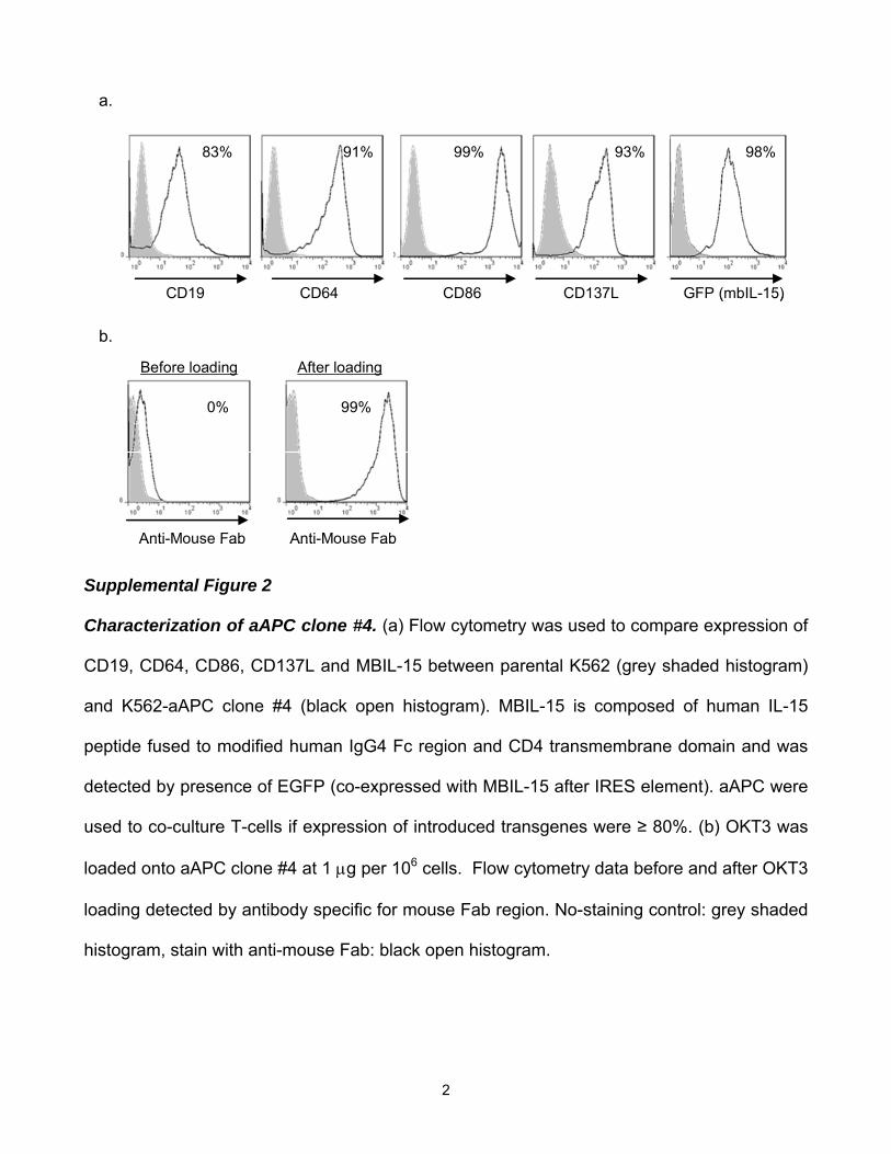
Artificial antigen presenting cells
K562-derived aAPC were previously modified by lentiviral transduction to constitutively co-
express CD19, CD64, CD86, CD137L, membrane-bound (MB) IL-15 and EGFP (the latter
For personal use only. at M D ANDERSON HOSP on April 24, 2012. bloodjournal.hematologylibrary.orgFrom

7
encoded following the EMCV IRES element). A clone (#4) was obtained by limiting dilution
and numerically expanded for use.20 For some experiments CD3-specific antibody (OKT3;
eBioscience, San Diego, CA) was used to activate T cells by pulsing the mAb onto the CD64+
(FcR) clone #4 (Supplement Figure 2). Expression of desired transgenes and bound OKT3
was validated weekly by flow cytometry before use in co-culture with T cells.
Propagation of primary T-cells
Healthy donor derived PBMC were isolated by density gradient separation using Ficoll-Paque
Plus (GE Healthcare, Pittsburgh, PA). T cells were numerically expanded in the presence of
50 IU/mL of recombinant human interleukin-2 ([rhIL-2] added three time a week; Chiron,
Emeryville, CA) on γ-irradiated (100 Gy) aAPC (clone #4, 1:2 T cell : aAPC ratio) that had
been pre-loaded with OKT3. T cells with aAPC were cultured in complete medium (CM)
defined as Hyclone-RPMI 1640 (Thermo Fisher Scientific, Waltham, MA) supplemented with
2 mmol/L L-glutamine (Glutamax-1: Invitrogen) and 10% heat-inactivated Hyclone-fetal
bovine serum (Thermo Fisher Scientific).
Generation and Propagation of CAR+ T cells
DNA supercoiled plasmids (15μg of CD19RCD28/pSBSO and 5μg of pKan-CMV-SB11)13
encoding the SB transposon (to stably express CD19RCD28) and the SB transposase (to
transiently express SB11) were electro-transferred using an Amaxa Nucleofector II device
(Lonza, Basel, Switzerland) at 2 x 10⁷ PBMC/cuvette as previously described (Supplement
Figure 1).12 T cells expressing CD19RCD28 were preferentially propagated in CM by
recursive addition every 7 or 14 days of clone #4 (not loaded with OKT3) at 1:2 T-cell : aAPC
(γ-irradiated to 100 Gy) ratio in the presence of rhIL-2 50 IU/mL, added three times a week.
For personal use only. at M D ANDERSON HOSP on April 24, 2012. bloodjournal.hematologylibrary.orgFrom

8
Electro-transfer of messenger RNA species into primary or CAR+ T cells
Six days after stimulation of unmodified T cells with OKT3-loaded clone #4 or 2 to 4 days
after the last stimulation of CD19RCD28+ T-cells with clone #4, 5x10⁶ T-cells were mixed with
2.5 to 10.0 μg of each ZFN mRNA in 100 μL of Human T-Cell Nucleofector solution (Cat
#VPA-1002, Lonza) and electroporated using the Nucleofector II device with program T-20.
Following electroporation, cells were immediately placed in 2 mL of pre-warmed CM and
cultured at 37ºC, 5% CO2 for 4 to 6 hours and then 50 IU/mL of rhIL-2 was added with 2 mL
of 20%FBS-RPMI. In some experiments to enhance ZFN-mediated enzymatic activity, after
overnight culture, cells were transferred to 30ºC, 5% CO2 and cultured for 2 days then
returned to 37ºC-5% CO2.
Enrichment of CD3neg T cells
Cells washed with PBS supplemented with 2% FBS and 2mM EDTA, were incubated for 10
minutes with CD3 microbeads (Cat # 130-050-101, MilteneyiBiotec, Auburn, CA) at 4°C. After
washing twice, cells were passed through an LD column (Cat # 130-042-901,
MilteneyiBiotec), and the flow-through fraction was collected for further use.
Surveyor Nuclease assay
The levels of genomic disruption of TRAC, TRBC1, and TRBC2 in T cells were determined by
Surveyor Nuclease assay (Transgenomics, Omaha, NE) using CEL I nuclease.21 The percent
target disruption was quantified by densitometry. The PCR primers used for the amplification
of target locus are:
TRAC forward 5’-GGGCAAAGAGGGAAATGAGA-3’
TRAC reverse 5’-CAATGGATAAGGCCGAGACC-3’
TRBC1 forward, 5’-CTGAACAAGGTGTTCCCACCC-3’
TRBC1 reverse 5’-GTGTGCGCTGGTTCCTTTCTT-3’
For personal use only. at M D ANDERSON HOSP on April 24, 2012. bloodjournal.hematologylibrary.orgFrom

9
TRBC2 forward, 5’-CCTGGCCACAGGCTTCTACC-3’
TRBC2 reverse 5’-CCACCTTGTCCACTCTGGCTT-3’
51 Chromium release assay
Target cells were labeled with 0.1 mCi of 51Cr (Perkin Elmer, Boston, MA) for 2 hours. After
washing thrice with ice-cold CM, labeled cells were diluted and plated at 103 cells/well in 100
μL CM in 96-well v-bottomed plates. T-cells were added in 100 μL/well at indicated effector
target ratios and the plate was spun (180 x g for 3minutes without brake) to facilitate cell-to-
cell contact. After 4 hours or 6 hours (when using primary tumor cells as targets) incubation
at 37°C, 5%CO2, 50 μL of supernatants were counted on TopCount (Perkin Elmer, Shelton,
CT). All assays were performed in triplicate. The percent specific lysis was calculated as
follows: ((experimental cpm - spontaneous cpm) / (maximum cpm - spontaneous cpm)) ×
100.
PKH-26 dilution assay
T-cells were incubated with 2.0 μM of the red-fluorescent lipophilic dye PKH-26 (Cat #
PKH26GL, Sigma-Aldrich) for 5 minutes at room temperature according to the manufacturer’s
instructions. Cells, 100% labeled with PKH-26, were stimulated with either OKT3 loaded
aAPC or CD19+ aAPC in CM supplemented with 50 IU/mL rhIL-2 (added every-other-day).
PKH-26-derived fluorescence was measured by flow cytometry 10 days after stimulation and
CD19RCD28+ T cells were revealed using anti-CAR mAb clone 136-20-1.
For personal use only. at M D ANDERSON HOSP on April 24, 2012. bloodjournal.hematologylibrary.orgFrom

10
RESULTS
Disruption of the αβ TCR-CD3 complex on T cells using ZFNs Two ZFN pairs targeting
the constant regions of TCR α (TRAC-ZFN-1 and TRAC-ZFN-2) or TCR β (TRBC-ZFN-1 and
TRBC-ZFN-2) (Figure 1) were developed and tested in primary human T-cells propagated ex
vivo for 6 days on OKT3-loaded aAPC (clone #4). Since transient expression of ZFNs is
sufficient to mediate gene knockout, we used a “hit-and-run” delivery strategy to transiently
express the ZFNs utilizing electro-transfer of in vitro transcribed mRNA species coding for the
ZFN pairs (Figure 2a). To measure TCR expression we used a mAb specific for CD3ε, which
is only present on the cell surface when TCRαβ is expressed. Nine days after electro-
transfer, flow cytometric analysis revealed that ZFN pairs targeting TRAC or TRBC eliminated
CD3ε expression on primary T cells at levels reaching 19.4% and 5.2% respectively. The
efficiency of TCR knockout correlated with the amount of electro-transferred mRNA (Figure
2b, upper panel). Electro-transfer of mRNA to primary T cells was generally well-tolerated,
though a slight reduction in cell viability was observed at higher doses. ZFN-mediated gene
disruption has been reported to be more efficient when cells are transiently exposed to mild
hypothermia.22 Thus, we cultured T cells for 2 days at 30°C after electro-transfer. ZFN-
mediated disruption of CD3ε was up to 2.4-fold higher when electroporated T cells were
cultured at 30°C versus 37°C. Using this approach, 37% and 15% of electroporated T-cells
lost expression of CD3ε using the ZFN pair targeting TRAC and TRBC, respectively, (Figure
2b, lower panel) with no change in the levels of CD3 negative cells in the untransfected
samples and without an appreciable decrease in viability (measured by Trypan blue).
To confirm that electroporated T cells had been genetically modified at the intended
ZFN target sites (TCR α or β loci), a Surveyor Nuclease assay was performed using specific
oligonucleotide primers flanking target sites within TRAC, TRBC1, or TRBC2. CEL I nuclease
For personal use only. at M D ANDERSON HOSP on April 24, 2012. bloodjournal.hematologylibrary.orgFrom

11
digestion products, representative of genetic changes induced by the ZFNs, were present
only after electro-transfer of the TCR-specific ZFN pairs and the percent disruption assessed
by densitometry correlated with loss of cell surface CD3ε expression (Figure 2c). These
experiments in primary T cells confirmed that ZFNs designed to target TRAC or TRBC lead to
permanent disruption of αβTCR expression, as assessed by the CEL I-mediated surveyor
nuclease assay and confirmed by flow cytometry analysis of CD3ε.
Enrichment of TCRneg T cells
For future clinical applications, rapid and robust methods for isolating sources of a TCR-
disrupted population will be needed. To address this issue, we enriched the TCR/CD3neg
population by negative selection using paramagnetic beads and a depletion column. With a
single depletion step, we enhanced the CD3εneg population to over 93% (Figure 3a). A
CD3εneg population could not be enriched from control T cells that were not genetically edited
with ZFNs. Back-to-back CD3-depletion resulted in >99% enrichment without skewing the
CD4+ or CD8+ T-cell subsets (Figure 3b). The depletion of CD3+ T cells will also deplete
remaining γδTCR+ T cells. A flow cytometry analysis of TCR Vβ repertoire in enriched TCRneg
T cells validated the elimination of TCRβ expression from the T-cell surface (Figure 3c). This
degree of depletion is clinically appealing as the loss of TCR on donor-derived T cells will
prevent GVHD in HLA-disparate recipients.
Generation of TCRnegCAR+ T cells by ZFNs
To test the ability of ZFN pairs to knock out TCR αβ expression from allogeneic
CD19RCD28+ T cells, we initially genetically modified PBMC to stably express the
CD19RCD28 CAR using the SB transposon/transposase system. The CD19RCD28+ T-cell
population was specifically propagated by stimulating with γ-irradiated CD19+ aAPC (clone
#4) every 7 days (Supplementary Figure 1b). After four rounds of stimulation, we observed
For personal use only. at M D ANDERSON HOSP on April 24, 2012. bloodjournal.hematologylibrary.orgFrom

12
over 90% CAR expression in T cells similar to our previously published results.12 Within 2 to 4
days after the fifth stimulation with CD19+ aAPC, when T cells were activated, we
electroporated the cells with mRNA encoding the TRAC or TRBC ZFNs (Figure 4a). Flow
cytometry analysis revealed that up to 30% and 26% of CD19RCD28+ T cells lost CD3ε
expression after transfection of the TRAC or TRBC ZFNs, respectively (Figure 4b). The
CD3εneg population was again readily enriched by paramagnetic beads, and the Surveyor
Nuclease assay confirmed that the CD3εneg population contained a high percentage of
modified alleles at the intended ZFN target sites within the TRAC and TRBC loci (Figure 4c).
The frequency of TRBC1 and TRBC2 disruption at the DNA level was approximately 20-25%
and that of TRAC disruption was approximately 60%. These numbers fit with the observed
frequencies of CD3εnegCD19RCD28+ T cells because in each cell only 1 out of 4 TRBC
alleles (2 TRBC1 and 2 TRBC2) is expressed. Similarly 1 of 2 TRAC alleles is expressed in
each T cell. Therefore, disruption of the expressed allele is sufficient to achieve the CD3
negative phenotype.
TCRnegCAR+ T cells do not respond to TCR stimulation, but do maintain CD19
specificity
We anticipated that TCRnegCAR+ T cells could not respond to TCR stimulation. To test this,
we measured the proliferative response of these cells after stimulation by cross-linking CD3
with OKT3 in comparison to activating CAR for sustained proliferation upon docking with
CD19. TCRnegCD19RCD28+ T cells proliferated in response to CD19, but not OKT3 (Figure
5a). Next, we assessed the ability of TCRnegCD19RCD28+ T cells to lyse CD19+ target cells
in a standard 4-hour 51Cr release assay (Figure 5b). The capacity of TCRnegCAR+ T cells to
specifically lyse CD19 target cells was similar to that observed for TCR+CD19RCD28+ T cells.
Moreover, these TCRnegCAR+ T cells maintain cytotoxicity against CD19+ primary tumors
For personal use only. at M D ANDERSON HOSP on April 24, 2012. bloodjournal.hematologylibrary.orgFrom

13
(Figure 5c). Together, these data confirmed that the absence of a measurable TCR on
TCRnegCD19RCD28+ T cells corresponds with abrogation of TCR activity, but does not
impact the ability of the CAR to activate genetically modified T cells for proliferation and
target cell killing.
TCRneg CD19RCD28+ T-cells can be propagated on CD19 expressing aAPCs
We validated that CD19RCD28+ T cells sustain their proliferative capacity to expand to the
cell numbers required for clinical applications. Both the TCRnegCD19RCD28+ and parental
TCR+CD19RCD28+ T cells exhibited similar growth kinetics in response to stimulation with
the CD19+ aAPC (Figure 6a). We did not observe any changes in CD3ε expression on
TCRnegCD19RCD28+ T cells after aAPC-mediated propagation (Figure 6b, top panel). As
predicted, these T cells failed to express TCRαβ on their cell surface (Figure 6b, middle
panel and Figure 6c). As expected, the ZFN-mediated disruption of αβTCR expression and
depletion of CD3+ T cells led to loss of γδTCR+ T cells (Figure 6b, middle panel). After
propagation on aAPC a subset of TCRnegCD19RCD28+ T cells exhibited memory phenotype
based on expression of CD62L and absence of CD45RA23 (Figure 6b, bottom panel) which
may benefit persistence and thus the therapeutic potential of our approach to “off-the-shelf”
adoptive T-cell therapy. These data confirm that TCRnegCAR+ T cells may be able to be
propagated to achieve sufficient cell numbers from a single donor-derived modified T-cell
pool for infusions into multiple recipients.
For personal use only. at M D ANDERSON HOSP on April 24, 2012. bloodjournal.hematologylibrary.orgFrom

14
DISCUSSION
We have demonstrated that T cells and indeed CAR+ T cells can be genetically edited by
ZFNs to eliminate expression of the endogenous αβ TCR. This has therapeutic implications
where donor-derived T cells are infused to achieve an anti-tumor effect. Therapeutic success
after allogeneic HSCT is defined as achieving a GVL-effect without causing clinically-
significant GVHD.24 Thus, separation of GVL and GVHD is a crucial issue following
engraftment of allogeneic hematopoietic stem cells and strategies to accomplish this are
based on infusing desired T-cell effector populations predicted to reduce unwanted allogeneic
effects. This includes the adoptive transfer of donor-derived memory T cells employing a
narrowed TCR Vβ repertoire compared with naïve T cells25,26 or in vitro depletion of T-cells
activated through allo-antigens27,28,29. Adding to this approach, we have previously
demonstrated that CAR+ T cells expressing alloreactive TCRs can be rendered anergic to
disparate HLA while maintaining specificity for CD19.13 This was achieved by blockade of co-
stimulatory molecules upon co-culture of genetically modified T cells with stimulator cells
expressing disparate HLA. An alternative to pre-selection includes conditional ablation of
infused allogeneic CAR+ T cells in the event that serious adverse events occur. This has
been accomplished by genetic modification of allogeneic T cells to express “suicide genes”
such as thymidine kinase (TK),30 iCasp9,31 CD20,32 thymidylate kinase,33 and a modified
Fas34 that can be triggered for conditional ablation via the administration of specific molecules
(e.g., ganciclovir to TK+ expressing cells).
We recognized that approaches to selectively deplete T cells expressing undesired
αβTCR may be incomplete and that complete knockout of the endogenous TCR might be
advantageous to prevent GVHD. Therefore, we undertook a genetic approach using designer
ZFNs to permanently disrupt the α and β constant region sequences in T cells thereby
For personal use only. at M D ANDERSON HOSP on April 24, 2012. bloodjournal.hematologylibrary.orgFrom

15
eliminating TCR expression. Since TCR αβ receptors need to form heterodimers to express a
functional cell surface molecule, knocking out either TRAC or TRBC was sufficient to
eliminate TCR αβ expression. This is supported by a recent publication showing that a
mutation in TRAC gene leads to the loss of TCR αβ expression.35
ZFNs have been demonstrated to disrupt target gene expression as a consequence of
error-prone DNA DSB repair by NHEJ, which in most cases results in a frame shift mutation
leading to a premature stop of translation.15 This technology is being evaluated in early stage
clinical trials infusing HIV-resistant T cells generated by ZFN-mediated disruption of the
CCR5 co-receptor for HIV-I.16,36 ZFNs target and thus disrupt gene expression at the
genomic level which is an advantage over techniques that involve transcriptional repression
and require sustained expression of the inhibiting factor (e.g., enforced expression of shRNA
to mediate TCR down regulation37). That ZFNs can permanently disrupt gene expression
after transient expression (without the inherent dangers of genomic integration) enabled our
use of in vitro transcribed mRNA species in a “hit-and-run” manner for electro-transfer of
ZFNs into T cells.
The human application of “universal” CAR+ T cells that have been genetically edited
with ZNFs will depend on efficacy as well as safety. The genetically modified T cells
specifically lysed primary targets and cell lines. Their therapeutic potential is also dependent
on persistence after adoptive transfer. The 2nd generation CAR chosen for this study
activates T cells through chimeric CD28 and CD3-ζ. It remains to be determined in side-by-
side clinical trials if other CAR designs, such as signaling through CD137 and CD3-ζ are
superior. Safety depends on selective elimination of endogenous TCR and minimizing ZFN-
mediated enzymatic activity at off-target sites. We evaluated the most likely “off-target” sites
using CEL-I nuclease and did not detect cleavage. We did observe that the efficiency of
For personal use only. at M D ANDERSON HOSP on April 24, 2012. bloodjournal.hematologylibrary.orgFrom

16
enzymatic activity at TRAC or TRBC genomic loci is approximately 20-40% after a single
electro-transfer of mRNA species coding for ZFN pairs. However, continued cell surface
expression of TCR from HLA-disparate candidate T-cell donors may cause GVHD after
adoptive immunotherapy. Therefore, to prevent GVHD after infusion we used CD3-specific
paramagnetic beads to deplete and re-deplete T cells with residual expression of TCR. This
is a clinically appealing strategy as this approach can readily be undertaken in compliance
with current good manufacturing practice for Phase I/II trials. Our planed clinical trials will
include release criteria of the manufactured T-cell product based on residual αβTCR
expression and an assessment of the maximum number of genetically modified T cells that
can be safely infused from an allogeneic donor into multiple recipients. In addition, the
wellbeing of the patients can also be safeguarded by co-expressing CAR with a transgene
capable of mediating conditional T-cell ablation.
Previous reports suggest that T-cell activation mediated through an endogenous TCR
is required to obtain a fully functional CAR in a model system using Jurkat cell lines38. In
contrast, we observed that knocking out TCR αβ expression from CD19RCD28+ T cells did
not appreciably alter the ability of these cells to specifically kill CD19+ targets or proliferate in
response to CD19. One reason for this discrepancy other than the difference in host cells
may be the use of a 2nd generation CAR, which includes signaling not only through CD3ζ
(signal 1) but also CD28 (signal 2; co-stimulation).39-41 A benefit to expressing a TCR with
known specificity is that activation through the endogenous immunoreceptor can be used to
propagate T cells to achieve an anti-tumor effect mediated by the CAR.42,43 It remains to be
tested in humans whether coordinated co-stimulation achieved through multiple CAR
signaling endodomains will be sufficient to sustain persistence in vivo or if triggering of T cells
through TCR is needed. However, the propagation of CD19RCD28+ T cells on aAPC
For personal use only. at M D ANDERSON HOSP on April 24, 2012. bloodjournal.hematologylibrary.orgFrom

17
modified to co-express CD19 along with co-stimulatory molecules results in the significant
expansion of CAR+ memory T-cell subsets predicted to have prolonged in vivo survival.44,45
Therefore, any loss of persistence of TCRnegCD19RCD28+ T cells may be off-set by co-
stimulatory properties of aAPC and the encoded CD28 intra-cellular domain within the CAR.
Preparing antigen-specific T cells from a third-party donor is clinically appealing as
these products can be generated, stored and validated before use and infused to multiple
patients immediately as needed.46 Indeed, third-party T cells have been successfully infused
into patients with post-transplantation lymphoproliferative diseases.47,48 Despite the fact that a
majority of the viral-antigen specific TCRαβ chains demonstrate cross-reactivity to allo-HLA in
vivo,49 clinically significant GVHD was not observed. This may be in part due to the ex vivo
repetitive antigen stimulation resulting in the emergence of either an oligoclonal or
monoclonal TCRαβ repertoire which decreases the chance of T-cell alloreactivity. On the
contrary, when we numerically expand CD19RCD28+ T cells through in vitro CD19
stimulation on γ-irradiated aAPC independent of TCR stimulation we did not observe skewing
of the TCR Vβ usage as assessed by a panel of Vβ-specific antibodies.
In conclusion, we demonstrate that TCRnegCAR+ T cells can be generated using a
genetic approach to remove (a) endogenous undesired TCR with ZFNs and (b) introduce a
desired CAR with the SB system using a common electro-transfer platform. Our approach
abolishes the danger of GVHD posed by adoptive transfer of large numbers of allogeneic T
cells while maintaining desired effector functions mediated by CD19RCD28 CAR to target
malignant B cells. This strategy provides an important step to developing a “universal” CAR+
T cell which can be manufactured from one donor and administered on demand to multiple
patients. Subsequent studies are focusing on preventing rejection of the infused allogeneic
TCRnegCAR+ T cells by the recipient’s immune system recognizing disparate HLA. This may
For personal use only. at M D ANDERSON HOSP on April 24, 2012. bloodjournal.hematologylibrary.orgFrom

18
be accomplished using genetic modifications including ZFN-mediated knockout of HLA and
over-expression of conserved HLA homologues to inhibit NK-cell activity.
For personal use only. at M D ANDERSON HOSP on April 24, 2012. bloodjournal.hematologylibrary.orgFrom

19
ACKNOWLEDGEMENTS
We thank Dr. Carl June at the University of Pennsylvania for assistance with K562-derived
aAPC clone #4 and Dr. Perry Hackett at the University of Minnesota for help with the SB
system. Support from: Cancer Center Core Grant (CA16672); DOD PR064229; RO1
(CA124782, CA120956, CA141303); R33 (CA116127); Burroughs Wellcome Fund; Cancer
Prevention Research Institute of Texas; CLL Global Research Foundation; Gillson
Longenbaugh Foundation; Harry T. Mangurian, Jr., Foundation, Institute of Personalized
Cancer Therapy; Leukemia and Lymphoma Society; Lymphoma Research Foundation; Miller
Foundation; Mr. and Mrs. Joe H. Scales; National Foundation for Cancer Research; Pediatric
Cancer Research Foundation; Production Assistance for Cellular Therapies (PACT); Sister
Institution Network Fund; Uehara Memorial Foundation; William Lawrence and Blanche
Hughes Children's Foundation.
AUTHORSHIP CONTRIBUTIONS
H.T., A.R. designed and performed experiments analyzed data and wrote the paper. P.L.,
Y.Z., L.Z., S.M., J.C.M. performed the experiments. H.H. supported experiments. P.K., B.R.,
D.A.L., R.E.C. contributed discussion and edited the paper. E.J.R., P.D.G, M.C.H. designed
experiments, analyzed data, and edited the paper. C.B., L.N. tested the TRAC and TRBC
target ZFNs developed by Sangamo BioSciences, and edited the paper. L.J.N.C. conceived
the idea, coordinated the project, designed experiments, analyzed data, and wrote the paper.
DISCLOSURE OF CONFLICTS OF INTEREST
The authors declare no competing financial interests.
For personal use only. at M D ANDERSON HOSP on April 24, 2012. bloodjournal.hematologylibrary.orgFrom
20
REFERENCES
1. Cooper LJ, Al-Kadhimi Z, DiGiusto D, et al. Development and application of CD19-specific T cells for adoptive immunotherapy of B cell malignancies. Blood Cells Mol Dis. 2004;33(1):83-89. 2. Jena B, Dotti G, Cooper LJ. Redirecting T-cell specificity by introducing a tumor-specific chimeric antigen receptor. Blood;116(7):1035-1044. 3. Kochenderfer JN, Dudley ME, Feldman SA, et al. B-cell depletion and remissions of malignancy along with cytokine-associated toxicity in a clinical trial of anti-CD19 chimeric-antigen-receptor-transduced T cells. Blood;119(12):2709-27201044. 4. Kochenderfer JN, Wilson WH, Janik JE, et al. Eradication of B-lineage cells and regression of lymphoma in a patient treated with autologous T cells genetically engineered to recognize CD19. Blood;116(20):4099-4102. 5. Kalos M, Levine BL, Porter DL, et al. T cells with chimeric antigen receptors have potent antitumor effects and can establish memory in patients with advanced leukemia. Sci Transl Med;3(95):95ra73. 6. Porter DL, Levine BL, Kalos M, Bagg A, June CH. Chimeric antigen receptor-modified T cells in chronic lymphoid leukemia. N Engl J Med;365(8):725-733. 7. Brentjens RJ, Riviere I, Park JH, et al. Safety and persistence of adoptively transferred autologous CD19-targeted T cells in patients with relapsed or chemotherapy refractory B-cell leukemias. Blood;118(18):4817-4828. 8. Kohn DB, Dotti G, Brentjens R, et al. CARs on Track in the Clinic. Mol Ther;19(3):432-438. 9. Izsvak Z, Hackett PB, Cooper LJ, Ivics Z. Translating Sleeping Beauty transposition into cellular therapies: victories and challenges. Bioessays;32(9):756-767. 10. Hackett PB, Largaespada DA, Cooper LJ. A transposon and transposase system for human application. Mol Ther;18(4):674-683. 11. Williams DA. Sleeping beauty vector system moves toward human trials in the United States. Mol Ther. 2008;16(9):1515-1516. 12. Singh H, Manuri PR, Olivares S, et al. Redirecting specificity of T-cell populations for CD19 using the Sleeping Beauty system. Cancer Res. 2008;68(8):2961-2971. 13. Davies JK, Singh H, Huls H, et al. Combining CD19 redirection and alloanergization to generate tumor-specific human T cells for allogeneic cell therapy of B-cell malignancies. Cancer Res;70(10):3915-3924. 14. Urnov FD, Rebar EJ, Holmes MC, Zhang HS, Gregory PD. Genome editing with engineered zinc finger nucleases. Nat Rev Genet;11(9):636-646. 15. Santiago Y, Chan E, Liu PQ, et al. Targeted gene knockout in mammalian cells by using engineered zinc-finger nucleases. Proc Natl Acad Sci U S A. 2008;105(15):5809-5814. 16. Perez EE, Wang J, Miller JC, et al. Establishment of HIV-1 resistance in CD4+ T cells by genome editing using zinc-finger nucleases. Nat Biotechnol. 2008;26(7):808-816. 17. Doyon Y, McCammon JM, Miller JC, et al. Heritable targeted gene disruption in zebrafish using designed zinc-finger nucleases. Nat Biotechnol. 2008;26(6):702-708. 18. Isalan M, Choo Y. Rapid, high-throughput engineering of sequence-specific zinc finger DNA-binding proteins. Methods Enzymol. 2001;340:593-609. 19. Provasi E, Genovese P, Lombardo A, et al. Editing T cell specificity towards leukemia by zinc finger nucleases and lentiviral gene transfer. Nat Med. Prepublished on 2012/04/03 20. Manuri PV, Wilson MH, Maiti SN, et al. piggyBac transposon/transposase system to generate CD19-specific T cells for the treatment of B-lineage malignancies. Hum Gene Ther;21(4):427-437.
For personal use only. at M D ANDERSON HOSP on April 24, 2012. bloodjournal.hematologylibrary.orgFrom

21
21. Guschin DY, Waite AJ, Katibah GE, Miller JC, Holmes MC, Rebar EJ. A rapid and general assay for monitoring endogenous gene modification. Methods Mol Biol;649:247-256. 22. Doyon Y, Choi VM, Xia DF, Vo TD, Gregory PD, Holmes MC. Transient cold shock enhances zinc-finger nuclease-mediated gene disruption. Nat Methods;7(6):459-460. 23. Sallusto F, Geginat J, Lanzavecchia A. Central memory and effector memory T cell subsets: function, generation, and maintenance. Annu Rev Immunol. 2004;22:745-763. 24. Bleakley M, Riddell SR. Molecules and mechanisms of the graft-versus-leukaemia effect. Nat Rev Cancer. 2004;4(5):371-380. 25. Chen BJ, Cui X, Sempowski GD, Liu C, Chao NJ. Transfer of allogeneic CD62L- memory T cells without graft-versus-host disease. Blood. 2004;103(4):1534-1541. 26. Foster AE, Marangolo M, Sartor MM, et al. Human CD62L- memory T cells are less responsive to alloantigen stimulation than CD62L+ naive T cells: potential for adoptive immunotherapy and allodepletion. Blood. 2004;104(8):2403-2409. 27. Amrolia PJ, Muccioli-Casadei G, Yvon E, et al. Selective depletion of donor alloreactive T cells without loss of antiviral or antileukemic responses. Blood. 2003;102(6):2292-2299. 28. Hartwig UF, Nonn M, Khan S, Meyer RG, Huber C, Herr W. Depletion of alloreactive T cells via CD69: implications on antiviral, antileukemic and immunoregulatory T lymphocytes. Bone Marrow Transplant. 2006;37(3):297-305. 29. Wehler TC, Nonn M, Brandt B, et al. Targeting the activation-induced antigen CD137 can selectively deplete alloreactive T cells from antileukemic and antitumor donor T-cell lines. Blood. 2007;109(1):365-373. 30. Bonini C, Ferrari G, Verzeletti S, et al. HSV-TK gene transfer into donor lymphocytes for control of allogeneic graft-versus-leukemia. Science. 1997;276(5319):1719-1724. 31. Straathof KC, Pule MA, Yotnda P, et al. An inducible caspase 9 safety switch for T-cell therapy. Blood. 2005;105(11):4247-4254. 32. Introna M, Barbui AM, Bambacioni F, et al. Genetic modification of human T cells with CD20: a strategy to purify and lyse transduced cells with anti-CD20 antibodies. Hum Gene Ther. 2000;11(4):611-620. 33. Sato T, Neschadim A, Konrad M, Fowler DH, Lavie A, Medin JA. Engineered human tmpk/AZT as a novel enzyme/prodrug axis for suicide gene therapy. Mol Ther. 2007;15(5):962-970. 34. Berger C, Blau CA, Huang ML, et al. Pharmacologically regulated Fas-mediated death of adoptively transferred T cells in a nonhuman primate model. Blood. 2004;103(4):1261-1269. 35. Morgan NV, Goddard S, Cardno TS, et al. Mutation in the TCRalpha subunit constant gene (TRAC) leads to a human immunodeficiency disorder characterized by a lack of TCRalphabeta+ T cells. J Clin Invest;121(2):695-702. 36. Holt N, Wang J, Kim K, et al. Human hematopoietic stem/progenitor cells modified by zinc-finger nucleases targeted to CCR5 control HIV-1 in vivo. Nat Biotechnol;28(8):839-847. 37. Okamoto S, Mineno J, Ikeda H, et al. Improved expression and reactivity of transduced tumor-specific TCRs in human lymphocytes by specific silencing of endogenous TCR. Cancer Res. 2009;69(23):9003-9011. 38. Bridgeman JS, Hawkins RE, Bagley S, Blaylock M, Holland M, Gilham DE. The optimal antigen response of chimeric antigen receptors harboring the CD3zeta transmembrane domain is dependent upon incorporation of the receptor into the endogenous TCR/CD3 complex. J Immunol;184(12):6938-6949.
For personal use only. at M D ANDERSON HOSP on April 24, 2012. bloodjournal.hematologylibrary.orgFrom

22
39. Maher J, Brentjens RJ, Gunset G, Riviere I, Sadelain M. Human T-lymphocyte cytotoxicity and proliferation directed by a single chimeric TCRzeta /CD28 receptor. Nat Biotechnol. 2002;20(1):70-75. 40. Kowolik CM, Topp MS, Gonzalez S, et al. CD28 costimulation provided through a CD19-specific chimeric antigen receptor enhances in vivo persistence and antitumor efficacy of adoptively transferred T cells. Cancer Res. 2006;66(22):10995-11004. 41. Savoldo B, Ramos CA, Liu E, et al. CD28 costimulation improves expansion and persistence of chimeric antigen receptor-modified T cells in lymphoma patients. J Clin Invest;121(5):1822-1826. 42. Cooper LJ, Al-Kadhimi Z, Serrano LM, et al. Enhanced antilymphoma efficacy of CD19-redirected influenza MP1-specific CTLs by cotransfer of T cells modified to present influenza MP1. Blood. 2005;105(4):1622-1631. 43. Pule MA, Savoldo B, Myers GD, et al. Virus-specific T cells engineered to coexpress tumor-specific receptors: persistence and antitumor activity in individuals with neuroblastoma. Nat Med. 2008;14(11):1264-1270. 44. Numbenjapon T, Serrano LM, Chang WC, Forman SJ, Jensen MC, Cooper LJ. Antigen-independent and antigen-dependent methods to numerically expand CD19-specific CD8+ T cells. Exp Hematol. 2007;35(7):1083-1090. 45. Butler MO, Friedlander P, Milstein MI, et al. Establishment of Antitumor Memory in Humans Using in Vitro-Educated CD8+ T Cells. Sci Transl Med;3(80):80ra34. 46. Cooper LJ. Off-the-shelf T-cell therapy. Blood;116(23):4741-4743. 47. Barker JN, Doubrovina E, Sauter C, et al. Successful treatment of EBV-associated posttransplantation lymphoma after cord blood transplantation using third-party EBV-specific cytotoxic T lymphocytes. Blood;116(23):5045-5049. 48. Haque T, Wilkie GM, Taylor C, et al. Treatment of Epstein-Barr-virus-positive post-transplantation lymphoproliferative disease with partly HLA-matched allogeneic cytotoxic T cells. Lancet. 2002;360(9331):436-442. 49. Amir AL, D'Orsogna LJ, Roelen DL, et al. Allo-HLA reactivity of virus-specific memory T cells is common. Blood;115(15):3146-3157. 50. Arden B, Clark SP, Kabelitz D, Mak TW. Human T-cell receptor variable gene segment families. Immunogenetics. 1995;42(6):455-500.
For personal use only. at M D ANDERSON HOSP on April 24, 2012. bloodjournal.hematologylibrary.orgFrom

23
FIGURE LEGENDS
Figure 1
ZFN pairs targeting sites within genomic loci of TCR-α and β constant region.
Each exon is shown by a block. Black blocks represent coding regions. Grey columns
represent non-coding regions. One ZFN pair was designed to bind exon 1 of the TCR α
constant region (TRAC) and another ZFN pair binds a conserved sequence on exon 1 of the
TCR β constant regions 1 (TRBC1) and 2 (TRBC2). Underlined nucleotide sequences
represent the intended binding sequence of each ZFN.
Figure 2
Disruption of the TCR αβ-CD3 complex in primary T cells
a. Schematic presentation of ZFN transfer
A pair of ZFN-encoding mRNA was electro-transferred 6 days after stimulation of
CARneg T cells. T cells were then cultured with 50 IU/mL of IL-2 and incubated at 30°C or
37°C-5%CO2, as indicated. CD3 expression was analyzed by flow cytometry on Day 7 to 9
after electroporation.
b. Down regulation of CD3 after electro-transfer of mRNA encoding the TCR αβ targeted
ZFNs
Day 9 after electro-transfer of the indicated doses of mRNA coding for TRAC or TRBC
targeted ZFN pairs, TCR αβ-CD3 expression was analyzed by co-staining for CD4, CD8, and
CD3ε. Representative flow data at Day 9 after ZFN electro-transfer is shown. Flow cytometry
data are gated on cells excluding propidium iodide. Numbers in the lower right quadrant
represent the percentage of CD3ε negative cells in T-cell populations.
For personal use only. at M D ANDERSON HOSP on April 24, 2012. bloodjournal.hematologylibrary.orgFrom

24
Top panels shows CD3ε expression in T cells cultured at 37°C after ZFN transfer and
bottom panels shows CD3ε expression in T cells transiently cultured at 30°C from Day 2 to 3
after ZFN transfer.
c. Surveyor Nuclease assay to detect ZFN-mediated modification of TCR target sites in T
cells
Arrows indicate the fragments produced by a Surveyor Nuclease digest of amplicons
bearing a mismatch at the intended site of ZFN cleavage in the TRAC or TRBC loci,
respectively. Lane headings indicate both the mRNA dose, specific ZFN pair delivered via
electro-transfer, and temperature of incubation for the different samples. Numbers beneath
each lane indicate the percentage of modified alleles in each sample.
Figure 3
TCRneg T cells can be enriched by depletion of CD3ε+T cells
a. CD3 expression before and after depletion using CD3-specific paramagnetic beads.
Flow cytometry reveals expression of CD3ε in CD4+ and CD8+ T cells 15 days after
stimulation by OKT3-loaded aAPC (9 days after ZFN transfection). Numbers in the lower right
quadrant represent the percentage of CD3εneg T cells. Representative results using in vitro
numerically expanded T cells.
b. CD3εneg T cells can be further enriched by additional round of depletion with CD3-specific
paramagnetic beads
Flow cytometry revealing expression of CD3ε in CD4+ and CD8+ T-cells after two
rounds of depletion of CD3εpos T-cells. Numbers in the lower right quadrant represents the
percentage of CD3ε negative cells in CD4+ and CD8+ T-cell populations.
c. Vβ repertoire analysis in T cells modified with ZFN.
For personal use only. at M D ANDERSON HOSP on April 24, 2012. bloodjournal.hematologylibrary.orgFrom

25
The Vβ usage clonogram was analyzed by a panel of TCR-specific mAbs, co-stained
with CD4 and CD8. Percentage of specific Vβ+ T-cell fractions within CD4 and CD8 gating is
shown. The nomenclatures of Vβ repertoire shown are based on Arden et al.50
Representative data from 3 independent experiment is shown.
Figure 4
Elimination of TCR αβ - CD3 complex from CD19-specific CAR+ T-cells
a. Schematic of electro-transfer of mRNA coding for ZFN pairs in CAR+ T cells
mRNA species encoding the indicated ZFN pairs were electro-transferred into CAR+ T
cells two days after stimulation with CD19+ aAPC. After electroporation, cells were
maintained with 50 IU/mL of IL-2 and incubated for two days at 30°C-5%CO2. CD3ε
expression was analyzed 9 days after electroporation by flow cytometry.
b. Disruption of TCR αβ - CD3 complex expression after electro-transfer of mRNA encoding
the TCR-specific ZFNs
Flow cytometry analysis of CD3ε expression in T cells 9 days after electro-transfer of
mRNA species encoding the indicated ZFN pairs, gated on the propidium iodide negative
population.
c. Surveyor Nuclease assay
Arrows indicate the fragments produced by a Surveyor Nuclease digest of amplicons
bearing a mismatch at the intended site of ZFN cleavage in the TRAC or TRBC loci,
respectively. Samples were analyzed 9 days after electroporation. The numbers at the
bottom represent percentages of modified alleles in each sample.
For personal use only. at M D ANDERSON HOSP on April 24, 2012. bloodjournal.hematologylibrary.orgFrom

26
Figure 5
Functional consequences of ZFN-mediated TCR knockout in CAR+ T cells
a. Loss of responsiveness of TCRneg CAR+ T cells to TCR stimulation
Dilution of PKH26 was measured 10 days after stimulation with aAPC loaded with OKT3
(upper panel) or expressing CD19 (lower panel). Flow cytometry data was gated on CAR+ T
cells. Parental: CAR+ T-cells without modification; no mRNA: mock electroporated CAR+ T
cells ; TRAC CD3neg: CAR+ T-cells electroporated with mRNA encoding ZFN pairs specific for
TRAC, and depleted CD3pos population; TRBC CD3neg: CAR+ T cells electroporated with
mRNA encoding ZFN pairs specific for TRBC, and depleted for CD3pos population.
b. Redirected specificity of TCRneg CAR+ T cells
Specific lysis by CAR+ T cells of an EL4 (mouse T-cell line) modified to express a truncated
version of human CD19 (closed symbols) was measured by standard 4 hour 51Cr release
assay. Specificity is shown by lack of lysis of CD19neg (parental) EL4 cells (open symbols).
CAR+ T-cells were modified by ZFNs (TRAC and TRBC) or unmodified CAR+ T-cells
(parental and no mRNA). The error bars represent the standard deviation.
c. Cytotoxicity by TCRneg CAR+ T cells against CD19+ primary B-cell tumors
Specific lysis by CAR+ T cells of B-cell malignances derived from patients was measured by 6
hour 51Cr release assay (effector : target ratio = 30:1). DLBCL: diffuse large B-cell lymphoma,
CLL: chronic lymphocytic lymphoma, and MCL: mantle cell lymphoma. The error bars
represent the standard deviation.
Figure 6
Propagation of TCRneg CAR+ T cells on CD19 expressing aAPC
a. Sustained proliferation of TCRneg CAR+ T cells
For personal use only. at M D ANDERSON HOSP on April 24, 2012. bloodjournal.hematologylibrary.orgFrom

27
CAR+ T cells with (TRAC and TRBC) or without (parental and no mRNA) TCR modification by
ZFNs were stimulated with γ-irradiated CD19+ aAPC every 2 weeks. Viable T cells were
enumerated every 7 days and inferred total numbers were calculated. Representative data
from 3 independent experiments is shown.
b. Analysis of TCRneg CAR+ T cells after propagation
Flow cytometry analysis of CD3ε expression (top panel), αβTCR and γδTCR expression
(middle panel), and subset analysis for memory pool (bottom panel) of TCRnegCAR+ T cells
after 28 days of propagation on aAPC. Numbers are percent expression for the quadrant.
c. Vβ repertoire analysis in TCRneg CAR+ T cells after propagation on aAPC
The Vβ usage clonogram was analyzed by a panel of TCR-specific mAbs, co-stained with
CD4 and CD8. Percentage of identified Vβ+ T-cell fractions within CD4 and CD8 flow
cytometry gates is shown. The nomenclatures of Vβ repertoire shown are based on Arden et
al.50 Representative data from 3 independent experiments is shown.
For personal use only. at M D ANDERSON HOSP on April 24, 2012. bloodjournal.hematologylibrary.orgFrom

Fig.1
TRAC:
Ex1Ex2 Ex3 Ex4
׀׀׀׀׀׀׀׀׀׀׀׀׀׀׀׀׀׀׀׀׀׀׀׀׀׀׀׀׀׀׀׀׀׀׀׀׀׀׀׀׀׀׀׀׀׀׀׀׀׀׀׀CAACAGTGCTGTGGCCTGGAGCAACAAATCTGACTTTGCATGTGCAAAGCCCGTTGTCACGACACCGGACCTCGTTGTTTAGACTGAAACGTACACGTTTGCGG
TRBC1:
Ex1Ex2Ex3 Ex4
TRBC2:
Chr. 7q34
Chr. 14q11.2
Ex1Ex2 Ex3 Ex4
Chr. 7q34
CGCTGTCAAGTCCAGTTCTACGGGCTCTCGGAGAATGACGAGTGGACCCAG
GCGACAGTTCAGGTCAAGATGCCCGAGAGCCTCTTACTGCTCACCTGGGTC׀׀׀׀׀׀׀׀׀׀׀׀׀׀׀׀׀׀׀׀׀׀׀׀׀׀׀׀׀׀׀׀׀׀׀׀׀׀׀׀׀׀׀׀׀׀׀׀׀׀׀
TRBC-ZFN-1
TRBC-ZFN-2
TRAC-ZFN-1
TRAC-ZFN-2
For personal use only. at M D ANDERSON HOSP on April 24, 2012. bloodjournal.hematologylibrary.orgFrom

b.
c.
CD
3ε
TRBC target ZFNs
5.0 g2.5 gNo mRNA
100 101 102 103 104100
101
102
103
104
5.2
100 101 102 103 104100
101
102
103
104
2.1
TRAC target ZFNs
5.0 g2.5 g
100 101 102 103 104100
101
102
103
104
19.4
100 101 102 103 104100
101
102
103
104
15.2
100 101 102 103 104100
101
102
103
104
2.4
100 101 102 103 104100
101
102
103
104
14.6
100 101 102 103 104100
101
102
103
104
3.8
100 101 102 103 104100
101
102
103
104
27.0
100 101 102 103 104100
101
102
103
104
37.0
100 101 102 103 104100
101
102
103
104
1.9
CD4 & CD8 (co-stain)
TRBCTRAC
no 2.5
g
5.0
g
2.5
g
5.0
g
30ºC 37ºC
no 2.5
g
5.0
g
2.5
g
5.0
g
30ºC 37ºC
37ºC
30ºC
Fig. 2
29 27 20 2529 27 20 25 3 11 1 5
a.
Day6Electro-transfer of in vitrotranscribed mRNAs coding for ZFNs
Day7-8 (Day1-2 after electro-transfer)Culture at 30°C-5%CO2
Day13-15 (7-9 days after electro-transfer)TCR expression analysis
IL-2
IL-2 Culture at 37°C-5%CO2
IL-2Day9 (Day3 after electro-transfer)37°C-5%CO2
Day0Stimulation with OKT3 loaded aAPC
IL-2
For personal use only. at M D ANDERSON HOSP on April 24, 2012. bloodjournal.hematologylibrary.orgFrom

a.
CD4 + CD8
CD
3ε
100 101 102 103 104100
101
102
103
104
100 101 102 103 104100
101
102
103
104
100 101 102 103 104100
101
102
103
104
100 101 102 103 104100
101
102
103
104
100 101 102 103 104100
101
102
103
104
100 101 102 103 104100
101
102
103
104
93.7
11.7
96.3
46.30.20.0
Isotype control No mRNA TRAC 2.5 g TRBC 5.0 g
CD3pos depletion x 1column
c. No mRNA TRAC 2.5 g
TRAC 2.5 g CD3pos depleted
TRBC 5.0 g
TRBC 5.0 g CD3pos depleted
% V
usag
e in
CD
4pos
CD
8pos
Frac
tion
0
2
4
6
8
Fig. 3
(co-stain)
Vb1
Vb2 Vb3
Vb4
VB
5.1
Vb5
.2V
b5.3
Vb7
.1
Vb7
.2
Vb8
Vb9
Vb1
1
Vb1
2
Vb1
3.1
Vb1
3.2
Vb1
3.6
Vb1
4
Vb1
6
Vb1
7
Vb1
8
Vb2
0
Vb2
1.3
Vb2
2
Vb2
3
0
2
4
6
8
99.6
14.4
99.7
29.71.30.0
Isotype control g g
99.6
14.4
99.7
29.71.30.0
Isotype control No mRNA TRAC 2.5 g TRBC 5.0 g
CD3pos depletion x 2columns
CD4 + CD8
CD
3ε
(co-stain)
b.
For personal use only. at M D ANDERSON HOSP on April 24, 2012. bloodjournal.hematologylibrary.orgFrom

b.
TRAC
TRBC1
TRBC2
a.
Isotype No mRNA
TRAC (2.5µg each) TRBC (5.0µg each)
CD3pos depletion x 1column
CAR
CD
3
100 101 102 103 104100
101
102
103
104
100 101 102 103 104100
101
102
103
104
100 101 102 103 104100
101
102
103
104
100 101 102 103 104100
101
102
103
104
100 101 102 103 104100
101
102
103
104
100 101 102 103 104100
101
102
103
104
0.2 0.2
30.0 26.1
93.4 90.1
CAR+ T-cells
Day2-4ZFN mRNAs electro-transfer
Day1 after electro-transferCulture at 30°C-5%CO2
for 2 days
7-9 days after ZFN electro-transferTCR expression analysis
IL-2
c.
Fig. 4
Day0Stimulation with CD19+ aAPC
IL-2
IL-2
267
TRB
CTR
BC
CD
3pos
depl
eted
TRA
C C
D3p
os d
eple
ted
TRA
C
no m
RN
A
8 20
24 62
For personal use only. at M D ANDERSON HOSP on April 24, 2012. bloodjournal.hematologylibrary.orgFrom

PKH26 dilution
OKT3
CD19
Parental No mRNA TRAC CD3neg TRBC CD3neg
Fig.5
a.
0
10
20
30
40
10:1 3:1 1:1E : T ratio
% S
peci
fic L
ysis
vs. CD19neg EL4
ParentalNo mRNATRAC CD3neg
TRBC CD3neg
ParentalNo mRNATRAC CD3neg
TRBC CD3neg
vs. CD19+ EL4b.
0
10
20
30
40
50
CD
19ne
gE
L4
DLB
CL
CLL
MC
L-1
ParentalNo mRNA
TRACTRBC
c.
CD
19po
sE
L4
MC
L-2
% S
peci
fic L
ysis
50
For personal use only. at M D ANDERSON HOSP on April 24, 2012. bloodjournal.hematologylibrary.orgFrom

1011
0 7Days after stimulation
Infe
rred
Tot
al C
ell n
umbe
rsParentalno mRNATRAC CD3negTRBC CD3neg
1010
109
108
107
106
0 14 21 28
Stim. Stim.
Fig.6
a.
b.
CD45RA
CD
62L
CD
3
0.7 97.6
36
60
3
1
0.6 97.3
47
51
2
0
47
52
1
0
49
50
1
0
TCR γδ
TCR
αβ
94
1
95
1
2
0
2
0
Parental No mRNA TRAC TRBC
CAR
c.No mRNA TRAC CD3pos depleted TRBC CD3pos depletedNo mRNA TRAC CD3pos depleted TRBC CD3pos depleted
0246
810
Vb2
VB
5.1
Vb1
3.2
Vb1
7V
b13.
1V
b8V
b20
Vb3
Vb1
Vb1
8V
b21.
3V
b12
Vb4
Vb1
3.6
Vb1
4V
b7.1
Vb7
.2V
b5.3
Vb9
Vb1
6V
b5.2
Vb2
2V
b11
Vb2
3
For personal use only. at M D ANDERSON HOSP on April 24, 2012. bloodjournal.hematologylibrary.orgFrom

1
SUPPLEMENTAL DATA
CAR+ T-cells
CD86 CD137LCD19 MBIL15
TCRαβ
CAR
CD28 CAR IL-15RCD137
Clone #4 aAPC
CAR+T-cellPBMC
Electro-transfer of DNA plasmids
coding for SB transposon and
transposase
Co-culture on irradiated aAPC
CD86 CD137LCD64 MBIL15
CD28TCR/CD3 IL-15RCD137
OKT3 loaded Clone #4 aAPC
T-cell
PBMC
Co-culture on irradiated aAPC
OKT3
a. CARneg T cells
b. CAR+ T cells
Supplemental Figure 1
Schematic of the approach to genetically modify and propagate T-cells from PBMC.
(a)T-cells were propagated by stimulation with OKT3 loaded γ-irradiated aAPC (clone #4) in
the presence of soluble IL-2 (b) DNA plasmids coding for SB transposon (CD19RCD28) and
SB transposase (SB11) were electro-transferred into primary human T-cells. CAR+ T cells
were selectively propagated by repeated additions of γ-irradiated aAPC (clone #4) in the
presence of rhIL-2.
2
a.
b.
99%
Anti-Mouse Fab
After loading
Anti-Mouse Fab
0%
Before loading
99%
Anti-Mouse Fab
After loading
Anti-Mouse Fab
0%
Before loading
98%93%99%91%83%
GFP (mbIL-15)CD19 CD64 CD86 CD137L
98%93%99%91%83%
GFP (mbIL-15)CD19 CD64 CD86 CD137L
Supplemental Figure 2
Characterization of aAPC clone #4. (a) Flow cytometry was used to compare expression of
CD19, CD64, CD86, CD137L and MBIL-15 between parental K562 (grey shaded histogram)
and K562-aAPC clone #4 (black open histogram). MBIL-15 is composed of human IL-15
peptide fused to modified human IgG4 Fc region and CD4 transmembrane domain and was
detected by presence of EGFP (co-expressed with MBIL-15 after IRES element). aAPC were
used to co-culture T-cells if expression of introduced transgenes were ≥ 80%. (b) OKT3 was
loaded onto aAPC clone #4 at 1 μg per 106 cells. Flow cytometry data before and after OKT3
loading detected by antibody specific for mouse Fab region. No-staining control: grey shaded
histogram, stain with anti-mouse Fab: black open histogram.


![Effective Adoptive Immunotherapy by T-LAK Cells Retargeted ... · (CANCER RESEARCH 58, 2838-2843. July I. 1998] Effective Adoptive Immunotherapy by T-LAK Cells Retargeted with Bacterial](https://img.dokumen.tips/doc/110x75/5f63e3f36c1d5541c3432813/effective-adoptive-immunotherapy-by-t-lak-cells-retargeted-cancer-research.jpg)


