Embed Size (px)
Citation preview

A Fasciclin-Like Arabinogalactan Protein, GhFLA1,Is Involved in Fiber Initiation and Elongationof Cotton1[C][W][OA]
Geng-Qing Huang2, Si-Ying Gong2, Wen-Liang Xu, Wen Li, Peng Li, Chao-Jun Zhang, Deng-Di Li,Yong Zheng, Fu-Guang Li, and Xue-Bao Li*
Hubei Key Laboratory of Genetic Regulation and Integrative Biology, College of Life Sciences, Central ChinaNormal University, Wuhan 430079, People’s Republic of China (G.-Q.H., S.-Y.G., W.-L.X., W.L., P.L., D.-D.L.,Y.Z., X.-B.L.); and State Key Laboratory of Cotton Biology, Institute of Cotton Research, Chinese Academy ofAgriculture Sciences, Anyang, Henan 455000, People’s Republic of China (C.-J.Z., F.-G.L.)
Arabinogalactan proteins (AGPs) are involved in many aspects of plant development. In this study, biochemical and geneticapproaches demonstrated that AGPs are abundant in developing fibers and may be involved in fiber initiation and elongation.To further investigate the role of AGPs during fiber development, a fasciclin-like arabinogalactan protein gene (GhFLA1) wasidentified in cotton (Gossypium hirsutum). Overexpression of GhFLA1 in cotton promoted fiber elongation, leading to an increasein fiber length. In contrast, suppression of GhFLA1 expression in cotton slowed down fiber initiation and elongation. As a result,the mature fibers of the transgenic plants were significantly shorter than those of the wild type. In addition, expression levels ofGhFLAs and the genes related to primary cell wall biosynthesis were remarkably enhanced in the GhFLA1 overexpressiontransgenic fibers, whereas the transcripts of these genes were dramatically reduced in the fibers of GhFLA1 RNA interferenceplants. An immunostaining assay indicated that both AGP composition and primary cell wall composition were changed in thetransgenic fibers. The levels of glucose, arabinose, and galactose were also altered in the primary cell wall of the transgenic fiberscompared with those of the wild type. Together, our results suggested that GhFLA1 may function in fiber initiation andelongation by affecting AGP composition and the integrity of the primary cell wall matrix.
Arabinogalactan proteins (AGPs) are one type ofglycoproteins that are universally distributed in theplant kingdom. AGP consists of a core-protein back-bone O-glycosylated by type II arabinogalactan gly-cans. Based on the core protein sequences, AGPs aredivided into six classes: classical AGPs with an N-terminalsignal sequence, a central Pro/Hyp-rich domain (alsonamed the AGP motif), and a hydrophobic C terminus;classical AGPs with Lys-rich domains; arabinogalactanpeptides with short protein backbones; fasciclin-like AGPs(FLAs), a class of chimeric AGPs that contain both AGPmotifs and one or two fasciclin domains; chimeric
plastocyanin AGPs; and chimeric nonclassical AGPs(Gaspar et al., 2001; Showalter et al., 2010).
In Arabidopsis (Arabidopsis thaliana), the AGP familyhas 85 distinct members, including 22 classical AGPs,three Lys-rich AGPs, 16 arabinogalactan peptides, 21FLAs, 17 plastocyanin AGPs, and six other nonclassicalAGPs (Showalter et al., 2010). AGPs are usually locatedon the cell surface, such as the plasma membrane, themembrane-cell wall interspace, the cell wall, and the in-tercellular matrix or space. The N-terminal secretion signalsequence and hydrophobic C terminus of AGPs determinewhere the AGPs are localized in the cell (Showalter, 2001;Ellis et al., 2010). The hydrophobic C terminus of mostAtAGPs can be posttranslationally modified by adding aglycosylphosphatidylinositol (GPI) membrane anchor thatcan be cleaved by phosphatidylinositol-specific phospho-lipase C and D (Borner et al., 2003; Elortza et al., 2006).GPI-anchored proteins are targeted to the plant cellsurface and are likely to be involved in extracellularmatrix remodeling and signaling (Borner et al., 2003;Lalanne et al., 2004; Gillmor et al., 2005). In Arabidopsis,14 FLAs are identified as GPI-anchored proteins, eight ofwhich are targets of phosphatidylinositol-specific phos-pholipase C and D (Borner et al., 2003; Elortza et al., 2003,2006).
Previous studies demonstrated that AGPs are involvedin various aspects of plant growth and development(Knox, 1999; Majewska-Sawka and Nothnagel, 2000;Gaspar et al., 2001; Showalter, 2001; Seifert and Roberts,
1 This workwas supported by the National Natural Sciences Foun-dation of China (grant nos. 30870142 and 30900073) and the Ministryof Agriculture of China Project for Transgenic Research (grant nos.2009ZX08009–117B and 2011ZX08009–003).
2 These authors contributed equally to the article.* Corresponding author; e-mail [email protected] author responsible for distribution of materials integral to the
findings presented in this article in accordance with the policy de-scribed in the Instructions for Authors (www.plantphysiol.org) is:Xue-Bao Li ([email protected]).
[C] Some figures in this article are displayed in color online but inblack and white in the print edition.
[W] The online version of this article contains Web-only data.[OA] Open Access articles can be viewed online without a subscrip-
tion.www.plantphysiol.org/cgi/doi/10.1104/pp.112.203760
1278 Plant Physiology�, March 2013, Vol. 161, pp. 1278–1290, www.plantphysiol.org � 2013 American Society of Plant Biologists. All Rights Reserved. www.plantphysiol.orgon June 8, 2018 - Published by Downloaded from
Copyright © 2013 American Society of Plant Biologists. All rights reserved.

2007; Ellis et al., 2010; Tan et al., 2012). It has beenreported that AGPs are involved in pollen tube growth(Jauh and Lord, 1996; Wu et al., 2000), cell differentiationand proliferation (Knox, 1997; Langan and Nothnagel,1997), programmed cell death (Gao and Showalter,1999), root epidermal cell expansion (Willats and Knox,1996), xylem development (Zhang et al., 2003), somaticembryogenesis (Chapman et al., 2000), as well as in-teraction with cytoskeleton (Nguema-Ona et al., 2007)and pectin (Immerzeel et al., 2006). Knockout of thePpAGP1 gene resulted in a reduction in cell length ofmoss (Physcomitrella patens) protonema (Lee et al.,2005). Studies revealed that AtAGP18 was essential forthe initiation of female gametogenesis (Acosta-Garcíaand Vielle-Calzada, 2004), while AtAGP19 functionedin plant development and reproduction (Yang et al.,2007). Overexpression of LeAGP1, a tomato (Solanumlycopersicum) Lys-rich AGP, promoted lateral branchingbut hampered reproductive development (Sun et al.,2004). AtAGP30 was required for root regeneration andseed germination (van Hengel and Roberts, 2003).Cucumber (Cucumis sativus) CsAGP1 was responsiveto GA, and overexpression of CsAGP1 resulted in tallerstature and earlier flowering compared with the wildtype (Park et al., 2003). Proteins containing a fasciclindomain have been shown to function as adhesionmolecules (Elkins et al., 1990). In Arabidopsis, FLA3 isinvolved in microspore development (Li et al., 2010a).Knockout of AtFLA4 resulted in abnormal cell expan-sion, thinner cell walls, increased sensitivity to salt,and a reduction in the rays of cellulose across the seedmucilage inner layer (Shi et al., 2003; Harpaz-Saad et al.,2011). AtFLA11 and AtFLA12 contribute to stem strengthby regulating cellulose deposition and to stem elasticityby affecting the integrity of the cell wall matrix (MacMillanet al., 2010).Cotton (Gossypium hirsutum) is the most important
fiber crop for the textile industry in the world. Cottonfibers are the trichomes differentiated from the outerinteguments of the ovule, and they undergo severaldistinctive but overlapping developmental stages: ini-tiation (from 2 d before anthesis to 5 DPA), elongation(3–20 DPA), secondary cell wall deposition (16–40DPA), and maturation (40–50 DPA; Basra and Malik,1984). Each cotton fiber is a single cell that elongateswithout the complication of cell division and multicel-lular development. Thus, cotton fibers may serve as anexcellent system for studying fundamental biologicalprocesses such as cell elongation and cell wall biogen-esis (Kim and Triplett, 2001). In the past decade, somegenes that are specifically/preferentially expressed infibers have been identified in cotton. Until now, how-ever, the limited numbers of cotton genes involved infiber development have been characterized, and the mo-lecular mechanism controlling fiber development still re-mains unclear. A previous study revealed that GhACT1,expressed predominantly in fiber cells, participates in fi-ber elongation (Li et al., 2005). GhDET2 plays a role in theinitiation and elongation of fiber cells (Luo et al., 2007).CottonMYB transcription factors function in fiber initiation
and elongation. Knockdown of GhMYB109 in cottondramatically reduced fiber elongation (Pu et al., 2008).Similarly, suppression of Suc synthase gene expressionhindered fiber initiation and elongation (Ruan et al., 2003).Furthermore, GhMYB25 and GhMYB25-like regulate earlyfiber development (Machado et al., 2009; Walford et al.,2011). A study reported that GhAGP4 plays a role infiber initiation and elongation of cotton, but its mole-cular mechanism in regulating fiber development stillremains unclear (Li et al., 2010b). In our previous study,several fiber-preferential genes encoding FLAs wereidentified in cotton (Huang et al., 2008). In this study, wefurther demonstrate that cotton FLA1 is involved in fiberinitiation and elongation.
RESULTS
b-Yariv Inhibits Cotton Fiber Elongation
As b-glucosyl Yariv reagent (b-Yariv) binds AGPs,1-DPA ovules of cotton were cultured in Beasley andTing (BT) liquid medium supplemented with 0 (con-trol), 10, 25, 50, and 100 mM b-Yariv, using a-glycosylYariv reagent (a-Yariv) as a negative control (a-Yarivis incapable of binding AGPs). As shown in Figure 1A,fiber elongation was remarkably inhibited when theovules were cultured in the medium containing dif-ferent concentrations of b-Yariv for 14 d. Fiber lengthwas 22.9, 20.0, 17.0, and 14.9 mm as the cultures weretreated with 10, 25, 50, and 100 mM b-Yariv, while fiberlength was 27 mm in BT medium and 25.8 and 22.8 mmin BT medium with 50 and 100 mM a-Yariv, respectively.Statistical analysis revealed that there was a significantdifference in fiber length between controls (BT mediumand a-Yariv) and b-Yariv treatments (Fig. 1B). Althoughhigh concentration (100 mM) of a-Yariv slightly inhibitedfiber elongation, no influence on fiber growth was foundwhen the fibers were treated with 50 mM a-Yariv(Supplemental Fig. S1). In addition to changes in fiberlength, the surface of partial ovules and fibers hadtaken up the b-Yariv reagent in a dose-specific man-ner. On the other hand, growth of cotton ovules waslittle interrupted by b-Yariv or a-Yariv.
Immunolocalization of AGPs and Polysaccharides duringCotton Fiber Initiation and Elongation
To investigate the presence of AGPs in initiating andelongating fibers, we immunolabeled 1-, 2-, and 4-DPAfibers with five anti-AGPmonoclonal antibodies (MAbs):JIM8 (for arabinogalactan), JIM13 (for arabinogalactanand AGP), LM2 (for AGP), MAC207 (for AGP), and LM6(for 1,5-Ara linkage AGPs or pectin). As shown in Figure2, A1 to B3, very strong signals of JIM8 and JIM13 epi-topes were found in fibers, especially at the 4-DPA stage.Strong signals of MAC207 epitopes were detected in 1-,2-, and 4-DPA fibers (Fig. 2, C1–C3), although its signalswere slightly weaker than those of JIM8 and JIM13. LM2epitopes showed no or weak signals in 1-, 2-, and 4-DPA
Plant Physiol. Vol. 161, 2013 1279
GhFLA1 Functions in Fiber Development
www.plantphysiol.orgon June 8, 2018 - Published by Downloaded from Copyright © 2013 American Society of Plant Biologists. All rights reserved.

fibers (Fig. 2, D1–D3), while relatively strong signals ofLM6 epitopes were observed in 1-, 2-, and 4-DPA fibers(Fig. 2, E1–E3). These data indicated that fiber cellscontain significant amounts of AGPs during fiber initi-ation and elongation of cotton.
Previous studies showed that AGPs bind to cell wallpectins and hemicelluloses (Kohorn, 2000). To inves-tigate the correlation between AGPs and pectins orhemicelluloses, two methyl-esterified homogalacturonan(Me-HG)-recognized MAbs (JIM5/JIM7) and one a-L-fucosylated xyloglucan-recognized MAb (CCRC-M1)were used to check the pectin and hemicellulose lo-calization during fiber initiation and elongation. Asshown in Figure 2, F1 to F3, relatively strong signals ofJIM5 epitopes were detected in 1-, 2-, and 4-DPA fibers,like LM6 and MAC207. JIM7 epitopes displayed verystrong signals in all 1-, 2-, and 4-DPA fibers, especiallyin tips of fibers (Fig. 2, G1–G3), while CCRC-M1 shareda similar signal pattern with JIM5 (Fig. 2, H1–H3). Incontrast, no signal was found in the control experimentswith only the secondary antibody (Fig. 2, I1–I3).
AGPs Are Abundant in Developing Fibers of Cotton
Total proteins were extracted from cotton fibers andseparated by SDS-PAGE, followed by probing withJIM8 and JIM13, respectively. As shown in Figure 3,the smear bands with molecular masses of 25 to300 kD were detected in the lanes with protein sam-ples from fibers at different developmental stages,using JIM13 as a probe. On the contrary, fewer bandswith greater size (114–250 kD) were found in the sameprotein samples when probed with JIM8. These resultsindicated that developing fibers contain a number ofAGPs.
Furthermore, we purified AGPs from fibers at dif-ferent developmental stages and dotted 2-mg AGPs innylon membrane. As shown in Figure 4A, the amountsof total AGPs were highly changed during fiber devel-opment. At the very early stage, a small quantity of AGPs
Figure 1. Inhibition of cotton fiber elongation in BT liquid medium byb-Yariv. A, Cotton ovules at 1 DPAwere cultured in vitro in BT mediumwith different concentrations of b-Yariv reagent for 14 d, using a-Yarivas a negative control. B, Statistical analysis of ovule and fiber length inBT medium without or with b-Yariv and a-Yariv reagents. The datashown were based on three repeats of the experiment (n $ 50 cottonovules in each repeat). Error bars represent SD. Independent Student’s ttests demonstrated that there was a significant difference (*P, 0.05) ora very significant difference (**P , 0.01) between b-Yariv-treated fi-bers and controls.
Figure 2. Immunohistochemical analysis of cotton fiber cells. Paraffinsections of 1-, 2-, and 4-DPA ovules of cotton were used for immu-nohistochemical assay. Immunolocalization was carried out with AGP,pectin, and hemicellulose antibodies JIM8 (A1–A3), JIM13 (B1–B3),MAC207 (C1–C3), LM2 (D1–D3), LM6 (E1–E3), JIM5 (F1–F3), JIM7(G1–G3), and CCRC-M1 (H1–H3) using PBS as a negative control (I1–I3). Bars = 75 mm.
1280 Plant Physiol. Vol. 161, 2013
Huang et al.
www.plantphysiol.orgon June 8, 2018 - Published by Downloaded from Copyright © 2013 American Society of Plant Biologists. All rights reserved.

was found in 2-DPA ovules with fibers. As fibersfurther developed, AGPs were gradually increasedand reached a peak value in 10-DPA fibers. Thereafter,AGPs remarkably declined in 15- to 20-DPA fibers. Dot-blot analysis showed that only weak signals of LM2epitopes were detected in developing fibers. StrongLM6 signals were found in AGP samples of 5- to 20-DPA fibers, but weak signals were found in 2-DPAovule/fiber AGPs. MAC207 labeling was strong inthe AGP samples isolated in 2-DPA ovules/fibers and20-DPA fibers but weak in the AGP samples isolated infibers at other developmental stages. In addition, JIM8-and JIM13-bound AGPs were abundant and widespreadin different fiber developmental stages (Fig. 4B).
Isolation and Characterization of Cotton GhFLA1
Through screening the fiber complementary DNA(cDNA) library and the public cotton EST database,seven FLA genes that are specifically or predominantlyexpressed in fibers were identified in cotton (Huanget al., 2008). To elucidate the role of the FLA proteins infiber development, we selected GhFLA1 (GenBank ac-cession no. EF672627), a fiber-specific gene, for furtherstudy. GhFLA1 protein shows 61.3% identity withAtFLA11, which belongs to group A (Johnson et al.,2003), and contains a signal peptide sequence, two AGPdomains, one fasciclin-like domain, and a C-terminalhydrophobic transmembrane domain (SupplementalFig. S2).
GhFLA1 Is Localized in the Cell Wall
AGPs are usually located in the cell wall and plasmamembrane or secreted outside of the cell. To investi-gate the subcellular localization of GhFLA1, an en-hanced GFP (eGFP) gene was inserted between thetwo sequences encoding GhFLA1 signal peptide andGPI-anchored signal or the truncated GhFLA1 GPIanchor to form GhFLA1(SP)-eGFP-GhFLA1(GPI) and
GhFLA1DGPI-eGFP (lacking the putative GPI anchoraddition signal sequence) constructs under the controlof the constitutive cauliflower mosaic virus 35S pro-moter. The constructs were stably transferred in Arabi-dopsis. As shown in Figure 5, A and B, GFP fluorescencewas localized on the peripheral (cell wall or plasmamembrane) of 5-d-old transgenic GhFLA1(SP)-eGFP-GhFLA1(GPI) Arabidopsis root cells. And in plasmo-lyzed root cells of GhFLA1(SP)-eGFP-GhFLA1(GPI)seedlings, GFP fluorescence was localized between thecell wall and the plasma membrane (Fig. 5, C and D),indicating that GhFLA1 protein is a cell wall protein. Onthe contrary, the localization of GFP fluorescence inGhFLA1DGPI-eGFP transgenic seedlings was differentfrom that in GhFLA1(SP)-eGFP-GhFLA1(GPI) seedlings.Although fluorescence seemed to be localized on theperipheral of the untreated cells (Fig. 5, E and F), GFPfluorescence was localized in cytoplasm of the cells whenGhFLA1DGPI-eGFP transgenic seedlings were plasmo-lyzed with 4% NaCl for 20 min (Fig. 5, G and H). Col-lectively, these data suggested that GhFLA1 protein is acell wall protein and that the GPI-anchoring signal isimportant for its localization.
GhFLA1 Functions in Fiber Cell Initiation and Elongation
To investigate the role ofGhFLA1 in fiber development,we constructed both GhFLA1 overexpression and RNAinterference (RNAi) vectors (Fig. 6A). The constructs were
Figure 3. Western-blot analysis of cotton fiber AGPs recognized byJIM8 and JIM13. An equal amount of total proteins was loaded andseparated by 12% SDS-PAGE. The left panel shows expression of JIM8epitopes in ovules and fibers, and the right panel shows expression ofJIM13 epitopes in ovules and fibers. Molecular mass (kD) is indicatedon the left.
Figure 4. Quantitative and immunodot-blot analyses of AGPs in cot-ton ovules and fibers. A, Quantitative analysis of the purified AGPs inovules and fibers. B, Immunodot-blot analysis of AGPs. AGPs extractedfrom fibers and ovules were spotted (2 mg of AGPs for each sample) onnylon membranes using 2 mg of gum arabic as a positive control andPBS as a negative control. Primary antibodies tested are indicated atleft. LM2, MAC207, JIM8, LM6, and JIM13 are AGP antibodies.
Plant Physiol. Vol. 161, 2013 1281
GhFLA1 Functions in Fiber Development
www.plantphysiol.orgon June 8, 2018 - Published by Downloaded from Copyright © 2013 American Society of Plant Biologists. All rights reserved.

introduced into cotton via Agrobacterium tumefaciens-mediated transformation. A total of 27 GhFLA1 over-expression and 32 GhFLA1 RNAi transgenic cottonplants (T0 generation) were generated. Among them,nine GhFLA1 overexpression and 11 GhFLA1 RNAitransgenic plants have only a single insertion in thegenome, while the others have two to six copies ofthe transgenes in the genome. Real-time reverse tran-scription (RT)-PCR analysis showed that the GhFLA1expression level in fibers of transgenic plants (T1–T3generations) overexpressing GhFLA1 was 2- to 7-foldhigher than that of the wild type (Fig. 6B), whereas itsexpression was dramatically repressed in GhFLA1RNAi transgenic lines (Fig. 6C) compared with thewild type. Further phenotypic observation indicatedthat cotton fiber initiation was postponed in the RNAitransgenic plants at 1 to 2 DPA, whereas fiber length inGhFLA1 overexpression transgenic plants was longerthan that of the wild type during early fiber develop-ment (Fig. 6, D and E). Measurement and statisticalanalysis revealed that the average length (31.5–35.3mm) of mature fibers of the GhFLA1 overexpressiontransgenic lines (T1–T3 generations) was significantlylonger than that of the wild type (30.4–31.1 mm; Fig. 6,F and H). In contrast, fibers of the RNAi transgeniclines (T1 and T2 generations) were much shorter (24.8–25.2 mm) than those of the wild type (Fig. 6, F and G).In addition, GhFLA1 overexpression transgenic linesshowed a similar phenotype to the wild type at thevegetative growth stage but had bigger ovules, higherstigmas, and lower rates of fruit setting than the wildtype. In contrast, flowers and bolls of GhFLA1 RNAi
lines were relatively smaller compared with the wildtype (Supplemental Fig. S3). These data indicated thatGhFLA1 may play an important role in reproductivegrowth, especially in fiber development of cotton.
AGP Composition Is Changed in Initiating and ElongatingFibers of Transgenic Cotton
To understand whether the up-regulation and down-regulation of GhFLA1 expression impact the expressionof the other GhFLAs, we further analyzed the expressionlevels of all the fiber-preferential GhFLAs in 10-DPAfibers of the overexpression and RNAi transgenic plantsby quantitative RT-PCR analysis using gene-specificprimers (Supplemental Table S1). As shown in Figure7A, more transcripts of the fiber-preferential GhFLAswere accumulated in the fibers of GhFLA1 overexpressiontransgenic lines, but expression levels of these genes de-clined in the RNAi transgenic fibers. As a result, totalAGP content and composition were altered in thetransgenic fibers (Fig. 7B; Supplemental Fig. S4A). Asshown in Figure 7B, a to c, MAC207 labeling signalswere mainly found in ovule epidermis cells and fibercells. Strong signals were observed in GhFLA1 over-expression transgenic ovules, whereas weak signalswere detected in the RNAi transgenic ovules. Simi-larly, strong JIM8 labeling signals were detected in theoverexpression transgenic fibers, but very weak sig-nals were seen in the RNAi plants (Fig. 7B, d–f). On thecontrary, strong JIM13 labeling signals were found inovule epidermis cells and fibers of the RNAi transgenicplants, but weak signals were observed in fibers of theoverexpression transgenic lines (Fig. 7B, g–i). In addition,signals of LM6 antibody were little altered in the GhFLA1overexpression and RNAi transgenic ovules and fiberscompared with wild-type plants (Fig. 7B, j–l). Collectively,immunofluorescence analysis indicated that various typesof AGPs, including GhFLA1, are required for rapid fiberdevelopment.
GhFLA1 Participates in Modulating Primary Cell WallComposition in Fibers
To evaluate if AGPs are correlated to the biosyn-thesis of cell wall polysaccharides, we analyzed theexpression levels of six genes involved in cell wall bio-synthesis in fibers by quantitative RT-PCR using gene-specific primers (Supplemental Table S1). The six geneswere GhGT47c (for glycosyltransferase47), GhCESA1 (forcellulose synthase1), GhSUS1 (for Suc synthase1), GhUGP1(for UDP-D-Glc pyrophosphorylase), GhUGD1 (for UDP-D-Glc dehydrogenase1), and GhUER1 (for UDP-4-keto-6-deoxy-D-Glc 3,5-epimerase 4-reductase1) and wereall expressed at high levels in elongating fibers. Comparedwith those in the wild type, GhGT47c,GhCESA1,GhSUS1,and GhUGP1, involved in hemicellulose and cellulosebiosynthesis, were up-regulated in GhFLA1 overexpressiontransgenic plants but down-regulated in the RNAi
Figure 5. Subcellular localization of GhFLA1 protein in root cells of7-d-old Arabidopsis transgenic seedlings. A to D, Transgenic root cellsexpressing GhFLA1(SP)-eGFP-GhFLA1(GPI). E to H, Transgenic rootcells expressing GhFLA1(noGPI)-eGFP. A, C, E, and G are GFP fluo-rescence images, and B, D, F, and H are the fluorescence imagesmerged with their respective bright-field images. A, B, E, and F,Unplasmolyzed root cells. C, D, G, and H, Root cells plasmolyzedwith 4% NaCl. Bars = 50 mm. [See online article for color version ofthis figure.]
1282 Plant Physiol. Vol. 161, 2013
Huang et al.
www.plantphysiol.orgon June 8, 2018 - Published by Downloaded from Copyright © 2013 American Society of Plant Biologists. All rights reserved.

transgenic lines. Similarly, transcripts of GhUGD1genes related to pectin biosynthesis were decreased inthe RNAi transgenic plants but increased in GhFLA1overexpression transgenic lines. However, GhUER1 ex-pression was altered only in the RNAi plants (Fig. 8A).We further assayed pectin and hemicellulose in
1-DPA ovules/fibers of the transgenic plants and thewild type (Fig. 8B; Supplemental Fig. S4B). The ex-perimental results revealed that signals of CCRC-M1antibody, which recognizes a-L-fucosylated xyloglu-can, were slightly stronger in the RNAi transgenic fi-bers than in wild-type and GhFLA1 overexpressiontransgenic fibers (Fig. 8B, a–c). JIM5 labeling of Me-HGshowed weak signals in fibers but relatively strongersignals in ovules. Furthermore, higher level of JIM5 sig-nals was detected in ovules of GhFLA1 overexpressiontransgenic lines compared with RNAi ovules (Fig. 8B,d–f). On the other hand, strong signals of JIM7 antibodythat partially labeled Me-HG were found in 1-DPA fiber
cells of both transgenic plants and the wild type (Fig.8B, g–i).
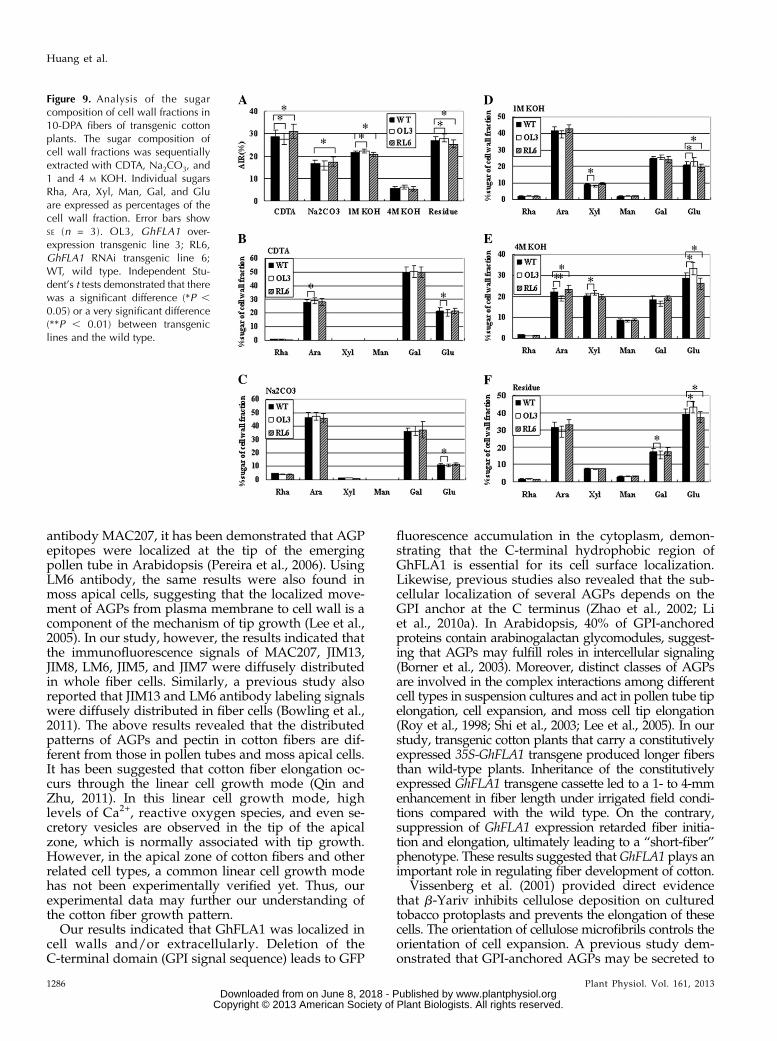
Additionally, fractions of pectin and hemicellulosewere chemically separated in GhFLA1 transgenic fibers.Cell walls derived from 10-DPA cotton fiber cells weresequentially extracted in 1,2-cyclohexanediaminetetra-acetic acid (CDTA), Na2CO3, and 1 and 4 M KOH. TheCDTA and Na2CO3 fractions are generally considered tobe enriched for ionically and covalently bound pectin,respectively. The 1 and 4 M KOH fractions representhemicellulose and hemicellulose-rich polymers, respec-tively, that are tightly bound to the cell wall. Residues aremainly composed of cellulose and associated polysac-charides. The experimental results showed that a higherrate of pectin and lower rates of hemicellulose and cellu-lose were seen in the GhFLA1 RNAi lines, whereas alower rate of pectin and higher rates of hemicellulose andcellulose were found in the GhFLA1 overexpressiontransgenic lines compared with the wild type (Fig. 9A).
Figure 6. Analysis of GhFLA1 expression andfunction in overexpression and RNAi transgeniccotton plants. A, Maps of the GhFLA1 over-expression construct (top) and the RNAi construct(bottom). LB, Left border; ORF, open readingframe; RB, right border. B, Real-time RT-PCRanalysis of the expression of GhFLA1 in over-expression plants (T2 generation). RNA was iso-lated from 10-DPA fibers. C, Real-time RT-PCRanalysis of the expression of GhFLA1 in RNAiplants (T2 generation). RNAwas isolated from 10-DPA fibers. D, Measurement of fiber length at1 and 2 DPA (n $ 50 cotton ovules per line). E,Paraffin sections of 1- and 2-DPA cotton ovules. F,Measurement of fiber length at maturation (n $
50 cotton ovules per line). G, Mature fibers ofGhFLA1 RNAi plants. H, Mature fibers of GhFLA1overexpression plants. Bars = 75 mm in E and1 cm in G and H. Values shown are means 6 SD.Independent Student’s t tests demonstrated thatthere was a significant difference (*P , 0.05) or avery significant difference (**P , 0.01) betweentransgenic fibers and the wild type (WT). OL,GhFLA1 overexpression transgenic lines; RL,GhFLA1 RNAi transgenic lines.
Plant Physiol. Vol. 161, 2013 1283
GhFLA1 Functions in Fiber Development
www.plantphysiol.orgon June 8, 2018 - Published by Downloaded from Copyright © 2013 American Society of Plant Biologists. All rights reserved.

We further analyzed the sugar composition of the fivefractions by measurement of alditol acetates using gaschromatography. The proportion of sugar compositionin CDTA and Na2CO3 fractions of the transgenic lineswas similar to that in the wild type (Fig. 9, B and C). Incontrast, the proportion of Glc in the 1 and 4 M KOHfractions and residues was significantly reduced inGhFLA1 RNAi transgenic lines but increased in GhFLA1overexpression transgenic lines compared with the wildtype. The proportion of Ara and Gal showed a contrarypattern in the transgenic fibers, compared with that ofGlc in the 1 and 4 M KOH fractions and residues (Fig. 9,D–F). These data suggested that GhFLA1 is involved in
modulating the biosynthesis of cell wall polysaccharidesduring fiber development.
DISCUSSION
The data presented in this study indicate the presenceof abundant AGP carbohydrate epitopes and AGP back-bone peptides during fiber development of cotton. Severalclasses of AGPs might contribute to the same process offiber initiation, elongation, and secondary cell wall bio-synthesis, while each of these AGPs may play a uniquerole and act together in regulating fiber development. In
Figure 7. Expression of GhFLA genes andimmunohistochemical localization of AGPsin fibers of the transgenic cotton plants. A,Quantitative RT-PCR analysis of the ex-pression of six fiber-specific/preferentialFLA genes in 10-DPA fibers of GhFLA1overexpression and RNAi transgenic cot-ton plants (T2 generation). Three individ-ual T2 plants were sampled for eachtransgenic line. Error bars show SE (n = 3).Independent Student’s t tests demonstratedthat there was a significant difference(*P , 0.05) or a very significant difference(**P , 0.01) between transgenic lines andthe wild type (WT) . Accession numbers inGenBank are as follows:GhFLA1 (EF672627),GhFLA2 (EF672628), GhFLA4 (EF672630),GhFLA6 (EF672632), GhFLA14 (EF672640),GhFLA15 (EF672641). B, Paraffin sectionsof 1-DPA cotton ovules used for immu-nohistochemical assay. Immunolocalizationwas carried out with antibodies MAC207,JIM8, JIM13, and LM6, using PBS as a neg-ative control. OL1, OL2, and OL3 (OE L3),GhFLA1 overexpression transgenic lines;RL3, RL4, and RL6 (RNAi L6),GhFLA1 RNAitransgenic lines. Bars = 75 mm.
1284 Plant Physiol. Vol. 161, 2013
Huang et al.
www.plantphysiol.orgon June 8, 2018 - Published by Downloaded from Copyright © 2013 American Society of Plant Biologists. All rights reserved.

Arabidopsis, 46 of all 85 AGPs coexpress with eachother and with glycosyltrasferase, glycosylhydrolase,and peroxidase that function in cell wall development(Showalter et al., 2010). Our previous study demon-strated the several GhFLAs were expressed at highlevels in 5- to 20-DPA fibers (Huang et al., 2008). Inaddition, bioinformatic analysis indicated the presenceof at least five classical AGP sequences in cotton fibers.These AGPs were strongly expressed in elongating fibercells and showed a coexpression pattern with GhFLAsand b-Yariv-binding AGPs (Fig. 4A; Supplemental Fig.S5). AGPs play important roles in cell elongation, andb-Yariv inhibited the extension of tip-growing lily(Lilium longiflorum) pollen tubes and extension of apicalcells of P. patens (Roy et al., 1998; Lee et al., 2005). Similarly,our results indicated a concentration-dependent inhibitoryeffect of b-Yariv on fiber elongation, suggesting that AGPsfunction in fiber development. A previous study showedthat some AGPs were located predominantly in the shootapex and the basal part of the embryos, which also
coincided with Yariv-binding sites using JIM8 andJIM13 antibodies (Tang et al., 2006). In this study,immunofluorescence assays indicated that MAC207,JIM8, and JIM13 epitopes were presented abundantlyin initiating and elongating fibers, implying that AGPsmay be required for normal fiber initiation and elongationof cotton.
Cell growth occurs in many dimensions, such as tipgrowth and diffuse growth. There are three intracel-lular components for tip growth of pollen tubes androot hairs: a tip high-calcium gradient, a polarizedactin cytoskeleton, and tip-directed vesicle trafficking(Cole and Fowler, 2006). The components needed toadd new plasma membrane and cell wall are deliveredto the apex and added to the tip by secretory vesiclesand the process of exocytosis. Microtubules, noncalciumion gradients and fluxes, pectin methylesterases, andactin-binding proteins that regulate actin dynamics alsoparticipate the tip growth process (Cole and Fowler,2006). Through labeling pollen tubes with monoclonal
Figure 8. Expression of the genes relatedto cell wall biosynthesis and immunohis-tochemical localization of pectin andhemicellulose in fibers of the transgeniccotton plants. A, Quantitative RT-PCRanalysis of genes related to cell wall bio-synthesis in 10-DPA fibers of GhFLA1overexpression and RNAi transgenic cot-ton plants (T2 generation). Three individ-ual T2 plants were sampled for eachtransgenic line. Error bars show SE (n = 3).Independent Student’s t tests demonstratedthat there was a significant difference(*P , 0.05) or a very significant difference(**P , 0.01) between transgenic lines andthe wild type (WT). Accession numbers inGenBank are as follows:GhUER1 (FJ415167),GhUGD1 (FJ415166), GhUGP1 (FJ415164),GhSUS1 (U73588), GhGT47c (ES807557),GhCESE1 (ES812699). B, Paraffin sections of1-DPA cotton ovules were used for immu-nohistochemical assay. Immunolocalizationwas carried out with antibodies CCRC-M1,JIM5, and JIM7, using PBS as a negativecontrol. OL1, OL2, and OL3 (OE L3),GhFLA1 overexpression transgenic lines;RL3, RL4, and RL6 (RNAi L6), GhFLA1RNAi transgenic lines. Bars = 75 mm.
Plant Physiol. Vol. 161, 2013 1285
GhFLA1 Functions in Fiber Development
www.plantphysiol.orgon June 8, 2018 - Published by Downloaded from Copyright © 2013 American Society of Plant Biologists. All rights reserved.
antibody MAC207, it has been demonstrated that AGPepitopes were localized at the tip of the emergingpollen tube in Arabidopsis (Pereira et al., 2006). UsingLM6 antibody, the same results were also found inmoss apical cells, suggesting that the localized move-ment of AGPs from plasma membrane to cell wall is acomponent of the mechanism of tip growth (Lee et al.,2005). In our study, however, the results indicated thatthe immunofluorescence signals of MAC207, JIM13,JIM8, LM6, JIM5, and JIM7 were diffusely distributedin whole fiber cells. Similarly, a previous study alsoreported that JIM13 and LM6 antibody labeling signalswere diffusely distributed in fiber cells (Bowling et al.,2011). The above results revealed that the distributedpatterns of AGPs and pectin in cotton fibers are dif-ferent from those in pollen tubes and moss apical cells.It has been suggested that cotton fiber elongation oc-curs through the linear cell growth mode (Qin andZhu, 2011). In this linear cell growth mode, highlevels of Ca2+, reactive oxygen species, and even se-cretory vesicles are observed in the tip of the apicalzone, which is normally associated with tip growth.However, in the apical zone of cotton fibers and otherrelated cell types, a common linear cell growth modehas not been experimentally verified yet. Thus, ourexperimental data may further our understanding ofthe cotton fiber growth pattern.
Our results indicated that GhFLA1 was localized incell walls and/or extracellularly. Deletion of theC-terminal domain (GPI signal sequence) leads to GFP
fluorescence accumulation in the cytoplasm, demon-strating that the C-terminal hydrophobic region ofGhFLA1 is essential for its cell surface localization.Likewise, previous studies also revealed that the sub-cellular localization of several AGPs depends on theGPI anchor at the C terminus (Zhao et al., 2002; Liet al., 2010a). In Arabidopsis, 40% of GPI-anchoredproteins contain arabinogalactan glycomodules, suggest-ing that AGPs may fulfill roles in intercellular signaling(Borner et al., 2003). Moreover, distinct classes of AGPsare involved in the complex interactions among differentcell types in suspension cultures and act in pollen tube tipelongation, cell expansion, and moss cell tip elongation(Roy et al., 1998; Shi et al., 2003; Lee et al., 2005). In ourstudy, transgenic cotton plants that carry a constitutivelyexpressed 35S-GhFLA1 transgene produced longer fibersthan wild-type plants. Inheritance of the constitutivelyexpressed GhFLA1 transgene cassette led to a 1- to 4-mmenhancement in fiber length under irrigated field condi-tions compared with the wild type. On the contrary,suppression of GhFLA1 expression retarded fiber initia-tion and elongation, ultimately leading to a “short-fiber”phenotype. These results suggested thatGhFLA1 plays animportant role in regulating fiber development of cotton.
Vissenberg et al. (2001) provided direct evidencethat b-Yariv inhibits cellulose deposition on culturedtobacco protoplasts and prevents the elongation of thesecells. The orientation of cellulose microfibrils controls theorientation of cell expansion. A previous study dem-onstrated that GPI-anchored AGPs may be secreted to
Figure 9. Analysis of the sugarcomposition of cell wall fractions in10-DPA fibers of transgenic cottonplants. The sugar composition ofcell wall fractions was sequentiallyextracted with CDTA, Na2CO3, and1 and 4 M KOH. Individual sugarsRha, Ara, Xyl, Man, Gal, and Gluare expressed as percentages of thecell wall fraction. Error bars showSE (n = 3). OL3, GhFLA1 over-expression transgenic line 3; RL6,GhFLA1 RNAi transgenic line 6;WT, wild type. Independent Stu-dent’s t tests demonstrated that therewas a significant difference (*P ,0.05) or a very significant difference(**P , 0.01) between transgeniclines and the wild type.
1286 Plant Physiol. Vol. 161, 2013
Huang et al.
www.plantphysiol.orgon June 8, 2018 - Published by Downloaded from Copyright © 2013 American Society of Plant Biologists. All rights reserved.
the cell surface in parallel with cellulose synthase.When AGPs are bound to cellulose, they may be re-leased from their GPI anchor and incorporate into thecell wall (Seifert and Roberts, 2007). Deficiency of BC10(for brittle culm10, a DUF266-containing and Golgi-located type II membrane protein) causes a reductionin the levels of cellulose and AGPs, leading to inferiormechanical properties (Zhou et al., 2009). AtFLA3 is in-volved in microspore development and may affect pol-len intine formation, possibly by participating incellulose deposition (Li et al., 2010a). It has been pro-posed that a subset of group A FLAs contribute to thestrength and stiffness of load-bearing plant materials(such as stems) in a partially redundant manner viatheir impact on cellulose synthesis and deposition andalso as structural components of the extracellular ma-trix. The same study also revealed that ArabidopsisFLA11 and FLA12 affected cellulose deposition andthe integrity of the cell wall matrix (MacMillan et al.,2010). Furthermore, a previous study revealed thatsome carrot (Daucus carota) AGPs from the mediumand cell wall may be covalently linked to pectincontaining a homogalacturonan structural element(Immerzeel et al., 2006). Pectin is an important con-stituent in the cell wall. It may account for 30% of thetotal sugar content in fiber cells but less than 18% in10-DPA ovules (Pang et al., 2010). In our study, theexpression of several GhFLAs and other genes related tohemicellulose and cellulose biosynthesis was dramaticallyup-regulated in fibers of GhFLA1 overexpression trans-genic cotton plants but down-regulated in GhFLA1 RNAitransgenic lines. Similarly, the expression of GhUER1 andGhUGD1 was down-regulated in fibers of the RNAitransgenic cotton plants. Meanwhile, immunofluores-cence analysis showed that the compositions of AGPsand hemicellulose were altered in transgenic fibers.The proportion of Glc, Ara, and Gal in 1 and 4 M KOHfractions and residues was also changed in the transgeniclines. These results suggested that GhFLA1 may be in-volved in modulating the biosynthesis of cell wall poly-saccharides during fiber development of cotton.
MATERIALS AND METHODS
Plant Growth Conditions and CottonGenetic Transformation
Cotton (Gossypium hirsutum ‘Coker312’) seeds were surface sterilized with70% (v/v) ethanol for 1 min and 10% hydrogen peroxide for 2 h, followed bywashing with sterile water. The sterilized seeds were germinated on one-half-strength Murashige and Skoog medium (12-h-light/12-h-dark cycle, 28°C).Hypocotyls were cut from sterile seedlings as explants for transformation asdescribed previously (Li et al., 2002). Tissues for DNA, RNA, and proteinextraction were collected from transgenic cotton plants (T1–T3 generations)grown in the field.
Treatment of In Vitro-Cultured Ovules with b-Yariv
Bolls at 1 DPA were harvested and surface sterilized by using 75% ethanolfor 1 min, followed by washing with sterile water. Ovules were dissected fromthe ovaries under sterile conditions and immediately floated on BT liquid
medium containing 5 mM indole-3-acetic acid and 0.5 mM GA3 (Beasley andTing, 1974) supplemented with 0 (control), 10, 25, 50, or 100 mM b-Yariv.Additional control cultures were grown in the same BT medium containing100 mM a-Yariv. The ovules were cultured at 32°C in the dark without agita-tion. Fiber length was measured after the ovules were cultured for 14 d. Ex-periments were repeated at least three times and run in three replicates eachtime. The total number of ovules counted in each group was 50, and P valueswere calculated according to the untreated series as controls.
DNA and Protein Sequence Analysis
Unless otherwise stated, nucleotide and amino acid sequences were ana-lyzed using DNAstar software (DNA Star), and protein sequence homologyanalysis was performed with ClustalW (http://www.ebi.ac.uk/Tools/msa/clustalw2/). The identification of protein domains and significant sites wasperformed with Motifscan (http://myhits.isb-sib.ch/cgi-bin/motif_scan).
Quantitative RT-PCR Analysis
Total RNA was extracted from roots, hypocotyls, cotyledons, leaves, petals,anthers, ovules, and developing fibers (2220 DPA). RNAs were purified usingthe Qiagen RNeasy kit according to the manufacturer’s instructions. First-strand synthesis of cDNA was performed using Moloney murine leukemiavirus reverse transcriptase (Promega) according to the manufacturer’s in-structions.
Expression of cotton genes in 10-DPA fibers of cotton was analyzed by real-time quantitative RT-PCR using the fluorescent intercalating dye SYBR Greenin the detection system (MJ Research; Option 2). A cotton polyubiquitin gene(GhUBI1; GenBank accession no. EU604080) was used as a standard control inthe RT-PCR. A two-step RT-PCR procedure was performed in all experimentsusing a method described earlier (Li et al., 2005). In brief, total RNA was re-verse transcribed into cDNA and used as templates in real-time PCR withgene-specific primers (Supplemental Table S1). PCR was performed usingSYBR Green Real-Time PCR Master Mix (Toyobo) according to the manu-facturer’s instructions. The relative target gene expression was determinedusing the comparative cycle threshold method. To achieve optimal amplifi-cation, PCR conditions for every primer combination were optimized forannealing temperature and Mg2+ concentration. PCR products were confirmedon an agarose gel.
Subcellular Localization
A pBI121-eGFP vector was constructed, and then the GhFLA1 N-signal peptidesequencewas cloned into pBI121-eGFP to form pBI121-GhFLA1SP-eGFP. Part of theGPI signal of GhFLA1 was also cloned into pBI121GhFLA1SP-eGFP to formpBI121-GhFLA1SP-eGFP-GhFLA1GPI. Also, a pBI121-GhFLA1DGPI-eGFP vector(GhFLA1 lacking the putative GPI anchor addition signal sequence) wasconstructed. The constructs were introduced into Arabidopsis (Arabidopsisthaliana) by the floral dip method. Homozygous transgenic plants wereused for GFP observation. eGFP fusion protein expression was visualizedin root cells of 7-d-old seedlings using a Leica SP5 confocal laser scanningmicroscope. To visualize the eGFP distribution, root cells were plasmo-lyzed in 4% NaCl for 20 min. All primers used are listed in SupplementalTable S2.
Construction of GhFLA1 Overexpression and RNAiRecombinant Plasmids
For the overexpression construct, the open reading frame of GhFLA1 wascloned in pBI121 vector. For the RNAi construct, a 265-bp specific sequence ofthe GhFLA1 gene was cloned into a pBluescript SK1 vector to create aninverted repeat transgene and then cloned into pBI121 vector. All primersused are listed in Supplemental Table S2.
Immunofluorescence Labeling with Anti-AGP, Anti-Pectin,and Anti-Hemicellulose MAbs
The distribution of AGPs, pectins, and hemicelluloses was investigatedwith the rat MAbs CCRC-M1, JIM5, JIM7, JIM8, JIM13, LM2, MAC207, andLM6. Samples were fixed overnight in 4% paraformaldehyde and 0.1 M
Plant Physiol. Vol. 161, 2013 1287
GhFLA1 Functions in Fiber Development
www.plantphysiol.orgon June 8, 2018 - Published by Downloaded from Copyright © 2013 American Society of Plant Biologists. All rights reserved.

phosphate-buffered saline (PBS), pH 7.5. After washing twice for 5 min eachtime in PBS buffer, the samples were blocked in 5% bovine serum albumin(BSA) in culture medium for 1 h at room temperature. Subsequently, theywere incubated with monoclonal antibodies (diluted 1:20 with 0.1 M PBScontaining 0.1% BSA) at room temperature for 2 h. After rinsing in PBS threetimes (5 min each), the samples were then incubated with fluoresceinisothiocyanate-labeled goat anti-rat IgG antiserum (Sigma) diluted 1:20 in thesame buffer at room temperature for 2 h. After a final rinse series in PBS, thesamples were mounted on slides with 0.1 M PBS for observation. The controlswere prepared following the same procedure but omitting the primary antibodyincubation.
A collection of MAbs (JIM8, JIM13, MAC207, and LM2) directed againstglycosyl moieties specific to AGPs was obtained as a gift from Dr. P. Knox(University of Leeds). The other MAbs (CCRC-M1, JIM5, and JIM7) were boughtfrom the Complex Carbohydrate Research Center. Fluorescein isothiocyanate-conjugated anti-rat IgG (F-1763; Sigma) was used as a secondary antibody.
Extraction of Total Proteins and Purification of AGPs fromCotton Ovules and Cotton Fibers
Extraction of total proteins and purification of AGPs from cotton ovules andfiber cells were performed by the method described by Schultz et al. (2000).Briefly, 10 g (fresh weight) of freeze-dried ovule or fiber tissue was ground tofine powders in liquid nitrogen, and then 10 mL of extraction buffer (50 mM
Tris-HCl, pH 8.0, 10 mM EDTA, 0.1% b-mercaptoethanol, and 1% [w/v] TritonX-100) was added. After incubation at 4°C for 3 h, the samples were centri-fuged for 10 min at 14,000g. The supernatant was collected and precipitatedwith 5 volumes of ethanol at 4°C overnight. The pellet was resuspended byvortex mixing in 5 mL of 50 mM Tris-HCl, pH 8.0. The insoluble material wasremoved by centrifugation, and the supernatant was retained. The pellet wasresuspended in an additional 5 mL of 50 mM Tris-HCl, pH 8.0, and the su-pernatant was pooled with the first supernatant. The buffer-soluble materialwas freeze dried overnight to concentrate the sample (total proteins). Thedried total proteins were resuspended in 300 mL of 1% (w/v) NaCl andtransferred to 1.5-mL microcentrifuge tubes. AGPs were precipitated with 300mL of b-Yariv by mixing the resuspended samples and leaving them overnightat 4°C. The insoluble Yariv-AGP complex was collected by centrifugation at14,000g for 1 h. The b-Yariv was removed by washing the pellet three times in1% (w/v) NaCl and then twice in methanol. The purified pellet was dissolvedin a minimum volume of dimethyl sulfoxide and mixed with solid sodiumdithionite. Water was added with vortex mixing until the mixture became aclear yellow color. The resulting yellow solution was then desalted on a PD-10column (Pharmacia) that had been equilibrated with water, and the eluate wasfreeze dried.
SDS-PAGE and Western-Blot Analysis for AGPs
Proteins were quantified using a Bradford protein assay kit (Bio-Rad).Twenty micrograms of total protein extract was loaded per lane, separatedby 10% SDS-PAGE, and electroblotted using semidry transfer to polyvinylidenedifluoride membranes. Membranes were blocked in 5% nonfat dry milk in Tris-buffered saline (50 mM Tris-HCl, 150 mM NaCl, pH 7.5) overnight at 4°C andthen incubated with the specific anti-AGP JIM8 and JIM13 antibodies (1:10dilution) in Tris-buffered saline containing 0.1% Tween 20 (TBST) with 5%nonfat dry milk for 2 h at room temperature. Membranes were washed threetimes with TBST and then incubated with goat anti-rat IgG secondary anti-bodies conjugated to horseradish peroxidase (Bio-Rad). Signals were visual-ized with a diaminobenzidine kit.
Dot-Blot Analysis for AGPs
AGPs were also characterized in terms of the binding capacity of severalantibodies. Since AGPs frequently coprecipitate with some pectin and mayshare common epitopes with pectin, we used MAC207, LM2/6, JIM8, andJIM13 antibodies. Antibody-binding tests were performed by the dot-blotmethod. Briefly, 2 mg of AGP sample from different cotton fibers (2-DPAovules/fibers and 5-, 10-, 15-, 18-, and 20-DPA fibers) were dotted onto nylonmembranes (Hybond Trans-Blot Transfer Medium; Bio-Rad) and air dried.The nylon membrane were then blocked in TBST buffer supplemented with1% BSA and 1% dry, nonfat milk for 2 h at room temperature. The incubationwith primary antibodies (all diluted 1:200 in TBST with 1% BSA and 1% milk)
was carried out for 12 h at room temperature, followed by washing in TBST.The blots were subsequently incubated for 2.5 h with secondary anti-rat an-tibody coupled to alkaline phosphatase (Sigma-Aldrich) that was diluted1:1,500 (v/v) in TBST with 1% milk. After washing in TBST and alkalinephosphatase buffer (100 mM Tris, 100 mM NaCl, and 5 mM MgCl2, pH 8.5), theblots were incubated with 5-bromo-4-chloro-3-indolyl phosphate/nitrobluetetrazolium in alkaline phosphatase buffer for 18 min. The reaction wasstopped by dipping the blots in distilled water. Controls were performed byomitting the incubation with primary antibodies. Gum arabic was used as apositive control.
The MAbs used in this project included (1) MAC207, which recognizesa-GlcA-(1,3)-a-GalA-(1,2)-a-Rha; (2) LM2, which recognizes AGP; (3) JIM8,which recognizes the sugar part of AGPs; (4) JIM13, which recognizes AGPs[recognizes the b-GlcA(1-3)-a-GalA(1-2)-Rha-region]; (5) LM6, which recog-nizes 1,5-a-L-arabinan epitopes of type I rhamnogalacturonans; (6) JIM5 andJIM7, which recognize Me-HG; and (7) CCRC-M1, which recognizes a-L-fucosylated xyloglucan.
Noncellulosic Neutral Monosaccharide Analysis
Cell wall fractionation was based upon the methods described by Brown et al.(2007) with minor modification. In brief, 10-DPA cotton fiber materials wereharvested, lyophilized, and ground into fine powders. Then, the samples werewashed three times with 70% ethanol, three times with 1:1 methanol:chloroform,and two times with acetone to obtain alcohol-insoluble residue. The alcohol-insoluble residue was subsequently destarched with amylase (A6380; Sigma-Aldrich) and extracted using (1) 50 mM CDTA containing 1% NaBH4, (2)50 mM Na2CO3 containing 1% NaBH4 (Na2CO3-soluble fraction), (3) 1 M KOHcontaining 1% NaBH4 (1 M KOH-soluble fraction), and (4) 4 M KOH containing1%NaBH4 (4 M KOH-soluble fraction) for 24 h at room temperature. All fractionswere filtered through a GF/C glass filter (Whatman). The alkali fractions wereneutralized with acetic acid. All cell wall fractions were then dialyzed exten-sively against deionized water for 5 d and lyophilized. The fractionation wasrepeated three times on three sets of plants grown independently, and the meanof these three independent replicates was calculated. All cell wall fractions weresubjected to 2 M trifluoroacetic acid at 120°C for 2 h to produce neutral mono-saccharides and subsequent derivatization of the solubilized monosaccharidesinto their corresponding alditol acetates. Finally, the different fractions were runon a gas chromatograph (6890N; Agilent Technologies) with nitrogen as thecarrier gas to determine their sugar composition by the method described pre-viously (Pang et al., 2010).
Sequence data from this article can be found in the GenBank/EMBL datalibraries under accession number GhFLA1 (EF672627).
Supplemental Data
The following materials are available in the online version of this article.
Supplemental Figure S1. No inhibition of 50 mM a-Yariv on fiber elongation.
Supplemental Figure S2. Analyses of the cotton FLA1 protein sequenceand phylogenetic relationship.
Supplemental Figure S3. Morphological alterations as a result of GhFLA1RNAi and overexpression transgenic plants.
Supplemental Figure S4. Bright-field micrographs of the images in Figures7B and 8B.
Supplemental Figure S5. Analysis of the expression of five classical AGPgenes in cotton tissues.
Supplemental Table S1. Gene-specific primers used in quantitative RT-PCR analysis.
Supplemental Table S2. Gene-specific primers used in vector construction.
ACKNOWLEDGMENTS
We thank Dr. P. Knox for the gift of LM2, JIM8, JIM13, LM6, and MAC207antibodies.
Received July 16, 2012; accepted January 22, 2013; published January 24, 2013.
1288 Plant Physiol. Vol. 161, 2013
Huang et al.
www.plantphysiol.orgon June 8, 2018 - Published by Downloaded from Copyright © 2013 American Society of Plant Biologists. All rights reserved.
LITERATURE CITED
Acosta-García G, Vielle-Calzada JP (2004) A classical arabinogalactanprotein is essential for the initiation of female gametogenesis in Arabi-dopsis. Plant Cell 16: 2614–2628
Basra AS, Malik CP (1984) Development of the cotton fiber. Int Rev Cytol89: 65–113
Beasley CA, Ting IP (1974) Effects of plant growth substances on in vitro fiberdevelopment from unfertilized cotton ovules. Amer J Bot 61: 188–194
Borner GHH, Lilley KS, Stevens TJ, Dupree P (2003) Identification ofglycosylphosphatidylinositol-anchored proteins in Arabidopsis: a pro-teomic and genomic analysis. Plant Physiol 132: 568–577
Bowling AJ, Vaughn KC, Turley RB (2011) Polysaccharide and glyco-protein distribution in the epidermis of cotton ovules during early fiberinitiation and growth. Protoplasma 248: 579–590
Brown DM, Goubet F, Wong VW, Goodacre R, Stephens E, Dupree P, TurnerSR (2007) Comparison of five xylan synthesis mutants reveals new insightinto the mechanisms of xylan synthesis. Plant J 52: 1154–1168
Chapman A, Blervacq AS, Vasseur J, Hilbert JL (2000) Arabinogalactan-proteins inCichorium somatic embryogenesis: effect of b-glucosyl Yariv reagent and epi-tope localisation during embryo development. Planta 211: 305–314
Cole RA, Fowler JE (2006) Polarized growth: maintaining focus on the tip.Curr Opin Plant Biol 9: 579–588
Elkins T, Zinn K, McAllister L, Hoffmann FM, Goodman CS (1990) Ge-netic analysis of a Drosophila neural cell adhesion molecule: interactionof fasciclin I and Abelson tyrosine kinase mutations. Cell 60: 565–575
Ellis M, Egelund J, Schultz CJ, Bacic A (2010) Arabinogalactan-proteins:key regulators at the cell surface? Plant Physiol 153: 403–419
Elortza F,MohammedS, Bunkenborg J, Foster LJ, Nühse TS, BrodbeckU, Peck SC,Jensen ON (2006) Modification-specific proteomics of plasmamembrane proteins:identification and characterization of glycosylphosphatidylinositol-anchored pro-teins released upon phospholipase D treatment. J Proteome Res 5: 935–943
Elortza F, Nühse TS, Foster LJ, Stensballe A, Peck SC, Jensen ON (2003)Proteomic analysis of glycosylphosphatidylinositol-anchored membraneproteins. Mol Cell Proteomics 2: 1261–1270
Gao M, Showalter AM (1999) Yariv reagent treatment induces pro-grammed cell death in Arabidopsis cell cultures and implicates arabi-nogalactan protein involvement. Plant J 19: 321–331
Gaspar YM, Johnson KL, McKenna JA, Bacic A, Schultz CJ (2001) Thecomplex structures of arabinogalactan-proteins and the journey towardsunderstanding function. Plant Mol Biol 47: 161–176
Gillmor CS, Lukowitz W, Brininstool G, Sedbrook JC, Hamann T, PoindexterP, Somerville C (2005) Glycosylphosphatidylinositol-anchored proteins arerequired for cell wall synthesis and morphogenesis in Arabidopsis. Plant Cell17: 1128–1140
Harpaz-Saad S, McFarlane HE, Xu S, Divi UK, Forward B, Western TL,Kieber JJ (2011) Cellulose synthesis via the FEI2 RLK/SOS5 pathwayand cellulose synthase 5 is required for the structure of seed coat mu-cilage in Arabidopsis. Plant J 68: 941–953
Huang GQ, Xu WL, Gong SY, Li B, Wang XL, Xu D, Li XB (2008) Char-acterization of 19 novel cotton FLA genes and their expression profilingin fiber development and in response to phytohormones and salt stress.Physiol Plant 134: 348–359
Immerzeel P, Eppink MM, de Vries SC, Schols HA, Voragen AGJ (2006)Carrot arabinogalactan proteins are interlinked with pectins. PhysiolPlant 128: 18–28
Jauh GY, Lord EM (1996) Localization of pectins and arabinogalactan-proteins in lily (Lilium longiflorum L.) pollen tube and style, and theirpossible roles in pollination. Planta 199: 251–261
Johnson KL, Jones BJ, Bacic A, Schultz CJ (2003) The fasciclin-like arabi-nogalactan proteins of Arabidopsis: a multigene family of putative celladhesion molecules. Plant Physiol 133: 1911–1925
Kim HJ, Triplett BA (2001) Cotton fiber growth in planta and in vitro:models for plant cell elongation and cell wall biogenesis. Plant Physiol127: 1361–1366
Kohorn BD (2000) Plasma membrane-cell wall contacts. Plant Physiol 124:31–38
Knox JP (1997) The use of antibodies to study the architecture and devel-opmental regulation of plant cell walls. Int Rev Cytol 171: 79–120
Knox JP (1999) Intriguing, complex and everywhere: getting to grips witharabinogalactan-proteins. Trends Plant Sci 4: 123–125
Lalanne E, Honys D, Johnson A, Borner GHH, Lilley KS, Dupree P,Grossniklaus U, Twell D (2004) SETH1 and SETH2, two components of
the glycosylphosphatidylinositol anchor biosynthetic pathway, are re-quired for pollen germination and tube growth in Arabidopsis. Plant Cell16: 229–240
Langan KJ, Nothnagel EA (1997) Cell surface arabinogalactan proteins andtheir relation to cell proliferation and viability. Protoplasma 196: 87–98
Lee KJ, Sakata Y, Mau SL, Pettolino F, Bacic A, Quatrano RS, Knight CD,Knox JP (2005) Arabinogalactan proteins are required for apical cellextension in the moss Physcomitrella patens. Plant Cell 17: 3051–3065
Li J, Yu M, Geng LL, Zhao J (2010a) The fasciclin-like arabinogalactanprotein gene, FLA3, is involved in microspore development of Arabi-dopsis. Plant J 64: 482–497
Li XB, Cai L, Cheng NH, Liu JW (2002) Molecular characterization of thecotton GhTUB1 gene that is preferentially expressed in fiber. PlantPhysiol 130: 666–674
Li XB, Fan XP, Wang XL, Cai L, Yang WC (2005) The cotton ACTIN1 gene isfunctionally expressed in fibers and participates in fiber elongation.Plant Cell 17: 859–875
Li YJ, Liu DQ, Tu LL, Zhang XL, Wang L, Zhu LF, Tan JF, Deng FL (2010b)Suppression of GhAGP4 gene expression repressed the initiation andelongation of cotton fiber. Plant Cell Rep 29: 193–202
Luo M, Xiao YH, Li XB, Lu XF, Deng W, Li DM, Hou L, Hu MY, Li Y, Pei Y(2007) GhDET2, a steroid 5alpha-reductase, plays an important role incotton fiber cell initiation and elongation. Plant J 51: 419–430
Machado A, Wu Y, Yang Y, Llewellyn DJ, Dennis ES (2009) The MYBtranscription factor GhMYB25 regulates early fibre and trichome de-velopment. Plant J 59: 52–62
MacMillan CP, Mansfield SD, Stachurski ZH, Evans R, Southerton SG(2010) Fasciclin-like arabinogalactan proteins: specialization for stembiomechanics and cell wall architecture in Arabidopsis and Eucalyptus.Plant J 62: 689–703
Majewska-Sawka A, Nothnagel EA (2000) The multiple roles of arabino-galactan proteins in plant development. Plant Physiol 122: 3–10
Nguema-Ona E, Bannigan A, Chevalier L, Baskin TI, Driouich A (2007)Disruption of arabinogalactan proteins disorganizes cortical microtu-bules in the root of Arabidopsis thaliana. Plant J 52: 240–251
Pang CY, Wang H, Pang Y, Xu C, Jiao Y, Qin YM, Western TL, Yu SX, ZhuYX (2010) Comparative proteomics indicates that biosynthesis of pecticprecursors is important for cotton fiber and Arabidopsis root hairelongation. Mol Cell Proteomics 9: 2019–2033
Park MH, Suzuki Y, Chono M, Knox JP, Yamaguchi I (2003) CsAGP1, agibberellin-responsive gene from cucumber hypocotyls, encodes aclassical arabinogalactan protein and is involved in stem elongation.Plant Physiol 131: 1450–1459
Pereira LG, Coimbra S, Oliveira H, Monteiro L, Sottomayor M (2006)Expression of arabinogalactan protein genes in pollen tubes of Arabi-dopsis thaliana. Planta 223: 374–380
Pu L, Li Q, Fan XP, Yang WC, Xue YB (2008) The R2R3 MYB transcriptionfactor GhMYB109 is required for cotton fiber development. Genetics 180:811–820
Qin YM, Zhu YX (2011) How cotton fibers elongate: a tale of linear cell-growth mode. Curr Opin Plant Biol 14: 106–111
Roy S, Jauh GY, Hepler PK, Lord EM (1998) Effects of Yariv phenyl-glycoside on cell wall assembly in the lily pollen tube. Planta 204:450–458
Ruan YL, Llewellyn DJ, Furbank RT (2003) Suppression of sucrose syn-thase gene expression represses cotton fiber cell initiation, elongation,and seed development. Plant Cell 15: 952–964
Schultz CJ, Johnson KL, Currie G, Bacic A (2000) The classical arabino-galactan protein gene family of Arabidopsis. Plant Cell 12: 1751–1768
Seifert GJ, Roberts K (2007) The biology of arabinogalactan proteins. AnnuRev Plant Biol 58: 137–161
Shi HZ, Kim YS, Guo Y, Stevenson B, Zhu JK (2003) The Arabidopsis SOS5locus encodes a putative cell surface adhesion protein and is required fornormal cell expansion. Plant Cell 15: 19–32
Showalter AM (2001) Arabinogalactan-proteins: structure, expression andfunction. Cell Mol Life Sci 58: 1399–1417
Showalter AM, Keppler B, Lichtenberg J, Gu D, Welch LR (2010) A bio-informatics approach to the identification, classification, and analysis ofhydroxyproline-rich glycoproteins. Plant Physiol 153: 485–513
Sun WX, Kieliszewski MJ, Showalter AM (2004) Overexpression of to-mato LeAGP-1 arabinogalactan-protein promotes lateral branching andhampers reproductive development. Plant J 40: 870–881
Plant Physiol. Vol. 161, 2013 1289
GhFLA1 Functions in Fiber Development
www.plantphysiol.orgon June 8, 2018 - Published by Downloaded from Copyright © 2013 American Society of Plant Biologists. All rights reserved.
Tan L, Showalter AM, Egelund J, Hernandez-Sanchez A, Doblin MS,Bacic A (2012) Arabinogalactan-proteins and the research challenges forthese enigmatic plant cell surface proteoglycans. Front Plant Sci 3: 140
Tang XC, He YQ, Wang Y, Sun MX (2006) The role of arabinogalactanproteins binding to Yariv reagents in the initiation, cell developmentalfate, and maintenance of microspore embryogenesis in Brassica napus L.cv. Topas. J Exp Bot 57: 2639–2650
van Hengel AJ, Roberts K (2003) AtAGP30, an arabinogalactan-protein inthe cell walls of the primary root, plays a role in root regeneration andseed germination. Plant J 36: 256–270
Vissenberg K, Feijó JA, Weisenseel MH, Verbelen JP (2001) Ion fluxes,auxin and the induction of elongation growth in Nicotiana tabacumcells. J Exp Bot 52: 2161–2167
Walford SA, Wu Y, Llewellyn DJ, Dennis ES (2011) GhMYB25-like: a keyfactor in early cotton fibre development. Plant J 65: 785–797
Willats WG, Knox JP (1996) A role for arabinogalactan-proteins in plantcell expansion: evidence from studies on the interaction of b-glucosylYariv reagent with seedlings of Arabidopsis thaliana. Plant J 9: 919–925
Wu HM, Wong E, Ogdahl J, Cheung AY (2000) A pollen tube growth-promoting arabinogalactan protein from Nicotiana alata is similar tothe tobacco TTS protein. Plant J 22: 165–176
Yang J, Sardar HS, McGovern KR, Zhang YZ, Showalter AM (2007) Alysine-rich arabinogalactan protein in Arabidopsis is essential for plantgrowth and development, including cell division and expansion. Plant J49: 629–640
Zhang Y, Brown G, Whetten R, Loopstra CA, Neale D, Kieliszewski MJ,Sederoff RR (2003) An arabinogalactan protein associated with sec-ondary cell wall formation in differentiating xylem of loblolly pine.Plant Mol Biol 52: 91–102
Zhao ZD, Tan L, Showalter AM, Lamport DT, Kieliszewski MJ (2002)Tomato LeAGP-1 arabinogalactan-protein purified from transgenic to-bacco corroborates the Hyp contiguity hypothesis. Plant J 31: 431–444
Zhou YH, Li SB, Qian Q, Zeng DL, Zhang M, Guo LB, Liu XL, Zhang BC,Deng LW, Liu XF, et al (2009) BC10, a DUF266-containing and Golgi-located type II membrane protein, is required for cell-wall biosynthesisin rice (Oryza sativa L.). Plant J 57: 446–462
1290 Plant Physiol. Vol. 161, 2013
Huang et al.
www.plantphysiol.orgon June 8, 2018 - Published by Downloaded from Copyright © 2013 American Society of Plant Biologists. All rights reserved.


![i5 Literature [081610DP] · Chicory), Beta Glucans, Oat Fiber) Sunflower oil, Proprietary Immune Blend, Natural Flavors, Medium Chain Triglycerides, Arabinogalactan, Broccoli Seed](https://img.dokumen.tips/doc/110x75/6013a3b270ad005e46206382/i5-literature-081610dp-chicory-beta-glucans-oat-fiber-sunflower-oil-proprietary.jpg)