Embed Size (px)
Citation preview
Nano Res
1
A facile synthesis of hierarchical Sn3O4 nanostructure
in an acidic aqueous solution and their strong visible
light-driven photocatalytic activity
Hui Song1, Su-Young Son1,2, Seul Ki Kim1, and Gun Young Jung1 ()
Nano Res., Just Accepted Manuscript • DOI 10.1007/s12274-015-0855-2
http://www.thenanoresearch.com on July 9, 2015
© Tsinghua University Press 2015
Just Accepted
This is a “Just Accepted” manuscript, which has been examined by the peer-review process and has been
accepted for publication. A “Just Accepted” manuscript is published online shortly after its acceptance,
which is prior to technical editing and formatting and author proofing. Tsinghua University Press (TUP)
provides “Just Accepted” as an optional and free service which allows authors to make their results available
to the research community as soon as possible after acceptance. After a manuscript has been technically
edited and formatted, it will be removed from the “Just Accepted” Web site and published as an ASAP
article. Please note that technical editing may introduce minor changes to the manuscript text and/or
graphics which may affect the content, and all legal disclaimers that apply to the journal pertain. In no event
shall TUP be held responsible for errors or consequences arising from the use of any information contained
in these “Just Accepted” manuscripts. To cite this manuscript please use its Digital Object Identifier (DOI®),
which is identical for all formats of publication.
Nano Research
DOI 10.1007/s12274-015-0855-2
1
A Facile Synthesis of Hierarchical Sn3O4
Nanostructures in an Acidic Aqueous
Solution and Their Strong Visible
Light-Driven Photocatalytic Activity
Hui Songa, Su-Younga,b, Seul Ki Kima, and
Gun Young Junga,*
aSchool of Materials Science and Engineering,
Gwangju Institute of Science and Technology
(GIST), Gwangju, South Korea.
bCarbon Covergence Materials Research
Center, Institute of Advanced Composite
Materials Korea Institute of Science and
Technology (KIST), Jeollabuk-do, South Korea
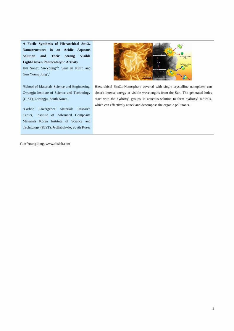
Hierarchical Sn3O4 Nanosphere covered with single crystalline nanoplates can
absorb intense energy at visible wavelengths from the Sun. The generated holes
react with the hydroxyl groups in aqueous solution to form hydroxyl radicals,
which can effectively attack and decompose the organic pollutants.
Gun Young Jung, www.alislab.com
2
A Facile Synthesis of Hierarchical Sn3O4 Nanostructure in an Acidic Aqueous Solution and Their Strong Visible Light-Driven Photocatalytic Activity
Hui Songa, Su-Young Son a,b Seul Ki Kima, and Gun Young Junga,* ()
a School of Materials Science and Engineering, Gwangju Institute of Science and Technology (GIST), Gwangju, South
Korea.
b Carbon Covergence Materials Research Center, Institute of Advanced Composite Materials Korea Institute of Science and
Technology (KIST), Jeollabuk-do, South Korea.
Received: day month year / Revised: day month year / Accepted: day month year (automatically inserted by the publisher)
© Tsinghua University Press and Springer-Verlag Berlin Heidelberg 2011
ABSTRACT Hierarchical Sn3O4 nanospheres were synthesized via hydrothermal reaction under strongly acidic ambient
conditions. The morphology of Sn3O4 was varied with decreasing values of pH; the prickly Sn3O4 nanosphere
was changed into a Sn3O4 nanosphere covered with single crystalline nanoplates with a high surface-to-volume
of ca. 55.05 m2g-1 and a band gap of ca. 2.25 eV. A small amount (0.05 g) of the hierarchical Sn3O4 nanostructures
completely decomposed a 30 % methyl orange (MO) solution in 100 ml deionized water in 15 min under
one-Sun condition (UV + Vis. light). The Sn3O4 photocatalyst exhibited a fast decomposition rate of 1.73 x 10⁻¹
min-1, which is a 90.86 % enhancement relative to that of the commercially available P25 photocatalyst. The
high photocatalytic activity of the hierarchical Sn3O4 nanostructure is attributed to its ability to absorb visible
light and its high surface-to-volume area.
KEYWORDS Sn3O4, Hierarchical structure, Hydrothermal, Morphology engineering, Photocatalyst
1. Introduction
Tin oxide has several advantages of controllable
size or shape via the simple hydrothermal
method,[1] excellent optical and electrical
properties (transparency at wavelengths of 300 ~
800 nm and a low electronic resistivity of 8.6 x 10⁻⁵
Ω-1), its non-toxicity[2] and so on. With these
merits, it has been utilized in various fields such as
gas sensors,[3,4] lithium ion batteries,[5]
dye-sensitized solar cells[6] and photocatalysts.[7-9]
Regarding its use as a photocatalyst, tin oxide has a
strong resistance against acidic/alkali solutions and
exhibits a good photocatalytic activity without
generating secondary pollutions under irradiation
with ultraviolet (UV) light.[8,9] However, there are
few works on solely using the tin oxides under
irradiation with visible light because of its wide
band gap. Therefore, co-catalysts with a narrow
Nano Res DOI (automatically inserted by the publisher)
Research Article
————————————
Address correspondence to [email protected]
3
energy band gap, such as CdS[10], Ru(bpy)32+[11]
and Pt[12], have been introduced to enhance the
photocatalytic activity of tin oxide under irradiation
with visible light. However, because the cost of
co-catalysts is expensive, the development of
photo-catalytic materials for generating
electron-hole pairs under visible light is necessary
to effectively utilize sun light.
Unlike SnO2 as a photocatalyst, Sn3O4 has been
used as a visible light-driven photocatalyst because
it has experimentally shown to absorb light at
visible wavelengths.[13-15] Therefore, the pollution
decomposition efficiency of Sn3O4 photocatalysts is
higher than that of SnO2 under the Sun.[13]
However, studies on the Sn3O4 have just begun,
including investigations into its growth
mechanism,[16-18] crystal structure and theoretical
properties.[19,20]
Sn3O4 was formed as one of intermediate tin
oxides (i.e., Sn3O4, Sn2O3 and Sn5O6) during the
disproportionation reaction of Tin(II) oxide
(SnO).[21] Sn3O4 was mainly generated by dry
chemical methods including carbothermal
evaporation,[15] thermal decomposition of SnO,[22]
carbothermal reduction.[23] Vilasi et. al reported
that Sn3O4 coexisted as a transition form at the
process temperature range of 400-500 oC during the
phase transformation from SnO to SnO2.[24]
However, only a small quantity of Sn3O4 was
obtained. Indeed, SnO2 was the major product in
the reaction, which was thermodynamically more
stable than Sn3O4 during the oxidation of Sn2+ at
high temperatures.
For this reason, hydrothermal methods were
used to generate Sn3O4 at temperatures below 200
oC without the production of unwanted SnO2.[25-29]
Li et. al synthesized hierarchical Sn3O4 structures
(diameter: ~ 1 μm) at 180 oC hydrothermally at pH 3
in an autoclave Teflon vessel, which made it
difficult to adjust the pH during the hydrothermal
process for controlling the morphology. They
showed that the hierarchical Sn3O4 structure had the
highest pollution decomposition rate among other
photocatalysts such as SnO, SnO2 and N-doped
TiO2.[13] However, only 10 ppm concentration of
methyl orange (MO) in 80 ml solution was
decomposed in 20 min by the photocatalytic
reaction of 0.04 g Sn3O4.
Herein, we proposed a facile hydrothermal
method in a 3-neck round flask to fabricate
hierarchical Sn3O4 nanospheres covered with
single-crystalline nanoplates at a temperature
below 100 oC. By adjusting the pH of the nutrient
solution during the process, the morphology and
phase transformation of SnO into the Sn3O4
nanostructure can be controlled. The photocatalytic
activity of the generated hierarchical Sn3O4
nanostructure was investigated and compared with
that of the commercially available TiO2 (Degussa,
P25) under one-Sun condition (i.e., Air Mass 1.5, 100
mWcm-2).
2. Experimental Section
2.1 Synthesis of hierarchical Sn3O4 nanostructure
1.23 g of tin oxalate (Sn(C2O4), M = 206.78 gmol-1)
was dissolved in 250 ml deionized (DI) water with
stirring for 30 min at room temperature. When the
temperature reached 70 oC, black precipitates of
SnO appeared. Then, the pH value (pH = 2, 3, 4 and
5) of the nutrient solution was adjusted by slowly
adding 0.5 M hydrochloric acid (HCl, M = 40.06
gmol-1, 37 %) to the solution having the black
precipitate while continuously stirring for 3 hrs at
95 oC. The color of precipitate was changed from
black to yellow. After completing the hydrothermal
reaction, the precipitate was collected by
centrifugation and then washed with DI water.
Finally, the product was dried inside a common
laboratory oven at 35 oC for 1 day.
4
2.2 Characterization of properties
The crystallographic information of hierarchical
Sn3O4 nanostructure was analyzed by X-ray
diffraction (XRD) using Cu K-alpha X-ray radiation
(40 kV, 100 mA, Rigaku D/max-2400) and Raman
spectroscopy with 514 nm laser beam (Horiba). The
morphology of sample was observed by scanning
electron microscopy (SEM, FE-SEM, JEOL 2010 F)
and high resolution transmission electron
microscopy (HRTEM, JEM-2100), operated at an
accelerating voltage of 200 kV. The specific surface
area was measured by the Brunauer-Emmett-Teller
(BET) method using a nanoporosity surface area
analyzer (nanoPOROSITY-XQ, Mirae Scientific
Instruments Inc.). A UV-Vis. spectrometer
(AvaSpec-ULS2048L-USB2 Spectrometer, Jinyoung
tech Inc.) was used to analyze the absorbance of the
hierarchical Sn3O4 nanostructure and the
decomposition of the MO solution. Photocatalytic
activity was characterized under illumination of an
AM 1.5 simulated sunlight source (SANEI solar
simulator, Class A) with a power density of 100 ±
2.5 mW cm-2.
2.3 Photocatalytic activity
0.05 g of as-produced catalysts of SnO, Sn3O4 and
commercially available P25 (TiO2, Degusa) were
added into a 30 vol. % MO solution in 100 ml DI
water. Prior to irradiation, the suspension was
violently stirred in the dark for 30 min to saturate
the solution with O2. The suspension was irradiated
with the light (UV + visible) from a solar simulator.
At given time intervals (15, 30, 45 and 60 min), a 3
ml aliquot was extracted and filtered to remove the
photocatalytic powder. The filtered solution was
analyzed by the UV-Vis. spectrometer to measure
the MO contents (Maximum absorption band, λ =
485 nm).
3. Results and discussion
3.1. The effect of pH on the formation of
hierarchical Sn3O4 nanostructure
The hierarchical Sn3O4 nanostructures were
synthesized by hydrothermal reaction at 95 oC. Tin
oxalate, Sn(C2O4), was used as the tin ions source.
Unlike the most commonly used tin salts such as tin
dichloride (SnCl2) and tin tetrachloride (SnCl4), the
Sn(C2O4) can easily offer abundant tin(II) ions (Sn2+)
in the nutrient solution because it can be dissociated
easily at a low temperature due to the relatively
weak electro-static attraction between the pure
metal ion (Sn2+) and the organic chelating reagent
(C2O4-).[6] Thus, SnO was easily synthesized at first
by using the Sn(C2O4) as a starting material at a low
temperature. Then, in an acidic conditions the SnO
was easily dissolved and changed into a tin
complex ion (Sn3(OH)42+). Finally, the Sn3O4
nanostructures were formed by dehydration of the
Sn3(OH)42+.[30]
When the temperature of nutrient solution
reached 70 oC, the color of the precipitate was
changed from white to black, indicating the
formation of SnO, and remained black until 95 oC.
However, the color of the precipitate became yellow
after adding a hydrochloric acid solution at 95 oC,
indicating that some changes to the SnO occurred.
The morphology and phase transformation of the
products were examined at different pH values of
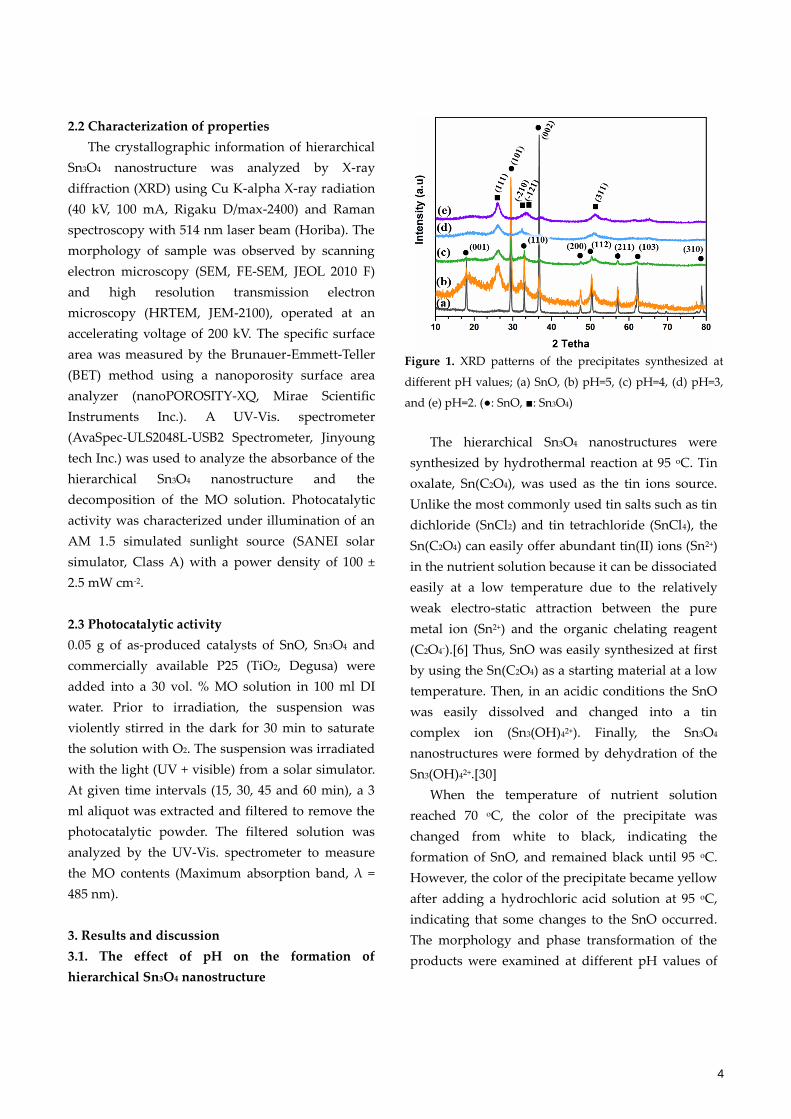
Figure 1. XRD patterns of the precipitates synthesized at
different pH values; (a) SnO, (b) pH=5, (c) pH=4, (d) pH=3,
and (e) pH=2. (: SnO, : Sn3O4)
5
the solution, which was controlled by varying the
amount of added hydrochloric acid. After
completing the hydrothermal reaction, the
precipitate was collected by centrifugation and then
dried to analyze the structure and determine its
photocatalytic ability.
The crystallographic structures of the
hierarchical Sn3O4 nanostructure at various pH
values was characterized by X-ray diffraction (XRD)
as shown in Figure 1. Figure 1a indicates the XRD
peaks of a tetragonal SnO marked with the
symbols at (001), (101) and (002), which are well
matched with the standard XRD data file
(JCPDS-06-0395). The phase transformation from
SnO to Sn3O4 occurred with decreasing pH, and the
XRD patterns indicated the coexistence of SnO and
Sn3O4 at pH 5 (Fig. 1b). Only Sn3O4 diffraction peaks
existed at pH of less than 3, as shown in Figure 1d
and e, where the symbols are assigned to (111),
(-210), (-121), and (311) of a triclinic Sn3O4
(JCPDS-20-1293).[21] Notably, several unknown
crystalline peaks were generated when the pH
approached 1. Thus, controlling the pH value
within 2 and 3 during the reaction was critical to
synthesize well-defined Sn3O4 nanostructure.
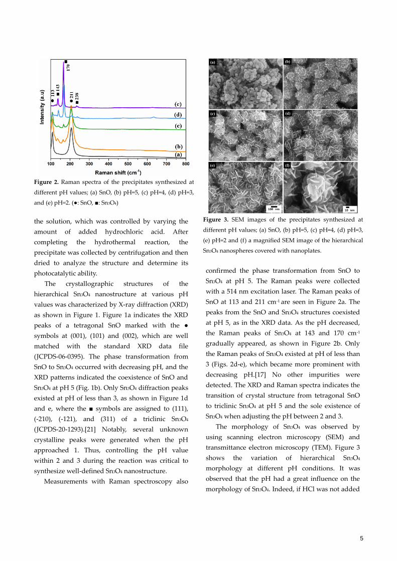
Measurements with Raman spectroscopy also
confirmed the phase transformation from SnO to
Sn3O4 at pH 5. The Raman peaks were collected
with a 514 nm excitation laser. The Raman peaks of
SnO at 113 and 211 cm-1 are seen in Figure 2a. The
peaks from the SnO and Sn3O4 structures coexisted
at pH 5, as in the XRD data. As the pH decreased,
the Raman peaks of Sn3O4 at 143 and 170 cm-1
gradually appeared, as shown in Figure 2b. Only
the Raman peaks of Sn3O4 existed at pH of less than
3 (Figs. 2d-e), which became more prominent with
decreasing pH.[17] No other impurities were
detected. The XRD and Raman spectra indicates the
transition of crystal structure from tetragonal SnO
to triclinic Sn3O4 at pH 5 and the sole existence of
Sn3O4 when adjusting the pH between 2 and 3.
The morphology of Sn3O4 was observed by
using scanning electron microscopy (SEM) and
transmittance electron microscopy (TEM). Figure 3
shows the variation of hierarchical Sn3O4
morphology at different pH conditions. It was
observed that the pH had a great influence on the
morphology of Sn3O4. Indeed, if HCl was not added
Figure 2. Raman spectra of the precipitates synthesized at
different pH values; (a) SnO, (b) pH=5, (c) pH=4, (d) pH=3,
and (e) pH=2. (: SnO, : Sn3O4)
Figure 3. SEM images of the precipitates synthesized at
different pH values; (a) SnO, (b) pH=5, (c) pH=4, (d) pH=3,
(e) pH=2 and (f) a magnified SEM image of the hierarchical
Sn3O4 nanospheres covered with nanoplates.
6
into the nutrient solution, then only SnO
nanoparticles aggregated together as shown in
Figure 3a.[31] At pH 5, the neighboring
nanoparticles aggregated into a prickly sphere with
a diameter of approximately 100 nm (Fig. 3b). As
the solution became more acidic (pH = 4, Fig. 3c),
the prickly surface became prominent. At pH less
than 3, in which the SnO was completely
transformed into the hierarchical Sn3O4, the prickles
were dissolved and recrystallized into nanoplates as
shown in Figure 3d and 3e.[31] Figure 3f shows a
magnified hierarchical Sn3O4 nanosphere having
irregularly oriented thin nanoplates with a radius of
20 nm.
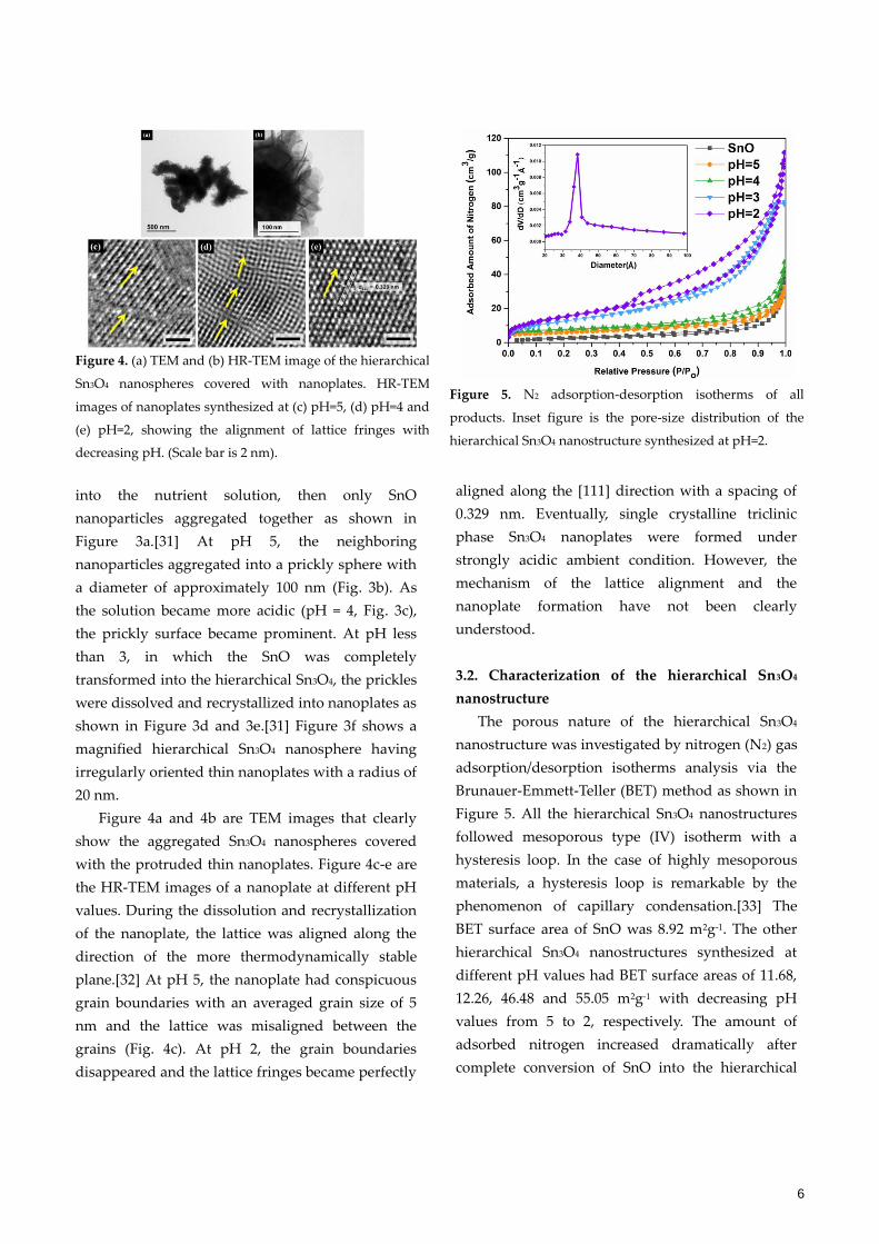
Figure 4a and 4b are TEM images that clearly
show the aggregated Sn3O4 nanospheres covered
with the protruded thin nanoplates. Figure 4c-e are
the HR-TEM images of a nanoplate at different pH
values. During the dissolution and recrystallization
of the nanoplate, the lattice was aligned along the
direction of the more thermodynamically stable
plane.[32] At pH 5, the nanoplate had conspicuous
grain boundaries with an averaged grain size of 5
nm and the lattice was misaligned between the
grains (Fig. 4c). At pH 2, the grain boundaries
disappeared and the lattice fringes became perfectly
aligned along the [111] direction with a spacing of
0.329 nm. Eventually, single crystalline triclinic
phase Sn3O4 nanoplates were formed under
strongly acidic ambient condition. However, the
mechanism of the lattice alignment and the
nanoplate formation have not been clearly
understood.
3.2. Characterization of the hierarchical Sn3O4
nanostructure
The porous nature of the hierarchical Sn3O4
nanostructure was investigated by nitrogen (N2) gas
adsorption/desorption isotherms analysis via the
Brunauer-Emmett-Teller (BET) method as shown in
Figure 5. All the hierarchical Sn3O4 nanostructures
followed mesoporous type (IV) isotherm with a
hysteresis loop. In the case of highly mesoporous
materials, a hysteresis loop is remarkable by the
phenomenon of capillary condensation.[33] The
BET surface area of SnO was 8.92 m2g-1. The other
hierarchical Sn3O4 nanostructures synthesized at
different pH values had BET surface areas of 11.68,
12.26, 46.48 and 55.05 m2g-1 with decreasing pH
values from 5 to 2, respectively. The amount of
adsorbed nitrogen increased dramatically after
complete conversion of SnO into the hierarchical
Figure 4. (a) TEM and (b) HR-TEM image of the hierarchical
Sn3O4 nanospheres covered with nanoplates. HR-TEM
images of nanoplates synthesized at (c) pH=5, (d) pH=4 and
(e) pH=2, showing the alignment of lattice fringes with
decreasing pH. (Scale bar is 2 nm).
Figure 5. N2 adsorption-desorption isotherms of all
products. Inset figure is the pore-size distribution of the
hierarchical Sn3O4 nanostructure synthesized at pH=2.
7
Sn3O4. The Sn3O4 structure synthesized at pH 2 had
the highest value of BET surface area, indicating a
highly porous structure caused by intensive etching
in such a strongly acidic ambient condition. The
inset of figure 5 demonstrates the pore size
distribution of hierarchical Sn3O4 synthesized at pH
2, revealing a maximum at 40 Å .
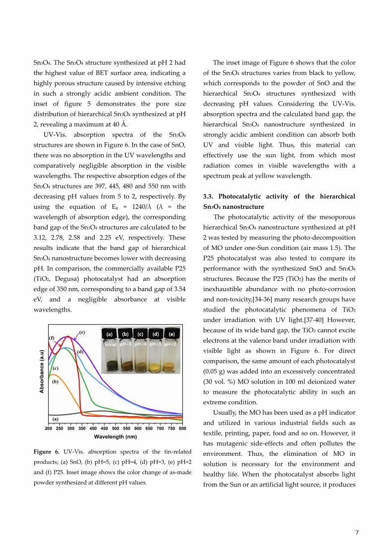
UV-Vis. absorption spectra of the Sn3O4
structures are shown in Figure 6. In the case of SnO,
there was no absorption in the UV wavelengths and
comparatively negligible absorption in the visible
wavelengths. The respective absorption edges of the
Sn3O4 structures are 397, 445, 480 and 550 nm with
decreasing pH values from 5 to 2, respectively. By
using the equation of Eg = 1240/λ (λ = the
wavelength of absorption edge), the corresponding
band gap of the Sn3O4 structures are calculated to be
3.12, 2.78, 2.58 and 2.25 eV, respectively. These
results indicate that the band gap of hierarchical
Sn3O4 nanostructure becomes lower with decreasing
pH. In comparison, the commercially available P25
(TiO2, Degusa) photocatalyst had an absorption
edge of 350 nm, corresponding to a band gap of 3.54
eV, and a negligible absorbance at visible
wavelengths.
The inset image of Figure 6 shows that the color
of the Sn3O4 structures varies from black to yellow,
which corresponds to the powder of SnO and the
hierarchical Sn3O4 structures synthesized with
decreasing pH values. Considering the UV-Vis.
absorption spectra and the calculated band gap, the
hierarchical Sn3O4 nanostructure synthesized in
strongly acidic ambient condition can absorb both
UV and visible light. Thus, this material can
effectively use the sun light, from which most
radiation comes in visible wavelengths with a
spectrum peak at yellow wavelength.
3.3. Photocatalytic activity of the hierarchical
Sn3O4 nanostructure
The photocatalytic activity of the mesoporous
hierarchical Sn3O4 nanostructure synthesized at pH
2 was tested by measuring the photo-decomposition
of MO under one-Sun condition (air mass 1.5). The
P25 photocatalyst was also tested to compare its
performance with the synthesized SnO and Sn3O4
structures. Because the P25 (TiO2) has the merits of
inexhaustible abundance with no photo-corrosion
and non-toxicity,[34-36] many research groups have
studied the photocatalytic phenomena of TiO2
under irradiation with UV light.[37-40] However,
because of its wide band gap, the TiO2 cannot excite
electrons at the valence band under irradiation with
visible light as shown in Figure 6. For direct
comparison, the same amount of each photocatalyst
(0.05 g) was added into an excessively concentrated
(30 vol. %) MO solution in 100 ml deionized water
to measure the photocatalytic ability in such an
extreme condition.
Usually, the MO has been used as a pH indicator
and utilized in various industrial fields such as
textile, printing, paper, food and so on. However, it
has mutagenic side-effects and often pollutes the
environment. Thus, the elimination of MO in
solution is necessary for the environment and
healthy life. When the photocatalyst absorbs light
from the Sun or an artificial light source, it produces
Figure 6. UV-Vis. absorption spectra of the tin-related
products; (a) SnO, (b) pH=5, (c) pH=4, (d) pH=3, (e) pH=2
and (f) P25. Inset image shows the color change of as-made
powder synthesized at different pH values.
8
pairs of electron and hole. The generated holes
attack the surrounding water molecules to generate
hydroxyl radicals (*OH) at the surface of
photocatalysts as depicted in the following
photo-oxidation mechanism;[41]
Metal oxide + hv → e-CB + h+VB (1)
h+VB +H2O → *OH + H+ (2)
It is known that the hydroxyl radical has the second
largest oxidation potential of 2.8 V among several
common oxidants including fluorine (3.03 V), ozone
(2.07 V), hydrogen peroxide (1.77 V) and chloride
(1.36 V). Therefore, the *OH can rapidly attack and
cleave the aromatic rings of organic pollutants.[42]
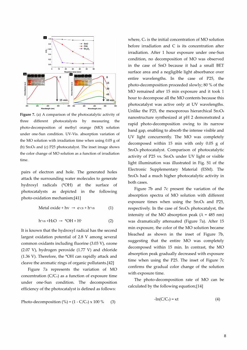
Figure 7a represents the variation of MO
concentration (C/Co) as a function of exposure time
under one-Sun condition. The decomposition
efficiency of the photocatalyst is defined as follows:
Photo-decomposition (%) = (1 - C/Co) x 100 % (3)
where, Co is the initial concentration of MO solution
before irradiation and C is its concentration after
irradiation. After 1 hour exposure under one-Sun
condition, no decomposition of MO was observed
in the case of SnO because it had a small BET
surface area and a negligible light absorbance over
entire wavelengths. In the case of P25, the
photo-decomposition proceeded slowly; 80 % of the
MO remained after 15 min exposure and it took 1
hour to decompose all the MO contents because this
photocatalyst was active only at UV wavelengths.
Unlike the P25, the mesoporous hierarchical Sn3O4
nanostructure synthesized at pH 2 demonstrated a
rapid photo-decomposition owing to its narrow
band gap, enabling to absorb the intense visible and
UV light concurrently. The MO was completely
decomposed within 15 min with only 0.05 g of
Sn3O4 photocatalyst. Comparison of photocatalytic
activity of P25 vs. Sn3O4 under UV light or visible
light illumination was illustrated in Fig. S1 of the
Electronic Supplementary Material (ESM). The
Sn3O4 had a much higher photocatalytic activity in
both cases.
Figure 7b and 7c present the variation of the
absorption spectra of MO solution with different
exposure times when using the Sn3O4 and P25,
respectively. In the case of Sn3O4 photocatalyst, the
intensity of the MO absorption peak (λ = 485 nm)
was dramatically attenuated (Figure 7a). After 15
min exposure, the color of the MO solution became
bleached as shown in the inset of Figure 7b,
suggesting that the entire MO was completely
decomposed within 15 min. In contrast, the MO
absorption peak gradually decreased with exposure
time when using the P25. The inset of Figure 7c
confirms the gradual color change of the solution
with exposure time.
The photo-decomposition rate of MO can be
calculated by the following equation;[14]
-ln(C/Co) = κt (4)
Figure 7. (a) A comparison of the photocatalytic activity of
three different photocatalysts by measuring the
photo-decomposition of methyl orange (MO) solution
under one-Sun condition. UV-Vis. absorption variation of
the MO solution with irradiation time when using 0.05 g of
(b) Sn3O4 and (c) P25 photocatalyst. The inset image shows
the color change of MO solution as a function of irradiation
time.
9
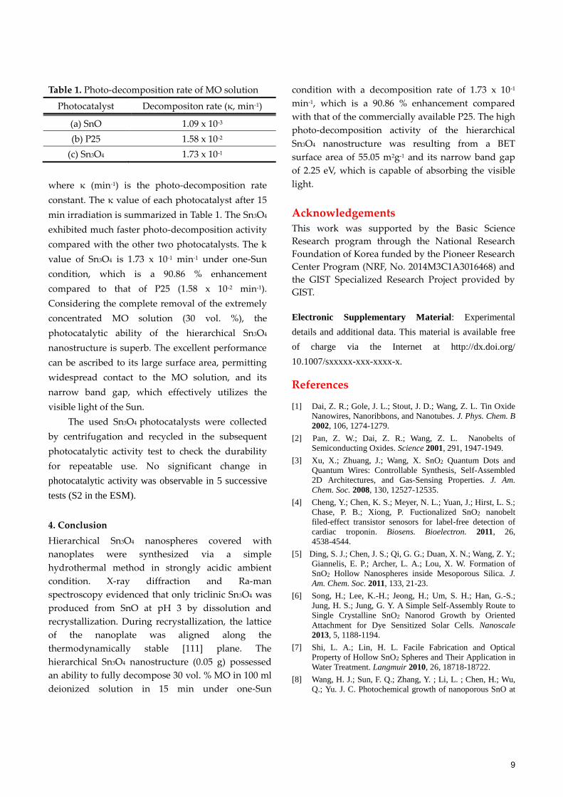
Table 1. Photo-decomposition rate of MO solution
Photocatalyst Decompositon rate (κ, min-1)
(a) SnO 1.09 x 10⁻³
(b) P25 1.58 x 10⁻²
(c) Sn3O4 1.73 x 10⁻¹
where κ (min-1) is the photo-decomposition rate
constant. The κ value of each photocatalyst after 15
min irradiation is summarized in Table 1. The Sn3O4
exhibited much faster photo-decomposition activity
compared with the other two photocatalysts. The k
value of Sn3O4 is 1.73 x 10⁻¹ min-1 under one-Sun
condition, which is a 90.86 % enhancement
compared to that of P25 (1.58 x 10⁻² min-1).
Considering the complete removal of the extremely
concentrated MO solution (30 vol. %), the
photocatalytic ability of the hierarchical Sn3O4
nanostructure is superb. The excellent performance
can be ascribed to its large surface area, permitting
widespread contact to the MO solution, and its
narrow band gap, which effectively utilizes the
visible light of the Sun.
The used Sn3O4 photocatalysts were collected
by centrifugation and recycled in the subsequent
photocatalytic activity test to check the durability
for repeatable use. No significant change in
photocatalytic activity was observable in 5 successive
tests (S2 in the ESM).
4. Conclusion
Hierarchical Sn3O4 nanospheres covered with
nanoplates were synthesized via a simple
hydrothermal method in strongly acidic ambient
condition. X-ray diffraction and Ra-man
spectroscopy evidenced that only triclinic Sn3O4 was
produced from SnO at pH 3 by dissolution and
recrystallization. During recrystallization, the lattice
of the nanoplate was aligned along the
thermodynamically stable [111] plane. The
hierarchical Sn3O4 nanostructure (0.05 g) possessed
an ability to fully decompose 30 vol. % MO in 100 ml
deionized solution in 15 min under one-Sun
condition with a decomposition rate of 1.73 x 10⁻¹
min-1, which is a 90.86 % enhancement compared
with that of the commercially available P25. The high
photo-decomposition activity of the hierarchical
Sn3O4 nanostructure was resulting from a BET
surface area of 55.05 m2g-1 and its narrow band gap
of 2.25 eV, which is capable of absorbing the visible
light.
Acknowledgements
This work was supported by the Basic Science
Research program through the National Research
Foundation of Korea funded by the Pioneer Research
Center Program (NRF, No. 2014M3C1A3016468) and
the GIST Specialized Research Project provided by
GIST.
Electronic Supplementary Material: Experimental
details and additional data. This material is available free
of charge via the Internet at http://dx.doi.org/
10.1007/sxxxxx-xxx-xxxx-x.
References
[1] Dai, Z. R.; Gole, J. L.; Stout, J. D.; Wang, Z. L. Tin Oxide
Nanowires, Nanoribbons, and Nanotubes. J. Phys. Chem. B
2002, 106, 1274-1279.
[2] Pan, Z. W.; Dai, Z. R.; Wang, Z. L. Nanobelts of
Semiconducting Oxides. Science 2001, 291, 1947-1949.
[3] Xu, X.; Zhuang, J.; Wang, X. SnO2 Quantum Dots and
Quantum Wires: Controllable Synthesis, Self-Assembled
2D Architectures, and Gas-Sensing Properties. J. Am.
Chem. Soc. 2008, 130, 12527-12535.
[4] Cheng, Y.; Chen, K. S.; Meyer, N. L.; Yuan, J.; Hirst, L. S.;
Chase, P. B.; Xiong, P. Fuctionalized SnO2 nanobelt
filed-effect transistor senosors for label-free detection of
cardiac troponin. Biosens. Bioelectron. 2011, 26,
4538-4544.
[5] Ding, S. J.; Chen, J. S.; Qi, G. G.; Duan, X. N.; Wang, Z. Y.;
Giannelis, E. P.; Archer, L. A.; Lou, X. W. Formation of
SnO2 Hollow Nanospheres inside Mesoporous Silica. J.
Am. Chem. Soc. 2011, 133, 21-23.
[6] Song, H.; Lee, K.-H.; Jeong, H.; Um, S. H.; Han, G.-S.;
Jung, H. S.; Jung, G. Y. A Simple Self-Assembly Route to
Single Crystalline SnO2 Nanorod Growth by Oriented
Attachment for Dye Sensitized Solar Cells. Nanoscale
2013, 5, 1188-1194.
[7] Shi, L. A.; Lin, H. L. Facile Fabrication and Optical
Property of Hollow SnO2 Spheres and Their Application in
Water Treatment. Langmuir 2010, 26, 18718-18722.
[8] Wang, H. J.; Sun, F. Q.; Zhang, Y. ; Li, L. ; Chen, H.; Wu,
Q.; Yu. J. C. Photochemical growth of nanoporous SnO at
10
the air-water interface and its high photocatalytic activity. J.
Mater. Chem. 2010, 20, 5641-5645.
[9] Nasr, C.; Kamat, P. V.; Hotchandani, S.
Photoelectrochemical behavior of coupled SnO2/CdSe
nanocrystalline semiconductor films. J. Electroanal. Chem.
1997, 420, 201-207.
[10] Kar, A.; Kundu, S.; Patra, A. Photocatalytic properties of
semiconductor SnO2/CdS heterostructure nanocrystal. RSC
Adv. 2012, 2, 10222-10230.
[11] Vlachopoulos, N.; Liska, P.; Augustynski, J.; Gratzel, M.
Very efficient visible light energy harvesting and
conversion by spectral sensitization of high surface area
polycrystalline titanium dioxide films. J. Am. Chem. Soc.
1988, 110, 1216-1220.
[12] Wrighton, M. S.; Ginley, D. S.; Wolczanski, P. T.; Ellis, A.
B.; Morse, D. L.; Linz, A. Photoassisted electrolysis of
water by irradiation of a titanium dioxide electrode. Proc.
Natl. Acad. Sci. USA. 1975, 729(4), 1518-1522.
[13] He, Y.; Li, D.; Chen, J.; Shao, Y.; Xian, J.; Zheng, X.;
Wang, P.; He, Y.; Li, D.; Chen, J.; Shao, Y.; Xian, J.; Zheng,
X.; Wang, P. Sn3O4: a novel heterovalent-tin photocatalyst
with hierarchical 3D nanostructures under visible light.
RSC Adv. 2014, 4, 1266-1269.
[14] Xia, W.; Wang, H.; Zeng, X.; Han, J.; Zhu, J.; Zhou, M.;
Wua, S. High-efficiency photocatalytic activity of type II
SnO/Sn3O4 heterostructures via interfacial charge transfer.
CrystEngComm 2014, 16, 6841-6847.
[15] Berengue, O. M.; Simon, R. A.; Chiquito, A. J.;
Dalmaschio, C. J.; Leite, E. R.; Guerreiro, H. A.;
Guimarães, F. E. G. Semiconducting Sn3O4 nanobelts:
growth and electronic structure. J. Appl. Phys. 2010, 107,
033717-4.
[16] Lawson, F. Tin Oxide-Sn3O4. Nature 1967, 215, 955-956.
[17] Wang, F.; Zhou, J.; Sham, T. K.; Ding, Z. Observation of
Single Tin Dioxide Nanoribbons by Confocal Raman
Microspectroscopy. J. Phys. Chem. C 2007, 111,
18839-18843.
[18] Seko, A.; Togo, A.; Oba, F.; Tanaka, I. Structure and
Stability of a Homlogous Series of Tin Oxides. Phys. Rev.
Lett. 2008, 100, 045702-4.
[19] Maki-jaskari, M. A.; Rantala, T. T. Possible structure of
nonstoichiometric tin oxide: the composition Sn2O3.
Modell. Simul. Mater. Sci. Eng. 2004, 12, 33-41.
[20] White, T. A.; Moreno, M. S.; Midgley, P. A. Strucutre
determination of the intermediate tin oxide Sn3O4 by
precession electron diffraction. Z. Kristallogr. 2010, 225,
56-66.
[21] Gauzzi, F.; Verdini, B.; Maddalena, A.; Principi, G. X-ray
Diffraction and Mossbauer Analyses of SnO Disproportion
Products. Inorg. Chim. Acta. 1985, 104, 1-7.
[22] Morenot, M. S.; Mercadert, R. C.; Bibilonit, A. G. Study of
intermediate oxides in SnO thermal decomposition. J.
Phys.: Condens. Matter 1992, 4, 351-355.
[23] Damaschio, C. J.; Berengue, O. M.; Stroppa, D. G.; Simon,
R. A.; Ramirez, A. J.; Schreiner, W. H.; Chiquito, A. J.;
Leite, E. R. Sn3O4 single crystal nanobelts grown by
carbothermal reduction. J. Cryst. Growth 2010, 312,
2881-2886.
[24] Cahen, S.; David, N.; Fiorani, J. M.; Maitre, A.; Vilasi, M.
Thermodynamic modelling of the O-Sn system.
Thermochimica Acta 2003, 403, 275-285.
[25] Ohgi, H.; Maeda, T.; Hosonno, E.; Fujihara, S.; Imai, H.
Evolution of Nanoscale SnO2 Grains, Flakes, and Plates
into Versatile Particles and Films through Crystal Growth
in Aqueous Solutions. Cryst. Growth Des. 2005, 5,
1079-1083.
[26] Uchiyama, H.; Ohgi, H.; Imai, H. Selective Preparation of
SnO2 and SnO Crystals with Controlled Morphologies in
an Aqueous Solution System. Cryst. Growth Des. 2006, 6,
2186-2190.
[27] Manikandan, M.; Tanbe, T.; Li, P.; Ueda, S.; Ramesh, G. V.;
Kodiyath, R.; Wang, J.; Hara, T.; Dakshanamoorthy, A.;
Ishihara, S.; Ariga, K.; Ye, J.; Umezawa, N.; Abe, H.
Photocatlytic Water Splitting under Visible Light by
Mixed-Valence Sn3O4. ACS Appl. Mater. Interfaces 2014, 6,
3790-3793.
[28] Xu, W.; Li, M.; Chen, X.; Zhao, J.; Tan, R.; Li, R.
Synthesis of hierarchical Sn3O4 microflowers
self-assembled by nanosheets. Mater. lett. 2014, 120,
140-142.
[29] Li, M.; Tan, R.; Li, R.; Song, W.; Xu, W. Effects of pH on
the microstrucutres and optical properties of Sn3O4
crystals prepared by hydrothermal method. Ceram. int.
2014, 40, 11381-11385.
[30] Davies, C. G.; Donaldson, J. D. The Mossbauer Effect in
Tin(ii) Compounds, Part 111. The Spectra of
Trihydroxostannates(ii) and of Basic Tin Salts. J. Chem.
Soc. (A) 1968, 946-948.
[31] Bavykin, D. V.; Friedrich, J. M.; Walsh, F. C. Protonated
Titanates and TiO2 Nanostructured Materials: Synthesis,
Properties, and Applications. Adv. Mater. 2006, 18,
2807-2824.
[32] Wu, J.; Hayakawa, S.; Tsuru, K.; Osaka, A. Porous titania
films prepared from interactions of titanium with hydrogen
peroxide solution. Scr. Mater. 2002, 46, 101-106.
[33] Sing, K. S. W.; Everett, D. H.; Haul, R. A. W.; Moscou, L.;
Pierotti, R. A.; Rouquerol, J.; Siemieniewska, T. Physical
and biophysical chemistry division commission on colloid
and surface chemistry including catalyst. Pure Appl. Chem.
1985, 57, 603-619.
[34] Ding, Y.; Zhang, P.; Long, Z.; Jiang, Y.; Xu, F.; Lei, J.
Fabrication and photocatalytic property of TiO2 nanofibers.
J. Sol-Gel. Sci. Technol. 2008, 46, 176-179.
[35] 0Bocarsly, A. B.; Bolts, J. M.; Cummins, P. G.; Wrighton, M.
S. Photoelectrolysis of water at high current density: Use
of ultraviolet laser excitation. Appl. Phys. Lett. 1977, 31,
568-570.
[36] Sclafani, A.; Mozzanega, M.-N.; Pichat, P. Effect of silver
deposits on the photocatalytic activity of titanium dioxide
samples for the dehydrogenation or oxidation of
2-propanol. J. Photochem. Photobiol. A: Chem. 1991, 59,
181-189.
[37] Doodeve, C. F.; Kitchener, J, A. The mechanism of
photosensitisation by solids. Trans. Faraday Soc. 1938, 34,
902-908.
[38] Fox, M. A.; Dulay, M. T. Heterogeneous photocatalysis.
Chem. Rev. 1993, 93, 341-357.
[39] Wang, R.; Hashimoto, K.; Fujishima, A.; Chikuni, M.;

11
Kojima, E.; Kitamura, A.; Shimohigoshi, M.; Watanabe, T.
Light-induced amphiphilic surface. Nature 1997, 388,
431-432.
[40] Han, C.; Wang, Y.; Lei, Y.; Wang, B.; Wu, N.; Wang, Y.;
Shi, Q.; Li, Q. In situ synthesis of graphitic-C3N4
nanosheet hybridized N-doped TiO2 nanofibers for
efficient photocatlytic H2 production and degradation.
Nano Res. 2015, 8(4), 1199-1209.
[41] Fujishima, A.; Honda, K. Electrochemical Photolysis of
Water at a Semiconductor Electrode. Nature 1972, 238,
37-38.
[42] Hoigne, J.; Bader, H. The role of hydroxyl radical reactions
in ozonation processes in aqueous solution. Water Res.
1976, 10, 377-386.