Embed Size (px)
Citation preview

MOL #72207
1
A dynamic pharmacophore drives the interaction between psalmotoxin-1
and the putative drug target acid-sensing ion channel 1a
Natalie J. Saez, Mehdi Mobli, Michael Bieri, Irène R. Chassagnon,
Alpeshkumar K. Malde, Roland Gamsjaeger, Alan E. Mark, Paul R. Gooley,
Lachlan D. Rash, and Glenn F. King
Institute for Molecular Bioscience, The University of Queensland,
St Lucia, QLD 4072, Australia (N.J.S., M.M., I.R.C., A.E.M., L.D.R. and G.F.K.)
Department of Biochemistry & Molecular Biology, Bio21 Molecular Science &
Biotechnology Institute, University of Melbourne, Parkville VIC 3010,
Australia (M.B. & P.R.G.)
School of Chemistry and Molecular Biosciences, The University of Queensland,
St Lucia, QLD 4072, Australia (A.K.M. and A.E.M.)
School of Molecular and Microbial Biosciences, The University of Sydney,
Sydney NSW 2006, Australia (R.G.)
Molecular Pharmacology Fast Forward. Published on August 8, 2011 as doi:10.1124/mol.111.072207
Copyright 2011 by the American Society for Pharmacology and Experimental Therapeutics.
This article has not been copyedited and formatted. The final version may differ from this version.Molecular Pharmacology Fast Forward. Published on August 8, 2011 as DOI: 10.1124/mol.111.072207
at ASPE
T Journals on N
ovember 21, 2018
molpharm
.aspetjournals.orgD
ownloaded from
MOL #72207
2
Running Title: Molecular basis of the π-TRTX-Pc1a:ASIC1a interaction
Address for correspondence: Prof. Glenn F. King, Institute for Molecular Bioscience,
University of Queensland, 306 Carmody Road, St Lucia, QLD 4072, Australia; Phone: +61 7
3346-2025; Fax: +61 7 3346-2021; Email: [email protected]
Words in Abstract: 247
Words in Introduction: 748
Words in Discussion: 1358
Number of text pages: 36
Number of tables: 3
Number of figures: 9
Number of references: 40
Non-standard abbreviations: ASIC, acid-sensing ion channel; CPMG, Carr-Purcell-
Meiboom-Gill; ICK, inhibitor cystine knot; IPTG, isopropyl β-D-1-thiogalactopyranoside; kex,
chemical/conformational exchange rate; NOE, nuclear Overhauser effect; π-TRTX-Pc1a,
π-theraphotoxin-Pc1a; PcTx1, psalmotoxin-1; R1, longitudinal relaxation rate; R2, transverse
relaxation rate; R2eff, effective transverse relaxation rate; Rex, chemical/conformational
exchange rate constant; rpHPLC, reverse-phase HPLC; TEV, tobacco etch virus.
This article has not been copyedited and formatted. The final version may differ from this version.Molecular Pharmacology Fast Forward. Published on August 8, 2011 as DOI: 10.1124/mol.111.072207
at ASPE
T Journals on N
ovember 21, 2018
molpharm
.aspetjournals.orgD
ownloaded from
MOL #72207
3
Abstract
Acid-sensing ion channel 1a (ASIC1a) is a primary acid sensor in the peripheral and central
nervous system. It has been implicated as a novel therapeutic target for a broad range of
pathophysiological conditions including pain, ischemic stroke, depression, and autoimmune
diseases such as multiple sclerosis. The only known selective blocker of ASIC1a is
π-TRTX-Pc1a (PcTx1), a disulfide-rich 40-residue peptide isolated from spider venom.
π-TRTX-Pc1a is an effective analgesic in rodent models of acute pain and it provides
neuroprotection in a mouse model of ischemic stroke. Thus, understanding the molecular
basis of the π-TRTX-Pc1a:ASIC1a interaction should facilitate development of
therapeutically useful ASIC1a blockers. We therefore developed an efficient bacterial
expression system in order to produce a panel of π-TRTX-Pc1a mutants for probing structure-
activity relationships as well isotopically-labeled toxin for determination of its solution
structure and dynamics. We demonstrate that the toxin pharmacophore resides in a β-hairpin
loop that was revealed to be mobile over a wide range of timescales using molecular
dynamics simulations in combination with NMR spin relaxation and relaxation dispersion
measurements. The toxin:receptor interaction was modeled by in silico docking of the toxin
structure onto a homology model of rat ASIC1a in a restraints-driven approach that was
designed to take account of the dynamics of the toxin pharmacophore and the consequent
remodeling of side-chain conformations upon receptor binding. The resulting model reveals
new insights into the mechanism of action of π-TRTX-Pc1a and provides an experimentally
validated template for the rational design of therapeutically useful π-TRTX-Pc1a mimetics.
This article has not been copyedited and formatted. The final version may differ from this version.Molecular Pharmacology Fast Forward. Published on August 8, 2011 as DOI: 10.1124/mol.111.072207
at ASPE
T Journals on N
ovember 21, 2018
molpharm
.aspetjournals.orgD
ownloaded from

MOL #72207
4
Introduction
It was recognized over 25 years ago that small diameter trigeminal neurons depolarize in
response to mild decreases in pH (Krishtal and Pidoplichko, 1981). However, acid-sensing
ion channels (ASICs) were not discovered and recognized as neuronal proton sensors until
1997 (Waldmann et al., 1997). ASICs are now thought to be a primary sensor of pain
associated with local tissue acidosis resulting from (patho)physiological conditions such as
inflammation, ischemia, infection, physical trauma, and tumors (Sluka et al., 2009).
ASICs belong to the epithelial sodium channel/degenerin (ENaC/DEG) superfamily of ion
channels, which have the same overall topology and selectivity for transporting sodium
(Kellenberger and Schild, 2002). However, ASICs are distinguished by their restriction to
chordates, their predominantly neuronal distribution, and their activation by a decrease in
extracellular pH (Gründer and Chen, 2010). There are four ASIC subtypes (ASIC1, ASIC2,
ASIC3 and ASIC4), encoded by four distinct genes, with two of these having alternatively
spliced forms (ASIC1a/ASIC1b and ASIC2a/ASIC2b). Functional ASIC channels consist of
homo- or heterotrimeric assemblies of these subunits (Jasti et al., 2007; Carnally et al., 2008),
each with unique pH sensitivities and kinetics (Hesselager et al., 2004).
ASIC1a is the most abundant ASIC subunit in the central nervous system and along with
ASIC3 it has the highest affinity for protons (Gründer and Chen, 2010). Homomeric ASIC1a
channels activate at pH 6.9 and the current amplitude increases as the proton concentration
increases down to pH 6.0 (Gründer and Chen, 2010). Various studies support a role for
ASIC1a in mediating both inflammatory and neuropathic pain, and PPC-5650, a selective
antagonist of ASIC1a, reduces thermal and mechanical hyperalgesia in a human inflammatory
pain model (Dube et al., 2009). In addition, ASIC1a appears to play an important role in fear
This article has not been copyedited and formatted. The final version may differ from this version.Molecular Pharmacology Fast Forward. Published on August 8, 2011 as DOI: 10.1124/mol.111.072207
at ASPE
T Journals on N
ovember 21, 2018
molpharm
.aspetjournals.orgD
ownloaded from

MOL #72207
5
conditioning, epileptic seizure termination, autoimmune disease, and ischemia-induced
neurodegeneration (Xiong et al., 2008; Dube et al., 2009; Sluka et al., 2009; Gründer and
Chen, 2010). Thus, ASIC1a has been touted as a novel therapeutic target for a broad range of
pathophysiological conditions including pain, ischemic stroke, depression and autoimmune
diseases (Xiong et al., 2008; Dube et al., 2009; Sluka et al., 2009; Gründer and Chen, 2010).
Several small molecule ASIC1a blockers have been reported, but they are mostly weak and
non-selective. The diuretic drug amiloride blocks all ASIC subtypes with moderate potency
(IC50 ~10–50 μM), while several non-steroidal anti-inflammatory drugs such as ibuprofen
weakly inhibit ASIC1a (IC50 ~350 μM) (Xiong et al., 2008). Anti-protozoal diarylamidines
also non-selectively block ASICs with low micromolar potency (Chen et al., 2010). The most
potent and selective blocker of ASIC1a is psalmotoxin-1 (PcTx1) a 40-residue peptide
isolated from the venom of the tarantula Psalmopoeus cambridgei. PcTx1 (hereafter called
π-theraphotoxin-Pc1a (π-TRTX-Pc1a) based on the rational nomenclature for naming peptide
toxins (King et al., 2008)) inhibits ASIC1a channels with an IC50 of ~1 nM, but does not
block other ASIC subtypes at concentrations up to 50 nM (Escoubas et al., 2000). At higher
concentrations (EC50 ~100–140 nM) π-TRTX-Pc1a also positively modulates rat ASIC1b
(Chen et al., 2006). π-TRTX-Pc1a is an effective analgesic, comparable to morphine, in
rodent models of acute pain (Mazzuca et al., 2007) and intranasal administration of π-TRTX-
Pc1a provided neuroprotection in a mouse model of ischemic stroke, even when administered
hours after injury (Pignataro et al., 2007).
A low-resolution solution structure of π-TRTX-Pc1a was determined previously and the
channel interaction surface was proposed to reside in a highly cationic β-hairpin loop
(Escoubas et al., 2003). In two recent studies (Pietra, 2009; Qadri et al., 2009), this structure
This article has not been copyedited and formatted. The final version may differ from this version.Molecular Pharmacology Fast Forward. Published on August 8, 2011 as DOI: 10.1124/mol.111.072207
at ASPE
T Journals on N
ovember 21, 2018
molpharm
.aspetjournals.orgD
ownloaded from
MOL #72207
6
of π-TRTX-Pc1a was docked onto a homology model of human ASIC1a and, in both cases,
the toxin was found to interact with the acidic pocket on the channel ectodomain that plays a
critical role in proton binding. However, the toxin orientation differs between the two models,
and the precise molecular details of the toxin:channel interaction remain uncertain.
In order to provide a better understanding of the mechanism of action of π-TRTX-Pc1a, we
developed a bacterial expression system for producing π-TRTX-Pc1a mutants for functional
analysis and isotopically labeled protein for determination of a high quality structure.
Mutagenesis studies, in combination with NMR relaxation data and molecular dynamics
simulations, revealed that the key pharmacophore residues are located in a β-hairpin loop that
undergoes significant motion in solution. Experimental restraints were used to dock our new
high-resolution structure of π-TRTX-Pc1a onto ASIC1a. The resulting model of the π-TRTX-
Pc1a:ASIC1a complex differs substantially from previously published models and it offers
new insights into the mechanism by which π-TRTX-Pc1a modulates the activity of ASIC1a.
Materials and Methods
Production of recombinant π-TRTX-Pc1a
A synthetic gene encoding π-TRTX-Pc1a, or point mutants thereof, with codons optimized
for expression in Escherichia coli, was cloned into a variant of the pLic-MBP expression
vector expression (Cabrita et al., 2006). This vector encodes a MalE signal sequence for
periplasmic export, a His6 affinity tag, a maltose binding protein (MBP) fusion tag (to aid
solubility), and a tobacco etch virus (TEV) protease recognition site directly preceding the
π-TRTX-Pc1a gene (Fig. 1A). Plasmids were transformed into E. coli strain BL21(λDE3) for
recombinant toxin production.
This article has not been copyedited and formatted. The final version may differ from this version.Molecular Pharmacology Fast Forward. Published on August 8, 2011 as DOI: 10.1124/mol.111.072207
at ASPE
T Journals on N
ovember 21, 2018
molpharm
.aspetjournals.orgD
ownloaded from

MOL #72207
7
Cultures were grown in LB medium at 37°C with shaking at 180 rpm. Expression of the toxin
gene was induced with 1 mM IPTG at an OD600 of 1.0–1.2 and cells were harvested 3 h later
by centrifugation for 10 min at 7741 g. For production of uniformly 13C/15N-labelled
π-TRTX-Pc1a, cultures were grown in minimal medium supplemented with 13C6-glucose and
15NH4Cl as the sole carbon and nitrogen sources, respectively.
The His6-MBP-toxin fusion protein was extracted from the bacterial periplasm by osmotic
shock using 30 mM Tris, 40% sucrose, 2 mM EDTA pH 8.0, and cold water. The His6-MBP-
toxin fusion protein was captured by passing the periplasmic extract (buffered in 20 mM Tris,
200 mM NaCl, 10% glycerol, pH 8.0) over Ni-NTA Superflow resin (Qiagen) followed by
washing with 15 mM imidazole to remove non-specific binders. The fusion protein was then
eluted with 250 mM imidazole. The buffer was exchanged to remove imidazole, then reduced
and oxidized glutathione were added to 3 mM and 0.3 mM, respectively, to activate TEV
protease and promote folding of the protein. Approximately 40 μg of His6-tagged TEV
protease was added per mg of π-TRTX-Pc1a, and the cleavage reaction was allowed to
proceed at room temperature for 12 h. The cleaved His6-MBP and His6-TEV were removed
by passing the solution over Ni-NTA Superflow resin, while the eluate containing cleaved
π-TRTX-Pc1a was collected for further purification using reverse-phase (rp) HPLC. rpHPLC
was performed on a Vydac C18 column (250 x 4.6 mm, particle size 5 μm) using a flow rate of
1 ml/min and a gradient of 20–40% Solvent B (0.1% trifluoroacetic (TFA) in 90%
acetonitrile) in Solvent A (0.1% TFA in water) over 40 min.
In order to facilitate comparisons with other studies on π-TRTX-Pc1a, residue numbers for
the native toxin are used throughout the text even though the recombinant toxin contains an
additional N-terminal serine residue that is a vestige of the TEV cleavage site.
This article has not been copyedited and formatted. The final version may differ from this version.Molecular Pharmacology Fast Forward. Published on August 8, 2011 as DOI: 10.1124/mol.111.072207
at ASPE
T Journals on N
ovember 21, 2018
molpharm
.aspetjournals.orgD
ownloaded from
MOL #72207
8
MALDI-TOF Mass Spectrometry
Toxin masses were confirmed by matrix assisted laser desorption ionization–time of flight
mass spectrometry (MALDI-TOF MS) using a Model 4700 Proteomics Bioanalyser (Applied
Biosystems, CA, USA). rpHPLC fractions were mixed (1:1, v:v) with α-cyano-4-hydroxy-
cinnamic acid matrix (5 mg/ml in 50/50 acetonitrile/H2O) and MALDI-TOF spectra were
collected in positive reflector mode. All masses given are for the monoisotopic M+H+ ions.
Electrophysiological measurements
Toxin activity was assessed using two-electrode voltage-clamp (TEVC) experiments
performed on Xenopus laevis oocytes expressing homomeric rat ASIC channels. Oocyte
preparation, cRNA injection, and electrophysiology were performed as described (Jensen et
al., 2009). Briefly, oocytes were injected with rat ASIC1a (0.25 ng), ASIC1b (1 ng), ASIC2a
(2 ng) or ASIC3 (2.5 ng) cRNA, and experiments performed at room temperature (21–22°C)
2–5 days after cRNA injection. Oocytes were clamped at –60 mV (Warner OC-725C oocyte
clamp; Warner Instruments, CT, USA) using two standard glass microelectrodes (0.5–2 MΩ)
filled with 3 mM KCl solution. Data acquisition and analysis were performed using pCLAMP
software, Version 8 (Axon Instruments, CA, USA). Currents were elicited by a drop in pH
from 7.45 to 6.0 every 60 s using a microperfusion system to allow rapid solution exchange.
Serial dilutions of π-TRTX-Pc1a (from 10 pM to 30 nM) were administered post-stimulation
at pH 7.45 and oocytes were bathed in the toxin solution until the next round of stimulation.
The effect of native and mutant π-TRTX-Pc1a on steady-state desensitization of rASIC1a was
determined by applying twice the determined IC50 for each peptide at various conditioning pH
values from 7.60 to 7.00 for 120 s prior to stimulation by a pH drop to 6.0. All experiments
were performed using ND96 solution spiked with 0.1% BSA in order to minimize adsorption
of π-TRTX-Pc1a to plastic tubing. Statistics were performed using Prism 5.0c for Mac OS X.
This article has not been copyedited and formatted. The final version may differ from this version.Molecular Pharmacology Fast Forward. Published on August 8, 2011 as DOI: 10.1124/mol.111.072207
at ASPE
T Journals on N
ovember 21, 2018
molpharm
.aspetjournals.orgD
ownloaded from
MOL #72207
9
Structure determination
Lyophilized recombinant π-TRTX-Pc1a was resuspended at a final concentration of 300 μM
in 10 mM sodium phosphate buffer, pH 6.0 constituted in either 92.5% H2O/7.5% D2O or
100% D2O. The sample was filtered using a low-protein-binding Ultrafree-MC centrifugal
filter (0.22 um pore size; Millipore, MA, USA), then 300 μL was added to a susceptibility-
matched 5 mm outer-diameter microtube (Shigemi Inc., Japan).
Data were acquired at 298 K using a 900 MHz NMR spectrometer (Bruker BioSpin,
Germany) equipped with a cryogenically cooled probe. Data for resonance assignments were
acquired using non-uniform sampling (NUS). Sampling schedules that approximated the
signal decay in each indirect dimension were generated using sched3D (Mobli et al., 2010).
Pulse programs were modified from the standard Bruker library for NUS mode and are
available from the authors upon request. NUS data were processed using the Rowland NMR
toolkit (www.rowland.org/rnmrtk/toolkit.html) and maximum entropy parameters were
automatically selected as previously described (Mobli et al., 2007).
13C- and 15N-edited HSQC-NOESY (mixing time of 250 ms), HNHB, and long-range HNCO
experiments were acquired using uniform sampling. All experiments were acquired in H2O
except for the 13C-edited HSQC-NOESY, which was acquired in D2O.
Dihedral-angles (20 φ, 22 ψ) were derived from TALOS chemical shift analysis (Cornilescu et
al., 1999), and the restraint range for structure calculations was set to twice the estimated
standard deviation. Eight additional φ restraints of –105 ± 75˚ were applied for residues for
which the intraresidue Hα-HN NOE was clearly weaker than that between the HN and Hα of
the preceding residue (Fletcher et al., 1997). All three X-Pro peptide bonds were identified as
This article has not been copyedited and formatted. The final version may differ from this version.Molecular Pharmacology Fast Forward. Published on August 8, 2011 as DOI: 10.1124/mol.111.072207
at ASPE
T Journals on N
ovember 21, 2018
molpharm
.aspetjournals.orgD
ownloaded from
MOL #72207
10
trans on the basis of characteristic NOEs and the Cα and Cβ chemical shifts of the Pro
residues. The magnitude of the 3JHNHβ couplings measured qualitatively from a 3D HNHB
spectrum were used in combination with Hα-Hβ and HN-Hβ NOESY crosspeak intensities to
obtain χ1 restraints and stereospecific assignments for 11 pairs of β-methylene protons, as well
as χ1 restraints for Ile4 and Val34. Additional stereospecific assignments were obtained from
preliminary structure calculations, including the methyl groups of all Val and Leu residues.
Hydrogen-bonds were identified by acquiring a long-range HNCO experiment, and by using
preliminary structures to identify the hydrogen-bond acceptor for slowly-exchanging amide
protons identified by their presence in the 13C-edited NOESY-HSQC spectrum acquired days
after reconstitution of the lyophilized protein in D2O. Hydrogen-bond and disulfide-bond
restraints were applied as described previously (Fletcher et al., 1997).
NOESY spectra were manually peak picked and integrated, then peaklists were automatically
assigned, distance restraints extracted, and an ensemble of structures calculated using the
torsion angle dynamics package CYANA (Güntert, 2004). The tolerances used for CYANA
were 0.03 ppm in the direct 1H dimension, 0.035 ppm in the indirect 1H dimension, and
0.25 ppm for the heteronucleus (13C/15N). During the automated NOESY assignment/structure
calculation process, CYANA assigned 89% of all NOESY crosspeaks (1391 out of 1566).
Spin relaxation data analysis
NMR experiments for measuring 1H-15N steady state NOE and longitudinal (R1) and
transverse (R2) 15N relaxation rates were acquired using single-scan interleaved pulse
sequences on 600 and 800 MHz Bruker spectrometers equipped with z-gradient cryoprobes.
Data were acquired at 298 K using a 450 μM sample of 13C/15N labelled π-TRTX-Pc1a. A 13C
This article has not been copyedited and formatted. The final version may differ from this version.Molecular Pharmacology Fast Forward. Published on August 8, 2011 as DOI: 10.1124/mol.111.072207
at ASPE
T Journals on N
ovember 21, 2018
molpharm
.aspetjournals.orgD
ownloaded from
MOL #72207
11
refocussing pulse was used during 15N evolution and a heat compensation cycle was placed in
the recovery time of R2 experiments. R1 spectra were recorded with eight delay times (2 ×
0.01, 0.05, 0.1, 2 × 0.3, 0.5, 0.7, 2 × 0.9, and 1.1 s) at both 600 and 800 MHz; R2 spectra were
recorded with seven delay times (600 MHz: 2 × 0.016064, 0.032128, 0.064256, 2 × 0.096384,
0.128512, 2 × 0.16064, 0.192768 s; 800 MHz: 2 × 0.0144256, 0.0288512, 0.0577024, 2 ×
0.0865536, 0.1154048, 2 × 0.144256, 0.1731072 s). 1H-15N NOE spectra were acquired
with a saturation delay of 4 s and an additional 5 s of relaxation. Spectra were processed using
NMRPipe and analysed using Sparky. Model-free analysis was performed using relaxGUI
(d'Auvergne and Gooley, 2008).
R2 relaxation dispersion data analysis
15N R2 relaxation rates were measured at 298 K using single-scan interleaved constant time
CPMG pulse sequences using a delay of 0.08 s (TCPMG) on a Bruker 600 MHz spectrometer.
Relaxation dispersion profiles were produced by recording spectra with varying CPMG field
strengths (25, 2 × 50, 75, 100, 2 × 150, 2 × 200, 300, 500, 600, 700, 900, 1000, 1500 and
2000 Hz). A reference spectrum without a CPMG pulse train was also recorded. Data fitting,
model selection, and simulation were performed using the software NESSY
(www.nessy.biochem.unimelb.edu.au).
Molecular dynamics simulations of π-TRTX-Pc1a
MD simulations of π-TRTX-Pc1a in explicit water were performed using GROMACS version
3.3.1 (van der Spoel et al., 2005) in conjunction with the GROMOS 53a6 force field
(Oostenbrink et al., 2004). The starting structure for simulations was the first model from the
ensemble of π-TRTX-Pc1a structures determined in the current study. The protonation states
This article has not been copyedited and formatted. The final version may differ from this version.Molecular Pharmacology Fast Forward. Published on August 8, 2011 as DOI: 10.1124/mol.111.072207
at ASPE
T Journals on N
ovember 21, 2018
molpharm
.aspetjournals.orgD
ownloaded from
MOL #72207
12
of ionizable sidechains were set to pH 6.0, the pH at which the structure was determined and
at which ASIC1a is activated under physiological conditions. The His14 residue in
π-TRTX-Pc1a could be positively charged or neutral at pH 6.0 and therefore two systems
were simulated. In system 1 both the Nδ and Nε of His14 were protonated (His14 positively
charged). In system 2 only the Nε was protonated (His14 neutral). As the experimental NMR
studies were performed at low salt concentration, systems 1 and 2 were simulated in the
absence of counter ions. Each system was simulated for 2 × 5 ns (four simulations in total)
using an integration time-step of 2 fs. Further details of the simulation parameters are
provided in Supplemental Data. Order parameters were calculated from the simulations using:
S 2 =1
23 μαμβ
2−1
β =1
3
∑α =1
3
∑⎡
⎣ ⎢
⎤
⎦ ⎥
where μα (α = 1, 2, 3) are the x, y, and z components of the normalized interatomic vector in
the molecular frame. In the experiment, the timeframe probed by S2 is dependent on the
rotational correlation time of the molecule. Given that the length of the trajectories is
insufficient to average over the rotational degrees of freedom, the correlation functions and
order parameters were calculated after a least-squares fit on the backbone atoms (i.e., Cα, C’,
and N). To correct for bias introduced using this approach the trajectory was analyzed by
averaging the values over 100, 200 and 500-ps windows.
Construction of a homology model of rat ASIC1a
Homology modelling was performed using Modeller 9v2 (Eswar et al., 2006). The 1.9 Å
resolution crystal structure of chicken ASIC1 (cASIC1) (2QTS; Jasti et al., 2007) was used as
a template for the extracellular domain (ECD) of rat ASIC1a (rASIC1a), whereas the 3 Å
resolution structure of a more complete cASIC1 construct (3HGC; Gonzales et al., 2009) was
used as the template for the transmembrane (TM) regions. Truncations were made to the N-
This article has not been copyedited and formatted. The final version may differ from this version.Molecular Pharmacology Fast Forward. Published on August 8, 2011 as DOI: 10.1124/mol.111.072207
at ASPE
T Journals on N
ovember 21, 2018
molpharm
.aspetjournals.orgD
ownloaded from
MOL #72207
13
and C-termini based on the residues missing from the crystal structures (Chain A: Δ1–40 and
458–526, Chain B: Δ1–40 and 461–526, Chain C: Δ1–38 and 457–526). An initial model
based on sequence similarity was refined based on predicted secondary structure. The ECD
and TM regions were modelled individually, combined in one PDB file, then the chain break
was fixed using Swiss PDB Viewer. Each subunit of the channel was modelled separately
then combined to create a single PDB file containing coordinates for the complete trimer.
Protonation states for the channel were set to mimic pH 7 based on the pKa values of Glu,
Asp, and His residues calculated previously for ASIC1a (Liechti et al., 2010); Glu and Asp
residues with a pKa > 7 were protonated in the model, while all His residues were
deprotonated. The model was energy minimized in water using GROMACS 3.3.1. Water was
then removed from the PDB file and poor rotamers were adjusted in Swiss PDB Viewer.
Model quality was monitored using MolProbity (Davis et al., 2007).
Modelling the π-TRTX-Pc1a:ASIC1a complex
HADDOCK v2.1 (Dominguez et al., 2003) was used for docking studies as it allows
incorporation of experimentally-derived information about residue flexibility and interaction
surfaces. Residues W24, R26 and R27 of π-TRTX-Pc1a were defined as “active” ambiguous
interactions restraints (AIRs) based on mutagenesis experiments described here. Toxin
residues 24–29 were allowed to be flexible during docking based on the solution dynamics
observed in NMR relaxation/dispersion experiments and MD simulations. In addition,
unambiguous distance restraints were included for the disulfide bridges and hydrogen bonds
in the toxin. All 25 models from the ensemble of π-TRTX-Pc1a structures were used for
cross-docking against rASIC1a. Prior to docking, the protonation states of π-TRTX-Pc1a
residues were set to mimic pH 7, with all Asp, Glu and His residues deprotonated, as this is
the optimal pH for binding of π-TRTX-Pc1a to ASIC1a (Salinas et al., 2006).
This article has not been copyedited and formatted. The final version may differ from this version.Molecular Pharmacology Fast Forward. Published on August 8, 2011 as DOI: 10.1124/mol.111.072207
at ASPE
T Journals on N
ovember 21, 2018
molpharm
.aspetjournals.orgD
ownloaded from
MOL #72207
14
HADDOCK was used to generate 2000 structures from rigid body docking, then semi-flexible
refinement and water refinement was performed to obtain the top 200 structures.
Results
Production of recombinant π-TRTX-Pc1a
Recombinant π-TRTX-Pc1a was produced previously using a Drosophila expression system
(Escoubas et al., 2003) but we explored the possibility of using E. coli as a more cost and time
effective method. Venom toxins generally pose a challenge for recombinant expression due to
the presence of multiple disulfide bonds, which cannot be formed within the cytoplasm of
most cells due to the reducing environment (Tedford et al., 2001). In order to obviate this
problem, we used an IPTG-inducible construct (Fig. 1A) that allowed an MBP-toxin fusion
protein to be exported to the E. coli periplasm, where the cell’s disulfide-bond processing
machinery is located (Tedford et al., 2001). A TEV protease cleavage site between the MBP
and toxin coding regions allowed facile removal of the MBP tag.
Using this system, π-TRTX-Pc1a was the major cellular protein produced after IPTG
induction (Fig. 1B) and the toxin could be purified to >98% homogeneity (as judged by SDS-
PAGE, HPLC, and MS) using a combination of nickel affinity chromatography and rpHPLC
(Fig. 1C). This bacterial expression system produces ~6 mg of correctly folded π-TRTX-Pc1a
per litre of culture, a significant improvement over the 0.5 mg/litre obtained using the
previously published Drosophila expression system (Escoubas et al., 2003). The recombinant
toxin blocked ASIC1a currents in Xenopus oocytes with an IC50 of 0.42 ± 0.05 nM
(Fig. 2A,B), which compares well with the previously published value of 0.9 nM (Escoubas et
al., 2000). As previously reported for the native toxin, recombinant π-TRTX-Pc1a had no
inhibitory effect on ASIC1b, ASIC2a and ASIC3 currents in Xenopus oocytes at
This article has not been copyedited and formatted. The final version may differ from this version.Molecular Pharmacology Fast Forward. Published on August 8, 2011 as DOI: 10.1124/mol.111.072207
at ASPE
T Journals on N
ovember 21, 2018
molpharm
.aspetjournals.orgD
ownloaded from
MOL #72207
15
concentrations up to 30 nM (data not shown).
Determination of a high-resolution solution structure of π-TRTX-Pc1a
The development of an efficient bacterial expression system allowed us to produce uniformly
13C/15N-labelled π-TRTX-Pc1a for structure determination using heteronuclear NMR. 1HN,
15N, 13Cα, 13Cβ, and 13C′ resonance assignments for the toxin were obtained from analysis of
amide-proton strips in 3D HNCACB, CBCA(CO)NH, and HNCO spectra. Sidechain 1H and
13C chemical shifts were obtained primarily from 3D H(CC)(CO)NH-TOCSY and
(H)CC(CO)NH-TOCSY spectra, respectively. However, some sidechain 1H resonances that
could not be unambiguously assigned from these spectra were assigned using a 4D
HCC(CO)NH-TOCSY experiment, which has the advantage of providing sidechain 1H-13C
connectivities (Mobli et al., 2010). A fully assigned 1H-15N HSQC spectrum of π-TRTX-Pc1a
is shown in Supplementary Fig. S1, and complete chemical shift assignments have been
deposited in BioMagResBank (accession number 16468).
Multiple hydrogen-bonds involving backbone amide protons were identified from a long-
range HNCO experiment, which has the advantage over hydrogen-deuterium exchange
experiments of unambiguously identifying the hydrogen-bond acceptor. Fig. 3 shows a
selected region from the long-range HNCO spectrum in which several hydrogen bonds can be
directly observed.
CYANA was used for automated NOESY assignment and structure calculation. 300
structures were calculated from random starting conformations, then the 25 conformers with
highest stereochemical quality as judged by MolProbity (Davis et al., 2007) were selected to
represent the solution structure of π-TRTX-Pc1a. Coordinates for the final ensemble of
This article has not been copyedited and formatted. The final version may differ from this version.Molecular Pharmacology Fast Forward. Published on August 8, 2011 as DOI: 10.1124/mol.111.072207
at ASPE
T Journals on N
ovember 21, 2018
molpharm
.aspetjournals.orgD
ownloaded from
MOL #72207
16
structures are available from the Protein Data Bank (accession number 2KNI).
Table 1 compares the precision and stereochemical quality of the π-TRTX-Pc1a structure
determined in the current study with the previously published structure determined using only
homonuclear NMR data (PDB accession number 1LMM). The average MolProbity score of
1.69 places the new ensemble of 25 structures in the 89th percentile relative to all other
structures ranked by MolProbity whereas the original ensemble ranks in the 4th percentile
with an average MolProbity score of 3.85. The higher overall stereochemical quality of the
new ensemble stems from a much lower clashscore (a measure of bad close contacts), higher
Ramachandran plot quality, and more favourable sidechain rotamers (summarized in Table 1).
The new π-TRTX-Pc1a structure (Fig. 4A) is more precisely determined than the original
ensemble (backbone rmsd of 0.25 ± 0.05 Å over residues D2–K36 compared with
1.00 ± 0.21 Å for the previous ensemble; Escoubas et al., 2003) due to the higher density of
experimental restraints used in the structure calculations (summarized in Table 1). As
demonstrated previously (Escoubas et al., 2003), the structure contains an inhibitor cystine
knot motif (Pallaghy et al., 1994) in which the C17–C33 disulfide bond pierces a 14-residue
ring formed by the other two disulfides and the intervening sections of the polypeptide
backbone (Fig. 4C). Thus, π-TRTX-Pc1a comprises four intercystine loops (numbered 1–4 in
Fig. 4A) bounded by N- and C-terminal “tails”. The dominant secondary structure feature is a
β hairpin (residues 21–35) comprising β strands 1 (L21–W24) and 2 (V32–K35) (Fig. 4).
Despite the significantly higher precision of the new ensemble, it is largely contained within
the bundle of previously determined π-TRTX-Pc1a structures (Fig. 4B). Importantly,
however, the higher precision of the new π-TRTX-Pc1a ensemble allows additional structural
This article has not been copyedited and formatted. The final version may differ from this version.Molecular Pharmacology Fast Forward. Published on August 8, 2011 as DOI: 10.1124/mol.111.072207
at ASPE
T Journals on N
ovember 21, 2018
molpharm
.aspetjournals.orgD
ownloaded from
MOL #72207
17
features to be recognized. Residues H14–D16 within loop 2 form a single turn of 310 helix
that is not apparent in the original structure, in which this region is poorly defined, and
residues C18–L21 at the outer edge of loop 3 form a well-defined β turn (Fig. 4A,C). Loops 1
and 4 are also much better defined than in the original ensemble (see Fig. 4B).
Definition of pharmacophore residues
The marked electrostatic anisotropy of π-TRTX-Pc1a, in which a strong dipole moment
emerges from the highly basic patch of Arg and Lys residues at the tip of loop 4 (residues 24
to 32), led to the suggestion that this hairpin loop is the functional surface of π-TRTX-Pc1a,
with the dipole moment being used to orient the toxin in the electric field of the ASIC1a
channel (Escoubas et al., 2003). Subsequent docking studies have lent credence to this
proposal (Pietra, 2009; Qadri et al., 2009). We decided to experimentally test this hypothesis
by using the newly developed bacterial expression system to produce recombinant versions of
π-TRTX-Pc1a in which residues in the β-hairpin loop were mutated to alanine. Specifically,
we tested the ability of W24A, K25A, R26A, and R27A mutants to block ASIC1a (see
Fig. 2C). The activity of a H14A mutant was also examined, since this residue lies adjacent to
the β-hairpin loop and contributes to the electrostatic anisotropy of π-TRTX-Pc1a. Natural
abundance 1H-15N HSQC spectra of the mutants were used to confirm that these point
mutations did not perturb the toxin structure (data not shown).
Based on the initial structure determination, K25 was predicted to be involved in the
interaction with ASIC1a (Escoubas et al., 2003) and this hypothesis appeared to be confirmed
by two published models of the toxin-ASIC1a complex (Pietra, 2009; Qadri et al., 2009).
Surprisingly, we found that the K25A mutant was almost equipotent with the wild type toxin
in blocking ASIC1a (IC50 of 1.19 nM versus 0.44 nM for wild-type toxin) (Fig. 2C). Thus, we
This article has not been copyedited and formatted. The final version may differ from this version.Molecular Pharmacology Fast Forward. Published on August 8, 2011 as DOI: 10.1124/mol.111.072207
at ASPE
T Journals on N
ovember 21, 2018
molpharm
.aspetjournals.orgD
ownloaded from
MOL #72207
18
conclude that K25 is not essential for the interaction of π-TRTX-Pc1a with ASIC1a and
probably makes few, if any, energetically favorable contacts with the channel. In contrast,
mutation of both W24 and R27 to Ala caused a profound diminution in the ability of
π-TRTX-Pc1a to block ASIC1a, with both mutations resulting in an ~150-fold increase in the
IC50 (Fig. 2C). We conclude that both residues are critical for the interaction of π-TRTX-Pc1a
with ASIC1a. Curiously, the R26A mutant was completely ineffective at blocking ASIC1a
but, at high concentrations (>100 nM), it behaved as a positive modulator, increasing the size
of the ASIC1a currents beyond control stimulations. We therefore conclude that R26 forms
part of the π-TRTX-Pc1a pharmacophore. The H14A mutant was equipotent with wild type
toxin in blocking ASIC1a (IC50 of 0.25 nM versus 0.44 nM for wild-type toxin; Fig. 2C),
suggesting that the toxin pharmacophore is restricted to residues in the β-hairpin loop.
Since π-TRTX-Pc1a inhibits ASIC1a by shifting its steady-state desensitization to more
alkaline pH, we investigated whether the mutations altered the functional effects of the toxin
in addition to affecting its binding. All the mutants tested promoted an alkaline shift in
steady-state desensitization similar to that caused by the wild-type peptide (see Table 3 and
Supplementary Fig. 2). These results suggest that the observed changes in efficacy are likely
due to a change in toxin binding affinity as opposed to an altered effect on channel gating. As
expected, the R26A mutant did not cause a substantial alkaline shift in the SSD curve but it
did shift the pH-dependence of activation to more alkaline values.
Structure and dynamics of the channel-binding loop
Since the mutagenesis studies described above revealed that the pharmacophore of
π-TRTX-Pc1a is most likely confined to the β-hairpin loop, we were particularly interested in
the orientation of sidechains within this loop as they were poorly defined in the original
This article has not been copyedited and formatted. The final version may differ from this version.Molecular Pharmacology Fast Forward. Published on August 8, 2011 as DOI: 10.1124/mol.111.072207
at ASPE
T Journals on N
ovember 21, 2018
molpharm
.aspetjournals.orgD
ownloaded from
MOL #72207
19
ensemble of structures (Escoubas et al., 2003). Although markedly more precise than the
previous structure, most sidechains in this loop (with the exception of F30) are still poorly
defined in the new ensemble (Fig. 5). There are two likely explanations for the poor definition
of these sidechains compared with the remainder of the peptide: (i) a lower density of distance
restraints, not because of intrinsic structural disorder, but due to ambiguous NOEs resulting
from overlapping chemical shifts for protons in the sidechains of K25, R26, R27, and R28;
(ii) enhanced sidechain dynamics. A number of observations made during the process of
compiling sequence-specific resonance assignments support the latter hypothesis. Resonances
from residues within loop 4, in particular those of K25, were often broad or not present in
various spectra, suggestive of interconversion between two or more conformations. For
example, there was a complete absence of sidechain 13C-1H correlations for K25 in the 13C-
edited NOESY-HSQC spectrum, and no correlations for the Cα of K25 and E31.
Given the critical importance of loop 4 for the interaction between π-TRTX-Pc1a and
ASIC1a, we chose complementary experimental and computational approaches to probe the
dynamics of this loop relative to other regions of the toxin. We first performed 5 ns MD
simulations of the motion of π-TRTX-Pc1a in water. The order parameters (S2) from the MD
simulations were then calculated using windows of 100, 200 or 500 ps (windows significantly
smaller than the estimated rotational correlation time of the peptide) and averaged over the
course of 5 ns. All analyses showed consistent results regardless of time window used. Thus,
for simplicity, results are only shown for analysis of the 100-ps windows (Fig. 6).
As expected from the new ensemble of structures, the N- and C-termini of the toxin have the
lowest order parameters, and these regions are presumed to be extremely flexible in solution.
There were only two other regions with S2 <0.7, namely residues 15–16 in loop 2 and residues
This article has not been copyedited and formatted. The final version may differ from this version.Molecular Pharmacology Fast Forward. Published on August 8, 2011 as DOI: 10.1124/mol.111.072207
at ASPE
T Journals on N
ovember 21, 2018
molpharm
.aspetjournals.orgD
ownloaded from
MOL #72207
20
25–30 in loop 4 (see Fig. 6), a result that is consistent with the disorder associated with loop 4
sidechains in the calculated ensemble of structures.
We next examined the backbone dynamics of π-TRTX-Pc1a using NMR spin relaxation and
relaxation dispersion measurements and compared this with the dynamics predicted by MD
simulations. The order parameter S2, which is indicative of motions on the ps to ns timescale,
and the chemical/conformational exchange rate Rex, which is indicative of motions on the low
μs timescale, were determined from model-free analysis of the 15N longitudinal relaxation rate
(R1), transverse relaxation rate (R2) and steady state 1H-15N NOE data (see Supplementary
Fig. S3). For most residues, the S2 values calculated from the MD simulations and NMR
relaxation data are in good agreement (see comparison in Fig. 6). The NMR data revealed
ps-ns scale motions for most residues in π-TRTX-Pc1a with particularly low S2 values for
residues at the N- and C-termini as well as residue 14 in loop 2 and residues 26–27 in loop 4
(Fig. 7A, top panel; also see Supplementary Table S2). Additionally, the spin relaxation
experiments yielded significant Rex values for most residues in loop 4, indicative of motion on
the low μs time scale (Fig. 7A, bottom panel).
To confirm the suspected motion in loop 4 over the μs-ms timescale, we performed R2
relaxation dispersion experiments which are better suited than spin relaxation experiments for
examining motion on this timescale. Analysis of the relaxation dispersion data revealed
significant motion on the μs-ms timescale in loop 2 (residues 8 and 12) and loop 4 (residues
25 and 30) (Fig. 7B; also see Supplementary Table S3). We therefore conclude, as
summarized schematically in Fig. 7C, that prior to binding ASIC1a, the pharmacophore-
containing loop 4 of π-TRTX-Pc1a undergoes significant motion over a wide range of
timescales. This motion clearly limits the utility of rigid-body docking approaches for
This article has not been copyedited and formatted. The final version may differ from this version.Molecular Pharmacology Fast Forward. Published on August 8, 2011 as DOI: 10.1124/mol.111.072207
at ASPE
T Journals on N
ovember 21, 2018
molpharm
.aspetjournals.orgD
ownloaded from

MOL #72207
21
modelling the π-TRTX-Pc1a:ASIC1a complex.
Modelling the π-TRTX-Pc1a:ASIC1a interaction
In order to perform restraints-based docking of π-TRTX-Pc1a and ASIC1a, we initially built a
homology model of rat ASIC1a. Although models of human ASIC1a were used in previous
docking studies (Pietra, 2009; Qadri et al., 2009), we chose instead to use rASIC1a as the
experimentally-derived docking restraints were derived from TEVC experiments on this
channel and because π-TRTX-Pc1a binds less avidly to human ASIC1a (data not shown). The
ECD of rASIC1a was modeled from the 1.9 Å resolution structure of a nonfunctional cASIC1
construct in which most of the cytoplasmic regions of the channel had been removed (Jasti et
al., 2007), leading to a non-physiological orientation for the TM helices (Gonzales et al.,
2009). Thus, the TM regions were instead modeled from a lower resolution structure of a
functional cASIC1 construct in which inclusion of more cytoplasmic regions led to a different
orientation of the TM helices that is considered more representative of the in vivo state of the
channel based on comparisons with the corresponding region of the P2X4 receptor (Gonzales
et al., 2009). The structure of the ECD in the rASIC1a model was very similar to that in the
high-resolution cASIC1 structure (rmsd of 0.5–0.7 Å over 416–420 residues, depending on
the chain), which is not surprising given the high level of sequence identity between the two
channels (90%). The stereochemical quality of the rASIC1a model is comparable with that of
the cASIC1 templates (see Supplementary Table S1).
Blind docking of our refined π-TRTX-Pc1a structure onto the homology model of rASIC1a
confirmed that the toxin binds to the acidic pocket of the channel as shown previously (Pietra,
2009; Qadri et al., 2009). In order to refine the molecular details of this interaction,
ambiguous interaction restraints derived from the mutagenesis studies were then used to drive
This article has not been copyedited and formatted. The final version may differ from this version.Molecular Pharmacology Fast Forward. Published on August 8, 2011 as DOI: 10.1124/mol.111.072207
at ASPE
T Journals on N
ovember 21, 2018
molpharm
.aspetjournals.orgD
ownloaded from

MOL #72207
22
the docking of π-TRTX-Pc1a onto the model of rASIC1a using HADDOCK. Residues W24,
R26 and R27 of π-TRTX-Pc1a were defined as “active” AIRs based on mutagenesis
experiments. In addition, the backbone and sidechains of toxin residues 24–29 were allowed
to be flexible during the final stages of docking due to the significant molecular motion
observed for these residues in NMR relaxation/dispersion experiments and MD simulations.
Very few studies have examined which residues on ASIC1a are required for interaction with
π-TRTX-Pc1a. However, a point mutant of D349 near the centre of the proton-binding pocket
was previously shown to decrease inhibition of ASIC1a by π-TRTX-Pc1a (Salinas et al.,
2006). This residue is adjacent to residues 167–185 on the neighboring subunit that were
shown to be critical for binding of π-TRTX-Pc1a (Chen et al., 2006). Thus, in order to define
a set of active AIRs on the channel, a point in the centre of the acidic pocket was chosen and
all residues within a 10 Å radius of this site with more than 30% solvent accessibility in the
rASIC1a model were defined as AIRs (see Supplementary Fig. S4). All active residues on the
channel were allowed to be semi-flexible during docking.
In all of the top 200 water-refined solutions, π-TRTX-Pc1a docked to the same region of the
channel, with loop 4 at the “tip” of the triangular-shaped toxin sandwiched in the acidic
pocket between adjacent subunits (Fig. 8A). Furthermore, the various poses differ only by
rotations around the functional tip of the toxin relative to the lowest energy structure
(Fig. 8B). Despite this rotation, the key functional residues in loop 4 (particularly W24-R28)
localize to the same site on the channel and overlay remarkably well. Thus, for the sake of
clarity, the following discussion will focus on the top 10 solutions from the lowest energy
cluster (i.e., the cluster with the lowest HADDOCK scores) (Fig. 8C).
This article has not been copyedited and formatted. The final version may differ from this version.Molecular Pharmacology Fast Forward. Published on August 8, 2011 as DOI: 10.1124/mol.111.072207
at ASPE
T Journals on N
ovember 21, 2018
molpharm
.aspetjournals.orgD
ownloaded from
MOL #72207
23
The mean buried interface area in the top ten structures (2000 ± 40 Å) was slightly higher
than the average buried surface area of 1600 ± 400 Å for all protein-protein complexes in the
PDB (Dominguez et al., 2003). Table 2 summarizes the observed hydrogen bond and
nonbonded interactions in the top 10 poses, and the key interactions are shown schematically
in Fig. 9. The interaction between the toxin and the channel is dominated by ionic interactions
and, as anticipated, each of the three residues in loop 4 that cause a major reduction in
π-TRTX-Pc1a activity when mutated to Ala (i.e., W24, R26, and R27) make significant
contributions. The positively charged guanidino group of R26 forms an ion pair with the
carboxyl group of D237 while the sidechain amide proton of R26 forms a hydrogen bond with
the carboxyl group of E235 (Fig. 9B). The guanidino group of R27 makes ionic interactions
with the carboxyl groups of both E242 on subunit A of the channel and D407 on subunit C
(Fig. 9C). The sidechain amide group of W24 forms a hydrogen bond with the sidechain
carboxyl group of D349 (Fig. 9A). This latter interaction presumably explains why mutation
of D349 decreases the ability of π-TRTX-Pc1a to inhibit ASIC1a (Salinas et al., 2006).
It was previously shown that π-TRTX-Pc1a modulation of human ASIC1a is completely
abolished by a F352L mutation (Sherwood et al., 2009). However, even though the
corresponding F350 residue in rASIC1a was included as an active restraint in the HADDOCK
docking, we never observed an interaction between this channel residue and the toxin. The
sidechain of this residue points away from the acidic pocket in both the crystal structure of
cASIC1 and the homology model of rASIC1a and therefore is not in a position to interact
with the toxin. Moreover, the F352L mutation in human ASIC1a causes an acidic shift in the
pH dependence of channel activation (Sherwood et al., 2009), suggesting that the channel
conformation/gating mechanism is perturbed. Thus, we propose that mutation of F352 to Leu
abrogates the binding of π-TRTX-Pc1a to human ASIC1a is because it perturbs the
This article has not been copyedited and formatted. The final version may differ from this version.Molecular Pharmacology Fast Forward. Published on August 8, 2011 as DOI: 10.1124/mol.111.072207
at ASPE
T Journals on N
ovember 21, 2018
molpharm
.aspetjournals.orgD
ownloaded from
MOL #72207
24
conformation of the helix in which it resides, thus altering the spatial disposition of the
adjacent D349 and P346 residues that are required for interaction with toxin residue Trp24.
While the interactions between W24, R26 and R27 and residues on the channel are conserved
in all low-energy clusters in the final docking solutions, the sidechain of K25 does not make
any consistent interaction with the channel. Although K25 interacts with channel residues
D349 and E353 in the lowest-energy cluster, in most clusters it points away from the channel
towards the solvent (see Supplementary Fig. S5). This is consistent with our data showing
that mutation of K25 to Ala does not reduce the ability of π-TRTX-Pc1a to inhibit ASIC1a.
Notably, W24, R26, and R27 mostly interact with the same subunit of the channel (defined as
subunit A in the current model). In contrast, although R28 was not included as an active AIR,
the sidechain of this toxin residue forms an ion pair with the carboxylate group of E219 on the
adjacent subunit of the channel (subunit C in the current model) (Fig. 9D), implicating R28 as
part of the toxin pharmacophore. We consequently predict that mutagenesis of R28 to
virtually any other residue, with the possible exception of His and Lys, will impair toxin
binding and activity. Other loop 4 residues that were identified from the docking as likely to
be important for the interaction include S29 and E31 (see Table 2 and Fig. 9E–F). C-terminal
residues K36, T37, K39 and T40 also make favorable contacts with the channel (summarized
in Table 2 and Fig. 9G–H).
Numerous residues from the adjacent subunit C interact with the toxin (see Table 2). Most of
these residues are conserved across all ASIC subtypes with H173 and A178 being the notable
exceptions (Fig. 9I). H173 appears to play a major role in π-TRTX-Pc1a binding as it
interacts with three toxin residues (R27, R28, and S29; see Table 2) and makes more
This article has not been copyedited and formatted. The final version may differ from this version.Molecular Pharmacology Fast Forward. Published on August 8, 2011 as DOI: 10.1124/mol.111.072207
at ASPE
T Journals on N
ovember 21, 2018
molpharm
.aspetjournals.orgD
ownloaded from

MOL #72207
25
intermolecular contacts than any other residue in subunit C. Since H173 and A178 are present
only in ASIC1a, we propose that they play a key role in determining the ability of
π-TRTX-Pc1a to specifically block homomeric ASIC1a channels, but not other homomeric or
heteromeric ASIC channels.
Discussion
The tarantula toxin π-TRTX-Pc1a is the most potent and selective blocker of ASIC1a
discovered to date. It has a unique mechanism of action, shifting the steady-state
desensitization of ASIC1a to more alkaline pH and rendering the channel inactive at
physiological proton concentrations (Chen et al., 2005). Since ASIC1a is now recognized as a
novel and broad ranging therapeutic target, understanding the molecular basis of its
interaction with π-TRTX-Pc1a should aid the rational development of potential analgesics
and neuroprotective drugs that target this channel. The recent determination of the crystal
structure of chicken ASIC1 (Jasti et al., 2007; Gonzales et al., 2009) has opened up the
possibility of using molecular docking studies to gain insight into this interaction. These
crystal structures were determined at low pH and likely represent the desensitized state of the
channel. This is fortuitous, as π-TRTX-Pc1a is believed to bind with higher affinity to the
desensitized state of ASIC1a than either the closed or open state (Chen et al., 2006).
The interaction between π-TRTX-Pc1a and a model of hASIC1a based on the cASIC1
structure was investigated in two previous docking studies (Pietra, 2009; Qadri et al., 2009) in
which no experimental restraints were used to guide the docking. The toxin was found to bind
to the same site on hASIC1a (i.e., the acidic pocket) in both studies, but in different
orientations. Consequently, no definitive molecular model is currently available to guide drug
development studies. We reasoned that molecular docking based on experimental restraints
This article has not been copyedited and formatted. The final version may differ from this version.Molecular Pharmacology Fast Forward. Published on August 8, 2011 as DOI: 10.1124/mol.111.072207
at ASPE
T Journals on N
ovember 21, 2018
molpharm
.aspetjournals.orgD
ownloaded from
MOL #72207
26
should produce a more reliable model for rational design of ASIC1a blockers.
To this end, we developed an efficient bacterial system for production of recombinant
π-TRTX-Pc1a that allowed us to: (i) determine a higher quality structure of the toxin; (ii)
examine its solution dynamics; and (iii) use a panel of point mutants to define key
components of the toxin pharmacophore. While the new structure of π-TRTX-Pc1a is very
precise and of high stereochemical quality, it appeared to show disorder in the β-hairpin loop
(loop 4) containing key pharmacophore residues. We used a combination of MD simulations,
NMR spin relaxation experiments, and NMR relaxation dispersion measurements to show
that there is significant motion in the pharmacophore loop over a wide range of timescales (ps
to µs; see Figs 6 and 7), supporting the apparent lack of precision for loop 4 residues in the
NMR-derived toxin structure. Although the sidechains of these residues are likely to be more
rigid when the toxin binds to ASIC1a, it is critical that their intrinsic flexibility is taken into
account in docking studies in order to prevent the formation of a structurally non-
physiological complex. While a structure of π-TRTX-Pc1a bound to ASIC1a would be
undoubtedly beneficial for rational structure-based design of mimetics that mimic the action
of the toxin, understanding the conformational states that the peptide can access may also aid
the design of mimetics.
Defining the π-TRTX-Pc1a pharmacophore
We used a panel of point mutants to demonstrate that W24, R26 and R27 in loop 4 are critical
for the ability of π-TRTX-Pc1a to inhibit ASIC1a. It was initially surprising that mutation of
K25, which is located in the middle of this positively charged loop, had no effect on the
ability of the toxin to inhibit ASIC1a. However, close inspection of the new toxin structure
reveals that the sidechain of K25 lies on the opposite face of the β-hairpin loop to the
This article has not been copyedited and formatted. The final version may differ from this version.Molecular Pharmacology Fast Forward. Published on August 8, 2011 as DOI: 10.1124/mol.111.072207
at ASPE
T Journals on N
ovember 21, 2018
molpharm
.aspetjournals.orgD
ownloaded from

MOL #72207
27
sidechains of W24, R26 and R27 (Fig. 5). Moreover, in many of the final docking poses
obtained for the π-TRTX-Pc1a:ASIC1a complex this residue points away from the channel,
thus providing an explanation for its lack of involvement in toxin activity.
Modeling the π-TRTX-Pc1a:ASIC1a interaction
There were several key differences between the ASIC1a model used in the current study and
those used in previous docking studies (Pietra, 2009; Qadri et al., 2009). First, we used rat
rather than human ASIC1a as π-TRTX-Pc1a binds more avidly to the rat channel (data not
shown) and the majority of structure-activity relationship studies examining the interaction
between π-TRTX-Pc1a and ASIC1a, including those reported here, used the rat channel.
Second, we incorporated explicit flexibility for interacting residues at the toxin:channel
interface based on our experimental data showing that the toxin pharmacophore is dynamic.
Third, we were able to incorporate physiologically relevant protonation states for key
ionizable residues in our model. Pietra used REDUCE for automatic protonation of His and
Asp residues (Pietra, 2009), while Qadri et al. (2009) did not describe the protonation states
used in their model. The pKa of His, Asp and Glu residues in ASIC1a were recently
determined using a Poisson-Boltzmann continuum approach and homology models of
hASIC1a (Liechti et al., 2010) and we used these values to yield the appropriate protonation
states for these residues at pH 7.
We used HADDOCK for docking as it enabled the incorporation of ambiguous interaction
restraints derived from mutagenesis data and movement of pharmacophore residues at the
interaction interface during the simulated annealing process The latter feature was considered
critical as the intrinsic flexibility of the toxin pharmacophore that was revealed in the current
study precludes the use of rigid body docking. Consistent with previous docking studies
This article has not been copyedited and formatted. The final version may differ from this version.Molecular Pharmacology Fast Forward. Published on August 8, 2011 as DOI: 10.1124/mol.111.072207
at ASPE
T Journals on N
ovember 21, 2018
molpharm
.aspetjournals.orgD
ownloaded from

MOL #72207
28
(Pietra, 2009; Qadri et al., 2009), π-TRTX-Pc1a was found to bind to the acidic pocket of
ASIC1a (i.e., one of the sites responsible for activation of the channel by protons at acidic
pH). Previous mutagenesis studies led to the conclusion that domains 3 and 5 of ASIC1a (i.e.,
residues 157–185 and 272–369, respectively) are intimately involved in binding
π-TRTX-Pc1a (Salinas et al., 2006), and the majority of channel residues at the interaction
interface in our docking model fall within these two domains.
In addition, residues from neighboring subunits were also involved in the interaction, and
several of these residues (i.e., H173 and A178; see Fig. 9I) are only present in ASIC1a; this
might at least partly explain why π-TRTX-Pc1a only inhibits homomeric ASIC1a channels.
However, it is important to note that π-TRTX-Pc1a is believed to bind more tightly to the
desensitized state of ASIC1a whereas the potentiating effect of π-TRTX-Pc1a on ASIC1b is
likely due to the toxin binding to the open (conducting) state of the channel. Since the open
state of ASIC1 may have a substantially different conformation to the desensitized state, it is
difficult to draw definitive conclusions from the current study about the interaction surface on
ASIC1b that is recognized by π-TRTX-Pc1a.
Consistent with the results from our mutagenesis experiments, the toxin residues that make
the largest number of interactions with the channel in the top ten docking solutions (i.e., W24,
R26, and R27) are those that have the largest impact on toxin function when mutated to Ala.
In contrast, the sidechain of K25 did not make any consistent interaction with the channel, in
agreement with our data demonstrating that mutation of K25 to Ala does not reduce the
ability of π-TRTX-Pc1a to inhibit ASIC1a.
A major contributor to the molecular events underlying the sensitivity of ASIC1a to acidic pH
This article has not been copyedited and formatted. The final version may differ from this version.Molecular Pharmacology Fast Forward. Published on August 8, 2011 as DOI: 10.1124/mol.111.072207
at ASPE
T Journals on N
ovember 21, 2018
molpharm
.aspetjournals.orgD
ownloaded from

MOL #72207
29
is the disruption of carboxyl-carboxylate pairs in the acidic pocket caused by the binding of
protons (Jasti et al., 2007; Gründer and Chen, 2010). We found that π-TRTX-Pc1a docks into
the acidic pocket of the channel and that arginine residues from the toxin mimic the action of
protons by interacting directly with several of the anionic residues in these carboxyl-
carboxylate pairs, including D237 and D349. Due to the sub-nanomolar affinity of
π-TRTX-Pc1a for this site on the channel, we propose that the toxin effectively mimics the
persistent activation by protons that leads to steady-state desensitization of ASIC1a when the
extracellular pH is incrementally decreased (Sherwood and Askwith, 2009). This is consistent
with the observation that π-TRTX-Pc1a shifts the steady-state desensitization of ASIC1a to
higher pH and renders the channel inactive under normal physiological conditions
(pH 7.3-7.4) (Chen et al., 2006).
In summary, our model of the ASIC1a:π-TRTX-Pc1a complex, which has been derived using
a variety of complementary experimental approaches, is consistent with all currently available
experimental data on the ASIC1a:π-TRTX-Pc1a interaction. This model greatly improves our
understanding of the molecular details of this important interaction and it should facilitate the
development of novel therapeutics that mimic the action of π-TRTX-Pc1a.
Acknowledgments
Clones of rat ASIC1a, ASIC2a, and ASIC3 were a kind gift from Prof. John Wood
(University College London) and rat ASIC1b was kindly provided by Prof. Stefan Gründer
(University of Würzburg).
This article has not been copyedited and formatted. The final version may differ from this version.Molecular Pharmacology Fast Forward. Published on August 8, 2011 as DOI: 10.1124/mol.111.072207
at ASPE
T Journals on N
ovember 21, 2018
molpharm
.aspetjournals.orgD
ownloaded from

MOL #72207
30
Authorship Contributions
Participated in research design: NJ Saez, IR Chassagnon, AE Mark, PR Gooley, LD Rash, &
GF King
Conducted experiments: NJ Saez, M Mobli, IR Chassagnon, M Bieri, AK Malde,
R Gamsjaeger, & LD Rash
Performed data analysis: NJ Saez, M Mobli, IR Chassagnon, M Bieri, AK Malde,
R Gamsjaeger, AE Mark, PR Gooley, LD Rash, & GF King
Wrote or contributed to the writing of the manuscript: NJ Saez, M Mobli, IR Chassagnon,
M Bieri, AK Malde, R Gamsjaeger, AE Mark, PR Gooley, LD Rash, & GF King
This article has not been copyedited and formatted. The final version may differ from this version.Molecular Pharmacology Fast Forward. Published on August 8, 2011 as DOI: 10.1124/mol.111.072207
at ASPE
T Journals on N
ovember 21, 2018
molpharm
.aspetjournals.orgD
ownloaded from

MOL #72207
31
References
Cabrita LD, Dai W and Bottomley SP (2006) A family of E. coli expression vectors for laboratory scale and high throughput soluble protein production. BMC Biotechnol 6:12.
Carnally SM, Dev HS, Stewart AP, Barrera NP, Van Bemmelen MX, Schild L, Henderson RM and Edwardson JM (2008) Direct visualization of the trimeric structure of the ASIC1a channel, using AFM imaging. Biochem Biophys Res Commun 372:752–755.
Chen X, Kalbacher H and Gründer S (2005) The tarantula toxin psalmotoxin 1 inhibits acid-sensing ion channel (ASIC) 1a by increasing its apparent H+ affinity. J Gen Physiol 126:71–79.
Chen X, Kalbacher H and Gründer S (2006) Interaction of acid-sensing ion channel (ASIC) 1 with the tarantula toxin psalmotoxin 1 is state dependent. J Gen Physiol 127:267–276.
Chen X, Qiu L, Li M, Durrnagel S, Orser BA, Xiong ZG and MacDonald JF (2010) Diarylamidines: high potency inhibitors of acid-sensing ion channels. Neuropharmacology 58:1045–1053.
Cornilescu G, Delaglio F and Bax A (1999) Protein backbone angle restraints from searching a database for chemical shift and sequence homology. J Biomol NMR 13:289–302.
d'Auvergne EJ and Gooley PR (2008) Optimisation of NMR dynamic models I. Minimisation algorithms and their performance within the model-free and Brownian rotational diffusion spaces. J Biomol NMR 40:107–119.
Davis IW, Leaver-Fay A, Chen VB, Block JN, Kapral GJ, Wang X, Murray LW, Arendall WB, 3rd, Snoeyink J, Richardson JS and Richardson DC (2007) MolProbity: all-atom contacts and structure validation for proteins and nucleic acids. Nucleic Acids Res 35:W375–W383.
Dominguez C, Boelens R and Bonvin AM (2003) HADDOCK: a protein-protein docking approach based on biochemical or biophysical information. J Am Chem Soc 125:1731–1737.
Dube GR, Elagoz A and Mangat H (2009) Acid sensing ion channels and acid nociception. Curr Pharm Des 15:1750–1766.
Escoubas P, Bernard C, Lambeau G, Lazdunski M and Darbon H (2003) Recombinant production and solution structure of PcTx1, the specific peptide inhibitor of ASIC1a proton-gated cation channels. Protein Sci 12:1332–1343.
Escoubas P, De Weille JR, Lecoq A, Diochot S, Waldmann R, Champigny G, Moinier D, Menez A and Lazdunski M (2000) Isolation of a tarantula toxin specific for a class of proton-gated Na+ channels. J Biol Chem 275:25116–25121.
Eswar N, Marti-Renom MA, Webb B, Madhusudhan MS, Eramian D, Shen M, Pieper U and A. Sali A (2006) Comparative protein structure modeling with MODELLER, in Current protocols in bioinformatics pp 5.6.1–5.6.30, John Wiley & Sons.
Fletcher JI, Smith R, O'Donoghue SI, Nilges M, Connor M, Howden MEH, Christie MJ and King GF (1997) The structure of a novel insecticidal neurotoxin, ω-atracotoxin-HV1, from the venom of an Australian funnel web spider. Nat Struct Biol 4:559–566.
Gonzales EB, Kawate T and Gouaux E (2009) Pore architecture and ion sites in acid-sensing ion channels and P2X receptors. Nature 460:599–604.
This article has not been copyedited and formatted. The final version may differ from this version.Molecular Pharmacology Fast Forward. Published on August 8, 2011 as DOI: 10.1124/mol.111.072207
at ASPE
T Journals on N
ovember 21, 2018
molpharm
.aspetjournals.orgD
ownloaded from

MOL #72207
32
Gründer S and Chen X (2010) Structure, function, and pharmacology of acid-sensing ion channels (ASICs): focus on ASIC1a. Int J Physiol Pathophysiol Pharmacol 2:73–94.
Güntert P (2004) Automated NMR structure calculation with CYANA. Methods Mol Biol 278:353–378.
Hesselager M, Timmermann DB and Ahring PK (2004) pH dependency and desensitization kinetics of heterologously expressed combinations of acid-sensing ion channel subunits. J Biol Chem 279:11006–11015.
Jasti J, Furukawa H, Gonzales EB and Gouaux E (2007) Structure of acid-sensing ion channel 1 at 1.9 Å resolution and low pH. Nature 449:316–323.
Jensen JE, Durek T, Alewood PF, Adams DJ, King GF and Rash LD (2009) Chemical synthesis and folding of APETx2, a potent and selective inhibitor of acid sensing ion channel 3. Toxicon 54:56–61.
Kellenberger S and Schild L (2002) Epithelial sodium channel/degenerin family of ion channels: a variety of functions for a shared structure. Physiol Rev 82:735–767.
King GF, Gentz MC, Escoubas P and Nicholson GM (2008) A rational nomenclature for naming peptide toxins from spiders and other venomous animals. Toxicon 52:264–276.
Krishtal OA and Pidoplichko VI (1981) A receptor for protons in the membrane of sensory neurons may participate in nociception. Neuroscience 6:2599–2601.
Liechti LA, Berneche S, Bargeton B, Iwaszkiewicz J, Roy S, Michielin O and Kellenberger S (2010) A combined computational and functional approach identifies new residues involved in pH-dependent gating of ASIC1a. J Biol Chem 285:16315–16329.
Mazzuca M, Heurteaux C, Alloui A, Diochot S, Baron A, Voilley N, Blondeau N, Escoubas P, Gelot A, Cupo A, Zimmer A, Zimmer AM, Eschalier A and Lazdunski M (2007) A tarantula peptide against pain via ASIC1a channels and opioid mechanisms. Nat Neurosci 10:943–945.
Mobli M, Maciejewski MW, Gryk MR and Hoch JC (2007) An automated tool for maximum entropy reconstruction of biomolecular NMR spectra. Nat Methods 4:467–468.
Mobli M, Stern AS, Bermel W, King GF and Hoch JC (2010) A non-uniformly sampled 4D HCC(CO)NH-TOCSY experiment processed using maximum entropy for rapid protein sidechain assignment. J Magn Reson 204:160–164.
Oostenbrink C, Villa A, Mark AE and van Gunsteren WF (2004) A biomolecular force field based on the free enthalpy of hydration and solvation: the GROMOS force-field parameter sets 53A5 and 53A6. J Comput Chem 25:1656–1676.
Pallaghy PK, Nielsen KJ, Craik DJ and Norton RS (1994) A common structural motif incorporating a cystine knot and a triple-stranded β-sheet in toxic and inhibitory polypeptides. Protein Sci 3:1833–1839.
Pietra F (2009) Docking and MD simulations of the interaction of the tarantula peptide psalmotoxin-1 with ASIC1a channels using a homology model. J Chem Inf Model 49:972–977.
Pignataro G, Simon RP and Xiong ZG (2007) Prolonged activation of ASIC1a and the time window for neuroprotection in cerebral ischaemia. Brain 130:151–158.
Qadri YJ, Berdiev BK, Song Y, Lippton HL, Fuller CM and Benos DJ (2009) Psalmotoxin-1 docking to human acid-sensing ion channel-1. J Biol Chem 284:17625–17633.
This article has not been copyedited and formatted. The final version may differ from this version.Molecular Pharmacology Fast Forward. Published on August 8, 2011 as DOI: 10.1124/mol.111.072207
at ASPE
T Journals on N
ovember 21, 2018
molpharm
.aspetjournals.orgD
ownloaded from
MOL #72207
33
Salinas M, Rash LD, Baron A, Lambeau G, Escoubas P and Lazdunski M (2006) The receptor site of the spider toxin PcTx1 on the proton-gated cation channel ASIC1a. J Physiol 570:339–354.
Sherwood TW and Askwith CC (2009) Dynorphin opioid peptides enhance acid-sensing ion channel 1a activity and acidosis-induced neuronal death. J Neurosci 29:14371–11480.
Sherwood TW, Franke R, Conneely S, Joyner J, Arumugan P and Askwith C (2009) Identification of protein domains that control proton and calcium sensitivity of ASIC1a. J. Biol. Chem. 284:27899–27907.
Sluka KA, Winter OC and Wemmie JA (2009) Acid-sensing ion channels: a new target for pain and CNS diseases. Curr Opin Drug Discov Devel 12:693–704.
Tedford HW, Fletcher JI and King GF (2001) Functional significance of the β-hairpin in the insecticidal neurotoxin ω-atracotoxin-Hv1a. J Biol Chem 276:26568–26576.
van der Spoel D, Lindahl E, Hess B, Groenhof G, Mark AE and Berendsen HJ (2005) GROMACS: fast, flexible, and free. J Comput Chem 26:1701–1718.
Waldmann R, Champigny G, Bassilana F, Heurteaux C and Lazdunski M (1997) A proton-gated cation channel involved in acid-sensing. Nature 386:173–177.
Xiong ZG, Pignataro G, Li M, Chang SY and Simon RP (2008) Acid-sensing ion channels (ASICs) as pharmacological targets for neurodegenerative diseases. Curr Opin Pharmacol 8:25–32.
This article has not been copyedited and formatted. The final version may differ from this version.Molecular Pharmacology Fast Forward. Published on August 8, 2011 as DOI: 10.1124/mol.111.072207
at ASPE
T Journals on N
ovember 21, 2018
molpharm
.aspetjournals.orgD
ownloaded from

MOL #72207
34
Footnotes
This work was supported by the National Health and Medical Research Council of Australia
[Project Grant 511067]; and the Australian Research Council [Discovery Grants DP0878608,
DP0987043, DP0879065]. N.J.S. was supported by an Australian Postgraduate Award from
the Australian Research Council; M.B. was supported by postdoctoral fellowships from the
Swiss National Science Foundation [Fellowships PBBEP3-125613 and PA00P3-134167].
This article has not been copyedited and formatted. The final version may differ from this version.Molecular Pharmacology Fast Forward. Published on August 8, 2011 as DOI: 10.1124/mol.111.072207
at ASPE
T Journals on N
ovember 21, 2018
molpharm
.aspetjournals.orgD
ownloaded from
MOL #72207
35
Figure legends
Figure 1: Expression and purification of recombinant π-TRTX-Pc1a. (A) Schematic of the
pLicC-NJS1 vector used for periplasmic expression of π-TRTX-Pc1a. The coding region
includes a MalE signal sequence (MalESS) for periplasmic export, a His6 affinity tag, an MBP
fusion tag, and a codon-optimized gene encoding π-TRTX-Pc1a, with a TEV protease
recognition site inserted between the MBP and toxin coding regions. The location of key
elements of the vector are shown, including the ribosome binding site (RBS). (B) SDS-PAGE
gel illustrating various steps in the purification of π-TRTX-Pc1a. Lanes are as follows: (M)
molecular weight markers; (1) cell extract prior to IPTG induction; (2) extract from IPTG-
induced cells; (3) extract from osmotic shock treatment (sucrose-enriched cells in water); (4)
soluble periplasmic extract; (5) fusion protein eluted from Ni-NTA column prior to TEV
cleavage; (6) Post-cleavage sample. (C) rpHPLC chromatogram showing the final step in the
purification of π-TRTX-Pc1a; the asterisk denotes the peak corresponding to correctly folded
recombinant toxin. The inset is a MALDI-TOF MS spectrum showing the M+H+ ion for the
purified recombinant toxin (obs. = 4774.17 Da; calc. = 4774.21 Da).
Figure 2: Electrophysiological characterization of recombinant π-TRTX-Pc1a. (A) Current
traces showing concentration-dependent inhibition of ASIC1a channels expressed in Xenopus
oocytes by π-TRTX-Pc1a. ASIC1a currents were elicited by a pH drop from 7.45 to 6.0 (toxin
applied for approximately 50 s between pH stimulations). (B) Logarithmic plot of the
concentration-dependence of ASIC1a block by π-TRTX-Pc1a yielded an IC50 value of
0.42 ± 0.05 nM. Each data point represents the mean ± standard error of mean (SEM) of 3–4
independent experiments. (C) Logarithmic plot of the concentration-dependence of ASIC1a
block by point mutants of π-TRTX-Pc1a. IC50 values (mean ± SEM) are given in the legend.
This article has not been copyedited and formatted. The final version may differ from this version.Molecular Pharmacology Fast Forward. Published on August 8, 2011 as DOI: 10.1124/mol.111.072207
at ASPE
T Journals on N
ovember 21, 2018
molpharm
.aspetjournals.orgD
ownloaded from
MOL #72207
36
Figure 3: Selected region from a long-range HNCO experiment. Hydrogen bonds are evident
between the backbone amide protons of residues Ile4, Trp7, Cys18, and Glu22, and the
backbone carbonyl oxygens of residues Asp16, Cys33, Ile4, and Val34, respectively. The
peak in the upper right with a carbonyl carbon chemical shift of ~173 ppm is from the
Cys3-Ile4 peptide bond (i.e., it is not indicative of a hydrogen bond).
Figure 4: (A) Stereoview of the ensemble of π-TRTX-Pc1a structures determined in this
study. The peptide backbone is shown in blue and the three disulfide bonds are highlighted in
red. The four intercystine loops and N- and C-termini are labelled. The structures are overlaid
for optimal superposition over the backbone atoms of residues of 2–36. (B) Overlay of the
ensemble of π-TRTX-Pc1a structures determined in this study (2KNI; blue) with the
previously determined ensemble (1LMM; red). The N- and C-termini are labeled. (C)
Schematic of the π-TRTX-Pc1a structure determined in this study. The disulfide bonds (red),
β-strands (blue), 310-helix (green), and N- and C-termini are labeled.
Figure 5: Schematic of the ensemble of π-TRTX-Pc1a structures highlighting the sidechains
of residues in the functionally critical β-hairpin loop (loop 4). The β-strands and 310-helix are
shown in blue and green, respectively. The sidechain of Glu31 has been omitted for the sake
of clarity. Loop 4 sidechains and the N- and C-termini are labeled.
Figure 6: Comparison of the average order parameters extracted from molecular dynamics
simulations on π-TRTX-Pc1a using 100-ps windows () with the order parameters obtained
from NMR relaxation experiments (). Error bars indicate standard deviations. Residue ‘0’ is
the vestigial N-terminal serine residue resulting from TEV cleavage of the fusion protein. The
secondary structure of the toxin is indicated at the top of the figure and the gray shading
highlights regions of significant flexibility (S2 < 0.7). “N-term” and “C-term” indicate the N-
and C-terminal regions.
This article has not been copyedited and formatted. The final version may differ from this version.Molecular Pharmacology Fast Forward. Published on August 8, 2011 as DOI: 10.1124/mol.111.072207
at ASPE
T Journals on N
ovember 21, 2018
molpharm
.aspetjournals.orgD
ownloaded from
MOL #72207
37
Figure 7: (A) Model-free order parameter (S2, top panel) and chemical/conformational
exchange rate (Rex, bottom panel) for π-TRTX-Pc1a extracted from NMR spin relaxation
experiments. Low S2 values for loop 4 (residues 25–31) indicate fast timescale motion in this
region of the peptide. Large Rex values for this region also suggest motion on the low μs
timescale. (B) Rex (top panel) and conformational/chemical exchange rate constant (kex,
bottom panel) for π-TRTX-Pc1a extracted from NMR relaxation dispersion experiments.
Motion over the µs-ms timescale is predominantly present within loop 4. Extracted values fit
best to fast exchange model, except residue 24 that is in slow exchange (minor population of
0.8%). (C) Structure of π-TRTX-Pc1a with degree of motion coded by color and width of the
polypeptide chain. More motion is indicated by a color gradient from yellow to red and by
increasing line thickness. The whole molecule is highly mobile on the ps-ns timescale as
indicated by the color coded S2 values (left panel). Only residues within the loop 4 are mobile
over the low μs time scale as indicated by the color coded Rex values extracted from
relaxation experiments (middle panel). Only a few residues, mostly within or adjacent to loop
4, were detected with motions on the μs-ms timescale as indicated by the color coded Rex
values from relaxation dispersion experiments (right panel).
Figure 8: (A) Representative poses from the top 200 water-refined solutions obtained from
restraints-based docking of π-TRTX-Pc1a onto a homology model of rASIC1a. Some
solutions have been omitted for the sake of clarity. The left panel shows a side-on view (i.e.,
parallel to the membrane surface) while the right panel shows the same poses viewed from
above the extracellular face of channel (i.e., orthogonal to the membrane). The
transmembrane region is highlighted in blue and the three subunits of the trimeric channel are
shown in black, light orange, and grey. The toxin orientation relative to the channel can be
grouped into seven clusters that are coloured green (the lowest-energy cluster), red, blue,
This article has not been copyedited and formatted. The final version may differ from this version.Molecular Pharmacology Fast Forward. Published on August 8, 2011 as DOI: 10.1124/mol.111.072207
at ASPE
T Journals on N
ovember 21, 2018
molpharm
.aspetjournals.orgD
ownloaded from

MOL #72207
38
orange, magenta, pink, and yellow. (B) Schematic of the toxin-channel complex. In all of the
top 200 poses, loop 4 of the triangular-shaped toxin (represented by the circled ‘4”) inserts
into the acidic pocket of the channel and is sandwiched between neighbouring subunits. The
N- and C-termini of the toxin (indicated by the symbols) are directed towards the solvent,
although the extended C-terminal segment makes sparse contacts with subunit A. The seven
clusters differ only by a rotation around the axis indicated, with loop 4 firmly embedded in
the acidic pocket in all cases. (C) A view of the lowest-energy cluster viewed from the
extracellular face of channel.
Figure 9. Summary of key intermolecular interactions in the lowest-energy structure obtained
from HADDOCK docking. The dominant interactions are shown for toxin residues W24 (A),
R26 (B), R27 (C), R28 (D), S29 (E), V11 and E31 (F), and K36, T37, K39 and T40 (G, H).
Toxin and channel residues are shown in green and grey, respectively. Ion pairs are indicated
by blue dashed lines, hydrogen bonds are represented as red dashed lines, and hydrophobic
interactions are indicated by two-sided red arrows. Channel residues are from subunit A
except those highlighted with an asterisk, which are from the adjacent subunit C. Note that
most toxin residues make multiple interactions with rASIC1a; for example, as shown in panel
(C), R27 makes ionic interactions with both E242 on subunit A and D407 on subunit C.
(I) Sequence alignment of domain 3 of rat ASIC1a (as defined in Salinas et al., 2006) with
other ASIC subtypes. Residues that are identical or conservatively substituted (relative to
ASIC1a) in at least four of the five subtypes are highlighted. Identical and conservatively
substituted residues are highlighted in red and orange, respectively. Note that ASIC1a
residues H173 and A178, which make key interactions with π-TRTX-Pc1a in the HADDOCK
model, are not conserved in the other ASIC subtypes.
This article has not been copyedited and formatted. The final version may differ from this version.Molecular Pharmacology Fast Forward. Published on August 8, 2011 as DOI: 10.1124/mol.111.072207
at ASPE
T Journals on N
ovember 21, 2018
molpharm
.aspetjournals.orgD
ownloaded from
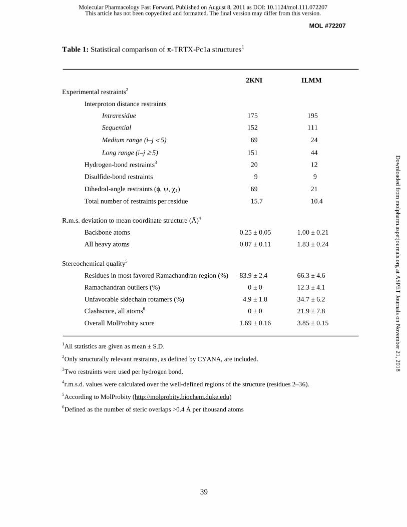
MOL #72207
39
Table 1: Statistical comparison of π-TRTX-Pc1a structures1
2KNI ILMM
Experimental restraints2
Interproton distance restraints
Intraresidue 175 195
Sequential 152 111
Medium range (i–j < 5) 69 24
Long range (i–j ≥ 5) 151 44
Hydrogen-bond restraints3 20 12
Disulfide-bond restraints 9 9
Dihedral-angle restraints (φ, ψ, χ1) 69 21
Total number of restraints per residue 15.7 10.4
R.m.s. deviation to mean coordinate structure (Å)4
Backbone atoms 0.25 ± 0.05 1.00 ± 0.21
All heavy atoms 0.87 ± 0.11 1.83 ± 0.24
Stereochemical quality5
Residues in most favored Ramachandran region (%) 83.9 ± 2.4 66.3 ± 4.6
Ramachandran outliers (%) 0 ± 0 12.3 ± 4.1
Unfavorable sidechain rotamers (%) 4.9 ± 1.8 34.7 ± 6.2
Clashscore, all atoms6 0 ± 0 21.9 ± 7.8
Overall MolProbity score 1.69 ± 0.16 3.85 ± 0.15
1All statistics are given as mean ± S.D.
2Only structurally relevant restraints, as defined by CYANA, are included.
3Two restraints were used per hydrogen bond.
4r.m.s.d. values were calculated over the well-defined regions of the structure (residues 2–36).
5According to MolProbity (http://molprobity.biochem.duke.edu)
6Defined as the number of steric overlaps >0.4 Å per thousand atoms
This article has not been copyedited and formatted. The final version may differ from this version.Molecular Pharmacology Fast Forward. Published on August 8, 2011 as DOI: 10.1124/mol.111.072207
at ASPE
T Journals on N
ovember 21, 2018
molpharm
.aspetjournals.orgD
ownloaded from
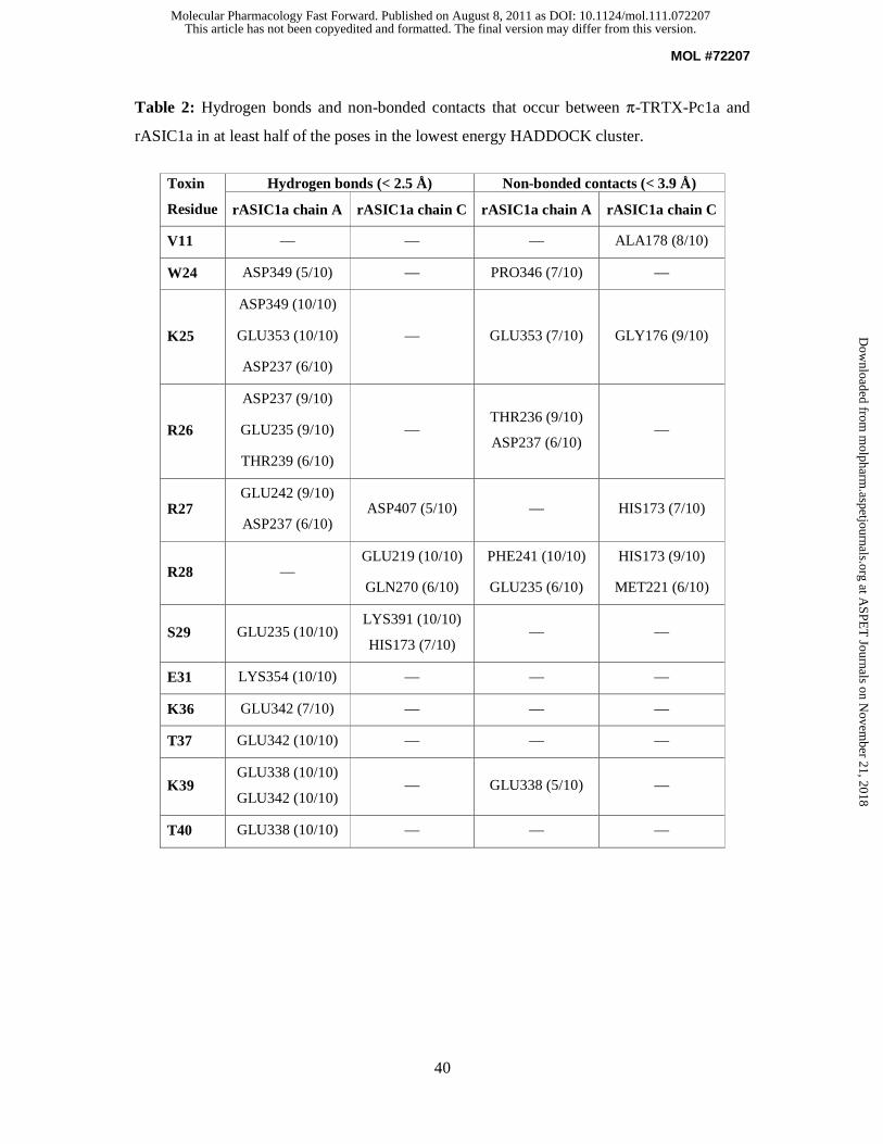
MOL #72207
40
Table 2: Hydrogen bonds and non-bonded contacts that occur between π-TRTX-Pc1a and
rASIC1a in at least half of the poses in the lowest energy HADDOCK cluster.
Toxin
Residue
Hydrogen bonds (< 2.5 Å) Non-bonded contacts (< 3.9 Å)
rASIC1a chain A rASIC1a chain C rASIC1a chain A rASIC1a chain C
V11 — — — ALA178 (8/10)
W24 ASP349 (5/10) — PRO346 (7/10) —
K25
ASP349 (10/10)
GLU353 (10/10)
ASP237 (6/10)
— GLU353 (7/10) GLY176 (9/10)
R26
ASP237 (9/10)
GLU235 (9/10)
THR239 (6/10)
— THR236 (9/10)
ASP237 (6/10) —
R27 GLU242 (9/10)
ASP237 (6/10) ASP407 (5/10) — HIS173 (7/10)
R28 — GLU219 (10/10)
GLN270 (6/10)
PHE241 (10/10)
GLU235 (6/10)
HIS173 (9/10)
MET221 (6/10)
S29 GLU235 (10/10) LYS391 (10/10)
HIS173 (7/10) — —
E31 LYS354 (10/10) — — —
K36 GLU342 (7/10) — — —
T37 GLU342 (10/10) — — —
K39 GLU338 (10/10)
GLU342 (10/10) — GLU338 (5/10) —
T40 GLU338 (10/10) — — —
This article has not been copyedited and formatted. The final version may differ from this version.Molecular Pharmacology Fast Forward. Published on August 8, 2011 as DOI: 10.1124/mol.111.072207
at ASPE
T Journals on N
ovember 21, 2018
molpharm
.aspetjournals.orgD
ownloaded from
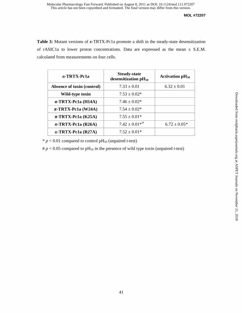
MOL #72207
41
Table 3: Mutant versions of π-TRTX-Pc1a promote a shift in the steady-state desensitization
of rASIC1a to lower proton concentrations. Data are expressed as the mean ± S.E.M.
calculated from measurements on four cells.
* p < 0.01 compared to control pH50 (unpaired t-test)
# p < 0.05 compared to pH50 in the presence of wild type toxin (unpaired t-test)
π-TRTX-Pc1a Steady-state desensitization pH50
Activation pH50
Absence of toxin (control) 7.33 ± 0.01 6.32 ± 0.01
Wild-type toxin 7.53 ± 0.02*
π-TRTX-Pc1a (H14A) 7.46 ± 0.02*
π-TRTX-Pc1a (W24A) 7.54 ± 0.02*
π-TRTX-Pc1a (K25A) 7.55 ± 0.01*
π-TRTX-Pc1a (R26A) 7.42 ± 0.01*/# 6.72 ± 0.05*
π-TRTX-Pc1a (R27A) 7.52 ± 0.01*
This article has not been copyedited and formatted. The final version may differ from this version.Molecular Pharmacology Fast Forward. Published on August 8, 2011 as DOI: 10.1124/mol.111.072207
at ASPE
T Journals on N
ovember 21, 2018
molpharm
.aspetjournals.orgD
ownloaded from

This article has not been copyedited and formatted. The final version may differ from this version.Molecular Pharmacology Fast Forward. Published on August 8, 2011 as DOI: 10.1124/mol.111.072207
at ASPE
T Journals on N
ovember 21, 2018
molpharm
.aspetjournals.orgD
ownloaded from
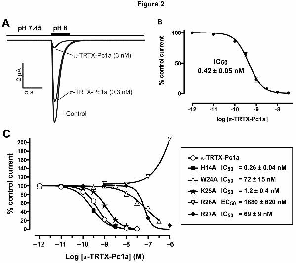
This article has not been copyedited and formatted. The final version may differ from this version.Molecular Pharmacology Fast Forward. Published on August 8, 2011 as DOI: 10.1124/mol.111.072207
at ASPE
T Journals on N
ovember 21, 2018
molpharm
.aspetjournals.orgD
ownloaded from
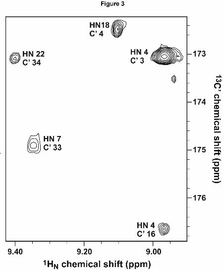
This article has not been copyedited and formatted. The final version may differ from this version.Molecular Pharmacology Fast Forward. Published on August 8, 2011 as DOI: 10.1124/mol.111.072207
at ASPE
T Journals on N
ovember 21, 2018
molpharm
.aspetjournals.orgD
ownloaded from
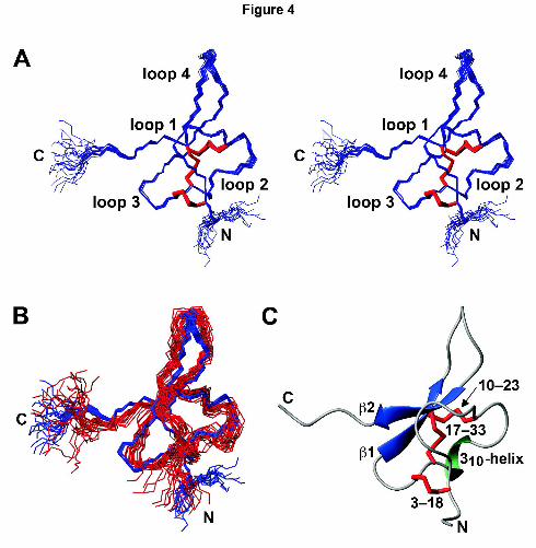
This article has not been copyedited and formatted. The final version may differ from this version.Molecular Pharmacology Fast Forward. Published on August 8, 2011 as DOI: 10.1124/mol.111.072207
at ASPE
T Journals on N
ovember 21, 2018
molpharm
.aspetjournals.orgD
ownloaded from

This article has not been copyedited and formatted. The final version may differ from this version.Molecular Pharmacology Fast Forward. Published on August 8, 2011 as DOI: 10.1124/mol.111.072207
at ASPE
T Journals on N
ovember 21, 2018
molpharm
.aspetjournals.orgD
ownloaded from
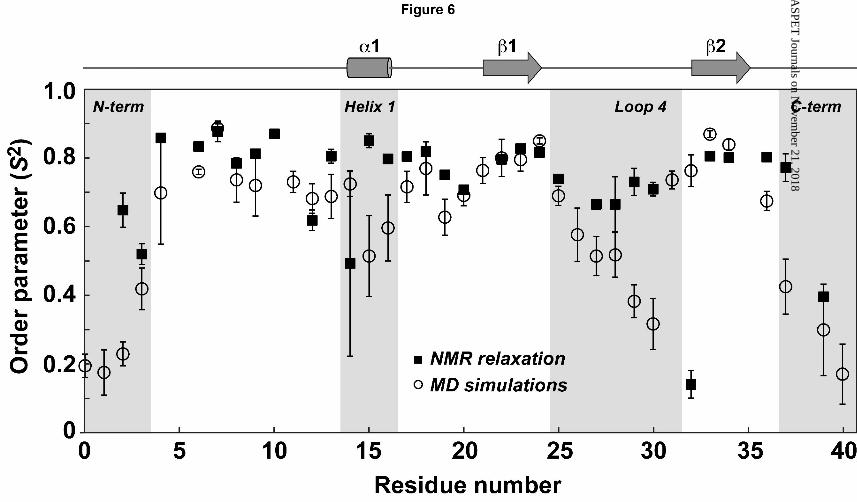
This article has not been copyedited and formatted. The final version may differ from this version.Molecular Pharmacology Fast Forward. Published on August 8, 2011 as DOI: 10.1124/mol.111.072207
at ASPE
T Journals on N
ovember 21, 2018
molpharm
.aspetjournals.orgD
ownloaded from
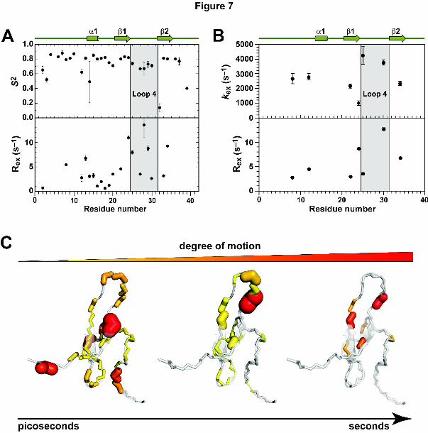
This article has not been copyedited and formatted. The final version may differ from this version.Molecular Pharmacology Fast Forward. Published on August 8, 2011 as DOI: 10.1124/mol.111.072207
at ASPE
T Journals on N
ovember 21, 2018
molpharm
.aspetjournals.orgD
ownloaded from
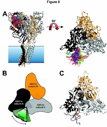
This article has not been copyedited and formatted. The final version may differ from this version.Molecular Pharmacology Fast Forward. Published on August 8, 2011 as DOI: 10.1124/mol.111.072207
at ASPE
T Journals on N
ovember 21, 2018
molpharm
.aspetjournals.orgD
ownloaded from
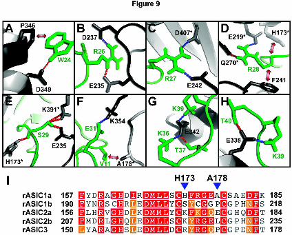
This article has not been copyedited and formatted. The final version may differ from this version.Molecular Pharmacology Fast Forward. Published on August 8, 2011 as DOI: 10.1124/mol.111.072207
at ASPE
T Journals on N
ovember 21, 2018
molpharm
.aspetjournals.orgD
ownloaded from