Embed Size (px)
Citation preview

1
A computational exploration of the color gamut of
nanoscale hollow scalene ellipsoids of Ag and Au
Catherine S. Kealley, Michael B. Cortie*
Institute for Nanoscale Technology, University of Technology Sydney, PO Box 123, Broadway, NSW
2007, Australia
Running head: color gamut of hollow scalene ellipsoids of Ag and Au
RECEIVED DATE (automatically inserted by publisher);
ABSTRACT
Hollow, nanoscale, scalene ellipsoids of Ag or Au provide an exceedingly tunable localized surface
plasmon resonance. Here we use numerical simulations to determine the limits of the color space that
would be possible from colloidal suspensions of these particles, and show that their color gamut will
exceed that possible with nanorods, nanoshells or nanorice. The important parameters are composition,
thickness of the shell and shape of the particle, in that order. The sensitivity of colors to geometry is
optimized for an aspect ratio of between 0.3 to 0.5 and was reduced for thinner shells. Shells of Ag will
have much wider and more vibrant gamut than those of Au. These findings indicate that hollow scalene
ellipsoids could be used as versatile pigments in materials or display systems that exploit plasmon
resonance to produce color.
KEYWORDS gold and silver nanoshells, scalene ellipsoid, plasmon resonance, color gamut

2
Introduction
Metallic nanostructures have attracted considerable attention because of their plasmonic, catalytic or
electronic properties. In particular, nanoparticles of Au and Ag have unique and interesting optical
properties in the visible and near-infrared regions of the spectrum (Kelly et al., 2003). The plasmonic
properties stem from the interaction of light with the free electrons in the metal nanoparticle giving rise
to localized surface plasmons, which lead to the selective absorption and scattering of particular
wavelengths of light. Hence, if the phenomena occur in the visible part of the spectrum, there is a
resulting color visible to the naked eye. Diverse applications, such as chemical and biomolecular
sensing, nanoscale optical components or devices, and surface-enhanced spectroscopies exploit this
phenomenon (Wang et al., 2006). The plasmon resonance frequencies of a metallic nanoparticle are
dependent on the particle’s composition, size, shape, and surface topography (Kelly et al., 2003).
It has been reported (Wang et al., 2006) that a ‘nanorice’ particle would combine the intense
local fields from nanorods with the highly tunable plasmon resonances of nanoshells. To tune the
plasmon resonance frequency of nanorods, the length or aspect ratio of the nanorod is varied (Yu et al.,
1997). For nanoshells, the relative size of the inner and outer radius of the metallic shell layer is
adjusted (Oldenburg et al., 1998, Prodan and Nordlander, 2004). In the reported ‘nanorice’ particle,
a=bc, where a and b are the equatorial radii and c the polar radius. For ‘nanorice’ consisting of a
spheroidal Au or Ag shell which encloses a spindle-shaped dielectric core, it was found that the
plasmon tunability arising from varying the thickness of the shell layer was far more greater than that
arising from varying the length of the nanostructure (ie. ratio of a to c) (Wang et al., 2006).
It is possible to envisage an even more flexible particle than a ‘nanorice’, specifically, a hollow
scalene ellipsoid in which abc, obtained by relaxing the geometry of the nanorice by one further
degree of freedom. In the present work we have sought to determine just how widely the optical
response of such an ellipsoidal nanoparticle can be tuned. The exercise has been carried out by
computer simulation of aqueous colloids of the hypothetical particles.

3
Methodology
1. Simulation of Concentric Ellipsoids
We have simulated the optical properties of the proposed nanoscale, hollow, scalene ellipsoids using
DDSCAT, the code of Draine and Flatau that exploits the discrete dipole approximation (Draine and
Flatau, 1994, Draine and Flatau, 2004). In this scheme the optical behavior is obtained numerically. The
applicability and accuracy of this technique has been previously verified by various
investigators (Brioude et al., 2005, Felidj et al., 1999, Kelly et al., 2003), and its particular advantage is
that it can be applied to arbitrarily-shaped particles. A linear response of the material to an electric field
is assumed in the code used. Furthermore, here we have used the bulk dielectric constant, and neglected
surface scattering. We will comment on the significance of this latter approximation later. The particle
is approximated by an array of polarisable points, so it is important to use a fine mesh in order to obtain
a reasonable rendition of fine detail. However, in practice, the array size (and hence accuracy) is limited
by available computational resources and time. At each point of the grid there is a dipole with
polarisability corresponding to the chosen material. The response of this array of dipoles to an incident
field is then determined self-consistently from which the extinction can be calculated.
Calculations were performed using the published complex refraction indices of gold, silver and
water (Weaver and Frederikse, 2001). A one nanometer dipole spacing (about 33,000 dipoles per
particle) was applied in the calculations. A typical target rendered at this resolution is shown in Figure
1(a). This is generally considered to be sufficiently fine for most shapes (He and Shi, 2005) but, to
check this, we repeated sample calculations at finer dipole spacings down to 0.5 nm, Figure 1(b). It is
apparent that the extinction peaks sharpen somewhat if re-calculated at a higher resolution. However,
given that the monodispersity assumed here for the colloidal particles is unattainable in practice
anyway, any actual extinction peaks that would be obtained in a real suspension would be less intense
and broader than even those calculated for the 1 nm dipole spacing. Therefore, we considered use of the

4
1 nm dipole spacing to be justified. The calculations were repeated for light polarized along each of the
axes of the target, and the results combined with a weighted average. The calculated optical properties
are expressed in terms of extinction efficiency, Qext. In the case of the DDSCAT code, this is the
ostensible optical extinction cross-section normalized by the nominal geometric cross-sectional area of
the particle, expressed as if all the matter in the particle was packed into a sphere (Draine and Flatau,
2004). Colors in the Commission Internationale de I’E Clairage (CIE) color model were directly
calculated from spectra using appropriate tables (American Society for Testing and Materials, 2001),
after converting the calculated Qext to a percentage transmission based on the density of nanoparticles
present in a colloidal suspension using:
(1)303.2
. .xCNA ext
where A is the solution absorbance, N the number of particles per unit volume, Cext the extinction cross-
section, and x the optical path length (Perez-Juste et al., 2005). Colors were assessed at a concentration
corresponding to 3x10¹⁶
nanoparticles per m3. In the CIE LAB system the L coordinate refers to the
luminance of the color, the a* measures the green-to-red tendency of the color, and the b
* the blue-to-
yellow tendency. The white point is given by a* = b
* = 0. The L-a
*-b
* colors were then converted
through CIE XYZ to RGB using the matrices given by Pascale (Pascale, 2003). Note that the calculated
colors err slightly on the side of being more intense than would be expected from real world particles.
This is because actual ellipsoidal particles would not be perfectly monodisperse and because, in the
case of very thin-walled targets, surface scattering of the plasmon would tend to reduce color saturation.
Color charts were drawn by sampling the parameter space at 10 nm intervals (or more closely if the
color varied rapidly with dimension).

5
2. Selection of Metals
A wide range of elements and compounds have dielectric properties that will provide a plasmon
resonance (Blaber et al., 2007). Sodium and potassium nanoshells, in particular, have dielectric
properties that would provide a strong localized plasmon resonance in the visible part of the
spectrum (Blaber et al., 2009). Unfortunately, the extreme chemical reactivity of these elements
precludes their use is atmospheric or aqueous environments, while nanoparticles of other, more noble
elements such as copper or platinum, have indifferent or absent plasmon resonances (Cortie et al.,
2006). Compounds such as AuAl2 (Supansomboon et al., 2008) and TiN (Reinholdt et al., 2004) also
possess properties that can generate a plasmon resonance in the visible, but their relatively large
extinction coefficients attenuate the resulting peaks. This leaves Ag and Au as the optimum candidates
for application in water or air. Actually, even Ag oxidizes in air, albeit slowly, so care would need to be
taken in any applications using this material to protect or passivate the particles.

6
Results and Discussion
Initial simulations found the frequency of the plasmon resonance to be very sensitive to aspect ratio, as
reported for nanorice by Wang et al. (Wang et al., 2006). An example for a particle size of L1 = 40 nm,
L2 = 50nm, L3 = 32 nm is given in Figure 2(a). As the core size increases (an increase in aspect ratio,
i.e. the particle becomes more like a shell with an over-all lower volume of metal), the color red-shifts.
Jackson and Halas (Jackson and Halas, 2001) reported that the extinction peak shifted to the blue when
the shell thickness was increased and the size of the dielectric cores kept constant. In our case, the
combination of the shell thickness decreasing and the dielectric core increasing has resulted in a red-
shift. The interesting result here is the sudden increase in this red-shift as the aspect ratio changes from
0.5 to 0.7 in both the Ag and Au particles. In the case of the Ag, this shift is associated with a drop in
Qext intensity. In general, the wavelength of the peak extinction of hollow objects exhibits a rapidly
increasing sensitivity to aspect ratio as the point of singularity (aspect ratio=1.0) is approached. An
example of this is shown in Figure 2(c), for a simple 40 nm outer diameter gold nanoshell, in vacuum.
There are, however, practical limits in regard to how thin a shell can be actually synthesized. In
addition, significant electron scattering will be experienced for aspect ratio>0.9 anyway, leading to an
attenuation of the extinction peaks (Blaber et al., 2009).
Overall, the Ag shell has greater color variability in comparison with the Au particle. The
reason for this is that the extinction peaks of the Ag nanoparticles move across the visible, from blue to
red end, whereas those of the gold, only start in the mid-visible and move out into the infra red. A
comprehensive set of simulations was then undertaken on selected aspect ratios. As the aspect ratio of
the shell approaches the extreme (for example, a very thin shell with an aspect ratio of 0.9), it was
found that the sensitivity of the color tunability decreased significantly. This is primarily because the
main plasmon resonance has been shifted in these instances into the near-infrared and is hence invisible
to the eye. However, for an aspect ratio of 0.5, the sensitivity of color to geometry was extensive.
Figure 3 shows the resulting color tunability for a particle with variable L2, constant L1 (40 nm), L3 (32

7
nm) and aspect ratio of 0.5. Figure 4 expands the geometry sensitivity by releasing both L2 and L3,
while holding L1 (40 nm) and aspect ratio (0.5) constant for Ag and Au. This can be directly compared
with the results in Figures 5 and 6 which were generated for particles with an aspect ratio of 0.3 and 0.7
respectively. For the aspect ratios in the 0.3 to 0.5 range, silver ellipsoids with L1L2L3, representing a
more spherical shape, produce a yellow plasmon resonance directly comparable with the color found
associated with Ag nanospheres (Lui and Guyot-Sionnest, 2004). However, as the ellipsoid is
elongated, the plasmon resonance red shifts, causing the color transmitted to shift towards blue. It can
also be seen that for the thicker shells (i.e. approaching a solid nanoparticle), in Figures 4 and 5 the
color produced is extremely sensitive to the length parameters. However, as the shell becomes quite
thin (Figure 6), the influence of the length parameters is greatly reduced as the plasmon resonance shifts
into the near-infrared.
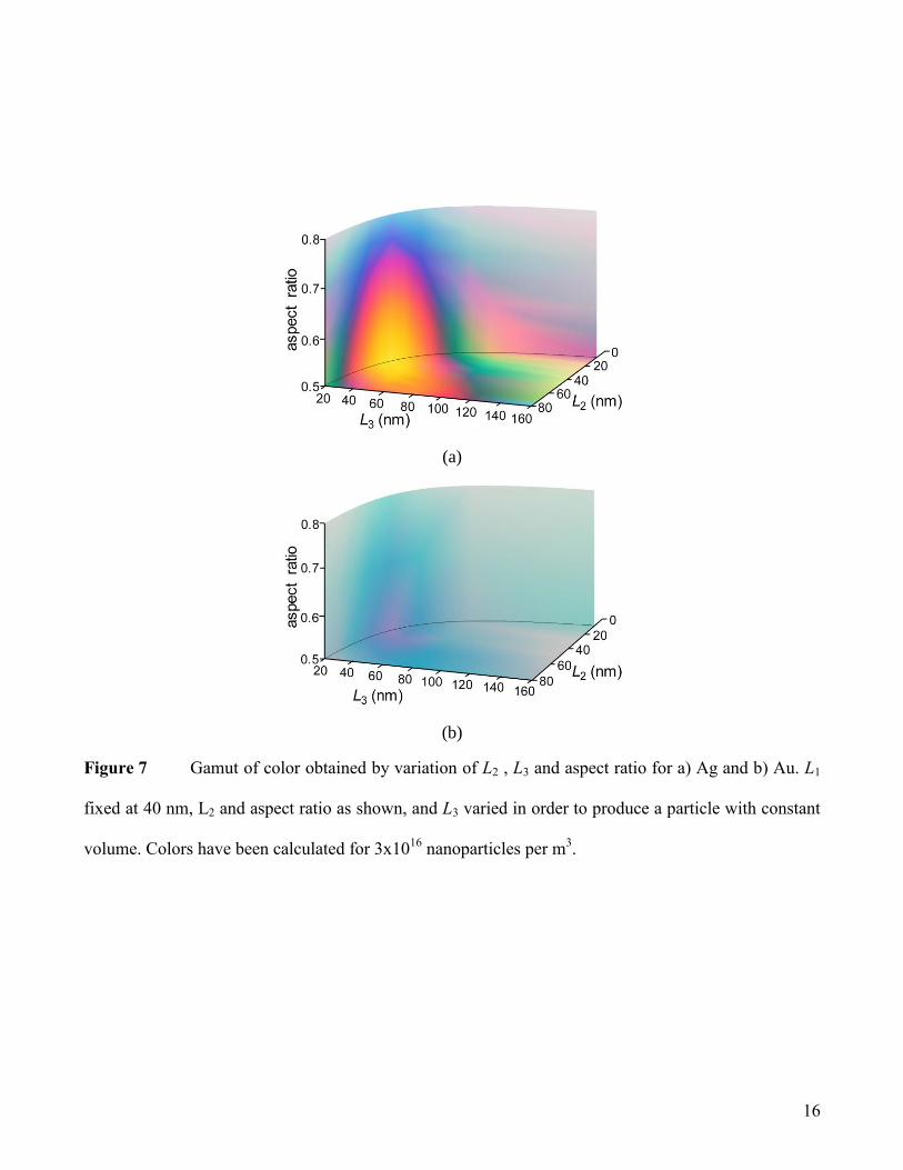
The results are combined in Figure 7, where the shell thickness variation is compared with a
variation in L2 and L3 (L1 is fixed at 40 nm), for both Ag and Au. Once again, it can be seen that the Ag
shell has far greater color variability in comparison with Au. For an aspect ratio of 0.5, a spherical Ag
particle is yellow in color, and it has the singular plasmon resonance of an isolated Ag sphere.
Elongating this particle (keeping the volume constant) results in either two or three resonance peaks
that are red-shifted in comparison with the plasmon resonance of the spherical particle, and produces a
blue color in transmission. As the sphere elongates (for example, L1= 40 nm, L2=20 nm, L3=40 nm) to
give a volume decrease, and the resonance peaks blue-shift. If the elongated particle has an increase in
the volume (for example, L1= 40 nm, L2=60 nm, L3=40 nm), then the resulting resonance is red-shifted.
For the higher aspect ratios of 0.6, 0.7 and 0.8 in Figure 7, L1 is 40 nm, while L2 and L3 are varied in
order to produce a particle with constant volume. At an aspect ratio of 0.6, the spherical Ag particle
(for example, L1=L2=L3=40 nm) is still yellow in color. However, by 0.7, the particle is becoming more
shell-like and the plasmon resonance of the spherical particle is red-shifted. Over-all, as the aspect ratio

8
increases in the Ag particles, the variability in the color produced by a change in the shape of a particle
is significantly diminished.
Finally, we concede that the synthesis of hollow particles might be difficult, so it is important to
also consider the tunability of core-shell ellipsoids, which are more readily fabricated. The metal shell
could be combined with any material, including a second metal, or an entirely difference material, for
example, an organic core. In order to simulate this, Ag (with the superior tunability) was chosen as the
shell material, and the refractive index of the hypothetical core material was varied (n=1.3, 1.37, 1.40,
1.45 and 1.50) (note: the absorption coefficient, k, has been kept constant at 0 for these calculations).
The results are shown in Figure 8. It can be seen that there is minimal change (a slight blue-shift) in the
plasmon resonances of the ellipsoid particle, as the refractive index of the core is increased. This
suggests that the optical response of the particle in this case is dominated by the metal shell.
Conclusion
Our simulations show that colloidal suspensions of hollow silver scalene nano-ellipsoids with aspect
ratios in the 0.3 to 0.5 range will exhibit a very extensive range of color as a function of small changes
in length parameters. Furthermore, Ag and Au are the optimum materials from which these colorful
nanoparticles might be constructed. The resulting gamut is wide and intense, particularly in the case of
the Ag particles. The simulations have confirmed that the aspect ratio is the most influential parameter.
The length parameters affect the color produced in shells with aspect ratios in the range of 0.3 to 0.5,
but are less critical outside of that range. Hollow scalene ellipsoids have the potential to be the ultimate
tunable colloidal particles, with multiple degrees of geometric freedom. However, given the extreme
sensitivity that the optical properties of these particles have in relation to their geometry, synthesis of
these materials would need to be controlled to a level that is perhaps not yet practical.

9
References
American Society for Testing and Materials A (2001) E308-01 Standard Practice for Computing the
Colors of Objects by using the CIE SystemMaterials.
Blaber MG, Arnold MD, Ford MJ (2009) Search for the iIdeal plasmonic nanoshell: the effects of
surface scattering and alternatives to gold and silver J. Phys. Chem. C 113:3041-3045.
Blaber MG, Arnold MD, Harris N, Ford MJ, Cortie MB (2007) Plasmon absorption in nanospheres: a
comparison of sodium, potassium, aluminium, silver and gold Phys. B 394:184-187.
Brioude A, Jiang XC, Pileni MP (2005) Optical Properites of gold nanorods:DDA simulations
supported by experiments J. Phys. Chem. B 109:13138-13142.
Cortie MB, Xu X, Ford MJ (2006) Effect of composition and packing configuration on the dichroic
optical properties of coinage metal nanorods Phys. Chem. Chem. Phys. 8:3520-3527.
Draine BT, Flatau PJ (1994) Discrete-dipole approximation for scattering calculations J. Opt. Soc. Am.
A 11:1491-1499.
Draine BT, Flatau PJ (2004) User Guide to the Discrete Dipole Approximation Code DDSCAT 6.1.
Felidj N, Aubard J, Levi G (1999) Discrete dipole approximation for ultraviolet-visible extinction
spectra simulation of silver and gold colloids J. Chem. Phys. 111:1195-1208.
He Y, Shi G (2005) Surface plasmon resonances of silver triangle nanoplates: graphic assignments of
resonance modes and linear fittings of resonance peaks J Phys. Chem. B 109:17503-17511.
Jackson JB, Halas NJ (2001) Silver nanoshells: variations in morphologies and optical properties J.
Phys. Chem. B 105:2743-2746.
Kelly KL, Coronado E, Zhao LL, Schatz GC (2003) The optical properties of metal nanoparticles: the
influence of size, shape and dielectric environment J. Phys. Chem. B 107:668-677.
Lui M, Guyot-Sionnest P (2004) Synthesis and optical characterization of Au/Ag core/shell nanorods J.
Phys. Chem. B 108:5882-5888.
Oldenburg SJ, Averitt RD, Westcott SL, Halas NJ (1998) Nanoengineering of optical resonances
Chem. Phys. Lett. 288:243-247.
Pascale D (2003) A review of RGB color spaces - from xyY to R'G'B'. Montreal, Canada.
Perez-Juste J, Pastoriza-Santos I, Liz-Marzan LM, Mulvaney P (2005) Gold nanorods: synthesis,
characterization and applications Coord. Chem. Rev. 249:1870-1901.
Prodan E, Nordlander P (2004) Plasmon hybridization in spherical nanoparticles J. Chem. Phys.
120:5444-5454.
Reinholdt A, Pecenka R, Pinchuk A, Runte S, Stepanov AL, Weirich TE, Kreibig U (2004) Structural,
compositional, optical and colorimetric characterization of TiN-nanoparticles Euro. Phys. J. D
31:69-76.
Supansomboon S, Maaroof A, Cortie MB (2008) “Purple glory”: The optical properties and technology
of AuAl2 coatings Gold Bull. 41:296-304.
Wang H, Brandl DW, Lel F, Nordlander P, Halas NJ (2006) Nanorice: a hybrid plasmonic
nanostructure Nano Lett. 6:827-832.
Weaver JH, Frederikse HPR (2001) IN LIDE, D. R. (Ed.) CRC Handbook of Chemistry and Physics. 82
ed. Boca Raton, Florida, CRC Press.
Yu Y-Y, Chang SS, Lee C-L, Chris Wang CR (1997) Gold nanorods: electrochemical synthesis and
optical properties J Phys. Chem. B 101:6661-6664.

10
(a)
(b)
Figure 1. Typical target and the effect of dipole spacing. (a) Hollowed-out concentric ellipsoidal model
used for all calculations. Outer dimensions L1, L2 and L3 are freely variable. Inner shell dimensions L4,
L5 and L6 are obtained from the corresponding outer shell dimensions by multiplying with the relevant
aspect ratio. Note: L1 was held constant at 40 nm for this work. The dipoles used to represent the shape
in the calculation are shown as little spheres. (b) Effect of dipole spacing on intensity of optical
extinction peaks. A particle with size as indicated was considered, and the dipole spacing varied from 1
nm to 0.5 nm. The effect is to slightly attenuate the optical extinction peaks.

11
(a)
(b)
(c)
Figure 2. Extinction efficiencies and resulting colors for particles with outer dimensions of 40 nm,
50 nm and 32 nm, where the aspect ratio has been varied. (a) Ag nanoparticles. (b) Au nanoparticles.
(c) Wavelength of the peak extinction of a 40 nm outer diameter gold nanoshell (in vacuum), as a
function of increasing aspect ratio.

12
(a)
(b)
Figure 3 Extinction efficiencies and resulting colors (small boxes) for a Ag particle with L2 varied
(some examples of the particle shapes are shown) for an aspect ratio of 0.5. (a) particles with L2<L1, (b)
particles with L1>L2.

13
(a)
(b)
Figure 4 Color tunability in particles with an aspect ratio of 0.5, with variable L2 and L3. (a) Ag
nanoparticles. (b) Au nanoparticles.

14
(a)
(b)
Figure 5 Color tunability from an aspect ratio of 0.3, with variable L2 and L3. (a) Ag
nanoparticles. (b) Au nanoparticles.

15
(a)
(b)
Figure 6 Color tunability from an aspect ratio of 0.7, with variable L2 and L3. (a) Ag
nanoparticles. (b) Au nanoparticles.
16
(a)
(b)
Figure 7 Gamut of color obtained by variation of L2 , L3 and aspect ratio for a) Ag and b) Au. L1
fixed at 40 nm, L2 and aspect ratio as shown, and L3 varied in order to produce a particle with constant
volume. Colors have been calculated for 3x10¹⁶
nanoparticles per m3.

17
Figure 8 Extinction efficiencies and resulting colors (small boxes) for a particle with outer
dimensions of 40 nm, 50 nm and 32 nm, and an aspect ratio 0.5, with variability in the refractive index
of the core material. Note: dotted line interpolates a region in which the software used would not
converge.


![COMPUTATIONAL MANUFACTURINGilp.mit.edu/images/conferences/2018/japan/presentations/Matusik... · [Maldovan et al. 2007] Microstructure Design. Material Gamut. SUMMARY: FROM DESIGN](https://img.dokumen.tips/doc/110x75/5ade39157f8b9a8b6d8df879/computational-maldovan-et-al-2007-microstructure-design-material-gamut-summary.jpg)