Embed Size (px)
Citation preview

Arch. Razi Ins. 54 (20021/7-29 /7
A Comparative Study on Histopathologic Effects of Iranian
Newcastle Disease Virus Isolates
Kianizadeh' l, M., Aini, 1.2 and Gholami, G.R.J
1. Avian Diseases Research & Diagnosis Dep/., Razi Vaccine & serum Research Ins/i/u/e,
P.O Box: 11365-1558, Tehran, Iran.
2. Faculty of Ve/erinary Medicine, University of Pu/ra Malaysia, 43400 UPM, Malaysia.
3. Pa/hology Dept., Razi Ins/i/u/e
Received 28 Mar 2002· accepted 17 Sep 2002
Summary
The histopathologic effects of Iranian Newcastle disease viruses isolated
From different outbreaks across the country wcre studied on difTerent organs
of specifie pathogen free chickens. Clinically, time and sequence of the
signs' occurrence were varied among the groups receiving different isolates.
Depression was the first c1inical sign observed by 48h postinfection (PI) in
ail groups except two groups, which showed depression by 97h PI. Grossly,
among the three systems, gastro-intestinal, respiratory and central nervous
system that were examined in infected groups, the latter two showed less
remarked lesions. Macroscopically, From 72h PI toward the end of
experiment the spleen showed atrophy in ail infected groups. At 96h PI and
later on, the brain was slightly hyperaemic in limited infected groups. The
early findings were observed in proventriculus, Iiver, small intestine and
spleen. Generally ail nine Iranian Newcastle disease viruses can affect
visceral organs [aster than other organs and could be placed in viscerotrop
velogenic group.
Key word: Newcastle disease, histopathology, Iran
Introduction
ND viruses (NDVs) distribution in various organs, and histopathologic changes in
organs have been studied as ways ofrevealing their pathogenicity (Shirai et al 1988,
• Aulhor for corrcspondence. E.mail: [email protected]

18 Kianizadeh et al./Arch. Raz; Ins. 54 (2002) 17-29
Ojok & Brown 1996, Hooper et al 1999). The virus enters the body via the
conjunctiva, digestive and respiratory tracts. The primary target organs for
attachment and replication are respiratory and intestinal tracts. After propagation at
the initial sites through the first viraemia, virus may reach the other organs such as
kidney, liver, bursa, spleen and lungs.
Pathological changes vary, based on several factors including infection route,
tropism and virulence of the virus, immune status and age of the host. It has been
shown that respiratory route is more sensitive compared to other routes of infection
(McFerran & McCrackem 1988). The reason NOV is less infectious when given
oralIy appears to be because the gizzard content is acidic (pH around 2.6), reducing
the infectivity 1000 fold (Alexander 1988). Hanson and Brandly (1955) have
grouped NOV strains based on tropism into velogenic, mesogenic and lentogenic
groups. While lentogenic strains are present in low titres in circulation, mesogenic
strains spread to sorne visceral organs by 24-44h PI. Virulent viruses can be found in
alI tissues with highest titer in thymus and lowest in muscles and brain. The reason
for this feature has been described (ColIins et al 1993) ofwhich, the cleaveability of
Fa is the main factor.
The objective ofthis study was to compare the pathogenesis among Iranian NOV
isolates in an experimental infection.
Materials and Methods
Viruses and inoculation procedures. Nine Iranian NOV isolates presented in table 1,
were inoculated as described by Ojok and Brown (1996). Briefly, groups often 6-
week-old specific pathogen free (SPF) chickens (Valo, Lohmann, Cuxhaven,
Germany) for each isolate were inoculated with 0.05ml of 107 embryo-Iethal doses
50% (EIDso) of virus by the ocular route. Additionally two birds were kept in seprate
pen during the experiment as negative controls. The birds were examined clinically
everyday.

Arch. Raz; Ins. 54 (20021 17-29 19
Table 1 Data on ND outbreaks in Iran
No Isolate Type Population
1 MK7 8roiler 10000
2 MKI2 8ro/layer 200
3 MKI3 Indigenous 200
4 MKI4 Indigenous 3500
5 Krd76 8roiler 2500
6 Kasra7 8roiler 16000
7 GH77 8roiler 5000
8 KH2178 8roiler 5000
9 ES 1/99 8roiler 10000
Sample collection. At intervals of 24h, birds were killed by injection of 1.5ml of
thiopental sodium (Specia, 16RUE GLISSON 75013 Paris, France) into the brachial
vein; one chicken (B) was killed at 24h PI, one (C) at 48h PI, two (0 and E) at 72h
PI, two (F and G) at 96h PI and three (H, l, J) at 120h PI. Necropsy was carried out
immediately. The gross lesions were recorded. A non-infected chicken (A) was
killed at the beginning of the experiment to supply control samples.
Samples preparation for microscopie sections. Samples were prepared as described
by Ojok and Brown (1996). Briefly, brain, lungs, trachea, liver, small intestine,
spleen and proventriculus from each bird were collected and fixed in 10% neutral
buffered formaI in for microscopic examination. Tissues were processed and
embedded in paraffin wax after fixation for 24h, sectioned 4-5Jlm and stained with
haematoxylin & eosin and observed with light microscope for histopathology
changes.
Results
Clinicat signs. Sorne generalized signs that were observed inc1uded depression,
ruffled feathers, loss of appetite, huddling, listless, swelling of eyelid of inoculated
eye. Oiarrhoea, paralysis, head shaking, torticollis, moribund state and death were

20 Kianizadeh et al./Arch. Razj [ns. 54 (2002) 17-29
others features, which usually occurred at the later stages. The time and sequence
of the signs' occurrence were varied among the groups receiving different isolates.
The negative control birds did not show any cIinical signs. The summarized data
are presented in table 2.
Table 2 Clinicat signs in experimenlat infection
lranian NDV h PI
isolales 24 48 72 96 120
MK7 I)A 2)1 3)B·4)B 5)B,F·6)B.G.D 7)H,B·8)H.B-9)G,J,K
MKI2 I)A 2)1 3)B-4)B 5)H.B-6)B 7)L<120-8)E-9)E
MKIJ I)A 2)B 3)B-4)B 5)F,K-6)L<96 7)L < 120-8 )E-)E
MKl4 I)A 2)B 3)B,C-4)B,C 5)E,K-6)E,K 7)L< 120-8)H,E-9)H,E
Krd76 I)A 2)B 3)B,C-4)B,C 5)E,K-6)E,K 7)L< 120-8)H.E-9)H,E
Kasra97 I)A 2)A 3 )B,C,I-4 )B,C,I 5)G,B-6)H.G,B 7)L< 120-8)L< 120-
9)H,G
GU77 I)A 2)B.C 3)E,C,F-4)G 5)H,L<96-6)L<96 7)L<120-8)H.L<120-
9)H.L<120
KlUn8 I)A 2)A 3)B,I-4)B,1 5)H,L<96-6)H,L<96 7)H,L<96-8)H,L<96-
9)H,L<96
ESI/99 I)A 2)B 3)B,C,D-4)B,C,D 5)H,L<96-6)H,L<96 7)H,L<96-8)H,L<96-
9)H,L<96
1-9: Chicken nlllnber; A: Normal slalue; B: Depression.C; RlIftled fealhers; D: Open mOlilh brealhing.;
E: Lislless; F: Head Iremors; G:Paralysis; H: Diarrhoea; 1: Eyelid swelling; J: Torlicollis; K: Sil on Ihe
hocks; L: Dealh.
Gross lesions, At each interval (24h), necropsy ofkilled or dead bird was carried
out immediately. No abnormalities were noted in the control bird (No 10). Grossly,
among the three systems; gastro-intestinal, respiratory and central nervous system
that were examined in infected groups, the latter two showed less remarked lesions.
The only features that were observed incIuded congestion in lungs, which began by
48hPI. In gastro-intestinal tract, the outstanding les ions were haemorrhagic foci

Arch. Raz; Ins. 54 (2002) 17-29 21
associated with necrosls ln the proventriculus, small intestine, caeca and large
intestine, which were visible mostly at 72hPI and later (Figure 1). The early les ions
in proventriculus were observed by 48 hpi in birds infected with isolate ES 1/99
(Figure 2).
Figure 1. Haemorrhage in small intestine of ND infeeted ehicken
• ••• :".: .. 1 ~
Figure 2. Haemorrhagie /esions in proven/rieu/us of ND infeeted ehicken

22 Kianizadeh el al./Arch. Raz; Ins. 54 (2002) 17-29
ln sorne cases the spleen had a stippled appearance on both capsular and cut
surfaces and was enlarged at 48hPI. Liver in ail infected groups was apparently
normal. Macroscopically, from 72hPI toward the end of experiment the spleen
showed atrophy in ail infected groups. At 96hPI and later on, the brain was slightly
hyperaemic in limited infected groups. During the experiment period no noticeable
effects on trachea were observed except for slight congestion in a few groups. Gross
les ions in each group caused by Iranian isolates are summarised in table 3.
Table 3 Gross lesions cou.\·ed by NDV isolales
Organs presenled wilh macroscopic lesions
No HPI Provenlri- liver Inlesline lung Trachea Spleen brain
culus
1 24 A A A A A A A
+ + + + + + +
B A A C A D A
2 48 ESI/99 + + KH2/78. + MIO. +
ESI/99 OH77.
MKl4
B A A C E D A
MK12. + + MK7. Kasra97 MK7 +
3,4 72 MKI3. KH2178,
KH2/78,OH77, ES 1/99
ESI/99,MKI4
B· A B C E F C
MK7, + OH77·50% MK12, MK12 + Kasra97
OH77, KH2178, MK13,
5,6 96 K12, Krd76, Kasra97,
MK13, ESI/99·, KH2/78,
KH2/78.ES 1/9 MKl2 ESI/99
9,MKI4, Kasra97
Krd76
B· A B C E F C
MK7,OH77, + MK7, MK7, MKI2, + MKI2,
MKI2,MKI3. OH77·, MK12. Kasra97 Kasra97,
7,8,9 120 Krd76, MKI4, MKI3, KH2/78
Kasra97, Krd76, Kasra97,
MKI4 Kasra97 ES 1/99
1-9: Chic ken number.; A: Apparently normal; B: Haemorrhagic; C:Congestion; D: splenomegaly:
E: Restricted hyperaemic areas; F: Atrophy.;·: Multiple site; +: Ali.

Arch. Raz; Ins. 54 (2002) /7-29 23
Microscopie findings. There were no abnorrnalities in non-infected control bird
(No. 10). At 24hPI no rnarked changes were observed in tissue sections in ail
infected groups. By 48hPI sorne changes were observed in different organs by
different isolates as follow: 1 isolate (ES 1/99) affected the proventriculus by
haernorrhagic necrosis, 6 isolates affected the liver by congestion and increased
rnononuclear cells around the blood vessels (Figure 3), 3 isolates affected the srnall
intestine by localized heterophils infiltration. At 72hPI the nurnber of the isolates
that could affect the organs increased. The affected organs included the following:
spleen, proventriculus, liver, small intestinal, and lungs that were affected by 8, 8, 6,
3 and 7 isolates, respectively. In intestine, lesions were haernorrhagic foci associated
with necrosis in intestinal wall (Figure 4). By 72hPI, trachea and brain, in ail
infected groups did not show any significant change. At 96hPI and later on, alrnost
ail the isolates could affect ail the organs with progressive lesions. The extent of
les ions in spleen and proventriculus were severe. The spleen showed rnultifocal
necrosis area, Iyrnphoid depletion with fibrin replacing (Figure 5).
Figure 3. Mononllclear cell infillralion in Il'D infecled /iver x400

24 Kianizadeh et al./Arch. Raz; /ns. 54 (2002) /7-29
Figure 4. Haemorrhagic inlesline of ND infecled chicken x4()()
Figure s. Necrosis and Iymphoid deplelion in spleen of ND infecled chicken x4()()
During the experimental period the traehea in most infeeted groups showed
lesser pathologie effeets exeept, in 3 groups infeeted with MK7, MKI2 and
Kasra97 ND isolates that showed haemorrhage and loss of eilia in infeeted group
Arch. Razi Ins. 54 (2002) 17-29 25
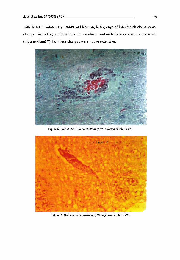
with MK 12 isolate. By 96hPI and later on, in 6 groups of infected chickens sorne
changes including endotheliosis in cerebrurn and rnalacia in cerebellurn occurred
(Figures 6 and 7), but these changes were not soextensive.
Figure 6. Endotheliosis in cerebel/um of ND infected chicken x400
Figure 7. Ma/acia in cerebel/um of ND infected chicken x400

26 Kianizadeh et al./Arch. Raz; [ns. 54 (2002) 17-29
Di.5cussion
ln this study the presence of virus in different tissues was examined by gross and
microscopy findings. The pathology of NOV infection varies from strain to strain.
Virulent NOV can multiply in many organs resulting in systemic infection and early
death without the bird showing ail the signs of the disease (Collins et al 1993). In ail
groups infected with Iranian isolates; spleen, proventriculus, liver and lung showed
the effects of the viral infection in the early phase of infection although the liver was
apparently normal and lesions in the lungs were not destructive and limited to
congestion. Grossly, virulent viruses cause predominantly haemorrhagic lesions in
various parts of intestinal tract, in particular in the mucosa, at the junction of
oesophagous/proventriculus and proventriculus/gizzard. The lesions in the posterior
half of the duodenum/jejunum and ileum are almost pathognomic for velogenic
viscerotropic NOV (Kouwenhoven 1993). The gastrointestinallesions that are seen
following a viscerotropic ND virus infection mostly developed in lymphoid
aggregates (Alexander 19S5). The outstanding les ions in infected groups with
Iranian NOV isolates were found in gastro intestinal tract (GIT). The first les ions in
GIT (proventticulus) and Iymphoid organ (spleen) were observed in 2 groups by
4ShPI of which, ESI/99 caused haemorrhagic lesions in proventriculus while the
spleen showed enlargement in groups infected by MK7, GH77 and MKI4, followed
by atrophy toward the end of the experiment. At the later stage of the infection (72-
120hPI), the les ions extended to intestine. This process was reported by Kaschula
(1961), in comparative study of virulent Asiatic and mi Id American forms of ND
and by Ojok and Brown (1996), In an experimental infection of virulent and
viscerotropic NOV in chicken.
Among the organs, trachea and brain in ail infected groups, showed changes in
the late phase of infection. This is matching with limited respiratory and nervous
c1inical signs during the experiment in ail groups. NOVs invaded the CNS after
multiplication in non-nervous tissues has ceased. However, virulent neurotropic

Arch. Raz; Ins. 54 (2002117-29 27
virus may be present in the CNS at the same time as in the respiratory or intestinal
tract (Kouwenhoven 1993
Respiratory disturbances were not observed except in groups infected with
ESI/99 and MK7, which showed open mouth breathing by 96hPI. The reason for
lack of respiratory signs in most groups may be related to the virulence of virus
causing fast progress of the disease or to the route of inoculation. The route of
inoculation may influence initial site of viral replication. Mortality varies greatly
depending on the property of the virus. Viruses of sorne strains reached vital organs
like liver and kidney very rapidly so that the birds may die before disease symptoms
are overt (Kouwenhoven 1993). The earliest death occurred in less th an 96hPI in 4
groups of which 2 groups were infected with KH2178 and ESI/99. The only NOV
isolate that did not cause death in the group even by 120hPI, was MK7. In this
group, torticollis was recorded as weil as the respiratory disturbance. Thus, the slow
trend of the disease progress has provided sufficient time for the birds to show ail
possible features of the disease. Waterson el al (\ 967) mentioned that the virulent
strains might kill so quickly that neurological signs may be seen for only a brief
period before death. The presence of microscopic changes since the early stages of
infection (48hPI) in most selected organs, represent the ability of Iranian NOV
isolates in inducing systemic infection. Based on microscopical findings, the
outstanding feature was the involving of the Iymphoid cells or organs in virus
multiplication after first viraemia. Congestion and focal necrosis in spleen were
recorded in 7 infected groups. Congestion and mononuc\ear Iymphoid cells
infiltration was seen in liver in 5 groups by 48hPI. Infiltration of heterophils was
present in intestinal sections. Ojok and Brown (1996) found Iymphoid areas in
spleen and thymus in infection with virulent viscerotropic NOV where they were
represented by massive destruction with extensive Iymphocytolysis and fibrin
replacing and in proventriculus with Iymphoid aggregates.
Congestion was the only lesions found in the lungs. This feature is matching with
lack of any c\inically significant respiratory disturbance in most of the infected

28 Kianizadeh et al./Arch. Razi lns. 54 (2002) J 7-29
groups. Lesions in brain included endotheliosis and neuronal degeneration occurred
late (96hPI) in comparison to the other organs. Such lesions are presented in birds
infected commonly with viscerotropic and mesogenic pathotypes (Alexander 1988).
Generally, during the experimental infection with ail Iranian NOV isolates, time
was the only variant regarding the pathology effects. Based on the results obtained,
ail isolates showed velogenic viscero neurotropic characteristics, which is in
agreement with the results ofpathogenicity indices (kianizadeh, et al 1999).
References
Alexander, D.J. (1988). Newcastle Disease. Massachusetts, Kluwer Academic
Publishers.
Collins, M.S., Bashiruddin, BJ. and Alexander, DJ. (1993). Deduced amino
acid sequences at the fusion prote in cIeavage of Newcastle disease viruses showing
variation in antigenicity and pathogenicity. Archives ofVirology 128:363-370.
Hooper, P.T., Hansson, E., Young, J.G., Russell, G.M. and Della-Porta, AJ.
(1999). Lesions in the upper respiratory tract in chickens experimentally infected
with Newcastle disease viruses isolated in Australia. Australian. Veterinary Journal
77: 50.
Kouwenhoven, B. (1993). Newcastle disease. In: J.B. McFerran and M.S.
McNuty (Eds.), Virus Infections of Birds. Pp:341-361. Amsterdam, The Netherland,
Elsevier Science Pubs.BV.
Kianizadeh, M., Ideris, A., Shahrabadi, M.S., Kargar,R., Pourbakhash, S.A.,
Omar, A.R. and Yusoff, K. (1999). Biological and Molecular Characterization of
Newcastle Disease Virus Isolates from Iran. Archives ofRazi Institute 50:1-9.
McFerran, J .B. and McKracken, R.M. (1988). Newcastle Disease pathogenesis.
ln: D.J. Alexander (Ed.), Newcastle Disease. Pp:161-183. Massachusetts, Kluwer
Academic Publishers.

Arch. Raz; Ins. 54 (2002) 17-29 29
Ojok, L., Brown, C. (1996). An immunohistochemical study of the pathogenesis
of virulent viscerotropic Newcastle disease in chicken. Journal of Comparative
Pathology 115:221-227.
Shirai, 1., Hihara, H. and Maeda, M. (1988). Virus distribution and
histopathologic changes in organs of chicken inoculated with Newcastle disease
virus (A vian Paramyxovirus-I) isolated from racing pigeon. Avian Diseases 3: 544-
547.
Waterson, M.D., Pennington, T.H. and Allan, W.H. (1967). Virulence in
Newcastle disease virus. British Medical Bulletin 23: 138-143.