Embed Size (px)
Citation preview
Brain (1998),121,889–905
A clinicopathological study of autism
A. Bailey,1 P. Luthert,2,* A. Dean,2 B. Harding,3 I. Janota,2 M. Montgomery,2,* M. Rutter1 andP. Lantos2
1MRC Child Psychiatry Unit, and2Department of Correspondence to: A. Bailey, MRC Child Psychiatry Unit,Neuropathology, The Institute of Psychiatry and The Institute of Psychiatry, De Crespigny Park, Denmark3Department of Histopathology, Institute of Child Health, Hill, London SE5 8AF, UKLondon, UK
*Present address: The Department of Pathology, TheInstitute of Ophthalmology, University College, London,UK
SummaryA neuropathological study of autism was established andbrain tissue examined from six mentally handicappedsubjects with autism. Clinical and educational recordswere obtained and standardized diagnostic interviewsconducted with the parents of cases not seen before death.Four of the six brains were megalencephalic, and areasof cortical abnormality were identified in four cases. There
Keywords: autism; neuropathology; megalencephaly; cortical dysgenesis
Abbreviations: ADI 5 Autism Diagnostic Interview; GFAP5 glial fibrillary acidic protein
IntroductionAutism is a severe developmental disorder characterized byimpairments in reciprocal social interaction and communica-tion, restricted and stereotyped patterns of behaviour andinterests, and an onset before 3 years of age (World HealthOrganization, 1992). The core disorder affects approximatelyfour in 10 000 children, and is much commoner in males, ina ratio of ~4 : 1. The syndrome was first described by LeoKanner (1943); he assumed that affected children were ofnormal intelligence and for several decades the disorder wasthought to be psychogenic. An organic basis was firstsuggested by the finding that three-quarters of sufferers arementally handicapped (Lockyer and Rutter, 1969) and thatat least one-quarter develop epilepsy (Rutter, 1970; Gillbergand Steffenburg, 1987). Subsequently autism was oftenassumed to be an unusual consequence of brain damage,caused either by medical disorders or obstetric hazards. Morerecently it has been appreciated that only a minority of casesof autism are associated with medical causes of mentalhandicap, then most commonly with tuberous sclerosis orFragile X (Rutteret al., 1994). The findings from twin andfamily studies suggest that the vast majority of idiopathiccases arise on the basis of strong specific genetic influences
© Oxford University Press 1998
were also developmental abnormalities of the brainstem,particularly of the inferior olives. Purkinje cell numberwas reduced in all the adult cases, and this reduction wassometimes accompanied by gliosis. The findings do notsupport previous claims of localized neurodevelopmentalabnormalities. They do point to the likely involvement ofthe cerebral cortex in autism.
(Baileyet al., 1996). Thus, autism usually appears to representa severe expression of a specific disease process.
Many regions of the brain have been implicated in thegenesis of autism, but the neurobiological basis of the disorderremains unknown. Autism is a rare disorder which wasdescribed relatively recently and there have been only a fewpost-mortem studies. Darby (1976) reviewed 33 diverse casesand found no consistent abnormalities. Two of the four casesreported by Williamset al. (1980) had associated disorders(phenylketonuria and probable Rett’s syndrome); in one ofthe two idiopathic cases pyramidal cell dendritic spine densitywas reduced in the mid-frontal gyrus and the number ofcerebellar Purkinje cells was also diminished. Colemanet al.(1985) undertook cell counts in several cortical regions froma single case and two control subjects. Consistent differenceswere not found, although the glia : neuron ratio was smallerin the autistic brain than in the two control subjects. Recentexamination of the brainstem of this case (Rodieret al.,1996) revealed a hypoplastic facial nucleus and superiorolive. Ritvo et al. (1986) measured Purkinje cell density inthe brains of four autistic and four control subjects andreported significantly lower counts in the autistic brains (the
890 A. Baileyet al.
histopathological findings in the cerebral hemispheres andbrainstem have not been reported). There have been two casereports of extremely retarded individuals who were alsoconsidered to have autism (Hofet al., 1991; Guerinet al.,1996).
Bauman and Kemper’s study of six brains is the mostcomprehensive post-mortem study of autism to-date (Kemperand Bauman, 1993). In all cases there was a reduction inPurkinje cell density, but this varied in severity (Bauman,1991). In four of the cases Purkinje cell density was decreasedby 50–95% in some areas (Arinet al., 1991); three of theseindividuals had a history of epilepsy (Kemper and Bauman,1993) which could be relevant. In two brains there was alsoa qualitative decrease in cerebellar granule cell density(Kemper and Bauman, 1993). The neurons of the cerebellarnuclei were enlarged in the brains of two children anddecreased in both size and number in those of three adults(Kemper and Bauman, 1993). The dentate nucleus wasdistorted in one brain (Bauman and Kemper, 1985). Inferiorolivary neurons were preserved; they were enlarged in theyounger individuals and unusually small in the adults. In fivebrains the inferior olivary neurons tended to cluster at theperiphery of the convolutions.
In the forebrain, abnormally small, densely packed neuronswere noted in all areas of the hippocampus, subiculum,mamillary body, septal nuclei and amygdala (Kemper andBauman, 1993). Hippocampal neuronal counts have beenpublished on only the first of these six cases (Bauman andKemper, 1985). The size of hippocampal pyramidal cells hasbeen measured in areas CA1 and CA4 of two cases; onlythe neurons in CA4 were significantly smaller than in thecontrol brains (Raymondet al., 1989). The only consistentcerebral cortical abnormalities were in the anterior cin-gulate region.
Because neuropathological abnormalities have been largelyconfined to the cerebellum and medial temporal structures,their possible involvement in autism has been the subject ofmuch interest. Bauman and Kemper (1985) argued thatdecreased Purkinje cell density, in the absence of either glialcell hyperplasia or retrograde olivary cell loss, suggested thatthe cerebellar abnormality developed at or before 30 weeksgestation. An MRI study of autistic individuals and controlsubjects (Courchesneet al., 1988) found hypoplasia ofcerebellar vermal lobules VI–VII, but not of vermal lobulesI–V. On the basis of the post-mortem and neuroimagingfindings, Courchesne’s group have argued that developmentalcerebellar abnormalities are the most consistent neuroanatom-ical lesion in autism, and that such abnormalities can lead tothe characteristic symptomatology by several different routes(Courchesneet al., 1994). Nevertheless the localized vermalabnormality has not been replicated using similar imagingprotocols (see Bailey and Cox, 1996). Temporal horn dilata-tion, visualized by pneumoencephalography (Hauseret al.,1975), has been cited in support of the hypothesis thatmedial temporal abnormalities underlie the autistic syndrome(Bauman and Kemper, 1985; DeLong, 1992; Kemper and
Bauman, 1993; Bachevalier, 1994). The finding of temporalhorn dilatation has not been replicated, and the only quantitat-ive MRI study of the posterior hippocampus did not find anydifferences between autistic and control subjects (Saitohet al., 1995).
The focus upon the cerebellum and medial temporal lobestructures arose largely because of the limited post-mortemevidence of abnormalities in other areas. Nevertheless,because autism is associated with epilepsy, EEG abnormalitiesand mental handicap, the possibility of neocortical involve-ment in autism has been raised (Minshew, 1991). Themain goals of the present study were to determine whetherneuropathological abnormalities are more extensive thanpreviously supposed, and to evaluate the previous observa-tions. The brain weights of the first four cases in this studyhave been previously reported (Baileyet al., 1993).
MethodsCase materialPost-mortem brain tissue was obtained from six individualswith autism. Contact was made with clinical colleaguesspecializing in the diagnosis and treatment of autism andadvertisements were placed in the publications of several ofthe national and international autistic societies. UK patholo-gists were also informed of the study. By these means post-mortem brain tissue from two cases (1 and 3) and wholebrains from four individuals who died since 1991 wereobtained. In addition, post-mortem findings, but no tissue,were available from a further 14 cases.
Diagnostic assessmentThe parents of five of the six cases included in the studywere interviewed using the Autism Diagnostic Interview(ADI), an investigator based instrument of known reliabilityand validity (Le Couteuret al., 1989). A diagnostic algorithmfor autism, based upon ICD-10 criteria, was completed usingthe information gathered with the ADI. Case 3 was reviewedby one of the authors (M.R.) in adulthood; because he diedin the 1970s the parents were not recontacted. The availableclinical and educational notes of all cases were reviewed.
Clinical detailsThe six patients were all male and diagnosed as showingautism in life. Cases 1, 2, 4, 5 and 6 all met ADI algorithmcriteria for autism. Relevant medical information and anypsychometric findings are noted below. Case histories areprovided in the Appendix. To maintain confidentiality detailsof the circumstances of death have been omitted.
Case 1, age 4 yearsBorn at 41 weeks gestation weighing 8 lb. No history ofperi- or neonatal brain damage. Head circumference just
A clinicopathological study of autism 891
above the 10th percentile at birth, above the 25th percentileby 9 months and above the 50th percentile by 22 months.Left convergent squint noticed from birth. Advice was soughtfor head lag and hypotonia at 9 months. He sat unaided at9 months, cruised at 24 months and walked slowly by29 months. Developmental ages (months) on the Griffithsscales at 24 months were: locomotor 11; personal–social18.5; hearing and speech 11.5; eye–hand co-ordination 13.5and performance 16. At 30.5 months the scores were:locomotor 16.5; personal–social 20.5; hearing and speech16.5; eye–hand co-ordination 19 and performance 17.5.Extensive biochemical investigations, chromosomal analysisand skull X-ray were normal. He was seen a few weeksbefore death by his paediatrician; he could run well butflapped his arms and was socially aloof.
Case 2, age 23 yearsInduced at 42 weeks gestation, weighed 7 lb 2 oz; no historyof peri- or neonatal brain damage. Head circumference at2 years 7 months was 51.5 cm (above 75th percentile). At3 years 6 months, head circumference was 54 cm (above97th percentile); a lumbar puncture and EEG were normal.Severe self-injury was a significant management problemand included autoamputation of part of a digit and analgouging. Medication, used in an effort to control his over-active and difficult behaviour, included: flupenthixol, chlor-promazine, chlorpheniramine, amitriptyline, lithium,carbamazepine and benzodiazepines. There was no definiteevidence of epilepsy but, ~6 months prior to death, thesubject had two falls accompanied by diminished awareness.
Case 3, age 27 yearsBorn at 38 weeks gestation weighing 5 lb 12 oz; no historyof peri- or neonatal brain damage. He sat at 11 months, stoodat 18 months and did not walk until 25 months. At the ageof 6 years he sustained a depressed right frontal fracture. Hehad two seizures at the age of 13 years which involved headdeviation to the right, and at the age of 18 years developedgeneralized seizures which occurred approximately oncea month. He had received phenytoin, phenobarbital andcarbamazepine. He completed several performance IQ testsat the age of 16 years and performed best on spatial items;his estimated IQ was 43. Using the Vineland scale his socialage was estimated as 1.35 years.
Case 4, age 24 yearsBorn at term weighing 8 lb 7 oz; no history of peri- orneonatal brain damage. His head circumference at 6 yearsand 10 months was 57 cm (above 97th percentile). An EEGat the age of 6 years was dysrhythmic and slow. His firstgrand mal seizure occurred at 19 years of age; an EEGshowed only minor diffuse abnormalities. His seizures con-tinued, were difficult to control and were preceded by
aggressive behaviour. He had been treated with sodiumvalproate, primidone, lamotrigine and clobazam.
Case 5, age 20 yearsBorn at term weighing 7 lb 11 oz; mother took a progesteronedrug during the first 16 weeks of pregnancy. No history ofperi- or neonatal brain damage. The first grand mal seizureoccurred at the age of 11 years. These seizures occurredapproximately weekly, mainly at night or on waking. AnEEG recording consisted mainly of slow wave activity; shortbursts of generalized spike and wave activity were also seen.He was initially treated with sodium valproate. This waschanged to carbamazepine and then vigabritim added. At theage of 10 years his mental age scores on the Griffiths Scaleswere (in months): locomotor 46, personal social 38, hearingand speech 22, eye–hand co-ordination 31.5 and perform-ance 52.
Case 6, age 24 yearsDelivered by forceps at 39 weeks gestation because of abroad head (head circumference not recorded); he weighed7 lb 15 oz. No history of peri- or neonatal brain damage. Inchildhood there were four febrile convulsions and sub-sequently four afebrile seizures. A convulsion at the age of22 years may have been a drop attack. He was prescribedritalin between the ages of 7 and 8 years but he neverreceived anticonvulsants.
Control materialFor the morphometric studies, identically processed, indi-vidual male control subjects for Cases 2, 4, 5 and 6 werechosen from the cases available at the Institute of Psychiatry.Potential control subjects were not included when intercurrentdisease could have affected neuronal counts. An identicallyprocessed male control for Case 3 was obtained from theInstitute of Neurology, but no cerebellum was available. Nosuitable, identically processed age-matched male controlsubjects were available at the Institute of Child Health forCase 1, and tissue from two age-matched females was used.The weight of the brain was not recorded for two control cases.
Tissue processing of whole brains (Cases 2, 4, 5and 6)Brains were weighed intact, and the brainstem and cerebellumwas separated and weighed. A cerebral hemisphere (left orright, chosen at random) was fixed intact and has not yetbeen examined histologically. The other hemisphere wasfreshly sliced and blocks removed for short fixation, cryopro-tection and electron microscopy when tissue preservationwas adequate. The remaining tissue was fixed in 10% bufferedformol saline, and blocks were subsequently taken for paraffin
892 A. Baileyet al.
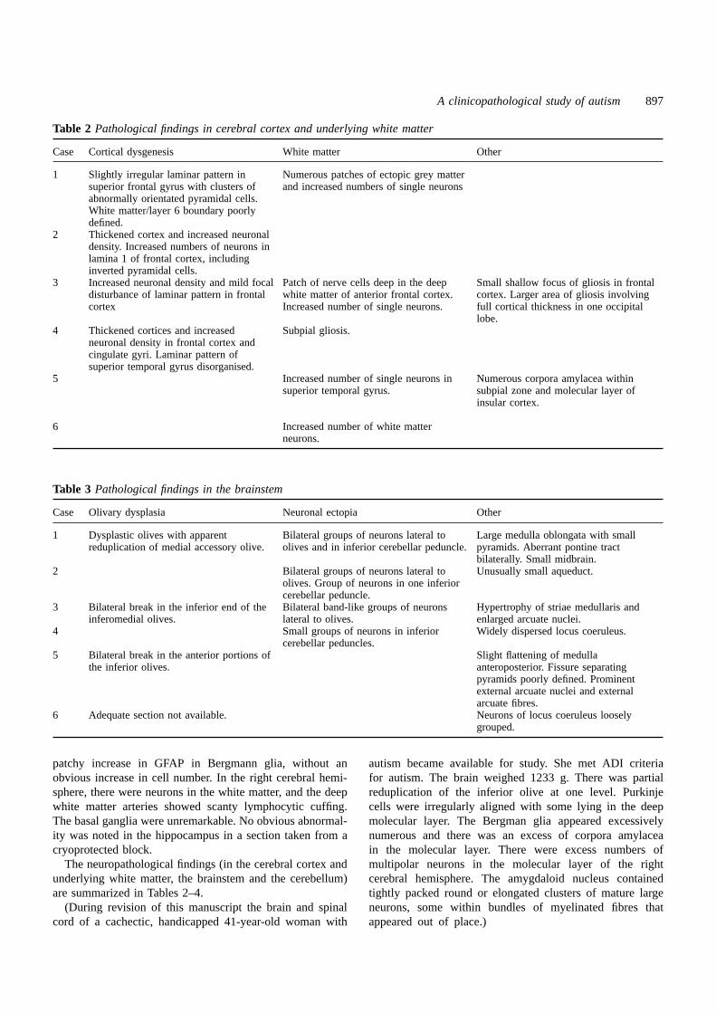
Table 1 Brain weight of six mentally handicapped autistic males
Case Age Brain Normal range* Weight of brainstem Ratio of total brain to(years) weight (kg) (kg) and cerebellum (kg) brainstem and
cerebellar weights
1 4 1.53 1.25–1.35 0.15 10.2 : 12 23 1.60 1.39–1.49 0.19 7.6 : 13 27 1.45 1.39–1.49 0.21 7.6 : 14 24 1.81 1.39–1.49 0.22 8.2 : 15 20 1.41 1.39–1.49 0.21 6.6 : 16 24 1.82 1.39–1.49 0.23 7.9 : 1
*Normal ranges given as mean6 2.5 SD (from Dekaban and Sadowsky, 1978).
embedding, routine staining and examination. Where pos-sible, the cerebral cortex, hippocampus and cerebellum werecompared histologically with material from identically pro-cessed age- and sex-matched control subjects.
Immunohistochemistry for glial fibrillary acidic protein(GFAP; DAKO, 1 : 1600) and phosphorylated neurofilaments(RT97; Courtesy of BH Anderton, 1 : 100) was performedusing the avidin–biotin complex method (DAKO) withdiaminobenzidine as the chromogen.
Morphometric studiesAs a supplement to subjective assessment, limited morphome-try was undertaken. Neuronal counts were performed onsections from three areas: (i) the medial aspect of the superiorfrontal gyrus at the level of the corpus callosum; (ii) theCA1, CA3 and CA4 sub-fields of the hippocampus (as closeas possible to the lateral geniculate body); and (iii) thePurkinje cell layer of the superior aspect of the cerebellarhemisphere.
Sections cut at ~14µm thickness were stained with cresylviolet and examined with a340 objective (310 eyepiece).Neurons were identified on the basis of classical morpholo-gical criteria. For neocortical counts, successive fields, asdefined by a rectangular eyepiece graticule were countedfrom pia to white matter. Neurons whose nucleus lay oneither of two adjacent borders of the field boundary (forbiddenlines) were excluded. Neurons were considered to lie withinthe thickness of the section if the mid-point of the nucleus,as defined by a sharply focussed nuclear outline, was present(Everallet al., 1991). All section thicknesses were measuredby focusing from the top to the bottom of the section with a3100 oil immersion objective and measuring the distancethe microscope stage travelled with a microcator. The productof field size and section thickness provided the referencevolume.
In the hippocampus, a single340 field of CA1, CA3 andCA4 was counted as described above. The number ofnucleolated Purkinje cells was counted and the length of thecounted Purkinje cell layer was measured using an IBAS2000 Kontron image analyser. The product of this length andsection thickness yielded a reference area. Purkinje cell
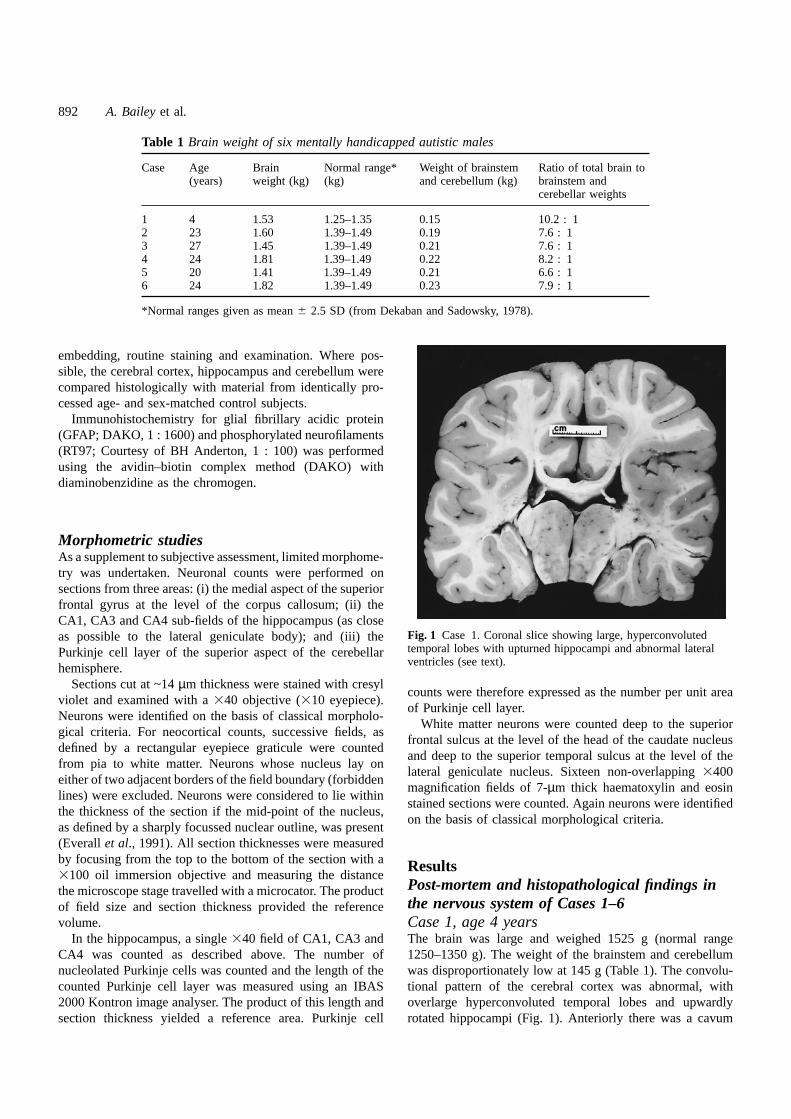
Fig. 1 Case 1. Coronal slice showing large, hyperconvolutedtemporal lobes with upturned hippocampi and abnormal lateralventricles (see text).
counts were therefore expressed as the number per unit areaof Purkinje cell layer.
White matter neurons were counted deep to the superiorfrontal sulcus at the level of the head of the caudate nucleusand deep to the superior temporal sulcus at the level of thelateral geniculate nucleus. Sixteen non-overlapping3400magnification fields of 7-µm thick haematoxylin and eosinstained sections were counted. Again neurons were identifiedon the basis of classical morphological criteria.
ResultsPost-mortem and histopathological findings inthe nervous system of Cases 1–6Case 1, age 4 yearsThe brain was large and weighed 1525 g (normal range1250–1350 g). The weight of the brainstem and cerebellumwas disproportionately low at 145 g (Table 1). The convolu-tional pattern of the cerebral cortex was abnormal, withoverlarge hyperconvoluted temporal lobes and upwardlyrotated hippocampi (Fig. 1). Anteriorly there was a cavum
A clinicopathological study of autism 893
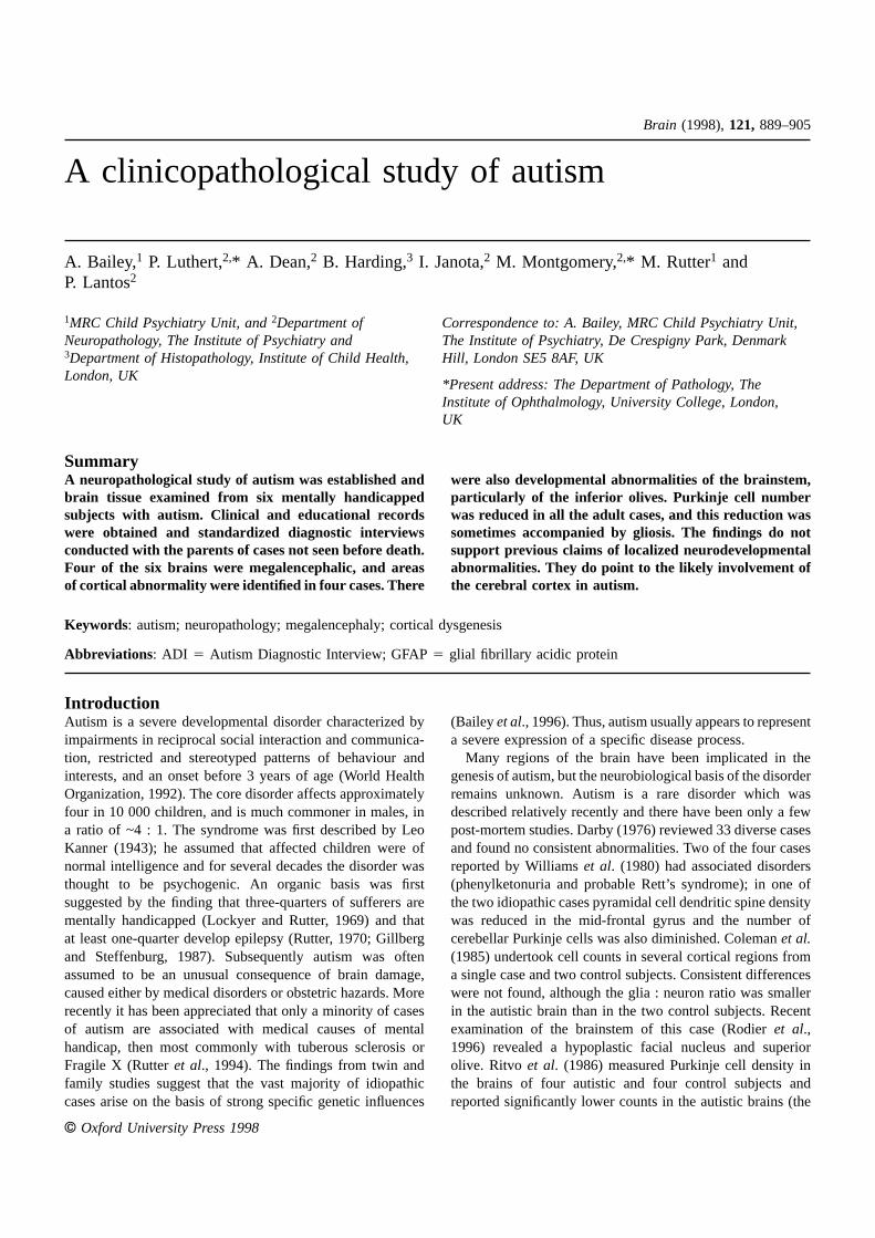
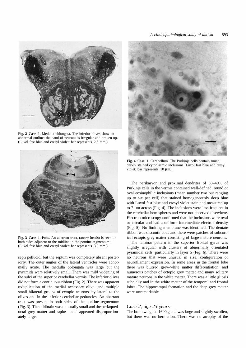
Fig. 2 Case 1. Medulla oblongata. The inferior olives show anabnormal outline; the band of neurons is irregular and broken up.(Luxol fast blue and cresyl violet; bar represents 2.5 mm.)
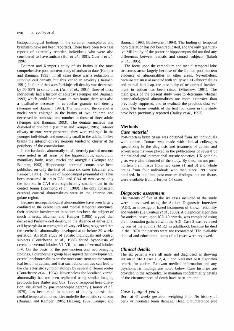
Fig. 3 Case 1. Pons. An aberrant tract, (arrow heads) is seen onboth sides adjacent to the midline in the pontine tegmentum.(Luxol fast blue and cresyl violet; bar represents 3.0 mm.)
septi pellucidi but the septum was completely absent poster-iorly. The outer angles of the lateral ventricles were abnor-mally acute. The medulla oblongata was large but thepyramids were relatively small. There was mild widening ofthe sulci of the superior cerebellar vermis. The inferior olivesdid not form a continuous ribbon (Fig. 2). There was apparentreduplication of the medial accessory olive, and multiplesmall bilateral groups of ectopic neurons lay lateral to theolives and in the inferior cerebellar peduncles. An aberranttract was present in both sides of the pontine tegmentum(Fig. 3). The midbrain was unusually small and the periaqued-uctal grey matter and raphe nuclei appeared disproportion-ately large.
Fig. 4 Case 1. Cerebellum. The Purkinje cells contain round,darkly stained cytoplasmic inclusions (Luxol fast blue and cresylviolet; bar represents 10µm.)
The perikaryon and proximal dendrites of 30–40% ofPurkinje cells in the vermis contained well-defined, round oroval eosinophilic inclusions (mean number two but rangingup to six per cell) that stained homogeneously deep bluewith Luxol fast blue and cresyl violet stain and measured upto 7 µm across (Fig. 4). The inclusions were less frequent inthe cerebellar hemispheres and were not observed elsewhere.Electron microscopy confirmed that the inclusions were ovalor circular and had a uniform intermediate electron density(Fig. 5). No limiting membrane was identified. The dentateribbon was discontinuous and there were patches of subcort-ical ectopic grey matter consisting of large mature neurons.
The laminar pattern in the superior frontal gyrus wasslightly irregular with clusters of abnormally orientatedpyramidal cells, particularly in layer 5 (Fig. 6). There wereno neurons that were unusual in size, configuration orneurofilament expression. In some areas in the frontal lobethere was blurred grey–white matter differentiation, andnumerous patches of ectopic grey matter and many solitarymature neurons in the white matter. There was a little gliosissubpially and in the white matter of the temporal and frontallobes. The hippocampal formation and the deep grey matterwere unremarkable.
Case 2, age 23 yearsThe brain weighed 1600 g and was large and slightly swollen,but there was no herniation. There was no atrophy of the

894 A. Baileyet al.
Fig. 5 Case 1. Cerebellum. The Purkinje cell at the top of the figure contains, on the right, a well-defined, homogeneous, moderatelyelectron-lucent inclusion. Note three granule cell neurons at the bottom of the figure. (Electron micrograph; bar represents 5µm.)
Fig. 6 Case 1. Lamina 5 of the superior frontal gyrus showingirregular orientation of pyramidal neurons. The top of the figure isclosest to the pia. (Luxol fast blue and cresyl violet; barrepresents 40µm.)
cerebellar vermis or hemisphere. The inferior olivary ribbonwas normally formed, but there were groups of ectopicneurons lateral to both olives. There was also a group ofectopic neurons in an inferior cerebellar peduncle adjacentto the brainstem. The cross-sectional area of the lower endof the aqueduct at the midbrain–pons junction was unusuallysmall (0.2 mm2). In the cerebellum the number of Purkinjecells was reduced, more so in the hemisphere than vermis,but no empty baskets were seen. There was an increase inGFAP in Bergmann glia. The parietal, frontal and cingulatecortices were thickened in the right cerebral hemisphere, aswas the cortex of the superior and middle temporal gyri. Thecortex was also unusually cellular. There was an increased
Fig. 7 Case 2. Layer 1 of frontal cortex. Note the increasednumber of neurons and, in particular, the misorientated pyramidalcell just beneath the pial surface (top). The bar represents 100µm and lies just above the junction between layers I and II.(Luxol fast blue and cresyl violet.)
number of small neurons in layer 1 of frontal cortex includingsome inverted pyramidal cells (Fig. 7); these were normalin configuration and neurofilament expression. The corpuscallosum was thin, measuring 2.5 mm in thickness justposterior to the genu. Neuronal density appeared to be
A clinicopathological study of autism 895
Fig. 8 Case 3. Medulla oblongata. The striae medullares are hypertrophied and the arcuate nuclei appear larger than usual. Compare theregular configuration of the inferior olives with the abnormal outline in Fig. 2. (Luxol fast blue and cresyl violet; bar represents 2 mm.)
increased in the hippocampus. The deep grey nuclei wereunremarkable. Electron microscopy of the frontal cortexrevealed moderately well-preserved tissue. No abnormalitiesof synapses, mitochondria, lysosomes or other organelleswere identified.
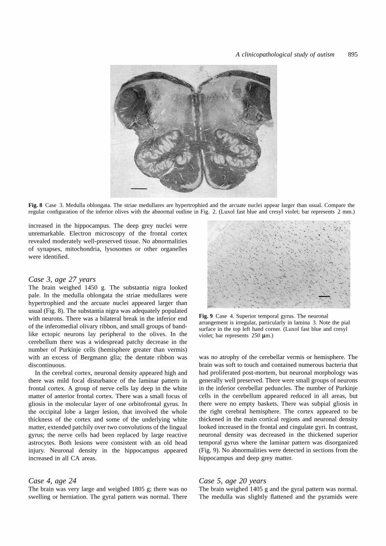
Case 3, age 27 yearsThe brain weighed 1450 g. The substantia nigra lookedpale. In the medulla oblongata the striae medullares werehypertrophied and the arcuate nuclei appeared larger thanusual (Fig. 8). The substantia nigra was adequately populatedwith neurons. There was a bilateral break in the inferior endof the inferomedial olivary ribbon, and small groups of band-like ectopic neurons lay peripheral to the olives. In thecerebellum there was a widespread patchy decrease in thenumber of Purkinje cells (hemisphere greater than vermis)with an excess of Bergmann glia; the dentate ribbon wasdiscontinuous.
In the cerebral cortex, neuronal density appeared high andthere was mild focal disturbance of the laminar pattern infrontal cortex. A group of nerve cells lay deep in the whitematter of anterior frontal cortex. There was a small focus ofgliosis in the molecular layer of one orbitofrontal gyrus. Inthe occipital lobe a larger lesion, that involved the wholethickness of the cortex and some of the underlying whitematter, extended patchily over two convolutions of the lingualgyrus; the nerve cells had been replaced by large reactiveastrocytes. Both lesions were consistent with an old headinjury. Neuronal density in the hippocampus appearedincreased in all CA areas.
Case 4, age 24The brain was very large and weighed 1805 g; there was noswelling or herniation. The gyral pattern was normal. There
Fig. 9 Case 4. Superior temporal gyrus. The neuronalarrangement is irregular, particularly in lamina 3. Note the pialsurface in the top left hand corner. (Luxol fast blue and cresylviolet; bar represents 250µm.)
was no atrophy of the cerebellar vermis or hemisphere. Thebrain was soft to touch and contained numerous bacteria thathad proliferated post-mortem, but neuronal morphology wasgenerally well preserved. There were small groups of neuronsin the inferior cerebellar peduncles. The number of Purkinjecells in the cerebellum appeared reduced in all areas, butthere were no empty baskets. There was subpial gliosis inthe right cerebral hemisphere. The cortex appeared to bethickened in the main cortical regions and neuronal densitylooked increased in the frontal and cingulate gyri. In contrast,neuronal density was decreased in the thickened superiortemporal gyrus where the laminar pattern was disorganized(Fig. 9). No abnormalities were detected in sections from thehippocampus and deep grey matter.
Case 5, age 20 yearsThe brain weighed 1405 g and the gyral pattern was normal.The medulla was slightly flattened and the pyramids were

896 A. Baileyet al.
Fig. 10 Case 5. Medulla Oblongata. The medulla is slightly flattened and the pyramids poorly demarcated from each other (Luxol fastblue and cresyl violet, magnification bar represents 2 mm.)
Fig. 11 Case 5. Cerebellar hemisphere. There is a reducednumber of Purkinje cells and a mild increase in Bergmann glia.(Haematoxylin and eosin; bar represents 100µm.)
poorly demarcated from each other (Fig. 10). There was noatrophy of the cerebellar vermis or hemisphere. Histologically,the external arcuate nuclei and the external arcuate fibreswere prominent. The anterior portions of the inferior olivaryribbons were interrupted and asymmetrically bilaterallyattenuated. In the cerebellum there was a diffuse decrease inPurkinje cell density in the hemisphere and vermis (Fig. 11).There was a slight increase in Bergmann glia number and amoderate patchy increase in GFAP staining in the molecularlayer. In the left cerebral hemisphere the leptomeningesshowed sparse perivascular lymphocytic cuffs, and there waswidespread capillary engorgement in the grey matter. Thewhite matter of the superior temporal gyrus contained manyscattered mature neurons (Fig. 12). There were numerouscorpora amylacea within the subpial zone and molecular
Fig. 12 Case 5. White matter in the superior temporal gyrus. Thedensity of neurons is 40/mm2 in this field. (Haematoxylin andeosin; bar represents 50µm.)
layer of the insular cortex. The striatum and internal capsulecontained a few lymphocytic cuffs. The hippocampus andparahippocampal gyrus were unremarkable.
Case 6, age 24 yearsThe brain was unusually large, swollen and weighed 1820 g,but there was no herniation. The gyral pattern was normal.The medulla oblongata had not been completely removed.There was no atrophy of the cerebellar vermis or hemisphere.There was no histological evidence of oedema. The neuronsof the locus coeruleus were loosely grouped and there wasa slight reduction in the number of nigral neurons. In thecerebellum there was a moderate reduction in the number ofPurkinje cells in the hemisphere and vermis, and moderate
A clinicopathological study of autism 897
Table 2 Pathological findings in cerebral cortex and underlying white matter
Case Cortical dysgenesis White matter Other
1 Slightly irregular laminar pattern in Numerous patches of ectopic grey mattersuperior frontal gyrus with clusters of and increased numbers of single neuronsabnormally orientated pyramidal cells.White matter/layer 6 boundary poorlydefined.
2 Thickened cortex and increased neuronaldensity. Increased numbers of neurons inlamina 1 of frontal cortex, includinginverted pyramidal cells.
3 Increased neuronal density and mild focal Patch of nerve cells deep in the deep Small shallow focus of gliosis in frontaldisturbance of laminar pattern in frontal white matter of anterior frontal cortex. cortex. Larger area of gliosis involvingcortex Increased number of single neurons. full cortical thickness in one occipital
lobe.4 Thickened cortices and increased Subpial gliosis.
neuronal density in frontal cortex andcingulate gyri. Laminar pattern ofsuperior temporal gyrus disorganised.
5 Increased number of single neurons in Numerous corpora amylacea withinsuperior temporal gyrus. subpial zone and molecular layer of
insular cortex.
6 Increased number of white matterneurons.
Table 3 Pathological findings in the brainstem
Case Olivary dysplasia Neuronal ectopia Other
1 Dysplastic olives with apparent Bilateral groups of neurons lateral to Large medulla oblongata with smallreduplication of medial accessory olive. olives and in inferior cerebellar peduncle. pyramids. Aberrant pontine tract
bilaterally. Small midbrain.2 Bilateral groups of neurons lateral to Unusually small aqueduct.
olives. Group of neurons in one inferiorcerebellar peduncle.
3 Bilateral break in the inferior end of the Bilateral band-like groups of neurons Hypertrophy of striae medullaris andinferomedial olives. lateral to olives. enlarged arcuate nuclei.
4 Small groups of neurons in inferior Widely dispersed locus coeruleus.cerebellar peduncles.
5 Bilateral break in the anterior portions of Slight flattening of medullathe inferior olives. anteroposterior. Fissure separating
pyramids poorly defined. Prominentexternal arcuate nuclei and externalarcuate fibres.
6 Adequate section not available. Neurons of locus coeruleus looselygrouped.
patchy increase in GFAP in Bergmann glia, without anobvious increase in cell number. In the right cerebral hemi-sphere, there were neurons in the white matter, and the deepwhite matter arteries showed scanty lymphocytic cuffing.The basal ganglia were unremarkable. No obvious abnormal-ity was noted in the hippocampus in a section taken from acryoprotected block.
The neuropathological findings (in the cerebral cortex andunderlying white matter, the brainstem and the cerebellum)are summarized in Tables 2–4.
(During revision of this manuscript the brain and spinalcord of a cachectic, handicapped 41-year-old woman with
autism became available for study. She met ADI criteriafor autism. The brain weighed 1233 g. There was partialreduplication of the inferior olive at one level. Purkinjecells were irregularly aligned with some lying in the deepmolecular layer. The Bergman glia appeared excessivelynumerous and there was an excess of corpora amylaceain the molecular layer. There were excess numbers ofmultipolar neurons in the molecular layer of the rightcerebral hemisphere. The amygdaloid nucleus containedtightly packed round or elongated clusters of mature largeneurons, some within bundles of myelinated fibres thatappeared out of place.)

898 A. Baileyet al.
Table 4 Pathological findings in the cerebellum
Case Purkinje cell layer Other
1 Cytoplasmic inclusions in Purkinje cells in the vermis and Break in dentate ribbon. Patches of subcortical ectopic greyhemisphere. matter.
2 Decreased numbers of Purkinje cells and increase in GFAP inBergmann glia.
3 Patchy decrease in numbers of Purkinje cells and proliferation Break in dentate ribbon.of Bergmann glia.
4 Decreased numbers of Purkinje cells.5 Decreased number of Purkinje cells. Areas of Bergmann glia
proliferation and moderate patchy increase in GFAP staining.6 Decreased numbers of Purkinje cells and moderate patchy
increase in GFAP in Bergmann glia.
Table 5 Neuronal density (10–2 3 n/mm3) in superiorfrontal gyrus
Case Autistic subjects Control subjects
Age Brain Count Age Brain Count(years) (kg) (years) (kg)
2 22 1.60 330 20 NA 2594 24 1.81 194 24 1.50 3115 20 1.40 144 40 1.47 1806 24 1.82 216 43 1.52 226IOP average† (221)* (244)3 27 1.45 365 35 NA 3421 4 1.53 336 4 1.18‡ 373
6 1.23‡ 395
NA 5 not available.†Figures in brackets are averages forInstitute of Psychiatry processed cases.‡Female. *ExactP 50.56, Mann–WhitneyU, Wilcoxon rank sum test.
MorphometryAmongst the normal control subjects there was considerablevariation in neuronal counts in the frontal cortex andhippocampal sub-fields, but more consistency in the Purkinjecell counts. The range of frontal counts in the brains ofautistic individuals was similar to that in the control subjects,although the count in Case 5, who had severe epilepsy, fellbelow the control range (Table 5). There were no consistentdifferences between autistic and control subjects in neuronalcounts across the different hippocampal sub-fields, althoughCase 3 had elevated counts in all CA fields compared withhis control (Table 6). Purkinje cell counts were consistentlylower in the adult autistic cases than in control subjects(Table 7). Statistical comparison of neuronal densitiesbetween control and autistic cases from the Institute ofPsychiatry (Mann–WhitneyU, Wilcoxon rank sum test) onlyrevealed significant differences in the density of Purkinjecells (exactP 5 0.02). White matter neuronal densities in theautistic brains were unremarkable in the areas systematicallycounted (data not shown).
Post-mortem findings in cases where tissue wasunavailable for studyPost-mortem findings, but not tissue, were available for afurther 14 individuals with a clinical diagnosis of autism.Brain weights were recorded in four young adult males(1530, 1450, 1400 and 1300 g), one young adult female(1400 g) and one 16-year-old female (1330 g). Eleven brainswere reportedly macroscopically normal. The meninges werethickened and adherent in one case with a history of meningitisin infancy. In the brain of the 16-year-old girl the corticalribbon was narrower than expected and subcortical whitematter was reduced in amount. The substantia nigra wasrather poorly pigmented. Microscopically, the cortex wasdescribed as well populated by nerve cells. No microscopicreports were available on any other brains.
DiscussionThe identified neuropathology in this series is already moreextensive than previously reported. The findings includedincreased brain size and developmental abnormalities of thecerebral cortex, brainstem and cerebellum; in some casesthere was also secondary pathology. Claims of consistentlyelevated neuronal density in the hippocampus have not beenreplicated. It seems unlikely that misdiagnosis is responsiblefor any discrepancies with previous findings. Although onlyone individual was seen during life (Case 3), all of the caseshad received a clinical diagnosis of autism. In addition casenotes were reviewed after death and the parents interviewedusing the ADI (Le Couteuret al., 1989). Nevertheless,because the cases in this study were all mentally handicapped,the applicability of the findings to more able individuals isuncertain. The heterogeneity of autism is emphasized byCase 1, which stands out because of the severe early motordifficulties, the severity of the malformations and the presenceof Purkinje cell inclusions.
Four brains were large with little evidence of significantoedema. In two cases macrocephaly was also noted duringchildhood. In Case 1 the temporal lobes were enlarged andhyperconvoluted, but there were no gyral abnormalities

A clinicopathological study of autism 899
Table 6 Neuronal density (10–2 3 n/mm3) in areas CA1, CA3 and CA4 of the hippocampus
Case Autistic subjects Control subjects
Age (years) CA1 CA3 CA4 Age (years) CA1 CA3 CA4
2 22 231 156 104 20 167 164 924 24 239 168 103 24 145 236 995 20 189 208 98 26 186 229 149
40 197 266 14543 216 231 93
IOP average† (220)* (177)* (102)(* ) (182) (225) (116)3 27 334 247 154 35 215 180 1241 4 306 279 306 4 298 322 279
6 212 237 162
†Figures in brackets are averages for Institute of Psychiatry processed cases. *ExactP 5 0.10 and(* )exactP 5 0.56, Mann–WhitneyU, Wilcoxon rank sum test.
Table 7 Purkinje cell densities (n/mm2) of Purkinje celllayer of the cerebellum
Case Autistic subjects Control subjects
Age Count Age Count(years) (n/mm2) (years) (n/mm2)
2 22 169 20 2204 24 196 26 2415 20 128 40 2206 24 160 43 268IOP average† (163)* (237)3‡ 27 1311 4 251 4 231
†Figures in brackets are averages for Institute of Psychiatryprocessed cases.‡No identically processed cerebellar tissue froma young male control was available. *ExactP 5 0.02, Mann–Whitney U, Wilcoxon rank sum test.
in the other cases. There were also several instances ofmacroscopically abnormal development of the brainstem, andof the corpus callosum in one brain.
There was microscopic pathology in the cerebral hemi-spheres, cortex and cerebellum. Abnormalities in corticaldevelopment were seen in individual cases, including: areasof increased cortical thickness, high neuronal density, neuronsin the molecular layer, neuronal disorganization, poor differ-entiation of the grey–white matter boundary, neuronal hetero-topias and focally increased numbers of single neurons inthe white matter (Table 2). In the brainstem (Table 3), theinferior olives were malformed in three brains and ectopicneurons related to the olivary complex were seen in a furthertwo. Olivary dysplasia was associated with enlarged arcuatenuclei in two cases. In one brain there was also a subtleabnormality of the locus coeruleus. Purkinje cell density wasdecreased in all the adult cases, but inclusions were seenonly in Case 1. In two cases there were minor developmentalcerebellar abnormalities.
Hippocampal neuronal density looked relatively high intwo cases, but only in Case 3 (Table 6) was there any
evidence of increased density in all CA subfields. There wasno evidence of a statistically significant increase in celldensity in the cases processed at the Institute of Psychiatrycompared with control subjects (Table 6); however, samplingand number of cases were limited. There was no hippocampalsclerosis or other pathology in any case. Examination of theamygdala has been limited by tissue sampling for neurochemi-stry but, with the exception of the most recent case, noabnormalities were identified.
In several brains there was also evidence of acquiredpathology. The number of Bergmann glia was increased inthree cases and there was also increased staining for GFAP,but no empty baskets were seen (the presence of groups ofbasket cell axons in the absence of the Purkinje cell perikaryonthat they normally ensheath is generally interpreted as evid-ence for acquired Purkinje cell loss). Cerebral subpial gliosiswas observed in two cases. There were increased numbersof corpora amylacea in the insular cortex of Case 5 (and inthe molecular layer of the cerebellum of the most recentlyidentified case). The two areas of cortical gliosis in Case 3were probably a consequence of the head injury in childhood.
Comparison with previous studiesThe most striking contrast with previous findings is that fourof the six brains were unusually large and heavy, althoughmegalencephaly was uncommon amongst the cases unavail-able for study. Brain weights were not reported by Ritvoet al. (1986) or by Kemper and Bauman (1993); however, inthis latter series they were apparently 100–200 g heavierthan expected in most subjects aged,12 years, but 100–200 g lighter than expected in the majority of adult subjects(Bauman, 1996). One of the cases in Darby’s literature review(1976) had a heavy brain, weighing 1550 g at 5 years of age(case 11 in that review); the brain of one of the two idiopathiccases described by Williamset al. (1980) weighed 1520 g(their case 1), and the other weighed 1430 g (their case 3);and the female described by Colemanet al. (1985) had abrain weight of 1380 g (Rodieret al., 1996). There are two
900 A. Baileyet al.
reports of autistic individuals with unusually small brains;one was associated with premature closure of the cerebralsutures and severe self injury (Hofet al., 1991); the otherwas also profoundly handicapped and physically disabled(Guerin et al., 1996). The association between autism andincreased brain weight contrasts with the finding of microce-phaly in many cases of mental handicap unaccompanied byautism (see for instance Coleet al., 1994).
There is some convergent evidence for increased brainsize in a proportion of individuals with autism. Three MRIstudies have found increased brain volume in child and adultsubjects. Filipeket al. (1992) reported increased brain volumein autistic children compared with normal subjects, develop-mental language disorder and non-autistic mentally handi-capped control subjects. Pivenet al. (1995, 1996) foundincreased total brain volume in adolescents and adults com-pared with normal control subjects. Increased head circumfer-ence in autistic individuals has been noted in several differentsamples (Hauseret al., 1975; Boltonet al., 1994; Baileyet al., 1995, Woodhouseet al., 1996; Lainhartet al., 1997).Together these data suggest that the finding of megalencephaly(brain weight. 2.5 SD above the mean) is in keeping witha tendency towards increased brain size in some adults andchildren with autism.
Cortical dysgenetic lesions have not been a prominentfeature of previous studies. They were not highlighted byKemper and Bauman (1993), with the exception that anteriorcingulate cortex was consistently unusually coarse and poorlylaminated, and associated with increased cell packing densityin one case (Kemper, 1988). Polymicrogyria has been seenin two post-mortem cases (Ritvoet al., 1986; Kemper, 1988)and several MRI studies have observed developmental corticalabnormalities in a small proportion of patients (Gaffney andTsai, 1987; Pivenet al., 1990; Schifteret al., 1994).
In this study there was no significant cerebellar atrophyor apparent granule cell loss; neuronal size in the dentatenucleus and olive appeared unremarkable; and there was notendency for olivary cells to cluster at the periphery of theconvolutions. The extent of the facial nuclei has not yet beenassessed. There was, however, clear evidence of develop-mental brainstem abnormalities that have not been reportedpreviously (Table 3). The MRI findings in the brainstem inautism are largely contradictory, probably reflecting method-ological differences and the small size of the relevantstructures.
Possible mechanismsPrimary megalencephaly may be associated with increasedcell number and increased cell size. Frontal cortical neuronaldensity appeared to be increased in three cases—includingin some areas of thickened cortex—but this is not evident inthe limited morphometry. The wide variation in corticalneuronal density in control subjects, and the possibilityof cortical neuronal loss secondary to epilepsy, limit theconclusions that can presently be drawn. Nevertheless, there
is no evidence of substantially decreased neuronal density inthe megalencephalic brains, suggesting that raised total cellnumber may contribute to brain enlargement. Increased cellreplication and impaired developmental cell death might bothlead to an excess of neurons. Programmed cell death is welldocumented in mammalian postnatal cerebral cortex, but alsoaffects a significant proportion of cells in proliferative and,to a lesser extent, postmitotic regions of murine foetal cerebralcortex (Blaschkeet al., 1996).
Different cortical dysgenetic lesions occurred either aloneor in combination, but there was no evidence of neuronalcytomegaly, abnormal neuronal configuration or abnormalneurofilament expression. Although focal increases in whitematter neuronal density were seen (Table 2 and Fig. 12),limited morphometry did not reveal a generalized increase.The co-occurrence of different patterns of dysgenesis is notuncommon (Prayson and Estes, 1995), and the findingssuggest that there may be abnormalities in cortical neuronalproliferation, migration and programmed cell death (Rorke,1994; Mischelet al., 1995).
Evidence of abnormal neuronal migration, and possiblyabnormal control of cell number, was also found in thebrainstem and cerebellum. Whether shared mechanismsunderlie the cortical and brainstem findings is unclear. Never-theless olivary anomalies seldom occur in isolation andheterotopias are usually associated with cortical develop-mental abnormalities, particularly megalencephaly, pachygy-ria and lissencephaly (Harding and Copp, 1997). Olivarydevelopment involves long distance migration from theprimary precerebellar neuropepithelium, which is also thesource of cells forming the arcuate nuclei and basis pontis(Essick, 1912); the cells forming the dentate nucleus arisefrom the superior portion of the rhombic lip. A tendency forinferior olivary neurons to cluster at the periphery of theconvolutions was not identified, but Kemper and Bauman’s(1993) observation may be related to the abnormalities thatwere observed in this series. In a case of Coffin–Sirissyndrome (DeBassioet al., 1985), peripheral clustering ofolivary neurons occurred in association with islands of ectopicolivary neurons, a large medial accessory olive, unusuallylarge arcuate nuclei and ectopic neurons in the white matterof the cerebellum at the level of the dentate nucleus.
There is only limited evidence, so far, that abnormaldevelopment also affects more rostral brainstem structures.An aberrant pontine tract was identified in Case 1 and therewas mild disorganization of the locus coeruleus in Case 6.Kemper and Bauman (1993) observed disorganization of thenucleus locus coeruleus and the nucleus raphe dorsalis inone brain and, in another case, the centrally placed neuronsof the basis pontis were apparently more densely packed andenlarged compared with control subjects (Kemper, 1988).
Decreased Purkinje cell density is a relatively consistentobservation across the post-mortem studies, although inseveral cases in this series many stretches of folia were wellpopulated with Purkinje cells. The frequency of develop-mental medullary abnormalities indirectly supports the hypo-
A clinicopathological study of autism 901
thesis that the Purkinje cell findings have a developmentalbasis. Kemper and Bauman (1993) argued that, in the absenceof glial cell hyperplasia or retrograde olivary cell loss,decreased Purkinje cell density pointed to a loss occurringat, or before, 30 weeks gestation. Nevertheless, if substantialPurkinje cell loss occurs only early in development, then theapparently normal development of the cerebellar cortex isslightly puzzling. Purkinje cells play a central role in normalcerebellar development and they control proliferation of cellsin the murine external granule layer (Feddersenet al., 1992;Smeyneet al., 1995). Postmitotic external granule layer cellsmigrate to form the internal granule layer (IGL), whichincreases in thickness in man from the 5–6th prenatal monthto the 4–5th postnatal month (Raaf and Kernohan, 1944),being very thin until 32 weeks gestation (Friede, 1973).Consequently, any substantial loss of Purkinje cells prior to32 weeks gestation could be associated with hypoplasticcerebellar folia. The modest patchy glial cell hyperplasiaseen in this series raises the additional possibility of postnatalloss of Purkinje cells, perhaps related to epilepsy (although,as yet, no empty baskets have been identified). Whilst olivarygliosis has not been observed, moderate loss of olivary cellsmay be difficult to identify, as the cells normally show greatvariation in density and lie relatively far apart (Brodal, 1940).The Purkinje cell inclusions seen in the only child in thisseries add a further complication. Such inclusions have notpreviously been reported and their aetiology is unknown.Purkinje cell density was unremarkable in this case, but weare not aware of any precedent for the complete disappearanceof inclusion-bearing cells.
In summary, developmental neuropathology was not local-ized to the limbic system, cerebellum (Kemper and Bauman,1993), or derivatives of a single hindbrain rhombomere(Rodieret al., 1996). Although there is evidence of abnormalneuronal migration, other factors influencing neuronal num-ber, survival and orientation also seem to be important. Thereis clearly a need for further study of the pathological basisof increased brain size. Of course, some developmentalpathology may be either a consequence of maldevelopmentat remote sites, or epiphenomena of more fundamentalabnormalities. Identifying the genes predisposing to autismmay help to clarify these relationships. With regard to timing,it would be premature to conclude that a single developmentalevent led to these findings. Nevertheless, abnormal develop-ment had sometimes begun by the time of olivary cellmigration, which occurs before the end of the 3rd month.
Relationship to symptomatologyExplanations of developmental cognitive and behaviouraldysfunction by neuropathology are precarious, as brain func-tion may not be impaired and inferences based upon localiz-ation in adults may not be pertinent. As yet, no singlepathology common to all cases of autism has been identified.Of course, autism is a complex behavioural disorder and it
would be too much to expect such specificity at this levelof analysis.
The finding of cortical dysgenetic lesions and megalence-phaly suggests that cerebral dysfunction may underlie somecognitive and behavioural abnormalities, and may providethe pathological substrate of epilepsy. The inconsistency ofthe neuropathological findings indicates, however, that theyare probably imperfect markers of abnormal cortical develop-ment and organization. Whilst brainstem abnormalities mightcause neurological impairments (Rodieret al., 1996), theyseem unlikely to lead directly to high level cognitive deficits,but possibly both types of impairments can occur in moreseverely affected individuals. No consistent hippocampalabnormalities were identified in this series and systematicexamination of the amygdala, medial septal nuclei, mamillarybodies and related structures has yet to be undertaken.Involvement of the amygdala remains an important possibil-ity, but the evidence of cortical and brainstem maldevelop-ment has removed the imperative to argue that all autisticsymptomatology arises from medial temporal and relatedstructures. The relationship of cerebellar abnormalities tosymptomatology (if any) remains uncertain. Although cere-bellar maldevelopment may prove to be a marker of thedisease process, behavioural consequences remain hypothet-ical until cerebellar dysfunction has been demonstrated unam-biguously. In summary, in this study we have found noevidence for a highly localized pathology that seems likelyto underlie autism. Instead, the findings raise the possibilitythat a combination of diverse, but related, neurodevelopmentalabnormalities give rise to the characteristic symptomatology,and the associated mental handicap and epilepsy.
AcknowledgementsWe are deeply indebted to all the parents and professionalswho have helped with this study. We wish to thank AndrewChadwick and Nigel Cairns of the MRC London DegenerativeBrain Bank, Department of Neuropathology, for technicalsupport; and Linda Wilkinson, Marion Gosden and DeborahGomer for secretarial help.
ReferencesArin DM, Bauman ML, Kemper TL. The distribution of Purkinjecell loss in the cerebellum in autism [abstract]. Neurology 1991;41 Suppl 1: 307.
Bachevalier J. Medial temporal lobe structures and autism: a reviewof clinical and experimental findings. [Review]. Neuropsychologia1994; 32: 627–48.
Bailey A, Cox T. Neuroimaging in child and developmental psychi-atry. In: Lewis S, Higgins JP, editors. Brain imaging in psychiatry.Oxford: Blackwell Scientific, 1996: 301–15.
Bailey A, Luthert P, Bolton P, Le Couteur A, Rutter M, Harding B.Autism and megalencephaly [letter]. Lancet 1993; 341: 1225–6.
Bailey A, Le Couteur A, Gottesman I, Bolton P, Simonoff E, Yuzda
902 A. Baileyet al.
E, et al. Autism as a strongly genetic disorder: evidence from aBritish twin study. Psychol Med 1995; 25: 63–77.
Bailey A, Phillips W, Rutter M. Autism: towards an integration ofclinical, genetic, neuropsychological, and neurobiological perspect-ives. [Review]. J Child Psychol Psychiatry 1996; 37: 89–126.
Bauman ML. Microscopic neuroanatomic abnormalities in autism.[Review]. Pediatrics 1991; 87 S Pt 2: 791–6.
Bauman ML. Neuroanatomic observations of the brain in pervasivedevelopmental disorders. [Review]. J Autism Dev Disord 1996; 26:199–203.
Bauman ML, Kemper TL. Histoanatomic observations of the brainin early infantile autism. Neurology 1985; 35: 866–74.
Blaschke A, Staley K, Chun J. Widespread programmed cell deathin proliferative and postmitotic regions of the fetal cerebral cortex.Development 1996; 122: 1165–74.
Bolton P, Macdonald H, Pickles A, Rios P, Goode S, Crowson M,et al. A case-control family history study of autism. J Child PsycholPsychiatry 1994; 35: 877–900.
Brodal A. Modification of Gudden method for study of cerebrallocalization. Arch Neurol Psychiatry 1940; 43: 46–58.
Cole G, Neal JW, Fraser WI, Cowie VA. Autopsy findings inpatients with mental handicap. J Intellect Disabil Res 1994; 38: 9–26.
Coleman PD, Romano J, Lapham L, Simon W. Cell counts incerebral cortex of an autistic patient. J Autism Dev Disord 1985;15: 245–55.
Courchesne E, Yeung-Courchesne R, Press GA, Hesselink JR,Jernigan TL. Hypoplasia of cerebellar vermal lobules VI and VIIin autism. N Engl J Med 1988; 318: 1349–54.
Courchesne C, Townsend J, Saitoh O. The brain in infantile autism:posterior fossa structures are abnormal [see comments]. Neurology1994; 44: 214–23. Comment in: Neurology 1994; 44: 203–8,Comment in: Neurology 1995; 45: 398–402.
Darby JK. Neuropathologic aspects of psychosis in children.J Autism Child Schizophr 1976; 6: 339–52.
DeBassio WA, Kemper TL, Knoefel JE. Coffin-Siris syndrome:neuropathologic findings. Arch Neurol 1985; 42: 350–3.
Dekaban AS, Sadowsky, D. Changes in brain weights during thespan of human life: relation of brain weights to body heights andbody weights. Ann Neurol 1978; 4: 345–56.
DeLong GR. Autism, amnesia, hippocampus, and learning.[Review]. Neurosci Biobehav Rev 1992; 16: 63–70.
Essick CR. The development of the nuclei pontis and the nucleusarcuatus in man. Am J Anat 1912: 25–54.
Everall IP, Luthert PJ, Lantos PL. Neuronal loss in the frontalcortex in HIV infection [see comments]. Lancet 1991; 337: 1119–21. Comment in: Lancet 1991; 338: 129–30.
Feddersen RM, Ehlenfeldt R, Yunis WS, Clark HB, Orr HT.Disrupted cerebellar cortical development and progressive degenera-tion of Purkinje cells in SV40 T antigen transgenic mice. Neuron1992; 9: 955–66.
Filipek PA, Richelme C, Kennedy DN, Rademacher J, Pitcher DA,
Zidel S, et al. Morphometric analysis of the brain in developmentallanguage disorders and autism [abstract]. Ann Neurol 1992; 32: 475.
Friede RL. Dating the development of human cerebellum. ActaNeuropathol (Berl) 1973; 23: 48–58.
Gaffney GR, Tsai LY. Magnetic resonance imaging of high levelautism. J Autism Dev Disord 1987; 17: 433–8.
Gillberg C, Steffenburg S. Outcome and prognostic factors ininfantile autism and similar conditions: a population-based study of46 cases followed through puberty. J Autism Dev Disord 1987; 17:273–87.
Guerin P, Lyon G, Barthelemy C, Sostak E, Chevrollier V, GarreauB, et al. Neuropathological study of a case of autistic syndromewith severe mental retardation. Dev Med Child Neurol 1996; 38:203–11.
Harding B, Copp AJ. Malformations. In: Graham DI, Lantos PL,editors. Greenfield’s neuropathology. 6th ed. London: Arnold, 1997:397–533.
Hauser S, DeLong GR, Rosman NP. Pneumographic findings in theinfantile autism syndrome: a correlation with temporal lobe disease.Brain 1975; 98: 363–88.
Hof PR, Knabe R, Bovier P, Bouras C. Neuropathological observa-tions in a case of autism presenting with self-injury behavior. ActaNeuropathol (Berl) 1991; 82: 321–6.
Kanner L. Autistic disturbances of affective contact. Nerv Child1943; 2: 217–50.
Kemper TL. Neuroanatomic studies of dyslexia and autism. In:Swann JW, Messer A, editors. Disorders of the developing nervoussystem: changing views on their origins, diagnoses and treatments.New York: Alan R. Liss, 1988: 125–54.
Kemper TL, Bauman ML. The contribution of neuropathologicstudies to the understanding of autism. Neurol Clin 1993; 11: 175–87.
Lainhart JE, Piven J, Wzorek M, Landa R, Santangelo SL, CoonH, et al. Macrocephaly in children and adults with autism. J AmAcad Child Adolesc Psychiatry 1997; 36: 282–90.
Le Couteur A, Rutter M, Lord C, Rios P, Robertson S, HoldgraferM, et al. Autism diagnostic interview: a standardized investigator-based instrument. J Autism Dev Disord 1989; 19: 363–87.
Lockyer L, Rutter M. A five- to fifteen-year follow-up study ofinfantile psychosis. Br J Psychiatry 1969; 115: 865–82.
Minshew NJ. Indices of neural function in autism: clinical andbiologic implications. [Review]. Pediatrics 1991; 87: 774–80.
Mischel PS, Nguyen LP, Vinters HV. Cerebral cortical dysplasiaassociated with pediatric epilepsy. Review of neuropathologic fea-tures and proposal for a grading system. [Review]. J NeuropatholExp Neurol 1995; 54: 137–53.
Piven J, Berthier ML, Starkstein SE, Nehme E, Pearlson G, FolsteinS. Magnetic resonance imaging evidence for a defect of cerebralcortical development in autism. Am J Psychiatry 1990; 147: 734–9.
Piven J, Arndt S, Bailey J, Havercamp S, Andreasen N, Palmer P.An MRI study of brain size in autism. Am J Psychiatry 1995; 152:1145–9.
Piven J, Arndt S, Bailey J, Andreasen N. Regional brain enlargement

A clinicopathological study of autism 903
in autism: a magnetic resonance imaging study. J Am Acad ChildAdolesc Psychiatry 1996; 35: 530–6.
Prayson RA, Estes ML. Cortical dysplasia: a histopathologic studyof 52 cases of partial lobectomy in patients with epilepsy. HumPathol 1995; 26: 493–500.
Raaf J, Kernohan JW. A study of the external granular layer in thecerebellum. Am J Anat 1944; 75: 151–72.
Raymond G, Bauman M, Kemper T. The hippocampus in autism:Golgi analysis [abstract]. Ann Neurol 1989; 26: 483–4.
Ritvo ER, Freeman BJ, Scheibel AB, Duong T, Robinson H, GuthrieD, et al. Lower Purkinje cell counts in the cerebellar of four autisticsubjects: initial findings of the UCLA-NSAC Autopsy ResearchReport. Am J Psychiatry 1986; 143: 862–6.
Rodier PM, Ingram JL, Tisdale B, Nelson S, Romano J. Embryolo-gical origin for autism: developmental anomalies of the cranialnerve motor nuclei. J Comp Neurol 1996; 370: 247–61.
Rorke LB. A perspective: the role of disordered genetic control ofneurogenesis in the pathogenesis of migration disorders. J Neuro-pathol Exp Neurol 1994; 53: 105–17.
Rutter M. Autistic children. Infancy to adulthood. Semin Psychiatry;1970; 2: 435–50.
Rutter M, Bailey A, Bolton P, Le Couteur A. Autism and known
AppendixCase historiesCase 1As a baby he was a poor feeder who disliked being held. A clinicalhearing test was failed at 7.5 months but the parents knew that hecould hear soft sounds and was sensitive to vibrations. He hadpersistent difficulties with gross motor control, was clumsy and didnot chew. He could be propped to stand at 2 years of age but couldnot move from this position. He acquired a few sounds but nospeech; he screamed frequently, especially if there was an echo. Hedid not turn to his name or speech, and never followed eye gaze orpointing. He could sometimes follow simple instructions, particularlyif context bound. He did not imitate or copy, but would sometimespoint to a picture in a book. In infancy he continued to dislikebeing held and sometimes urinated when picked up. He took nointerest in people and would only look at his parents if they jumpedand waved their arms. He appeared to focus on parts of people andwas more interested in his parents’ glasses and earrings than theirfaces; he was particularly interested in buckles and zips. He couldspot small items such as milk bottle tops and paper clips but wouldignore large objects in the environment. He would not seek comfortif hurt. He would bite his parents and other children, and appearedto enjoy the chaotic reaction that this provoked. He becameincreasingly destructive and overactive. He was interested inmechanical things and would spend most of the day in minuteexamination and manipulation of tiny objects; his fine motor co-ordination appeared unimpaired, although he acquired few finemotor skills. He liked to fiddle with bunches of keys, and wouldattempt to put these in locks. He enjoyed watching a spinning top,and would spin wheels for hours; he also liked watching credits at
medical conditions: myth and substance. [Review]. J Child PsycholPsychiatry 1994; 35: 311–22.
Saitoh O, Courchesne E, Egaas B, Lincoln AJ, Schreibman L.Cross-sectional area of the posterior hippocampus in autistic patientswith cerebellar and corpus callosum abnormalities. Neurology 1995;45: 317–24.
Schifter T, Hoffman JM, Hatten HP Jr, Hanson MW, Coleman RE,DeLong GR. Neuroimaging in infantile autism. [Review]. J ChildNeurol 1994; 9: 155–61.
Smeyne RJ, Chu T, Lewin A, Bian F, S.-Crisman S, Kunsch C,et al. Local control of granule cell generation by cerebellar Purkinjecells. Mol Cell Neurosci 1995; 6: 230–51.
Williams RS, Hauser SL, Purpura DP, DeLong GR, Swisher CM.Autism and mental retardation: neuropathologic studies performedin four retarded persons with autistic behavior. Arch Neurol 1980;37: 749–53.
Woodhouse W, Bailey A, Rutter M, Bolton P, Baird G, Le CouteurA. Head circumference in autism and other pervasive developmentaldisorders. J Child Psychol Psychiatry 1996; 37: 665–71.
World Health Organization. ICD-10 classification of mental andbehavioural disorders: clinical descriptions and diagnostic guide-lines. Geneva: World Health Organization, 1992.
Received June 4, 1997. Accepted January 5, 1998
the end of television programmes. He flicked light switches repeat-edly. He would often flap his arms and pant, particularly if excited,and this could be accompanied by rocking on his toes. He liked tolook at the ceiling and spin, and also enjoyed going on roundabouts.In the 1st year he rubbed his feet together and clenched his handstogether in the midline; when older he engaged in hand stereotypiesclose to his face. He gnawed at his fingers and nails, head-bangedand pulled at his penis. He appeared intrigued by pain; he wentback repeatedly to an exposed mains socket to get a shock and hecut himself with a razor. He would occasionally cry if he hurthimself but appeared insensitive to temperature. He had markedpica and would drink the water in a paddling pool until sick.
Case 2He sat at 7 months and walked at 1 year. He was dry and cleanby 2 years, but 6 months later developed secondary enuresis.Parental concern was aroused at 24 months by speech delay andby his habit of sitting on a table and spinning a record. There wasno pre-speech babble and, although he would repeat ‘one, two,three’ when climbing steps at 18 months of age, he developed noother speech until the age of 7 years. High tone hearing loss wasqueried, but was thought to be insufficient to interfere with speechacquisition. He did not react to voices or people although he couldhear the rustle of sweet papers. He was able to hum tunes and hehad favourite records. He did not imitate or point or use gesturesto communicate; as a child he would take his mothers hand andlead her to something he wanted. Later in life he acquired a smallvocabulary, but would only talk when pressed; most of the time hewas mute. He would usually reply with a single word, but had a
904 A. Baileyet al.
few direct phrases such as ‘go away’ or ‘I want my mummy’. Hisarticulation was poor. As a young child he did not understand evensimple instructions, but this improved with intensive teaching. Thereis no history of echolalia. As a young child he avoided eye contact,but this had been taught by the age of 11 years. He took no interestin his parents and was unconcerned by their absence. He avoidedother children, made no social overtures and would not seek orrespond to comfort when hurt or upset. He did not cry until the ageof 6 years, and had a limited range of facial expression whichcould be inappropriate. As a child he did not engage in pretendplay. He would spend many hours on a swing, fiddling with straws(which mother had to carry with her), watching a spinning top orfiddling with pliable objects or pieces of string. He was good atshapes and completed simple jigsaw puzzles upside down. He likedmanipulating small objects and was skilled at spotting small objectsthat people had lost. Certain aspects of his life were routinized. Hedisliked changes in the furniture at home; he would usually returnpieces to their original place. He avoided going to bed unlessaccompanied by his mother. When older, walks and his mother’svisits (when he was in residential care) had to be conducted in astereotyped manner. He had many hand mannerisms which wereaccompanied by swaying, he also frequently looked at his handsand became upset if he was stopped. He liked to spin and bounceand had a dancing routine when music was played; he would rockin stressful situations. He was socially disinhibited and as an adultcould attack other residents without provocation. He attended anumber of different educational centres until he was 9 years of agewhen he was transferred to a residential school for autistic childrenuntil the age of 14 years. He was then moved to a long-stay hospitalwhere he remained until his death.
Case 3In infancy, he was inaccessible and detached. He had no languageexcept for the word ‘no’, but he did make some noises and imitatedenvironmental sounds such as a dog barking. He understood simpleinstructions. His needs were anticipated by his mother whom hewould follow around. He was aloof and would not play with otherchildren; he preferred to line up small articles such as pins andneedles in rows. As a child he could be indiscriminately affectionatetowards strangers but as an adult he resented the company of others.In childhood he had rigid likes and dislikes concerning food, andhad to sit at the same spot and use the same spoon; a crooked tablecloth distressed him. He collected matches, pins, knives and bustickets, and enjoyed breaking up razor blades into little pieces andholding them in his mouth. He also enjoyed music. Later he hadan obsession for string, thread and shoelaces which he flicked infront of his eyes and sometimes ate. When older, he flicked his facewith the empty end of a sleeve which he watched. He had stereotypedpostures and mannerisms when excited; he flicked his fingers andflapped his hands from the elbows. He sometimes walked on histoes. When younger he was constantly active, wringing his handsand slapping his head, but became underactive in adulthood. Heshowed no response to pain and sometimes tore out his own hair.He entered a residential facility at the age of 7 years.
Case 4He sat at 6 months and walked at 16 months. The parents wereconcerned at 18 months because of a lack of interest in his
mother and her activities, his tendency to wander and the needto wake him for feeding. His first word was yoghurt at 36months. He subsequently developed a small vocabulary of singlewords but these were not used between the ages of 5 and 12years. He did not point or use gestures to communicate and didnot attend to speech. He echoed some single words and acquiredsome stereotyped phrases such as ‘cup of tea please’ and ‘tiemy shoelace’; he would say ‘goodbye’ when he wanted othersto leave. Most speech was related to food needs and was poorlyarticulated. He understood single but not double commands. Heliked music, and could hum tunes, sing songs and fill in missingwords from songs. He ignored people around him, was unconcernedby his parents absence and disliked people coming too close;however, he could enjoy rough and tumble. He did not makesocial approaches for either physical needs or comfort, but asan adult would sometimes touch his parents. He was sociallydisinhibited and could also laugh inappropriately. Between theages of 2 and 3 years he carried a hairbrush and waved a pokerin front of his eyes like a windscreen wiper; similarly, hetwiddled with sticks and wire. He also liked to put objects inthe fire and watch them burn. He lined up items and enjoyedjigsaws. He would touch velvet, petals and leaves. He hadcomplex hand and finger mannerisms in front of his eyes whichwere accompanied by a rigid facial expression. He bottom hoppeduntil the age of 16 years and rocked violently, breaking foursofas. He was routinized and disliked change; he objected to hismother not having her legs crossed, a door not being fully closedand new clothes. He would notice if objects were moved. As achild he was overactive and had good balance. He was taughtto ride a bicycle over the course of 2 years but could neverkick a ball. When older, he chewed objects and could not passcigarette butts without picking them up and eating them. Whenupset he would bite the back of his fingers and sometimes head-banged; he was insensitive to cold and pain.
Case 5His parents were concerned at 14 months because of his unusuallanguage development. At 4 months he did not alert to noises andlater did not respond to pointing. First words were acquired at 10months but were used for a few weeks and disappeared, as didsubsequent new words. He sat at 8 months, walked at 16 monthsand was first assessed at 22 months because he walked with hishands raised. He did not usually respond to voice, and deafnesswas queried. He retained the ability to say a few single words, butdid not speak on a daily basis, and any language usually related tofood or drink. He did not use his small vocabulary to communicate,neither did he gesture, point or use eye gaze communicatively.Articulation was poor. There was no imaginative or imitative playand little curiosity. As an adult he could say ~100 words, knew afew signs and understood single words. His parents were alsoconcerned in childhood about hyperactive unpredictable behaviourand lack of awareness of danger. Bladder and bowel control werenot acquired until the age of 10 years. As a child he did not raisehis arms to be lifted, gaze directly at others or check back; hisresponse to other people was unpredictable and he did not greet hisparents, show them objects or direct their attention. Facial expressionwas limited and often inappropriate. He was relatively insensitiveto pain and did not offer others comfort. As a young child he wasunaffectionate and even in adulthood socially disinhibited. He had
A clinicopathological study of autism 905
no childhood peer relationships but formed an attachment to afellow resident in adulthood. He lacked curiosity and as a childplayed with the doors and wheels of toy cars, and enjoyed thetexture of sand and pebbles. He was preoccupied by the Ladybirdseries of books and knew all the local stockists; he liked to carry abook and if left alone would flick its corners all day. He enjoyedrepetitively listening to stories on cassettes. He flapped when runningor excited, and in enclosed spaces walked in circles. In childhoodchewing was immature, he frequently dribbled and chewed ormouthed most things.
Case 6The parents sought advice at 24 months because of speech delay.There was no pre-speech babble. First words were acquired at 10months, numbered,10, were not used communicatively and haddisappeared by the age of 4 years. There was no social use ofsounds, no gesturing or pointing and no imitation of activities.Usually he did not respond to voice, although he did put his hands
over his ears to certain domestic sounds. He learnt a limited numberof social gestures, understood single words and in adulthood hadfive to six signs. In childhood he took little notice of his parentsand there was no greeting, showing or sharing. He was aloof andwould not come for comfort. Any eye contact was fleeting andfacial expressions were sometimes inappropriate and reduced inrange. He did not check back and there was no separation anxiety.He could be shy with strangers but also socially disinhibited. Hetook no interest in other children, but sometimes enjoyed beingchased or engaging in rough and tumble with his parents. Therewas no imaginative play. He was an overactive child. He enjoyedtearing paper and later flicking grass against his ear. He always hadto have a ‘flicker’ of some sort but was not interested in thisvisually. His parents took efforts to prevent him becoming tooroutinized. He was obsessed by water and would flush a line ofschool toilets repeatedly. He would empty anything that was onlyhalf full, had to break panes of glass that were cracked and wouldunravel any clothes with imperfections. He smelt most thingsand from the age of 14 years compulsively touched objects. Heoccasionally flapped whilst walking and there was some moderateself injury.