Embed Size (px)
Citation preview

1
A CLINICAL STUDY, DIAGNOSIS AND MANAGEMENT
OF LIVER ABSCESS PRESENT IN OUR INSTITUTION
CHENGALPATTU MEDICAL COLLEGE
A PROSPECTIVE STUDY
Dissertation submitted to
THE TAMILNADU Dr. M. G. R. MEDICAL UNIVERSITY
In partial fulfilment of the regulations
for the award of the degree of
M. S. GENERAL SURGERY (BRANCH I)
CHENGALPATTU MEDICAL COLLEGE
THE TAMILNADU Dr. M. G. R. MEDICAL UNIVERSITY
CHENNAI, TAMILNADU
APRIL 2016
2
DECLARATION
I, Dr.MURUGESAN.V, solemnly declare that the dissertation “A
CLINICAL STUDY,DIAGNOSIS AND MANAGEMENT OF LIVER
ABSCESS PRESENT IN OUR INSTITUTION CHENGALPATTU
MEDICAL COLLGE” A bonafide work done by me in the Department of
General Surgery, Chengalpattu Medical College, Chengalpattu, Under the
guidance of PROF. Dr.C.SRINIVASAN. M.S, professor, Department of
General Surgery, Chengalpattu Medical College, Chengalpattu.
Place: Chengalpattu.
Date:
(DR.V.MURUGESAN)

3
CERTIFICATE
This is to certify that this dissertation titled “A CLINICAL STUDY,
DIAGNOSIS AND MANAGEMENT OF LIVER ABSCESS PRESENT
IN OUR INSTITUTION CHENGALPATTU MEDICAL COLLGE” has
been prepared by DR.MURUGESAN.V, under my supervision in the
Department of General Surgery, Chengalpattu Medical College, Chengalpattu,
during the academic period 2013 – 2016, and is being submitted to The
Tamilnadu Dr. M.G.R. Medical University, Chennai, in partial fulfilment of
the University regulation for the award of the Degree “Master Of Surgery”
(M. S., General Surgery) and his dissertation is a bonafide work.
PROF.Dr. K. MUTHURAJ M.S., PROF.Dr. K.MUTHURAJ M.S
Professor & HOD, DEAN,
Dept of general surgery, Chengalpattu Medical College,
Chengalpattu Medical College, Chengalpattu
Chengalpattu

4
GUIDE CERTIFICATE
This is to certify that the dissertation entitled, “A CLINICAL STUDY,
DIAGNOSIS AND MANAGEMENT OF LIVER ABSCESS PRESENT
IN OUR INSTITUTION CHENGALPATTU MEDICAL COLLGE
submitted by the candidate Dr.MURUGESAN.V partial fulfilment for the
award of the degree of MASTER OF SURGERY by The Tamilnadu
Dr.M.G.R. Medical University, Chennai is a record of original work done by
him under my guidance and supervision in the Department of general surgery,
Chengalpattu Medical College, Chengalpattu during the academic
year 2013-16.
Place: Chengalpattu PROF.Dr.C.SRINIVASAN.M.S.,
Date: Professor of surgery,
Department of General surgery,
Chengalpattu Medical College,
Chengalpattu.
5
ACKNOWLEDGEMENT
I wish to express my sincere thanks to PROF.
Dr.K.MUTHURAJ.M.S, Dean, Chengalpattu Medical College & Hospital,
Chengalpattu, for having kindly permitted me to utilize the hospital facilities.
I wish to express my grateful thanks to PROF.Dr.C.SRINIVASAN
M.S, PROFESSOR, Department of General Surgery, Chengalpattu Medical
College, Chengalpattu for their immense help, encouragement and constant
supervision.
I Am Greatly Thankful To Our Asst. Professors Dr. P.PANDIANM.S,
Dr.G.RAMESH. M.S, Dr.K.SENTHIL KUMAR. M.S, Dr.M.SENTHIL
KUMAR. M.S, Dr. J. KIRANKUMAR. M.S, for their Valuable Suggestions
and guidance and great care and attention to prepare this dissertation.
I owe great debt of gratitude to all the Assistant Professors for their able
help and support. They have been a source of great encouragement throughout
my Post graduate course.
And I can never forget theatre personnel for their willing co-operation
and assistance. I thank all the patients who took part in my study and their
relatives.
6

7
8
9
10
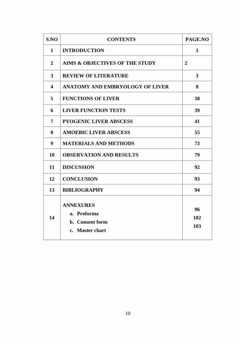
S.NO CONTENTS PAGE.NO
1 INTRODUCTION 1
2 AIMS & OBJECTIVES OF THE STUDY 2
3 REVIEW OF LITERATURE 3
4 ANATOMY AND EMBRYOLOGY OF LIVER 8
5 FUNCTIONS OF LIVER 38
6 LIVER FUNCTION TESTS 39
7 PYOGENIC LIVER ABSCESS 41
8 AMOEBIC LIVER ABSCESS 55
9 MATERIALS AND METHODS 73
10 OBSERVATION AND RESULTS 79
11 DISCUSSION 92
12 CONCLUSION 93
13 BIBLIOGRAPHY 94
14
ANNEXURES
a. Proforma
b. Consent form
c. Master chart
96
102
103

11
LIST OF TABLES
TABLE
NO.
TITILE
1. COMPARISION OF AMOEBIC AND PYOGENIC LIVER
ABSCESS
2. AGE DISTRIBUTION
3. SEX DISTRIBUTION
4. ABDOMINAL PAIN
5. FEVER
6. JAUNDICE
7. OTHER MANIFESTATIONS
8. INTERCOSTAL TENDERNESS
9. ABDOMINAL PAIN IN AMOEBIC AND PYOGENIC ABSCESS
10. FEVER IN AMOEBIC AND PYOGENIC ABSCESS
11. JAUNDICE IN AMOEBIC AND PYOGENIC ABSCESS
12. OTHER MANIFESTATIONS IN AMOEBIC AND PYOGENIC
ABSCESS
13. LOCATION OF ABSCESS
14. NO.OF ABSCESS
15. CORRELATION OF MEDICAL MANAGEMENT
16. CORRELATION OF IMAGE GUIDED DRAINAGE
17. CORRELATION OF SURGICAL DRAINAGE
18. TREATMENT OF LIVER ABSCESS
19. FREQUENCY OF AMOEBIC LIVER ABSCESS
20. FREQUENCY OF PYOGENIC LIVER ABSCESS
21. COMPLICATIONS OF LIVER ABSCESS
12
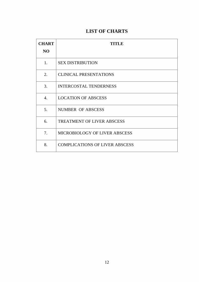
LIST OF CHARTS
CHART
NO
TITLE
1. SEX DISTRIBUTION
2. CLINICAL PRESENTATIONS
3. INTERCOSTAL TENDERNESS
4. LOCATION OF ABSCESS
5. NUMBER OF ABSCESS
6. TREATMENT OF LIVER ABSCESS
7. MICROBIOLOGY OF LIVER ABSCESS
8. COMPLICATIONS OF LIVER ABSCESS
13
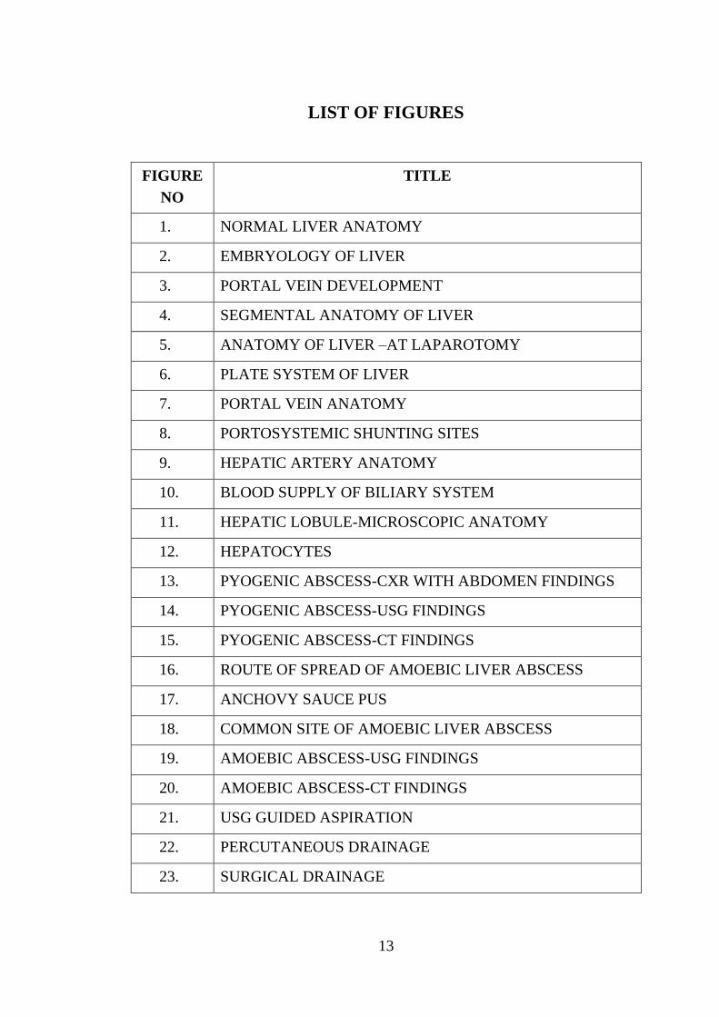
LIST OF FIGURES
FIGURE
NO
TITLE
1. NORMAL LIVER ANATOMY
2. EMBRYOLOGY OF LIVER
3. PORTAL VEIN DEVELOPMENT
4. SEGMENTAL ANATOMY OF LIVER
5. ANATOMY OF LIVER –AT LAPAROTOMY
6. PLATE SYSTEM OF LIVER
7. PORTAL VEIN ANATOMY
8. PORTOSYSTEMIC SHUNTING SITES
9. HEPATIC ARTERY ANATOMY
10. BLOOD SUPPLY OF BILIARY SYSTEM
11. HEPATIC LOBULE-MICROSCOPIC ANATOMY
12. HEPATOCYTES
13. PYOGENIC ABSCESS-CXR WITH ABDOMEN FINDINGS
14. PYOGENIC ABSCESS-USG FINDINGS
15. PYOGENIC ABSCESS-CT FINDINGS
16. ROUTE OF SPREAD OF AMOEBIC LIVER ABSCESS
17. ANCHOVY SAUCE PUS
18. COMMON SITE OF AMOEBIC LIVER ABSCESS
19. AMOEBIC ABSCESS-USG FINDINGS
20. AMOEBIC ABSCESS-CT FINDINGS
21. USG GUIDED ASPIRATION
22. PERCUTANEOUS DRAINAGE
23. SURGICAL DRAINAGE
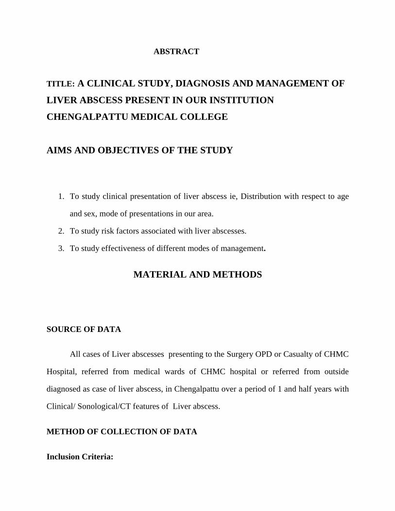
ABSTRACT
TITLE: A CLINICAL STUDY, DIAGNOSIS AND MANAGEMENT OF
LIVER ABSCESS PRESENT IN OUR INSTITUTION
CHENGALPATTU MEDICAL COLLEGE
AIMS AND OBJECTIVES OF THE STUDY
1. To study clinical presentation of liver abscess ie, Distribution with respect to age
and sex, mode of presentations in our area.
2. To study risk factors associated with liver abscesses.
3. To study effectiveness of different modes of management.
MATERIAL AND METHODS
SOURCE OF DATA
All cases of Liver abscesses presenting to the Surgery OPD or Casualty of CHMC
Hospital, referred from medical wards of CHMC hospital or referred from outside
diagnosed as case of liver abscess, in Chengalpattu over a period of 1 and half years with
Clinical/ Sonological/CT features of Liver abscess.
METHOD OF COLLECTION OF DATA
Inclusion Criteria:

1. All cases of liver abscess diagnosed clinically and/or ultrasonographically.
2. All cases of bacterial and parasitic liver abscess
3. All cases in evolving, liquefied & ruptured stage with or without peritonitis
4. All cases of Diagnosed Liver Abscess being referred to CHMC.
Exclusion Criteria:
Traumatic Liver Abscess
Past history of liver abscess
Sample size: 60cases.
Study design: Prospective study
Duration of study: 1 1/2 years.
This study was done from MARCH 2014 to AUGUST 2015 on 60 patients. Case
selection for the study will be done in the initial 1 yr followed by follow up totally for 6
months.
CONCLUSION
Most common liver abscess presenting in our institution was amoebic in nature.
Most common age of presentation was 45 years. Most common presentation of liver
abscess was abdominal pain. Most common clinical sign was intercostal tenderness.

Alcoholism becomes the most frequently associated risk factor. Ultrasound and CT
scan abdomen plays an important role in diagnosing most of the liver abscess patients
presented in our institution. Eighteen percent (18%) of the liver abscess patients were
managed successfully with medical management alone.
Those who are all not responding to medical management were treated with image
guided drainage/aspiration. Forty five percent (45%) of the patients presented in our
institution were successfully treated by image guided drainage/aspiration.
Those who developed complications (ruptured liver abscess) and who are all not
responding to conservative line of management required emergency open drainage. Thirty
seven percent (37%) of patients who developed complications on presentation or later were
treated with open drainage in our institution.

KEY WORDS
LIVER ABSCESS
AMOEBIC LIVER ABSCESS
PYOGENIC LIVER ABSCESS
ULTRASONOGRAM
CT ABDOMEN
MEDICAL MANAGEMENT
IMAGE GUIDED ASPIRATION/DRAINAGE
OPEN DRAINAGE


14
LIST OF ABBREVIATIONS
CBD - COMMON BILE DUCT
CHD - COMMON HEPATIC DUCT
PT - PROTHROMBIN TIME
INR - INTERNATIONAL NORMALISED RATIO
A:G - ALBUMIN:GLOBULIN
IM - INTRAMUSCULAR
IVC - INFERIOR VENA CAVA
AST - ASPARTATE TRANSAMINASES
ALT - ALANINE TRANSAMINASES
CXR - CHEST X RAY
USG - ULTRASONOGRAM
CT - COMPUTED TOMOGRAM
PTC - PERCUTANEOUS TRANSHEPATIC
CHOLANGIOGRAM
INTRODUCTION
Liver abscess was described early in 460-377 B.C. by Hippocrates, still
it remains a challenging situation (especially in tropical countries due to poor

15
hygiene & sanitation, alcoholism and reduced literacy rate) due to its highly
variable presentation, causing diagnostic difficulties.
As India is a one of the tropical country and home to 400 million
people harbouring E.histolytica, the causative organism of amoebic liver
abscess, it is important to thoroughly understanding of the liver abscess.
• Due to the rising incidence in alcoholism, diabetics &
immunocompromised status, liver abscess becomes a matter of grave
concern as complications rate are high especially in this sub-group,
leading to increased morbidity and mortality.
• Due to the advancement in imaging modalities, a more concrete picture
to treat liver abscesses is slowly evolving. However, much work
remains to be done. The story has not ended: it has just begun.
• All these factors has inspired me to select Liver Abscess as my thesis
topic which assumes more importance in our country where rural
population constitutes approximately 70% and therefore it is mandatory
to develop appropriate & realistic guidelines for early diagnosis and
management strategies for liver abscesses in order to reduce the
morbidity and mortality associated with it.
AIMS AND OBJECTIVES OF THE STUDY
1. To study clinical presentation of liver abscess ie, Distribution with
respect to age and sex, mode of presentations in our area.
16
2. To study risk factors associated with liver abscesses.
3. To study effectiveness of different modes of management.
17
REVIEW OF LITERATURE
The liver is one of the most common organ subjected to the
development of abscesses. In one study of 540 intraabdominal abscesses over
a 12 year period liver abscesses made 48% of all visceral abscesses.
In 400.B.C, Hippocratesestablished drainage of liver abscess as a form
of therapy.
Oschner & Debakey described Pyogenic liver abscess in 47 cases in the
year 1938 in their classic paper and reviewed the world literature.
In 1953, McFadzean and associates advocated closed aspiration and
antibiotics for treatment of solitary pyogenic liver abscess.
In 1846, Waller described Pyogenic abscess as a diseased characterized
by suppurative thrombophlebitis of the portal vein and formation of single or
multiple abscesses.
In older days, Pyogenic liver abscess was largely a disease of people of
20-30yrs age group but now the spectrum of disease has changed to age group
50-60yrs with biliary tract diseases & cryptogenic as the main etiology.
With the advent & development of improved antibiotics over the
passage of time, incidence of pyogenic liver abscess would be expected to
decrease, but the incidence is increasing as indicated by studies of Huang &

18
Associates(1996) in which there were 20-22 cases per 100,000 hospital
admissions, appears to be double those of previous 2 decades.
In 1883, Koch initially demonstrated amoebas in the capillaries and
tissues adjoining the wall of the liver abscess.
In 1887 Entamoeba Histolytica was first recovered from the wall of a
hepatic abscess by Kartulis.
Yeoh Kg et al-National University Hospital, Singapore, Reviewed 41
cases from 1994 to 199827 (67%) pyogenic, 6 (15%) amoebic, 2 (5%)
tuberculosis, 6(15%) intermediate. Percutanious needle aspiration was
performed for 85% of pyogenic abscess, and surgery was indicated in only
two cases because of complications. They found that percutaneeous aspiration
of liver abscess not only help to confirm the diagnosis but also to uncover
clinically unsuspected conditions like malignancy and tuberculosis which may
mimic liver abscess.
Hai aa singh a et al Department of Surgery Patna Medical College
Hospital India reviewed 220 cases of ameobic liver abscess between 1981-
86.the majorities were young middle aged males belonging to lower
socioeconomic group and 85% gave history of drinking toddy fermented palm
juice. Over 88%responded well to conservative treatment with aspiration4.
Laparotomy was required in slightly over 10% of cases and in these the
mortality was 12% as compared to 2% with conservative treatment.
Alvarez Perez et al-Depatment of Surgery, San Aagustin Hospital
Avilies Spain Reveiwed pyogenic liver abscess, 133patients treated in 5
19
hospitals during the year 1985 to 1987were studied. 63(47%) were subjected
to percutanious drainage, 45 (34%) were treated by open drainage (surgical)
and the remaining 25 cases(19%)received antibiotic therapy only. Prognostic
variables were the presence of shock, anaemia, elevated prothrombin time and
mixed infection. Treatment of pyogenic liver abscess should be tailored to
each patient, the most of them can be successfully treated with antibiotics and
percutaneous methods. Those with signs of multiorgan failure or septicemia
should preferably be managed in ICU.
Amoebiasis are present in 10% of the population, (most commonly
affects young adults 20-30yr age group with 19:1 male to female ratio) around
500 million persons in developing countries with 50 million cases of invasive
disease, and may account for as many as 100,000 deaths . Amoebic abscess is
one of the most common complication of intestinal amoebiasis and can occur
many years after exposure in endemic areas.
Indirect haemagglutination test is a reliable serologic test for hepatic
amoebiasis, yielding positive results in atleast 85% of patients with extra
intestinal disease and titres often exceed 1:256 almost always noted by 2
weeks into disease and may remain high for many years following successful
therapy.
Leukocytosis with white cell counts averaging between 18,000 –
20,000 is noted in great majority of the patients with liver abscess (mostly
pyogenic), Anaemia associated with long standing infection, the most
significant chemical abnormality is an elevation of serum alkaline
phosphatase. Grossly raised serum levels of vitamin B12 (2000 to 4000 pg/ml)
20
have been reported present in pyogenic liver abscess. The “chocolate sauce,”
“anchovy paste” aspirate is considered pathognomic of an amoebic abscess.
Most hepatic abscesses involve the right lobe of liver (postero-superior
segment), accounting for three-fouth of the cases , in 20% of cases the left
lobe is involved and in rare cases caudate lobe is involved.
Most of the pyogenic abscesses are polymicrobial in nature and account
for about 40% of the cases. In pyogenic liver abscess Eschericia.Coli and
Klebsiella.pneumonia most commonly cultured organisms.
The classic description of the presenting symptoms of liver abscess are
fever, jaundice, and right upper quadrant pain & tenderness. A recent study
from Taiwan of 133 patients found fever in 96% of the patients, chills in 80%,
abdominal pain in 53%, and jaundice in 29%.
Patients with liver abscess who require aspiration irrespective of the
etiology includes abscess with size > 5 cm, both lobes of the liver involved
and duration of symptoms >1 week and advanced age.
Abscesses smaller than 5cm size were treated with parenteral antibiotic
therapy while those larger than 5cm size were treated by image guided
percutaneous aspiration/drainage.
Large abscesses>10cm and multi-loculated abscesses with exaggerated
necrotic process were managed by open surgery.
Hepatic abscesses, both pyogenic and amoebic, becomes the important
cause of morbidity and mortality in tropical countries. Although the primary
21
mode of treatment of amoebic liver abscess is medical, amoebic abscesses
(15%) are not responding to medical therapy. Secondary bacterial infection
occurs in 20% of amoebic liver abscesses. Thus drainage may be required in
those cases with amoebic liver abscesses. For most intra abdominal abscesses
percutaneous drainage is now considered as the standard treatment of choice.
Ultrasound & CT are the mainstay in the diagnostic modalities for
hepatic abscess. The sensitivity of ultrasound in diagnosing hepatic abscess is
around 80-95%. The sensitivity of CT abdomen in diagnosing hepatic abscess
is around 95-100%.
Factors independently associated with poor outcome in Amoebic Liver
Abscess are 1)elevated serum bilirubin (>3.5mg/dl),
2) Encephalopathy,
3) Hypoalbuminaemia (<2.0gm/dl),
4) Multiple abscess cavities,
5) Abscess volume greater than 500ml,
6) Anaemia& diabetes.

22
ANATOMY
General Description and Topography of Liver
The liver is a solid gastrointestinal organ whose mass (1.2kg-1.6kg)
largely occupies the right hypochondrium and part of epigastrium.
The lower margin of liver coincides with costal margin and the superior
surface is draped over by the diaphragm.
Most of the right liver and most of the left liver is covered by the
thoracic cage.
The liver extends superiorly to the height of the 5th rib on the right and
the 6th rib on the left. Posterior surface of the liver straddles the inferior vena
cava (IVC).
A wedge of liver extends to the left half of the abdomen across the
epigastrium to lie just above the anterior aspect of the stomach and under the
central and left diaphragm.
The superior surface of the liver is convex and lies underneath the
diaphragm, whereas the inferior surface is somewhat concave and extends
upto sharp anterior border.
23
The liver is invested in the peritoneum in all areas except
1. The gallbladder bed,
2. The porta hepatis and
3. Posteriorly on either side of the IVC in two wedge-shaped areas (The
bare area of liver to the right of IVC).
Ligaments in relation to Liver
The peritoneal reflections on the liver surface are referred as ligaments.
1. Coronary and triangular ligament
The diaphragmatic peritoneal duplications are referred to as the coronary
ligament, whose lateral margins on either side are
1. Right triangular ligament and
2. Left triangular ligament.
2. Falciform ligament
From the center of the coronary ligament emerges the falciform
ligament, which extends anteriorly as a thin membrane connecting the liver
surface to the diaphragm, abdominal wall, and umbilicus.
3. Ligamentum teres
The ligamentum teres (the obliterated umbilical vein) runs along the
inferior aspect of the falciform ligament from the umbilicus to the umbilical
fissure.
24
The umbilical fissure is on the inferior surface of the left liver and
contains the left portal triad.
The falciform ligament, the most obvious surface marking of the liver,
historically was used to mark the division of the right and left lobes of the
liver in early descriptions of hepatic anatomy.
However the description is inaccurate and it has minimal utility to the
hepatobiliary surgeon.
4. Ligamentum venosum
On the posterior surface of left liver, running from the left portal vein
in the porta hepatis toward the left hepatic vein and the IVC, is the
ligamentum venosum (obliterated sinus venosus), which also runs in a fissure.
Hepatic arterial and portal venous blood flow enter the liver at the
hilum and branch throughout the liver as a single unit that also includes the
bile ducts (portal triad). These portal triads are invested in a peritoneal sheath
that invaginates at the hepatic hilum. Venous drainage is through hepatic veins
that empty directly into the IVC.
25
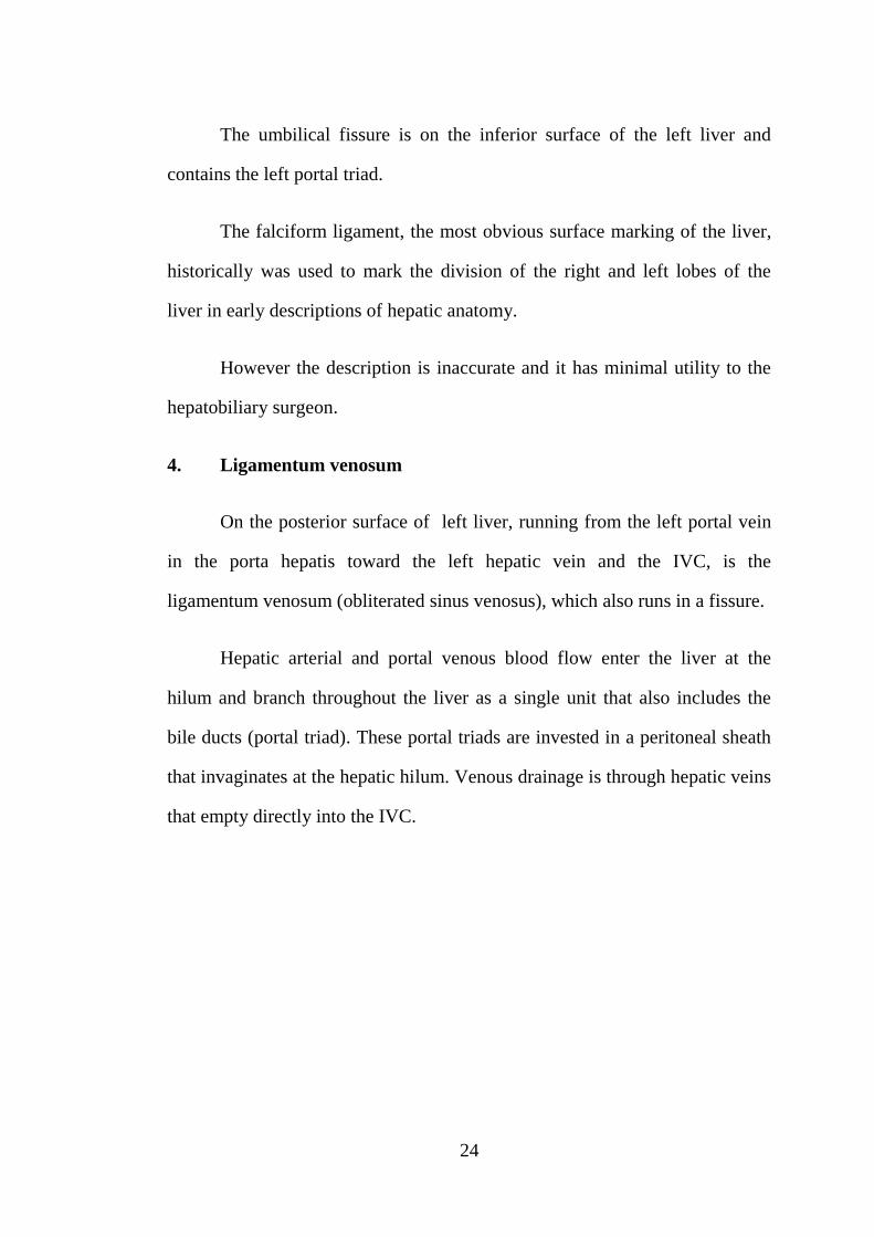
Figure.1
A, Historically, the liver was divided into right and left lobes by the
external marking of the falciform ligament. On the inferior surface of the
falciform ligament, the ligamentum teres can be seen entering the umbilical
fissure. B, The posterior and inferior surface of the liver is shown. The liver
embraces the inferior vena cava (IVC) posteriorly in a groove. The lumens of
the three major hepatic veins and the right adrenal vein can be seen directly
26
entering the IVC. The bare area, bounded by the right and left triangular
ligaments, is illustrated. To the left of the IVC is the caudate lobe, which is
bounded on its left side by a fissure containing the ligamentum venosum. The
lesser omentum terminates along the edge of the ligamentum venosum; thus,
the caudate lobe lies within the lesser sac, and the rest of the liver lies in the
supracolic compartment. A layer of fibrous tissue can be seen bridging the
right lobe to the caudate lobe posterior to the IVC, encircling the IVC. This
ligament of tissue must be divided on the right side when mobilizing the right
liver off of the IVC
Normal Development and Embryology
The liver primordium is formed in the 3rd week of gestation as an
outgrowth of endodermal epithelium (known as the hepatic diverticulum, or
liver bud). It arises from foregut.
The connection between the duodenum and hepatic diverticulum
narrows to form the bile duct, and the gallbladder and cystic duct are formed
by the out pouching from the bile duct.
Hepatic cells develop cords and intermingle with the umbilical and
vitellineveins to form hepatic sinusoids. Simultaneously, Kupffercells
,hematopoietic cells, and connective tissue form from mesoderm of the
septum transversum.
The septum transversum (mesodermal origin) connects the liver to the
anterior abdominal wall and to the foregut. As the liver protrudes into the
abdominal cavity, these structures are stretched into thin membranes,
27
ultimately forming the falciform ligament and the lesser omentum,
respectively.
The mesoderm on the surface of the developing liver differentiates into
visceral layer of the peritoneum except superiorly, where contact between the
liver and mesoderm (future diaphragm) is maintained, forming a bare area
devoid of visceral peritoneum.
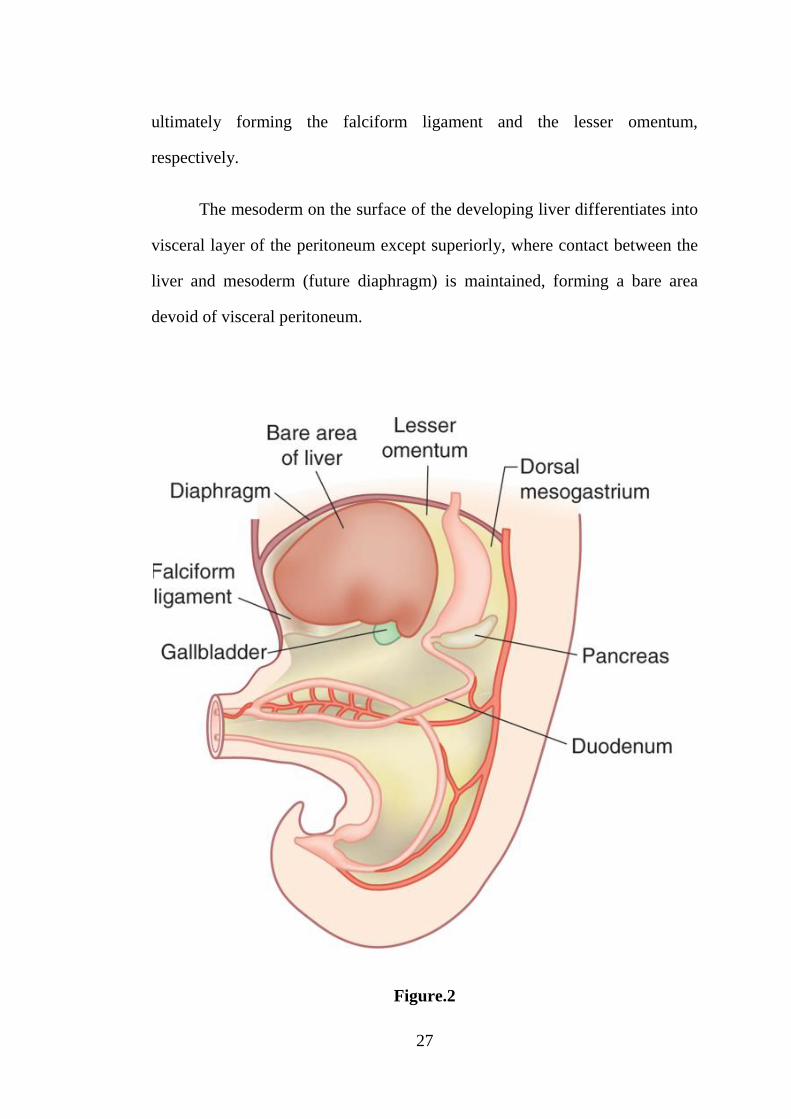
Figure.2
28
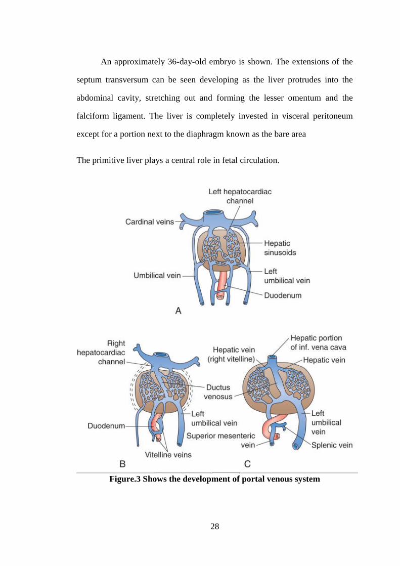
An approximately 36-day-old embryo is shown. The extensions of the
septum transversum can be seen developing as the liver protrudes into the
abdominal cavity, stretching out and forming the lesser omentum and the
falciform ligament. The liver is completely invested in visceral peritoneum
except for a portion next to the diaphragm known as the bare area
The primitive liver plays a central role in fetal circulation.
Figure.3 Shows the development of portal venous system

29
The fetal liver is one of the most important organ of hematopoiesis. In
the 10th week of gestation, the liver is 10% of the body weight, which is due
to developing hepatic sinusoids and active hematopoiesis.
During the last 8 weeks of intrauterine life, hepatic hematopoiesis
decreases, and the liver weight is decreased to 5% of total body weight.
By the 12th week of gestation, bile forms in hepatic cells, along with
the simultaneous development of the gall-bladder and bile duct, allowing
drainage of bile into the foregut.
Functional Anatomy
Historically, the liver was divided into left and right lobes by the
obvious external landmark of the falciform ligament. Not only was this
description oversimplified, but it was also anatomically incorrect in
relationship to the blood supply to the liver.
Later, more accurate descriptions of the liver lobar anatomy were
developed. The liver was separated into right and left lobes determined by
portal and hepatic vein branches.
Briefly, a plane without any surface markings running from the gall-
bladder to the left side of the IVC (known as the cantlie’s line or portal
fissure) divided the liver into
1. The right and
2. The left lobes.
30
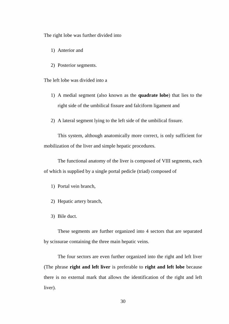
The right lobe was further divided into
1) Anterior and
2) Posterior segments.
The left lobe was divided into a
1) A medial segment (also known as the quadrate lobe) that lies to the
right side of the umbilical fissure and falciform ligament and
2) A lateral segment lying to the left side of the umbilical fissure.
This system, although anatomically more correct, is only sufficient for
mobilization of the liver and simple hepatic procedures.
The functional anatomy of the liver is composed of VIII segments, each
of which is supplied by a single portal pedicle (triad) composed of
1) Portal vein branch,
2) Hepatic artery branch,
3) Bile duct.
These segments are further organized into 4 sectors that are separated
by scissurae containing the three main hepatic veins.
The four sectors are even further organized into the right and left liver
(The phrase right and left liver is preferable to right and left lobe because
there is no external mark that allows the identification of the right and left
liver).
31
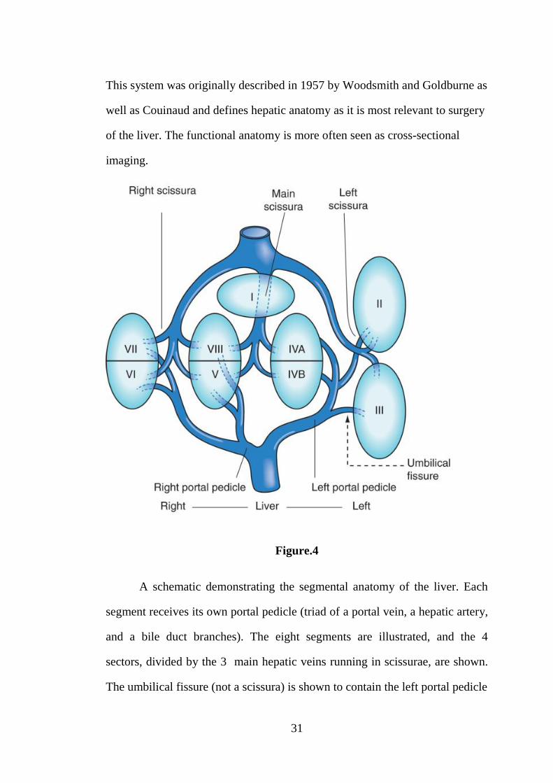
This system was originally described in 1957 by Woodsmith and Goldburne as
well as Couinaud and defines hepatic anatomy as it is most relevant to surgery
of the liver. The functional anatomy is more often seen as cross-sectional
imaging.
Figure.4
A schematic demonstrating the segmental anatomy of the liver. Each
segment receives its own portal pedicle (triad of a portal vein, a hepatic artery,
and a bile duct branches). The eight segments are illustrated, and the 4
sectors, divided by the 3 main hepatic veins running in scissurae, are shown.
The umbilical fissure (not a scissura) is shown to contain the left portal pedicle

32
Figure.5
Segmental anatomy of the liver as seen at laparotomy in the anatomic
position (A) and in the ex vivo position (B).
The main scissura which contains the middle hepatic vein, which runs
in a direction anteroposteriorly from the gallbladder fossa to the left side of the
inferior vena cava and divides the liver into right and left hemi-livers. The line
of the main scissura is also known as Cantlie's line.
Right liver
The right scissura divides the liver into an anterior (segments V and
VIII) and posterior (segments VI and VII) sector by , which contains right
hepatic vein.
33
The right portal pedicle, composed of the right portal vein, hepatic
artery, and bile duct, splits into right anterior and posterior pedicles that supply
the segments of the anterior and posterior segments.
Left liver
The left liver has a visible fissure along its inferior surface called the
umbilical fissure.
The ligamentum teres (containing the remnant of the umbilical vein)
runs into this fissure. The falciform ligament is contiguous with the umbilical
fissure and ligamentum teres. The umbilical fissure is not a scissura, does not
contain a hepatic vein, and in fact, contains the left portal pedicle (triad
containing the left portal vein, hepatic artery, and bile duct), which runs in this
fissure, branching to feed the left liver.
The left scissura runs in a direction posterior to the ligamentum teres
and which contains the left hepatic vein. The left scissura splits the liver into
an anterior (segments III and IV) and posterior (segment II—the only sector
composed of a single segment) sector.
At the hilum of the liver, the right portal triad has a short extrahepatic
course of about 10mm to 15 mm before entering the substance of the liver and
branching into anterior and posterior sectoral branches. The left portal triad,
however, has a long extrahepatic course of up to 3 or 4 cm and runs
transversely along the base of segment IV in a peritoneal sheath that is the
upper end of lesser omentum.
34
The left portal triad, as it runs along the base of segment IV, is
separated from the liver substance by connective tissue known as the hilar
plate. The continuation of the left portal triad runs anteriorly and caudally in
the umbilical fissure and gives branches to the segments II and III and
recurrent branches to the segment IV.
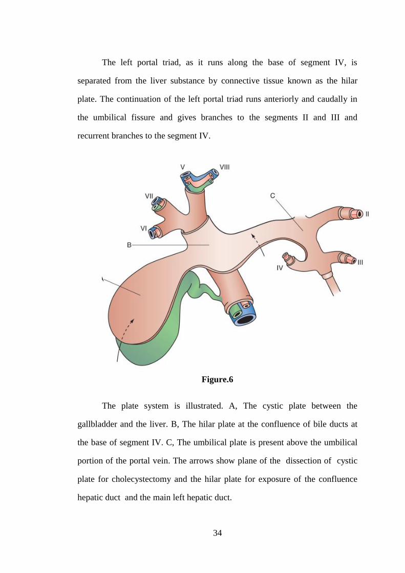
Figure.6
The plate system is illustrated. A, The cystic plate between the
gallbladder and the liver. B, The hilar plate at the confluence of bile ducts at
the base of segment IV. C, The umbilical plate is present above the umbilical
portion of the portal vein. The arrows show plane of the dissection of cystic
plate for cholecystectomy and the hilar plate for exposure of the confluence
hepatic duct and the main left hepatic duct.
35
Caudate lobe
The segment I (caudate lobe) is the dorsal portion of the liver and
embraces the Inferior Vena Cava on its posterior surface and lies posterior to
left portal triad inferiorly and left and middle hepatic veins superiorly.
The most part of the caudate lobe is on the left side of the IVC, but
inferiorly, it traverses between the left portal triad and IVC, where it fuses to
the right liver (segments VI and VII). This part of the caudate lobe is known as
the caudate process (right portion). The left portion of caudate lobe lies in the
lesser omental bursa and is covered anteriorly by the gastrohepatic ligament
(lesser omentum) that separates it from segments II and III anteriorly. The
gastrohepatic ligament attaches to the ligamentumvenosum (sinus venosus
remnant) along the left side of the left portal triad.
The vascular inflow and biliary drainage to the caudate lobe comes
from both right and left systems. The caudate process largely derives its portal
venous supply from the bifurcation of the main portal vein or the right portal
vein, whereas the left part of the caudate derives its portal venous supply
from the left main portal vein.
The biliary drainage and the arterial supply of the right portion are
generally through the right posterior sectoral system and the left portion
through the left main vessels.
The hepatic venous drainage of the caudate is somewhat unique in that
multiple small veins drain posteriorly into the IVC directly.
36
The posterior edge of left side of caudate terminates into a fibrous
component that attaches to the crura of the diaphragm and also runs
posteriorly, wrapping behind the IVC and attaching to segment VII of the right
liver.
IVC ligament
Up to 50% of the time, this fibrous component is composed either
partially or completely of liver parenchyma, and thus liver tissue may
completely encircle the IVC. This important structure is known as the IVC
ligament and is important when mobilizing the right liver or the caudate lobe
off of the IVC.
Riedel’s lobe
A tongue of tissue extending inferiorly off of the right liver.
Portal Vein
The 75% of hepatic blood flow is through portal venous system, and
although it is postcapillary and largely deoxygenated, its large-volume flow
rate provides 50% to 70% of the liver's oxygenation.
The lack of valves in the portal venous system provides a system that
can accommodate high flow at low pressure because of the low resistance and
allows measurement of portal venous pressure anywhere along the system.
The portal vein forms behind the pancreas neck at the confluence of the
splenic vein and superior mesenteric vein at the height of the L2 vertebra. The

37
length of the main portal vein ranges from 5.5 to 8 cm, and its diameter is
usually about 1 cm.
Cephalad to its formation behind the pancreas neck, the portal vein runs
behind the 1stportion of the duodenum and into the hepatoduodenal ligament,
where it runs along the right border of the lesser omentum, usually posterior to
the bile duct and hepatic artery. The portal vein divides into main right and left
branches at the hilum of the liver.
The portal vein’s left branch runs transversely along the base of
segment IV and into the umbilical fissure, where it gives off branches to
segments II and III and feedback branches to segment IV.
The left portal vein also gives off posterior branches to the left side of
the caudate lobe.
The right portal vein has a short extrahepatic course and usually enters
the substance of the liver, where it splits into anterior and posterior sectoral
branches. These sectoral branches can occasionally be seen extrahepatically
and can come off the main portal vein before its bifurcation.
There is usually a small branch off the right portal vein or at the
bifurcation that comes off posteriorly to supply the caudate process.

38
Figure.7
The anatomy of the portal vein is demonstrated. The superior
mesenteric vein (SMV) joins the splenic vein (SV) posterior to the pancreas
neck (shaded) to form the portal vein. Note the entrance of the inferior
mesenteric vein (IMV) into the splenic vein—the most common anatomic
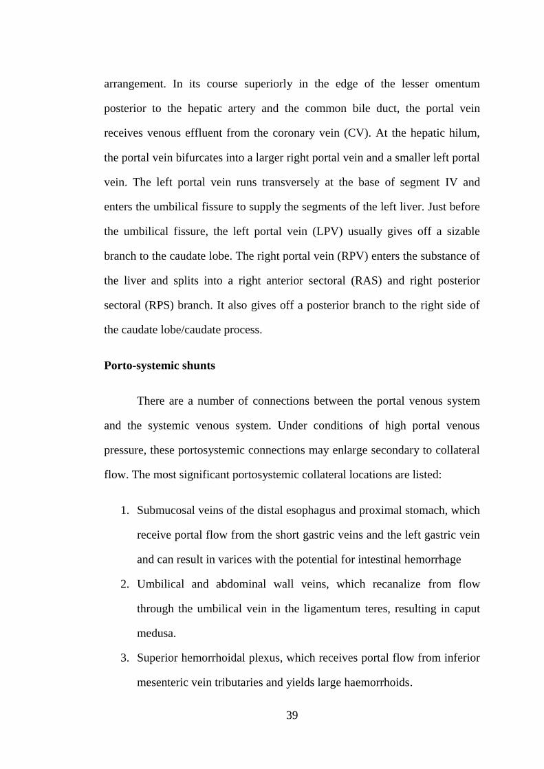
39
arrangement. In its course superiorly in the edge of the lesser omentum
posterior to the hepatic artery and the common bile duct, the portal vein
receives venous effluent from the coronary vein (CV). At the hepatic hilum,
the portal vein bifurcates into a larger right portal vein and a smaller left portal
vein. The left portal vein runs transversely at the base of segment IV and
enters the umbilical fissure to supply the segments of the left liver. Just before
the umbilical fissure, the left portal vein (LPV) usually gives off a sizable
branch to the caudate lobe. The right portal vein (RPV) enters the substance of
the liver and splits into a right anterior sectoral (RAS) and right posterior
sectoral (RPS) branch. It also gives off a posterior branch to the right side of
the caudate lobe/caudate process.
Porto-systemic shunts
There are a number of connections between the portal venous system
and the systemic venous system. Under conditions of high portal venous
pressure, these portosystemic connections may enlarge secondary to collateral
flow. The most significant portosystemic collateral locations are listed:
1. Submucosal veins of the distal esophagus and proximal stomach, which
receive portal flow from the short gastric veins and the left gastric vein
and can result in varices with the potential for intestinal hemorrhage
2. Umbilical and abdominal wall veins, which recanalize from flow
through the umbilical vein in the ligamentum teres, resulting in caput
medusa.
3. Superior hemorrhoidal plexus, which receives portal flow from inferior
mesenteric vein tributaries and yields large haemorrhoids.

40
4. Other retroperitoneal communications yielding collaterals that can
make abdominal surgery hazardous
The anatomy of the portal vein and its branches is relatively constant
and has much less variation than the ductal and hepatic arterial system.
The portal vein is rarely found anterior to the neck of the pancreas and
the duodenum. Entrance of the portal vein directly into the vena cava has also
been described.
Figure.8
Portosystemic collateral (shunting) pathways develop where the portal
venous and systemic venous systems are in close apposition.

41
Hepatic Artery
The hepatic artery, representing high-flow oxygenated systemic arterial
flow, provides about 25% of the blood flow to the liver and 30% to 50% of its
oxygenation.
Figure.9
The anatomy of the celiac axis and hepatic arterial system is illustrated.
The celiac axis, lies just below the diaphragmatic hiatus, divides into1)the
splenic,2) left gastric, and 3)common hepatic arteries. The common hepatic
artery runs in the right direction and turns superiorly toward the hilum. At the
point of this turn, the proper hepatic artery is formed after giving the
gastroduodenal artery. The hepatic artery proper gives off right and left
hepatic arteries in the hilum. Note the middle hepatic artery off of the

42
proximal part of the left hepatic artery, which goes on to supply segment IV.
The cystic artery most commonly comes off the right hepatic artery within the
triangle of Calot.
Hepatic Veins
The three major hepatic veins drain from the superior and posterior
surface of the liver directly into the IVC.
The right hepatic vein runs in the right scissura and drains most of the
right liver after a short (1-cm) extrahepatic course into the right side of the
IVC.
The middle and left hepatic veins usually join intrahepatically and enter
the left side of the IVC as a single vessel, although they may drain separately.
The left hepatic vein runs in the left scissura and drains segments II and III,
and the middle hepatic vein runs in the portal scissura (between segment IV
and the anterior sector of the right liver) draining segment IV and some of the
anterior sector of the right liver.
The umbilical vein is an additional vein that runs under the falciform
ligament, between the left and middle veins, and usually empties into the left
hepatic vein.
Multiple small venous branches from the right posterior sector and
caudate lobe drain posteriorly directly into the IVC. There is often a venous
tributary from caudate that drains superiorly into the left hepatic vein.
43
Biliary System
The intrahepatic bile ducts are terminal branches of the main right and
left hepatic ductal branches that invaginate Glisson's capsule at the hilum
along with corresponding portal vein and hepatic artery branches, forming the
peritoneal covered portal triads. Along these intrahepatic portal pedicles, the
bile duct branches are usually superior to the portal vein, whereas the hepatic
artery branches run inferiorly.
Left Hepatic Duct
The left hepatic bile duct drains segments 2,3and 4, which constitute
the left liver. The intrahepatic ductal branches of the left liver join to form the
main left duct at the base of the umbilical fissure, where the left hepatic duct
courses transversely across the base of segment 4 to join the right hepatic duct
at the hilum. In its transverse portion, the left hepatic duct drains 1 to 3 small
branches from segment 4.
Right Hepatic Duct
The right hepatic duct drains the right liver and is formed by the joining
of the anterior sectoral duct (draining segments 5 and 8) and the posterior
sectoral duct (draining segments 6 and 7). The posterior sectoral duct runs in a
horizontal and posterior direction, whereas the anterior sectoral duct runs
vertically. The main right hepatic duct bifurcates just above the right portal
vein. The short right hepatic duct meets the longer left hepatic duct, forming
the confluence anterior to the right portal vein, constituting the common
hepatic duct.
44
The segment I (caudate lobe) has its own biliary drainage, which is
usually through both right and left systems, although in up to 15% of cases,
drainage is through the left system only, and in 5%, it is through the right
system only.
Common bile duct
The common hepatic duct drains inferiorly, and below the takeoff of
the cystic duct is referred to as the common bile duct.
The common bile or hepatic duct runs along the right side of the
hepatoduodenal ligament to the right side of the hepatic artery and anterior to
the portal vein. The common bile duct continues inferiorly (usually ∼10-15
cm in length and 6 mm in diameter) behind the 1st part of the duodenum and
into the head of the pancreas in an inferior and slightly rightward direction.
The intrapancreatic distal common bile duct then joins with the main
pancreatic duct (of Wirsung), with or without a common channel, and enters
the second portion of the duodenum through the major duodenal papilla of
Vater.
At the choledochoduodenal junction, a complex muscular complex
known as the sphincter of the oddi regulates bile flow and prevents reflux of
contents of the duodenum into the biliary tree. There are three major parts to
this sphincter: the sphincter choledochus, which is a circular muscle that
serves to regulate bile flow and the filling of the gallbladder; the pancreatic
sphincter, present to variable degrees, which surrounds the intraduodenal
45
pancreatic duct; and the sphincter ampullae, made up of longitudinal muscle,
which serves to prevent duodenal reflux.[4]
Gallbladder
The gallbladder is a biliary reservoir that lies against the inferior
surface of segments IV and V of the liver, usually making an impression
against it. A peritoneal layer covers most of the gallbladder except for the
portion adherent to the liver. Where the gallbladder is adherent to the liver,
there is a layer of fibroconnective tissue known as the cystic plate, which is an
extension of the hilar plate. Variable in size, but usually about 10 cm long and
3 to 5 cm wide, the gallbladder is composed of a1) fundus, 2) body, 3)
infundibulum, and4)neck that ultimately empties into the cystic duct. The
fundus usually projects just slightly beyond the liver edge anteriorly and when
folded on itself is described as a Phrygian cap. Continuing toward the bile
duct, the body of the gallbladder is usually in close proximity to the 2nd portion
of the duodenum and the transverse colon. The infundibulum (or Hartmann's
pouch) hangs forward along the free edge of hepatoduodenal ligament and can
fold in front of the cystic duct. The portion of gallbladder between the
infundibulum and the cystic duct is the neck of the gallbladder. The cystic duct
is variable in its length, its course, and its insertion into the main biliary tree.
The first portion of the cystic duct is usually tortuous and contains mucosal
duplications, referred to as the fold of Heister, that regulate the filling and
emptying of the gallbladder. Most commonly, the cystic duct joins the
common hepatic duct to form the common bile duct.
46
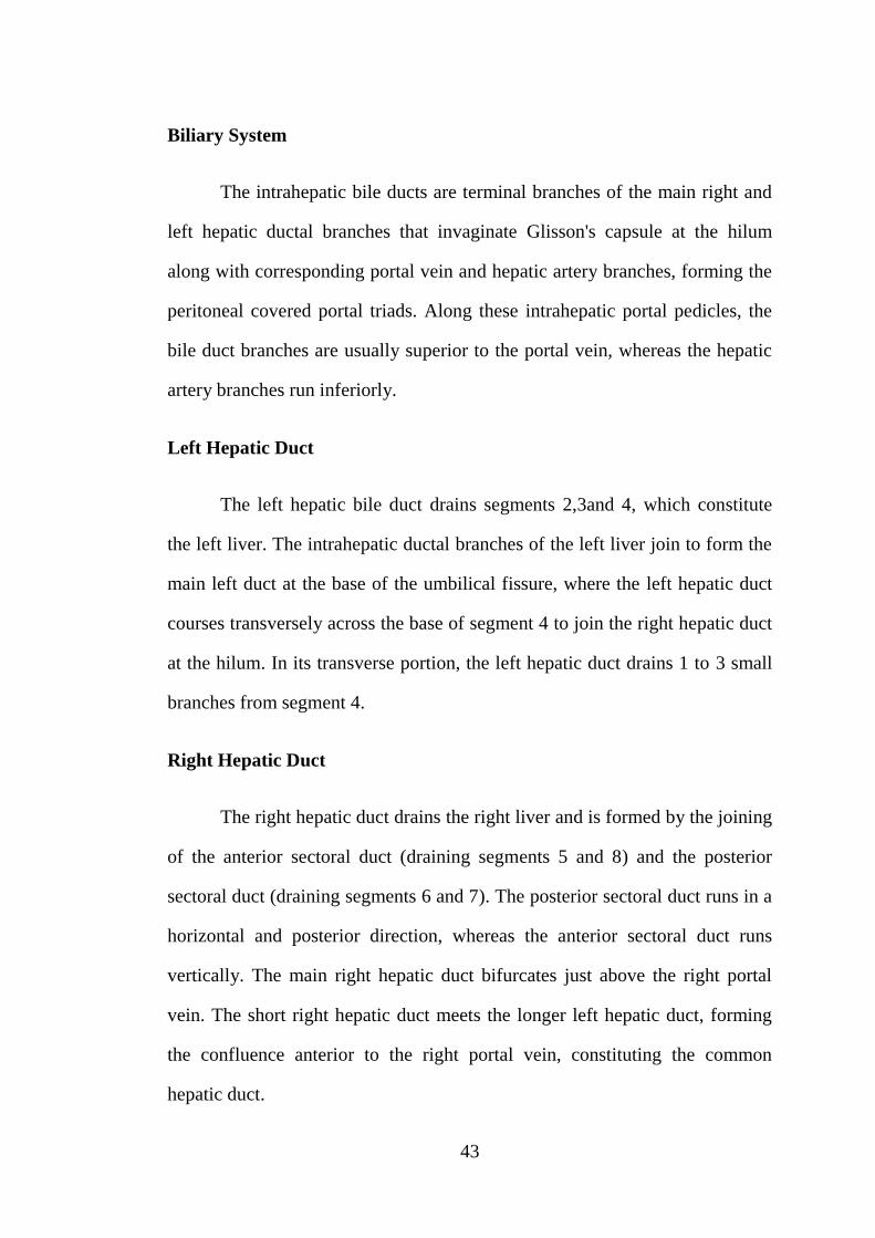
Blood supply of biliay system
Figure.10
The blood supply to the CBD and CHD is illustrated. a) The Right
hepatic artery; b) artery at 9’o clock; c) the retroduodenal artery; d) the left
hepatic artery; e) the proper hepatic artery; f) artery at 3’o clock; g) the
common hepatic artery; h, the gastroduodenal artery.

47
Nerves supply
The innervation of the liver and biliary tract is by
1) sympathetic fibers originating from T7 through T10
2) parasympathetic fibers from both vagal nerves.
The sympathetic fibers pass through celiac ganglia before giving off
postganglionic fibers to the liver and bile ducts. The right-sided celiac ganglia
and right vagal nerve form an anterior hepatic plexus of nerves that runs along
the hepatic artery. The left-sided celiac ganglia and left vagal nerve form a
posterior hepatic plexus that runs posterior to common bile duct and portal
vein. The hepatic arteries are supplied by sympathetic fibers, and the
gallbladder and extrahepatic bile ducts receive innervation from both
sympathetic and parasympathetic fibers. The clinical significance of these
nerves is still not well understood. Pain elicited from acute distention of the
liver (and thus the liver capsule) is referred to the right shoulder because of
innervation of the capsule from the phrenic nerve.
Lymphatics drainage
Most lymph node drainage from the liver is to the lymph nodes of
hepatoduodenal ligament. Lymphatic drainage usually continues along the
hepatic artery to the celiac lymph nodes and from there to the cisterna chyli.
Lymphatic drainage can also follow the hepatic veins to lymph nodes in the
area of the suprahepatic IVC and through the diaphragmatic hiatus. The
lymphatic drainage of the gallbladder and most of the extrahepatic biliary tract

48
is generally into the lymph nodes of the hepatoduodenal ligament. This
drainage can also follow along the hepatic artery to the celiac lymph nodes,
but can also run to lymph nodes behind the head of the pancreas or in the
inter-aortocaval groove.
49
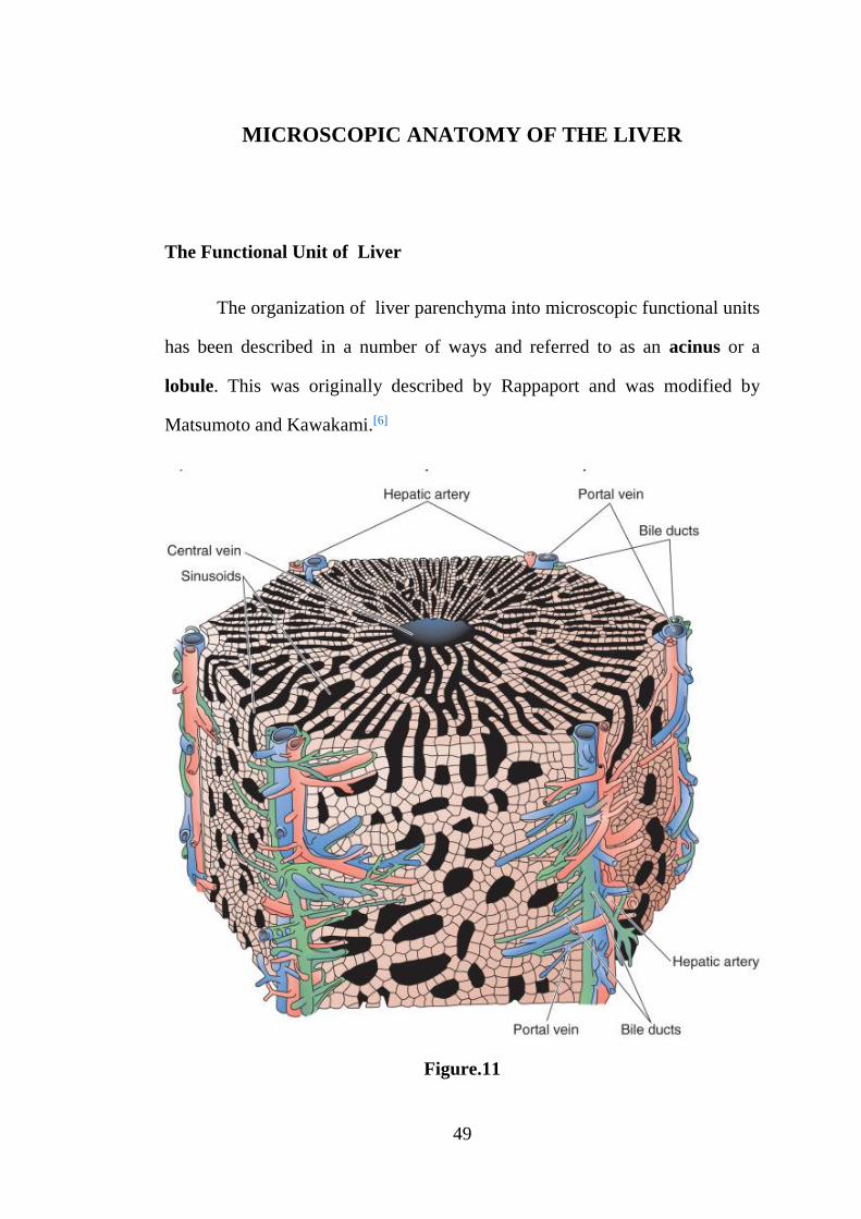
MICROSCOPIC ANATOMY OF THE LIVER
The Functional Unit of Liver
The organization of liver parenchyma into microscopic functional units
has been described in a number of ways and referred to as an acinus or a
lobule. This was originally described by Rappaport and was modified by
Matsumoto and Kawakami.[6]
Figure.11
50
Schematic illustration of a hepatic lobule seen as a three-dimensional
polyhedral unit. The terminal portal triads ( the portal vein, hepatic artery, and
bile duct) are at each corner and give off branches along the sides of the
lobule. Hepatocytes are in single-cell sheets with sinusoids presenton either
end aligned radially toward a central hepatic venule.
Between the portal triad and the central hepatic venule in the centre of
the lobule, there are three zones that differ in their enzymatic makeup and
exposure to nutrients and oxygenated blood. Although there is debate as to the
shape of these zones and their relationship to the basic lobular unit, in general,
zones 1 through 3 fan out from the terminal portal triad toward the central
hepatic venule.
Zone 1, the periportal zone, is exposed to an environment rich in
nutrients and oxygen. Zones 2 (intermediate zone) and Zone 3 (perivenular
zone) are exposed to environments less rich in oxygen and nutrients.
The cells of the different zones differ enzymatically and respond
differently to toxin exposure as well as hypoxia. This anatomic arrangement
also explains the phenomenon of centrilobular necrosis from hypotension
because zone 3 is the most susceptible to decreases in oxygen delivery.

51
Figure.12
Hepatocytes –microscopic anatomy
52
FUNCTIONS OF THE LIVER
1. Removal of gut endotoxins and foreign antigens-liver acts as the first
filter.
2. Drug and hormone metabolism.
3. Formation of bilirubin and its metabolism.
4. Formation of urea from protein catabolism.
5. Glucose metabolism, glycolysis, and gluconeogenesis.
6. Clotting factors synthesis.
7. pH balance and correction of lactic acidosis.
8. Maintaining body temperature.
9. Storage of vit. B12, vit. A, Cu, Fe
53
LIVER FUNCTION TESTS (LFT)
1. Serum bilirubin which includes both direct and indirect. Test is known
as van den Bergh’s test.
2. Serum albumin, globulin and A : G ratio; serum albumin is the
indicator of chronic liver disease.
3. Prothrombin time: Normal value is 12-16 seconds.
Difference between control and test more than 4 seconds or test being
more than 1½ times the control is significant. When it is altered it is corrected
by injecting vitamin K, 10 mg IM for 5 days or by fresh frozen plasma
(FFP)—For cell synthesis.
4. Alkaline phosphatase - Secretary function.
5. Aspartate amino transaminase 5-40 IU/litre (AST, SGOT)-signifies
inflammation.
6. Alanine transaminase 5-40 IU/litre (ALT, SGPT).
7. 5 nucleotidase.
8. Gamma glutamyl transpeptidase (GGT) 10-48 IU/L.
9. Immunological tests: Antimitochondrial or antinuclear antibodies.
10. AFP.
11. Specific tests: (a) For haemochromatosis-serum iron, total iron binding
capacity, serum ferritin. (b) Wilson’s disease: Serum copper, urinary
copper, serum ceruloplasmin.

54
12. Flurodeoxy glucose—positron emission tomography (FDG-PET): It is
to find out the uptake of labeled glucose which varies in different
diseases of liver, i.e., benign, malignant and inflammatory.
13. Technetium-99 m labelled radioisotope scan shows the uptake and
excretion of bile.
14. A sulphur colloid liver scan shows specifically Kupffer cell activity.
Sulphur colloid will not show uptake in adenoma and haemangioma as
Kupffer cells are absent in these lesions.
15. Urine for bile salts (Hay’s test), for bile pigments (Fouchet’s test) and
for urobilinogen (Ehrlich’s aldehyde test).
Other investigations for liver diseases
1. U/S abdomen
2. Angiography
3. CT Scan
4. PTC
5. ERCP
6. MRI
7. Laparoscopy and laparoscopic U/S
8. Liver biopsy

55
INFECTIOUS DISEASES
PYOGENIC LIVER ABSCESS
Pathogenesis
The liver is probably exposed to portal venous bacterial loads on a
regular basis and clears this bacterial load without problems in the usual
circumstance.
The development of a hepatic abscess occurs when the inoculum of
bacteria, irrespective of the route of exposure, exceeds the liver's ability to
clear it. This results in tissue invasion, and the formation of an organized
abscess after neutrophil infiltration.
The potential routes of hepatic exposure to bacteria follow:
1. Biliary tree route.
2. Portal vein route.
3. Hepatic artery route.
4. Direct extension of a nearby focus of infection.
5. Trauma.
1. Bilairy route
Biliary tree infections are presently the most common identifiable cause
of hepatic abscess. Biliary obstruction results in bile stasis with the potential
for subsequent bacterial infection, colonization, and ascension into the liver.
56
This process is called as ascending suppurative cholangitis. The nature of
biliary obstruction is mostly related to malignancy or stone disease.
In Asia, stones in the hepatic ductal system and cholangitis are a
common cause, whereas in the Western world, malignancy causing
obstruction is becoming a more predominant factor.The common link between
all causes of hepatic abscess from the biliary tree obstruction and bacteria in
the biliary tree.
Previous biliary-enteric anastomosis has also been associated with
hepatic abscess formation, likely due to unimpeded exposure of the biliary tree
to enteric organisms.
2. Portal venous route
The portal venous system drains the most of the gastrointestinal tract,
and any infections of the gastrointestinal tract can result in pyelophlebitis
(ascending portal vein infection ) with exposure of the liver to large amounts
of bacteria. The most common causes of pyelophlebitis are appendicitis,
diverticulitis, pancreatitis, inflammatory bowel disease, pelvic inflammatory
disease, hollow viscus perforation, or omphalitis in the newborn. Hepatic
abscess has also been associated with colorectal malignancy.
3. Hepatic artery route
Any systemic infection (pneumonia, endocarditis, osteomyelitis) can
result in bacteremia and infection of the liver through the hepatic artery.
Hepatic abscess from systemic infections may also reflect an altered immune
57
response, such as in patients with malignancy, acquired immunodeficiency
syndrome, or disorders of granulocyte function. Children with chronic
granulomatous disease are particularly susceptible.
4. Direct extension
Hepatic abscess can occurs as the result of direct extension of an
infective process. Common examples of this include subdiaphragmatic
abscess, suppurative cholecystitis, perinephric abscess, and perforation of the
hollow viscera directly into the liver.
5. Trauma
Penetrating injury and blunt trauma can result in anan area of necrotic
liver or intrahepatic hematoma, which can subsequently develop into an
abscess. Bacteria may have been introduced from the trauma, or the affected
area may be seeded from systemic bacteremia. Hepatic abscesses associated
with trauma have a delayed presentation, up to weeks after the injury.
Iatrogenic hepatic necrosis causes include hepatic artery embolization or
thermal ablative procedures, can be complicated by abscess.
6. Cryptogenic
Cryptogenic abscesses are the most common cause in recent case series.
Possible theories for cryptogenic hepatic abscess are resolved infective
process at the time of presentation ,undiagnosed abdominal pathology, and
host factors such as malignancy or diabetes rendering the liver more
susceptible to transient hepatic artery or portal vein bacteremias.

58
In patients with cryptogenic hepatic abscess who have had computed
tomography (CT) and ultrasonography, it has been argued that a diligent
search for a cause should ensue.
Predisposing Factors
Pyogenic liver abscesses occur more commonly in adults with
comorbid conditions including diabetes , cirrhosis, pancreatitis, inflammatory
bowel disease, pyelonephritis, and peptic ulcer disease. Solid organ cancers as
well as lymphoma and leukemia are present in 17–36% of patients with liver
abscesses. The combination of chemotherapy and steroid use is thought to be
responsible in these cases.
In children, pyogenic liver abscesses tend to occur in patients with host-
defense abnormalities or immune disorders. Complement deficiencies, chronic
granulomatous disease, and leukemia and other malignancies place these
children at increased risk for liver abscess. Hepatic abscesses are also seen in
sickle cell anemia, congenital hepatic fibrosis, polycystic liver disease, and
after liver transplantation
Pathology and Microbiology
Most commonest site of hepatic abscess is the right lobe of the
liver75%.It is due to preferential laminar flow of blood to the right side. The
left lobe is involved in 20% of the time, and the caudate lobe is uncommonly
involved (5%). About 50% of hepatic abscesses are solitary. The size of
hepatic abscesses can vary from <1 mm to several centimeters in diameter and

59
can be multiloculated or a single cavity.Surrounding inflammation can cause
adhesion to local structures.
In early series, sterile abscesses were commonly reported but probably
reflected inadequate culture techniques, whereas in modern series, few
abscesses are sampled before the administration of antibiotics. Abscesses from
pyelophlebitis or cholangitis tend to be polymicrobial, with a high
preponderance of gram-negative rods. Systemic infections are usually the
cause infection with a single organism.
Most of the hepatic abscesses are polymicrobial in nature (40%).
Solitary abscesses are more likely to be polymicrobial. Anaerobes are
involved about 40% to 60% of the time. The most common micro organisms
cultured are E.coli and Kleb.pneumoniae. Other common organisms
encountered are Staph.aureus, Enterococcus species, Strep.viridans, and
Bacteroides species. Klebsiella is frequently associated with gas-forming
abscesses. Uncommonly encountered organisms (<10% of cultures) include
Pseudomonas, Enterobacter, Proteus, Citrobacter, Serratia, β-hemolytic
streptococci, microaerophilic streptococci, Fusobacterium, Clostridium, and
other rare anaerobes. Blood cultures are positive in about 50% to 60% of
cases. Fungal and mycobacterial hepatic abscesses are rare and are almost
always associated with immunosuppression, usually from chemotherapy.
Clinical Features
Fever, jaundice, Weight loss, Pain, Nausea and vomiting, Malaise,
chills, anorexia, Cough and pleurisy, pruritus, diarrohea.
60
Physical Examination –
Right upper quadrant tenderness, hepatomegaly, Jaundice, Right upper
quadrant mass, Ascites, Pleural effusion or Rub.
Laboratory investigation
Increased Alkaline phosphatase, TC count>10000/mm3, Albumin
<3g/dl, Hematocrit <36%, Bilirubin >2mg/dl.
Imaging studies
The most essential element to making the diagnosis of hepatic abscess
is radiographic imaging.
Chest x-ray
Chest x-rays are abnormal in about 50% of the time, and findings
generally reflect subdiaphragmatic pathology such as right pleural effusion, an
elevated right hemidiaphragm, or atelectasis. Occasionally, these can be left-
sided findings in the case of an abscess involving the left liver.
Xray abdomen erect
Plain abdominal x-rays, in rare cases, can be helpful. They can show
portal venous gas or air-fluid levels.

61
Figure. 13
Plain abdominal x-ray demonstrating an abnormal collection of air in
the rt upper quadrant consistent with a pyogenic hepatic abscess (black arrow).
Ultrasound and CT are the mainstays in diagnostic modalities for hepatic
abscess.
Ultrasonogram abdomen
Ultrasound usually demonstrates
1) A oval or round area that is less echogenic than the liver and
2) Distinguish solid from cystic lesions.
62
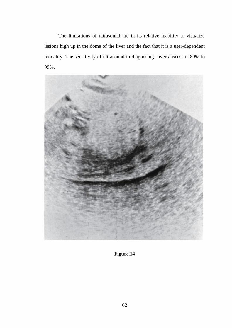
The limitations of ultrasound are in its relative inability to visualize
lesions high up in the dome of the liver and the fact that it is a user-dependent
modality. The sensitivity of ultrasound in diagnosing liver abscess is 80% to
95%.
Figure.14
63
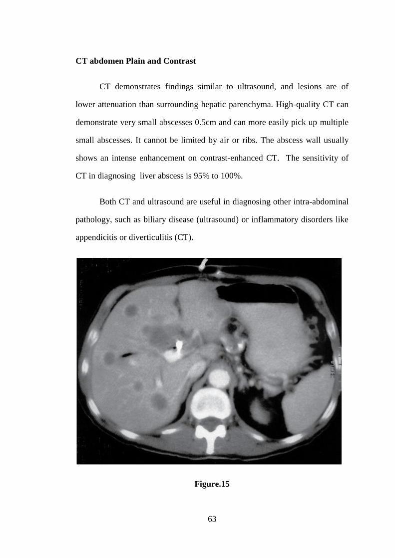
CT abdomen Plain and Contrast
CT demonstrates findings similar to ultrasound, and lesions are of
lower attenuation than surrounding hepatic parenchyma. High-quality CT can
demonstrate very small abscesses 0.5cm and can more easily pick up multiple
small abscesses. It cannot be limited by air or ribs. The abscess wall usually
shows an intense enhancement on contrast-enhanced CT. The sensitivity of
CT in diagnosing liver abscess is 95% to 100%.
Both CT and ultrasound are useful in diagnosing other intra-abdominal
pathology, such as biliary disease (ultrasound) or inflammatory disorders like
appendicitis or diverticulitis (CT).
Figure.15
64
Differential Diagnosis
1) Amoebic abscess
2) Echinococcal cyst.
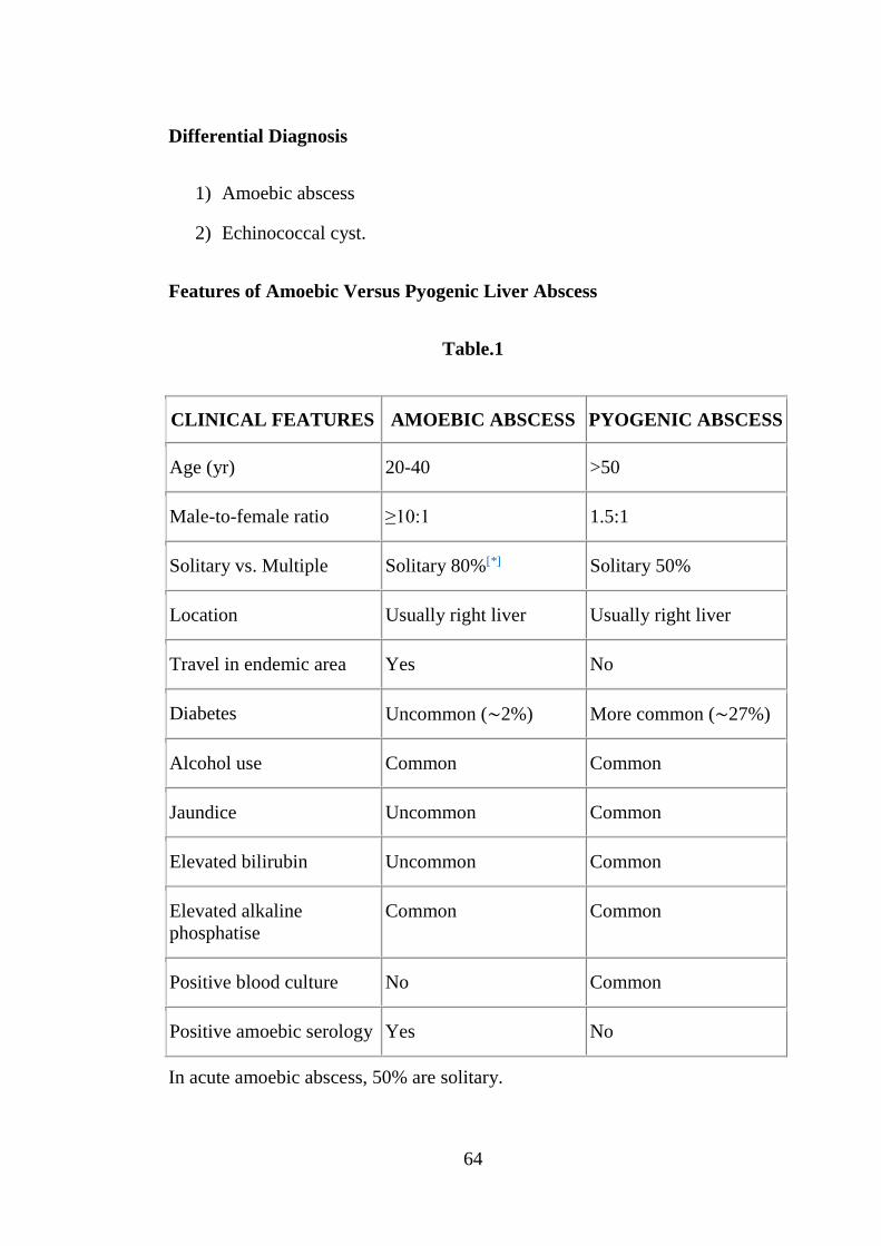
Features of Amoebic Versus Pyogenic Liver Abscess
Table.1
CLINICAL FEATURES AMOEBIC ABSCESS PYOGENIC ABSCESS
Age (yr) 20-40 >50
Male-to-female ratio ≥10:1 1.5:1
Solitary vs. Multiple Solitary 80%[*] Solitary 50%
Location Usually right liver Usually right liver
Travel in endemic area Yes No
Diabetes Uncommon (∼2%) More common (∼27%)
Alcohol use Common Common
Jaundice Uncommon Common
Elevated bilirubin Uncommon Common
Elevated alkaline
phosphatise
Common Common
Positive blood culture No Common
Positive amoebic serology Yes No
In acute amoebic abscess, 50% are solitary.
65
Treatment
Before the availability of antibiotics and the routine use of drainage
procedures, untreated hepatic pyogenic abscess was almost uniformly fatal. It
was not until the classic review by Ochsner and DeBakey in 1938 that routine
surgical drainage was employed and dramatic reductions in mortality were
noted. Open surgical drainage of pyogenic abscesses was the sole treatment
(with the addition of antibiotics eventually) for hepatic abscess until the 1980s.
Since the 1980s, less invasive percutaneous drainage techniques, along with
the use of intravenous (IV) antibiotics, have been employed. Laparotomy is
generally reserved for failures of percutaneous drainage.
When the diagnosis of pyogenic hepatic abscess is suspected, broad-
spectrum IV antibiotics are started immediately to control ongoing bacteremia
and its associated complications.
Blood cultures and cultures of the abscess from aspiration are sent for
aerobic and anaerobic cultures. In immunosuppressed patients, mycobacterial
and fungal cultures of the aspirate need to be considered.
Patients who are at risk for amoebic infections have amoebic serologies
drawn. Until cultures have specifically identified the offending organism,
broad-spectrum antibiotics covering gram-negatives, gram-positives, and
anaerobes are used.
66
Medical management
Combinations such as ampicillin, an aminoglycoside, and
metronidazole or a third-generation cephalosporin with metronidazole are
appropriate. The optimal duration of antibiotic treatment is not well defined
and must be individualized depending on the success of the drainage
procedure. Antibiotics are certainly continued while there is evidence of
ongoing infection, such as fever, chills, or leukocytosis. Beyond this, it is
unclear how long to continue antibiotics, but recommendations usually are for
2 or more weeks.
Percutaneous Drainage Procedures
During the past 20 years, percutaneous catheter drainage has become
the treatment of choice for most patients. Success rates range from 69% to
90%.
Advantages
1. The simplicity of treatment (usually employed at the time of radiologic
diagnosis)
2. Avoidance of general anesthesia and
3. Avoidance of laparotomy.
Contraindications
1. Ascites,
2. Coagulopathy, or
3. Proximity to vital structures..
67
A recent retrospective study comparing surgical with percutaneous
drainage for large (>5 cm) abscesses showed a better success rate with surgical
drainage. Despite this, two thirds of the percutaneous treatments were
successful, and the overall morbidity and mortality rates were similar. There
has never been a randomized prospective comparison between percutaneous
and surgical therapy for hepatic abscess, but case series suggest that for most
cases, there are similar success rates and mortality rates.
Modern series attempting to compare these two techniques
retrospectively must be read with caution because most patients treated
surgically have failed other, less invasive techniques. In general, surgery is
reserved for patients who require surgical treatment of the primary pathology
(e.g., appendicitis) or for those who have failed percutaneous techniques.
Percutaneous aspiration
Percutaneous aspiration without the placement of a drain is another
treatment modality. Success rates are generally 60% to 90% and are somewhat
similar to those for percutaneous catheter drainage. Usually, however, more
than one aspiration is required, and one fourth of patients require three or more
aspirations. One randomized trial has evaluated percutaneous aspiration versus
percutaneous catheter drainage. Success rates were 60% in the aspiration
group and 100% in the catheter group, but all but one patient in the aspiration
group had a single aspiration. Another recent randomized trial compared
aspiration alone to catheter drainage. Sixty-four randomized patients were
analyzed, and there were similar outcomes in terms of treatment success rate,
hospital stay, antibiotic duration, and mortality. In the aspiration-only group,
68
40% required two aspirations, and 20% required three aspirations. In general,
catheter drainage remains the treatment of choice, although a trial of a single
aspiration is reasonable to consider.
Some investigators have reported success with antibiotics alone. Most
of these patients, however, have had a diagnostic aspiration and thus at least a
partial drainage. Additionally, other series have reported that antibiotic
treatment without drainage carries a prohibitively high mortality rate (59%-
100%). In patients who are not surgical candidates or who absolutely refuse
any invasive procedure, an attempt at antibiotic treatment is reasonable;
however, this is not recommended in all other situations.
Open drainage
Ruptured liver abscesses
Liver resection
Liver resection is occasionally required for hepatic abscess. This may
be required for an infected hepatic malignancy, hepatolithiasis, or intrahepatic
biliary stricture. If hepatic destruction from infection is severe, some patients
may benefit from resection.
Outcome
Mortality from pyogenic hepatic abscess has dramatically improved
during the past 6 decades. Before the routine use of surgical drainage,
pyogenic abscess was uniformly fatal. With the routine use of surgical
drainage and the use of IV antibiotics, mortality was reduced to about 50%, a

69
figure that stayed relatively constant from 1945 until the early 1980s. Since
the 1980s, mortality has been reported from 10% to 20%, and series from the
1990s now routinely demonstrate a mortality rate of less than 10%.
Factors predictive of a poor outcome
The presence of malignancy,
Factors associated with malignancy (jaundice, markedly elevated
LFTs), or
Signs of sepsis
Hypoalbuminemia
Signs of severe infection, such as marked leukocytosis
Acute Physiology and Chronic Health Evaluation (APACHE) II scores,
Abscess rupture
Bacteremia, and
Shock.
Amoebic Liver Abscess
Epidemiology
Amoebic liver abscesses tend to be more common in Hispanic males,
aged 20 to 40 years, with a history of travel to (or origination from) an
endemic area.
Poverty and cramped living conditions are associated with higher rates
of infection. A male preponderance of greater than 10:1 is seen. Heavy alcohol

70
consumption is commonly reported and may render the liver more susceptible
to amoebic infection.
Patients with impaired host immunity also appear to be at higher risk
for infection and have higher mortality rates. Patients with amoebic liver
abscess without a travel history to an endemic area often have an associated
immunosuppression, such as human immunodeficiency virus (HIV) infection,
malnutrition, chronic infection, or chronic steroid use.
Pathogenesis
Entamoeba. histolytica is a protozoa that exists as a trophozoite or as a
cyst.
All other species in the genus Entamoeba are found to be
nonpathogenic, and not all strains of E. histolytica are considered virulent.
Ingestion of cysts of Entamoeba histolytica occurs i.e through a fecal-oral
route. Humans are the principal host, and the main source of infection is
human contact with a cyst-passing carrier.
Contaminated water and vegetables are also a route of human infection.
Once ingested, the cysts are not degraded in the stomach and pass to the
intestines where the trophozoite is released and passed on to the colon. In the
colon, the trophozoite can invade mucosa, resulting in disease.

71
Route of spread
Figure.16
It is believed that the trophozoites reach the liver through the portal
venous system. There is no evidence for trophozoites passing through
lymphatics. As implied by its name, E. Histolytica trophozoites have the
capacity to lyse tissues through a complex set of events, including cell
adherence, cell activation, and subsequent release of multiple enzymes
resulting in necrosis. The major mechanism is probably enzymatic cellular
hydrolysis. Amoebic liver abscesses are thus formed by progressing, localized
hepatic necrosis resulting in a cavity containing acellular proteinaceous debris

72
surrounded by a rim of invasive amoebic trophozoites. Early development of
an amoebic liver abscess is associated with an accumulation of
polymorphonuclear leukocytes, which are then lysed by the trophozoites.
Antiamoebic antibodies develop rapidly in patients with invasive
disease or amoebic hepatic abscess. Secretory immunoglobulin A (IgA)
antibodies have been shown in vitro to inhibit adherence to colonic
epithelium; however, the development of these antibodies does not halt the
progression of disease. Interestingly, children who lack antiamoebic IgG have
innate resistance to invasive infection, suggesting an alternative immune-
mediated re-sponse. There is now evidence that a cell-mediated T-helper
response is probably the major mechanism of resistance.
Pathology
Hepatic amoebic abscess is essentially the result of liquefaction
necrosis of the liver, producing a cavity full of blood and liquefied liver tissue.
The appearance of this fluid is typically described as anchovy sauce,
and the fluid is odorless unless secondary bacterial infection has taken place.
The progressive hepatic necrosis continues until Glisson's capsule is
reached because the capsule is resistant to hydrolysis by the amoebae and thus
amoebic abscesses tend to abut the liver capsule. Because of the resistance of
Glisson's capsule, the cavity is typically crisscrossed by portal triads protected
by this peritoneal sheath. Early on, the formed cavity is ill defined, with no
real fibrous response around the edges, but a chronic abscess can ultimately

73
develop a fibrous capsule and may even calcify. Like pyogenic abscesses,
amoebic abscesses tend to occur mainly in the right liver.
ANCHOVY SAUCE PUS
Figure.17
Most common location
Figure.18
Most common site of amoebic liver abscess was right posterosuperior
segment.

74
Clinical Features
It is common in males (20:1), may be after an attack of amoebic
dysentery or many months after the attack or history of dysentery may
not be there at all.
They present with fever, loss of weight, chills and rigors, non
productive cough, shoulder pain.
Pain in the right hypochondrium.
Soft, tender, smooth, liver with increased liver span.
Intercostal tenderness is elicited which is a useful clinical sign.
Right sided pleural effusion may be evident.
Mild jaundice is not uncommon especially in cirrhotics and multiple
abscesses which may signify poor prognosis.
Tenderness, rigidity and skin oedema in right hypochondrium may be
present in acute cases.
In chronic amoebic liver abscess, smooth, firm/hard, nontender liver
may be palpable.
Amoebic liver abscess may be –
Acute – present with high fever, chills, rigors, tender, soft palpable
liver, with intercostal tenderness.
Chronic – present with firm/hard, smooth, non-tender palpable liver
without acute features.
75
Features may be of –
Systemic – present with fever, chills and rigors, loss of appetite,
reduced weight, and jaundice.
Abdominal – present with pain and tenderness, localized guarding and
rigidity, mass in right upper abdomen (tender, soft liver), ascites,
splenomegaly, abdominal wall oedema.
Thoracic – present with dry cough, chest pain in right lower part, right
shoulder pain, pleural effusion, and intercostal tenderness.
Features of complications – rupture/infection/ septicaemia/liver
failure.
Differential diagnosis
For Acute type
1. Acute cholecystitis
2. Acute presentation of hepato-cellular carcinoma
3. (HCC) due to haemorrhage or necrosis.
4. Subphrenic abscess.
Chronic amoebic liver abscess mimics hepatoma in every respect
Investigations
Total count may be increased. Patients typically have a mild to
moderate leukocytosis without eosinophilia Anemia is common
Liver function tests may show altered bilirubin and albumin level.

76
Prothrombin time may be widened and if it is so Inj. vit K 10 mg IM for
5 days should be given. Even with this if P.T. remains widened then
fresh frozen plasma (FFP) is needed to rectify the P.T.
Serum alkaline phosphatase, ALT, AST levels are altered.
Indirect haemagglutination test (95% positive rate),
ELISA and gel diffusion precipitative test are reliable serological tests.
Serological tests are reliable in nonendemic areas than endemic areas. Counter
immunoelectrophoresis is more useful in active disease. This test has largely
been replaced by enzyme immunoassays (EIAs), which are simple, rapidly
performed, and inexpensive. The EIA has 99% sensitivity and 90% specificity
in patients with hepatic abscess. Unfortunately, the presence of antibodies may
reflect old infection, and interpretation can be difficult in endemic areas.
Ongoing studies are focusing on identifying specific E. histolytic antigens in
an attempt to identify acute infection.
Chest X-ray findings:
- Raised fixed diaphragm (tenting)
- Pleural effusion
- Soft tissue shadow
- Atelectasis
77
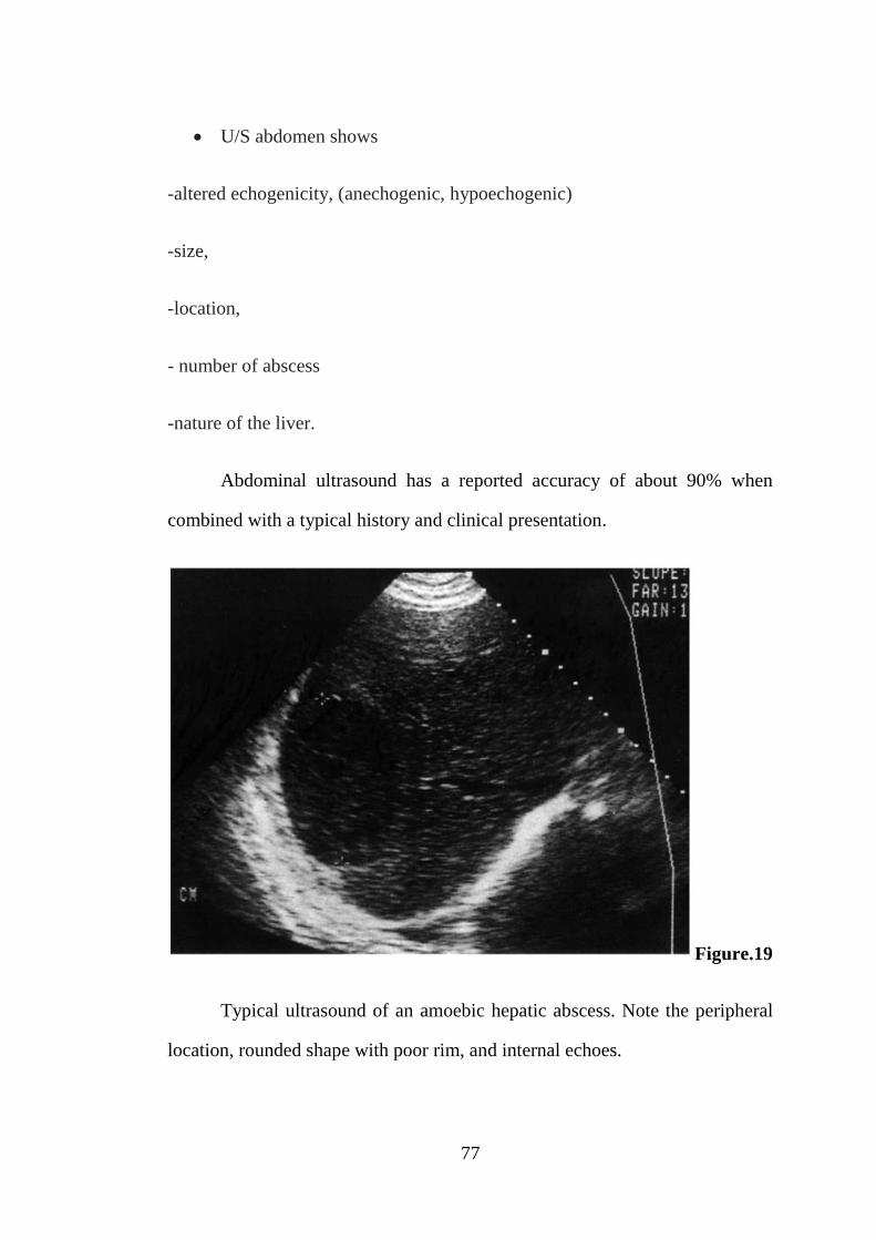
U/S abdomen shows
-altered echogenicity, (anechogenic, hypoechogenic)
-size,
-location,
- number of abscess
-nature of the liver.
Abdominal ultrasound has a reported accuracy of about 90% when
combined with a typical history and clinical presentation.
Figure.19
Typical ultrasound of an amoebic hepatic abscess. Note the peripheral
location, rounded shape with poor rim, and internal echoes.

78
CT scan-contrast study. CT scan shows raised diaphragm; abscess
cavity (low density area) – its size, location, number; presence of
effusion; changes in the lung.
Abdominal CT scan is probably more sensitive than ultrasound and is
helpful in differentiating amoebic from pyogenic abscess, with rim
enhancement noted in the latter. CT can also be helpful in identifying simple
cysts and necrotic tumors.
Figure.20
CT scan of amoebic abscess. The lesion is peripherally located and
round. Rim is nonenhancing but shows peripheral edema (black arrows). Note
the extension into the intercostal space (white arrow).
79
Sigmoidoscopy/colonoscopy
-used to identify the active ulcers. Scrapings of the ulcer show trophozoites.
When the previously outlined workup is still not definitive and
diagnostic uncertainty persists, two options are considered. A therapeutic trial
of antiamoebic drugs in which rapid improvement occurs in most cases of
amoebic abscess can be helpful. In situations in which amoebic serology is
inconclusive and therapeutic trial of antibiotics is either deemed inappropriate
or has failed to improve symptoms, consideration is given to diagnostic
aspiration. A pyogenic abscess would have bacteria and leukocytes, whereas
an amoebic abscess would contain the typical anchovy sauce appearance.
Cultures of amoebic abscess are usually negative and do not contain
leukocytes.
In cases in which neoplasm or hydatid disease is given serious
consideration, aspiration should not be performed.
Management
The mainstay of treatment for amoebic abscesses is metronidazole (750
mg orally tds per day for 10 days), which is curative in more than 90% of
patients. Clinical improvement is usually seen within 3 days. If response to
metronidazole is poor or the drug is not tolerated, other agents can be used.
Emetine hydrochloride is effective against invasive amebiasis
(particularly in the liver) but requires intramuscular injections and has serious
cardiac side effects. A more attractive option is chloroquine, but this is a less
effective agent. After treatment of the liver abscess, it is recommended that

80
luminal agents such as iodoquinol, paromomycin, and diloxanide furoate are
administered to treat the carrier state.
Aspiration
In case of large abscess and infected abscess aspiration with a wide
bore needle is done under U/S guidance after correcting the P.T.
Previously without U/S, aspiration used to be done by passing needle in
right 6th intercostal space in midaxillary line.
Figure.21
Aspiration of the amoebic liver abscess under US guidance or through
right 8th intercostal space in midclavicular line.
81
Therapeutic needle aspiration of amoebic abscesses has been proposed.
Small randomized trials comparing metronidazole with or without aspiration
have shown minor benefits with aspiration, but no major improvement to
justify routine aspiration. In general, aspiration is recommended for diagnostic
uncertainty, failure to respond to metronidazole therapy in 3 to 5 days, or in
abscesses felt to be at high risk for rupture. Abscesses greater than 5 cm in
diameter and in the left liver are thought to be a higher risk for rupture, and
aspiration needs to be considered.
Percutaneous Drainage
Under U/S guidance pigtail catheter is placed into the abscess cavity
percutaneously to drain the pus. Catheter tube and abscess cavity has to be
washed and irrigated at regular intervals with normal saline. It may fail if there
is thick pus, multiloculated abscess, and multiple abscesses. Procedure may
cause bleeding and infection.
Note: Aspirated fluid is sent for C/S, cytology and for study of trophozoites
(last part of the aspirated fluid should be sent for this).

82
Open Drainage
Figure.22
Through transperitoneal approach, abscess area is opened, pus is
evacuated. Malecot’s catheter is placed and brought out through a separate
stab incision. The catheter is kept in situ until drainage stops completely.
Complete drainage of pus will be confirmed by repeat U/S. During discharge,
advice is given to avoid alcohol, Chloroquine 250 mg BD for 10 days and
Diloxanide furaote 500 mg tid is given for 10-14 days.
Complications of surgery – anesthetic problems; bleeding; liver
failure (in cirrhotic patients); intraperitoneal abscess formation; bile leak – bile
peritonitis and fistula.
83
Surgery
Indications for surgery
1) Even after repeated aspirations if abscess cavity
2) fills again
3) Thick pus
4) Multiloculated abscess
5) Left lobe abscess, because of danger of rupture into
6) pericardial cavity
7) Ruptured abscess
8) Caudate lobe abscess
9) Multiple abscesses
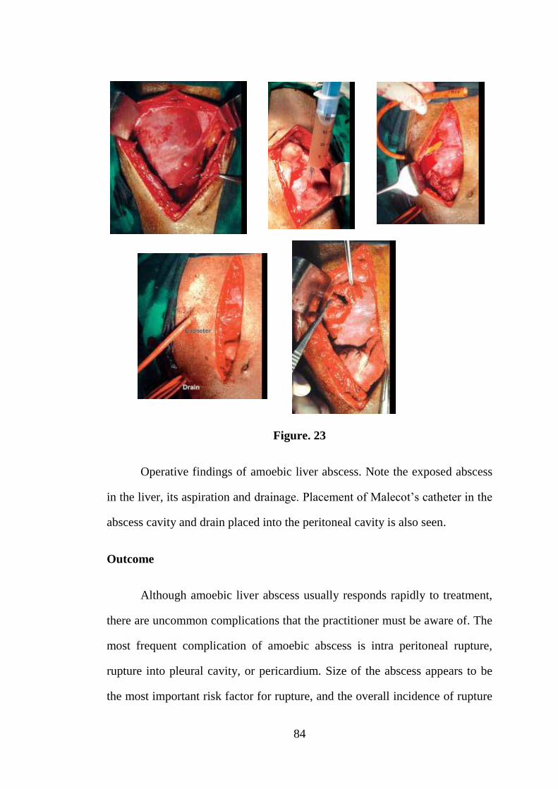
84
Figure. 23
Operative findings of amoebic liver abscess. Note the exposed abscess
in the liver, its aspiration and drainage. Placement of Malecot’s catheter in the
abscess cavity and drain placed into the peritoneal cavity is also seen.
Outcome
Although amoebic liver abscess usually responds rapidly to treatment,
there are uncommon complications that the practitioner must be aware of. The
most frequent complication of amoebic abscess is intra peritoneal rupture,
rupture into pleural cavity, or pericardium. Size of the abscess appears to be
the most important risk factor for rupture, and the overall incidence of rupture
85
ranges from 3% to 17%. Most peritoneal ruptures tend to be contained by the
diaphragm, abdominal wall or omentum, but rupture can fistulize into a
hollow viscus. A peritoneal rupture usually presents as abdominal pain,
peritonitis, and either a mass or generalized distention. Laparotomy was
advocated in the past for this complication, but now most cases are managed
successfully with percutaneous drainage. Laparotomy is indicated in cases of
doubtful diagnosis, failure of conservative therapy, hollow viscus perforation,
fistulization resulting in hemorrhage or sepsis. Rupture into the pleural space
usually results in a large and rapidly accumulated effusion that collapses the
involved lung. Treatment consists of thoracentesis, but if secondary bacterial
infection ensues, more aggressive surgical approaches may be necessary.
Rupture can occur into the bronchi and is usually self-limited with postural
drainage, bronchodilators. Rarely, a left-sided abscess may rupture into the
pericardium and can present as an asymptomatic pericardial effusion or even
cardiac tamponade. This must be treated with aspiration. Other complications
include compression of the biliary tree or inferior vena cava from a very large
abscess and development of a brain abscess.
The mortality rate for all patients with amoebic liver abscess is about
5% and does not appear to be affected by the addition of aspiration to
metronidazole therapy or chronicity of symptoms. When an abscess ruptures,
the mortality rate is reported to be from 6% to as high as 50%. Factors
independently associated with poor outcome are elevated serum bilirubin
(>3.5 mg/dL), encephalopathy, hypoalbuminemia (<2.0 g/dL), multiple
abscess cavities, abscess volume greater than 500 mL, anemia, and diabetes.

86
Although clinical improvement after adequate treatment with antiamoebic
agents is the rule, radiologic resolution of the abscess cavity is usually
delayed. The average time to radiologic resolution is 3 to 9 months and can
take as long as years in some patients. Studies have shown that more than 90%
of the visible lesions disappear radiologically, but a small percentage of
patients are left with a clinically irrelevant residual lesion.

87
MATERIAL AND METHODS
SOURCE OF DATA
All cases of Liver abscesses presenting to the Surgery OPD or
Casualty of CHMC Hospital, referred from medical wards of CHMC hospital
or referred from outside diagnosed as case of liver abscess, in Chengalpattu
over a period of 1 and half years with Clinical/ Sonological/CT features of
Liver abscess.
METHOD OF COLLECTION OF DATA
Patient data collection and evaluation.
Data will be collected from all patients attending CHMC Hospital,
Chengalpattu in General Surgery OPD, Casualty and Inpatient
department, irrespective of their gender/ background /socio economic
status. Age above 18years included in the study. The patients will be
evaluated and followed up according to protocol.
Detailed history of patient will be entered in proforma.
Complete haemogram, LFT, Prothrombin time, Serology for amoebic
antigen will be sent immediately on presentation.
Ultrasound of Abdomen and Pelvis will be done routinely on the same
day of presentation.

88
Patient will be treated by medical/image guided drainage/surgical(open
drainage) based on its site, size and other clinical parameters.
Patient will be followed up daily clinically and LFT.
USG Abdomen will be repeated on the 3rd day if patient
symptomatically not relieved.
If the patient develops any of the complications like ruptured liver
abscess into any of the serosal cavity, patient will be immediately take
up for surgery.
Patient will be informed about any surgical procedure and proper
informed consent will be taken.
Patient data collected regarding:
Age :
Sex :
Occupation :
Chief presenting complaints :
Past-surgical history :
Past history of similar illness :
History of alcoholism :
History of diabetes :
89
History of immunodefiency states :
History of biliary tract disorder :
History of amoebic dysentery :
History of jaundice :
General examination of systems :
Respiratory system :
Per abdomen :
Inspection :
Palpation :
Percussion :
Per rectal :
If the patient is referred from elsewhere the details of the same will be
considered at the time of admission.
Blood investigations
Total count :
Bilirubin :
PT/INR :
Albumin :

90
Serology for amoebic antigen :
Imaging studies
CXR
X ray abdomen erect
USG abdomen
CT abdomen
Complications if developed will be assessed in detail.
Management strategies for liver abscesses followed
Medical management
Image guided drainage
Open drainage
Complications –followed up.
Follow-up of patients:
Patients will be followed up over a period of 6 months
1) Once in 2 weeks for first 2 months
2) Then on a monthly basis after discharge , To look for recurrent attacks
or To look for the development of complications and To monitor the
efficacy of the treatment given.

91
Inclusion Criteria:
1. All cases of liver abscess diagnosed clinically and/or
ultrasonographically.
2. All cases of bacterial and parasitic liver abscess
3. All cases in evolving, liquefied & ruptured stage with or without
peritonitis
4. All cases of Diagnosed Liver Abscess being referred to CHMC.
Exclusion Criteria:
Traumatic Liver Abscess
Past history of liver abscess
Sample size: 60cases.
Study design: Prospective study
Duration of study: 1 1/2 years.
This study was done from MARCH 2014 to AUGUST 2015 on 60
patients. Case selection for the study will be done in the initial 1 yr followed
by follow up totally for 6 months.
Study place: CHMC, Chengalpattu
Does the study required any investigations or interventions to be
conducted on patients? If so, please describe briefly.
92
Routine blood investigations, X rays Chest and Abdomen, USG
Abdomen & LFT and Prothrombin time, Serology for amoebic antigen [CT
Abdomen if required]. Surgical intervention if the complication necessitates
the same.
Has ethical clearance been obtained from your institution?
Yes

93
OBSERVATION AND RESULTS
Totally 60 patients who met the inclusion criteria were studied.
AGE DISTRIBUTION
Table. 2
AGE
N 60
Mean 45.95
Median 44
Std. Deviation 12.423
Minimum 24
Maximum 73
The mean age of distribution is 45.95.
SEX DISTRIBUTION
Table. 3
SEX Frequency Percent
Male 47 78.3
Female 13 21.7
Total 60 100

94
Chart.1
In this study, out of 60 patients, liver abscess are present in 47 males
(78.3%) and 13 are females (21.7%).
CLINICAL FEATURES
Table.4
ABDOMINAL PAIN Frequency Percent
Present 44 73.3
Absent 16 26.7
Total 60 100
47
13
0
5
10
15
20
25
30
35
40
45
50
Male Female
SEX
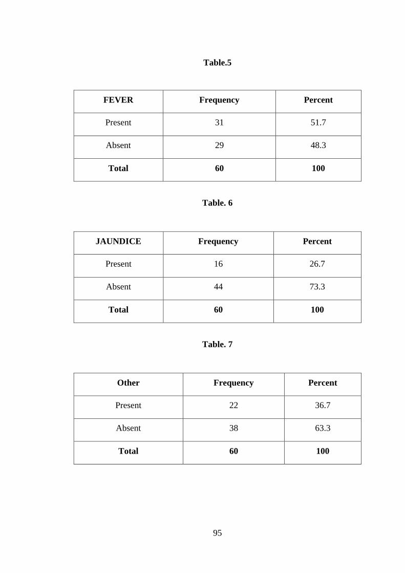
95
Table.5
FEVER Frequency Percent
Present 31 51.7
Absent 29 48.3
Total 60 100
Table. 6
JAUNDICE Frequency Percent
Present 16 26.7
Absent 44 73.3
Total 60 100
Table. 7
Other Frequency Percent
Present 22 36.7
Absent 38 63.3
Total 60 100
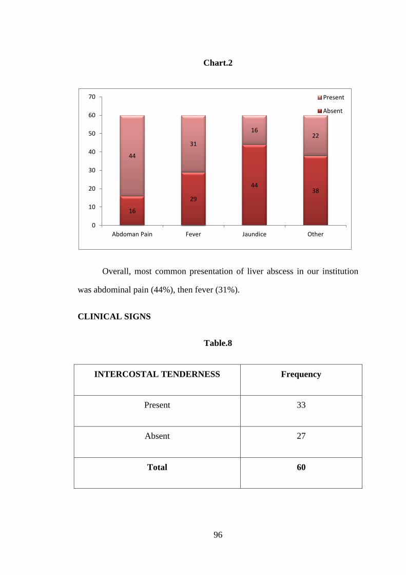
96
Chart.2
Overall, most common presentation of liver abscess in our institution
was abdominal pain (44%), then fever (31%).
CLINICAL SIGNS
Table.8
INTERCOSTAL TENDERNESS Frequency
Present 33
Absent 27
Total 60
16
29
4438
44
31
1622
0
10
20
30
40
50
60
70
Abdoman Pain Fever Jaundice Other
Present
Absent
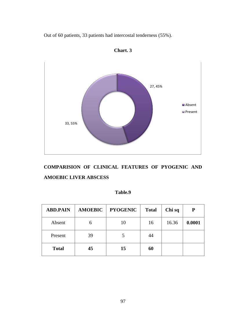
97
Out of 60 patients, 33 patients had intercostal tenderness (55%).
Chart. 3
COMPARISION OF CLINICAL FEATURES OF PYOGENIC AND
AMOEBIC LIVER ABSCESS
Table.9
ABD.PAIN AMOEBIC PYOGENIC Total Chi sq P
Absent 6 10 16 16.36 0.0001
Present 39 5 44
Total 45 15 60
27, 45%
33, 55%
Absent
Present
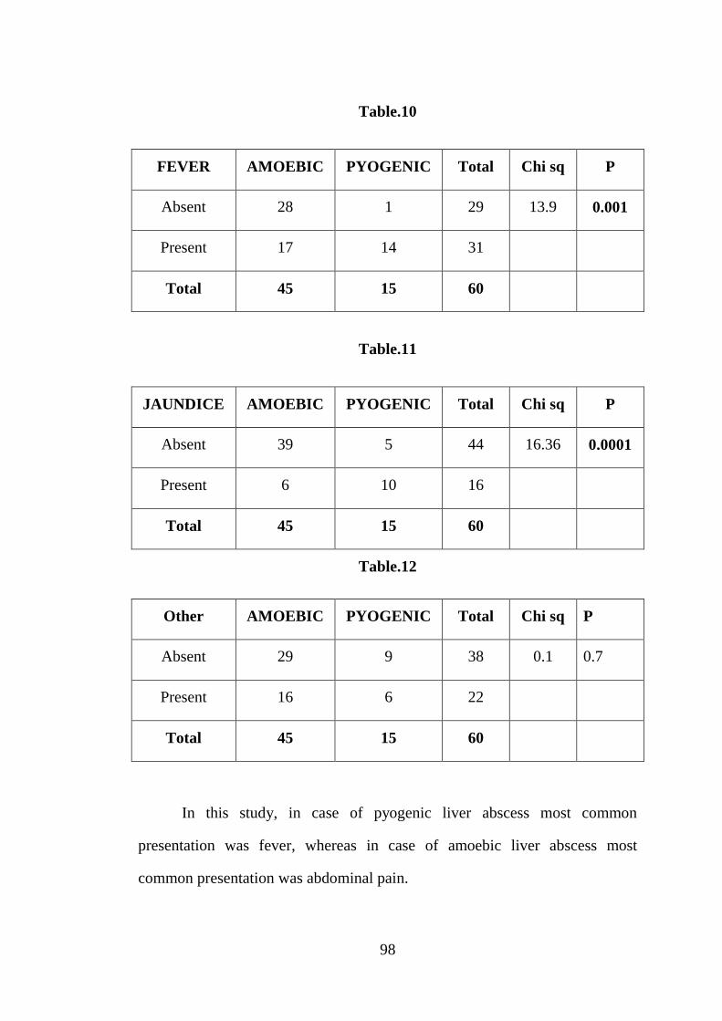
98
Table.10
FEVER AMOEBIC PYOGENIC Total Chi sq P
Absent 28 1 29 13.9 0.001
Present 17 14 31
Total 45 15 60
Table.11
JAUNDICE AMOEBIC PYOGENIC Total Chi sq P
Absent 39 5 44 16.36 0.0001
Present 6 10 16
Total 45 15 60
Table.12
Other AMOEBIC PYOGENIC Total Chi sq P
Absent 29 9 38 0.1 0.7
Present 16 6 22
Total 45 15 60
In this study, in case of pyogenic liver abscess most common
presentation was fever, whereas in case of amoebic liver abscess most
common presentation was abdominal pain.

99
LOCATION OF ABSCESS
Table.13
SITE Frequency
Left 7
Right 53
Total 60
Chart.4
Out of 60 cases, in this study abscess located in right lobe was 53 cases
(88.3%)
7, 12%
53, 88%
Left
Right

100
SOLITARY OR MULTIPLE
Table.14
Frequency
Single 54
Multiple 6
Total 60
Chart. 5
In this study, most of the abscesses were solitary (90%).
54, 90%
6, 10%
Single
Multiple
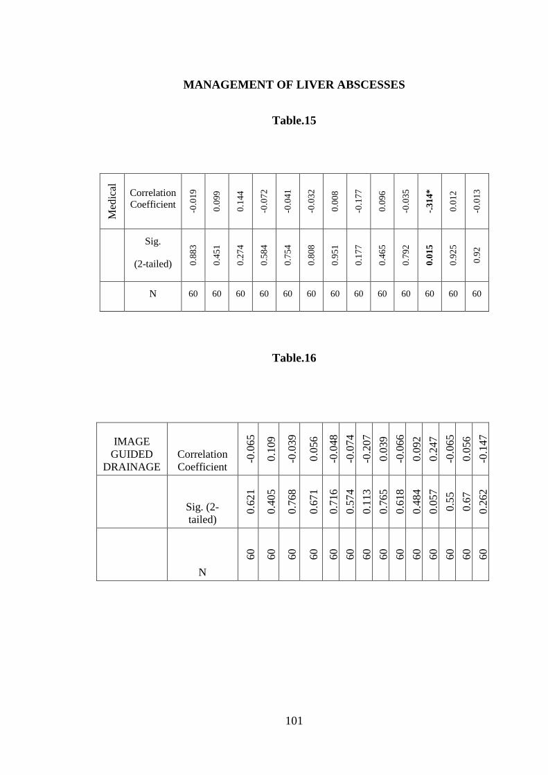
101
MANAGEMENT OF LIVER ABSCESSES
Table.15
Med
ical
Correlation
Coefficient
-0.0
19
0.0
99
0.1
44
-0.0
72
-0.0
41
-0.0
32
0.0
08
-0.1
77
0.0
96
-0.0
35
-.3
14*
0.0
12
-0.0
13
Sig.
(2-tailed) 0.8
83
0.4
51
0.2
74
0.5
84
0.7
54
0.8
08
0.9
51
0.1
77
0.4
65
0.7
92
0.0
15
0.9
25
0.9
2
N 60 60 60 60 60 60 60 60 60 60 60 60 60
Table.16
IMAGE
GUIDED
DRAINAGE
Correlation
Coefficient
-0.0
65
0.1
09
-0.0
39
0.0
56
-0.0
48
-0.0
74
-0.2
07
0.0
39
-0.0
66
0.0
92
0.2
47
-0.0
65
0.0
56
-0.1
47
Sig. (2-
tailed)
0.6
21
0.4
05
0.7
68
0.6
71
0.7
16
0.5
74
0.1
13
0.7
65
0.6
18
0.4
84
0.0
57
0.5
5
0.6
7
0.2
62
N
60
60
60
60
60
60
60
60
60
60
60
60
60
60

102
Table.17
OPEN
DRAI
NAGE
Correlation
Coefficient
-0.0
29
-.2
93
*
0.1
18
.29
3*
-0.0
24
0
.26
4*
.27
0*
0.1
31
-0.0
85
.72
8*
*
0.0
37
0.1
18
-.3
38
**
0.1
9
Sig. (2-
tailed)
0.8
28
0.0
23
0.3
7
0.0
23
0.8
53
1
0.0
42
0.0
37
0.3
17
0.5
17
0.0
001
0.7
81
0.3
7
0.0
08
0.1
47
N 60
60
60
60
60
60
60
60
60
60
60
60
60
60
60
Table.18
MEDICAL IMAGE SURGICAL
11 27 22

103
Chart. 6.
Out of 60 patients in our study, 11 patients (18%) responded to medical
management alone. In this study 27 patients (45%) were treated by image
guided aspiration or drainage. Those who required surgical drainage were 22
patient (37%), who are all not responding to the above modalities of treatment
and those with complication.
MICROBIOLOGY
Table.19
AMOEBIC Frequency
Absent 15
Present 45
Total 60
18%
45%
37%
MEDICAL IMAGE SURGICAL
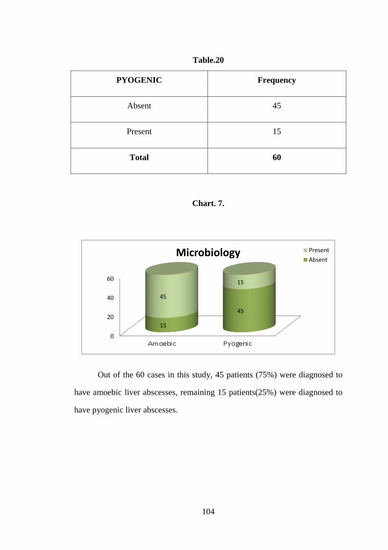
104
Table.20
PYOGENIC Frequency
Absent 45
Present 15
Total 60
Chart. 7.
Out of the 60 cases in this study, 45 patients (75%) were diagnosed to
have amoebic liver abscesses, remaining 15 patients(25%) were diagnosed to
have pyogenic liver abscesses.
0
20
40
60
15
45
45
15
Microbiology Present
Absent
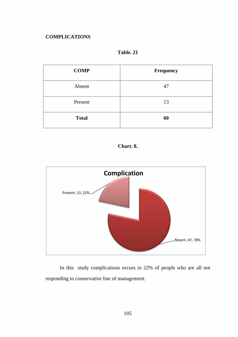
105
COMPLICATIONS
Table. 21
COMP Frequency
Absent 47
Present 13
Total 60
Chart. 8.
In this study complications occurs in 22% of people who are all not
responding to conservative line of management.
Absent, 47, 78%
Present, 13, 22%
Complication
106
DISCUSSION
In this study 60 patients who met the inclusion criteria were included.
In our study, the mean age of distribution of liver abscess is 45.95.
In the study, liver abscess are present in 47 males (78.3%) and 13 are
females (21.7%).
In our study, most common presentation was abdominal pain (44%),
then fever (31%). Out of 60 patients, 33 patients had intercostal tenderness
(55%).
In this study, in case of pyogenic liver abscess most common
presentation was fever, whereas in case of amoebic liver abscess most
common presentation was abdominal pain.
Out of 60 cases, abscess located in right lobe is 53 cases (88.3%). In
this study, most of the abscesses were solitary (90%). Out of 60 patients in our
study, 11 patients (18%) responded to medical management alone .Then 27
patients (45%) were treated by image guided aspiration or drainage. Those
who required and treated by surgical drainage was 22 patient (37%), who are
all not responding to the above modalities of treatment and those with
complication.
Out of the 60 cases in this study, 45 patients (75%) were diagnosed to
have Amoebic liver abscesses, remaining 15 patients (25%) were diagnosed to
have pyogenic liver abscesses. In this study complications occurs in 22% of
people who are all not responding to conservative line of management.

107
CONCLUSION
Most common liver abscess presenting in our institution was amoebic
in nature. Most common age of presentation was 45 years. Most common
presentation of liver abscess was abdominal pain. Most common clinical sign
was intercostal tenderness.
Alcoholism becomes the most frequently associated risk factor.
Ultrasound and CT scan abdomen plays an important role in diagnosing most
of the liver abscess patients presented in our institution. Eighteen percent
(18%) of the liver abscess patients were managed successfully with medical
management alone.
Those who are all not responding to medical management were treated
with image guided drainage/aspiration. Forty five percent (45%) of the
patients presented in our institution were successfully treated by image guided
drainage/aspiration.
Those who developed complications (ruptured liver abscess) and who
are all not responding to conservative line of management required emergency
open drainage. Thirty seven percent (37%) of patients who developed
complications on presentation or later were treated with open drainage in our
institution.

108
BIBLIOGRAPHY
1. Principles of Internal Medicine – Harrison’s 16th edition pg. no. 752,
Infectious diseases, Liver abscess.
2. 3. Oshsner A. Pyogenic abscess of liver. Am J Surg 1938;40:292
3. McFadzean AJS, Chang KPS, Wong CC. Solitary pyogenic abscess
of liver treated by closed aspiration and antibiotics: A repoprt of 14
consecutive cases and recovery. Br J Surg 1953;41: 141-152
4. Maingoat’s abdominal operations 9th edition volume 2, Seymour I.
Schwartz and Harold ellis pg.no. 1215 Section XI , Liver, Pyogenic &
Amoebic abscesses.
5. Blumgart – Surgery of the Liver, Biliary Tract and Pancreas 4th
Edition Vol.2 Pg.no. 928 Liver & Biliary Infection & Infestation,
Pyogenic Liver Abscess, R.W.Strong.
6. Maingoat’s abdominal operations 9th edition volume 2, Seymour I.
Schwartz and Harold ellis pg.no. 1218 Section XI , Liver, Pyogenic &
Amoebic abscesses.
7. Manual of Clinical problems in infectious disease 4th edition,
Lippincott Williams & Wilkins, pg.no.78,79
8. Maingoat’s abdominal operations 9th edition volume 2, Seymour I.
Schwartz and Harold ellis pg.no. 1218 Section XI , Liver, Pyogenic &
Amoebic abscesses.

109
9. Maingoat’s abdominal operations 9th edition volume 2, Seymour I.
Schwartz and Harold ellis pg.no. 1216-1220 Section XI , Liver,
Pyogenic & Amoebic abscesses.
10. Sebastin (Townsend) Textbook Of Surgery Pg. no. 1487 Infectious
Diseases, Pyogenic Liver Abscess.
11. Sebastin(Townsend_ Textbook Of Surgery Pg. no. 1487 Infectious
Diseases, Pyogenic Liver Abscess.
12. World J Gastroenterol 2008 April 7; 14(13): 2089-2093
Gastroenterology ISSN 1007-9327
13. McFadzean AJS, Chang KPS, Wong CC. Solitary Pyogenic abscess
of the liver treated by closed aspiration and antibiotics: a report of 14
consecutive cases with recovery. Br J Surg 1953; 41: 141-52.
14. Indian Journal Of Surgery, Medknow Publications on Behalf Of
Associations Of Surgeons Of India ISSN: 0972-2068, Vol.64, No.6,
Nov-Dec. 2002, pp. 516-519
15. Sebastin(Townsend) Textbook Of Surgery Pg. no. 1488 Infectious
Diseases, Pyogenic Liver Abscess.
16. Sebastin(Townsend) Textbook Of Surgery Pg. no. 1494 Infectious
Diseases, Pyogenic Liver Abscess.
110
PROFORMA
PARTICULARS OF PATIENT:
Name : Case no :
Age :
Sex : Date of Admission :
Religion : Date of Operation :
IP.No : Date of Discharge :
Address :
Chief Complaints:
Abdominal pain
Fever
Jaundice
Other manifestions
History of presenting complaints:
Abdomen pain:
Duration
Time of onset
Mode of onset
111
Site of pain
Radiation of pain
Character of pain
Aggravating factors
Relieving factors
Fever:
Duration
Type
Severity
Jaundice
Loss of appetite
Loss of weight
Past history:
Similar complaints
Abdominal surgery
Personal history:
Diet
Appetite
Sleep
Alcohol intake
Smoking
Bowel/Bladder
112
Family history:
Menstrual history:
Menarche
Lmp
Menstrual cycles
PHYSICAL EXAMINATION
General physical examination:
Built, Pallor
Nourishment, Icterus
Lymhpadenopathy, Cyanosis
Clubbing , Pedal edema
VITALS:
Pulse
Blood pressure
Respiratory rate
Temperature

113
ABDOMINAL EXAMINATION
Inspection:
Contour of abdomen
Movements of all quadrants with abdomen
Visible pulsation and peristalsis
Skin
Hernial orifices
Umbilicus
Scrotum
Palpation:
Local rise of temperature
Local Tenderness
Right hypochondrial tenderness
Intercostal tenderness
Any organomegaly
Percussion:
Auscultation:
Rectal examination:
114
SYSTEMIC EXAMINATION
Cardiovascular system :
Respiratory system :
Central Nervous system :
Provisional diagnosis :
Blood investigations :
Total count
Bilirubin
PT/INR
Albumin
Serology for amoebic antigen.
Imaging studies
CXR
X ray abdomen erect
USG abdomen
CT abdomen
Complications if developed will be assessed in detail.

115
Management strategies for liver abscesses followed
Medical management
Image guided drainage
Open drainage
Complications –followed up.
Follow up of patients:
Patients will be followed up over a period of 6 months
1. once in 2 weeks for first 2 months
2. then on a monthly basis after discharge, To look for recurrent attacks or
To look for the development of complications and To monitor the
efficacy of the treatment given.

116
CONSENT FORM
For Operation / Anaesthesia/ Study
I Hosp. No. in my
full Senses hereby give my complete consent for ___________ or any other
Procedure deemed at which is a / and diagnostic procedure / biopsy /
Transfusion/ operation to be performed on me / my ward
age under any anaesthesia deemed fit. The nature and
risks. Involved in the procedure have been explained to me to my satisfaction.
For academic and scientific purpose, the operation / procedure may be.
Televised or photographed.
Date:
Signature / Thumb Impression
Name :
of patient / Guardian
Designation :
Guardian
Relationship :
Full address
117
KEYWORDS TO MASTER CHART
S.No- SERIAL NUMBER
SEX
1. MALE
2. FEMALE
ABDOMINAL PAIN
1-PRESENT
0-ABSENT
FEVER
1-PRESENT
0-ABSENT
JAUNDICE
1-PRESENT
0-ABSENT
OTHER SYMPTOMS
1-PESENT
0-ABSENT’
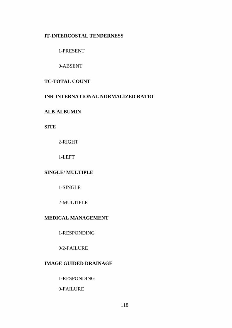
118
IT-INTERCOSTAL TENDERNESS
1-PRESENT
0-ABSENT
TC-TOTAL COUNT
INR-INTERNATIONAL NORMALIZED RATIO
ALB-ALBUMIN
SITE
2-RIGHT
1-LEFT
SINGLE/ MULTIPLE
1-SINGLE
2-MULTIPLE
MEDICAL MANAGEMENT
1-RESPONDING
0/2-FAILURE
IMAGE GUIDED DRAINAGE
1-RESPONDING
0-FAILURE
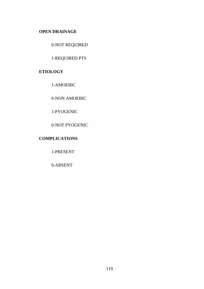
119
OPEN DRAINAGE
0-NOT REQUIRED
1-REQUIRED PTS
ETIOLOGY
1-AMOEBIC
0-NON AMOEBIC
1-PYOGENIC
0-NOT PYOGENIC
COMPLICATIONS
1-PRESENT
0-ABSENT
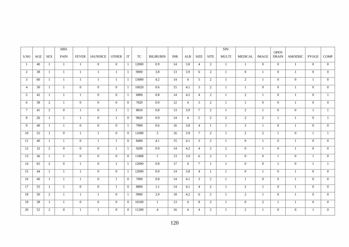
120
S.NO AGE SEX
ABD.
PAIN FEVER JAUNDICE OTHER IT TC BILIRUBIN INR ALB SIZE SITE
SIN/
MULTI MEDICAL IMAGE
OPEN
DRAIN AMOEBIC PYOGE COMP
1 40 1 1 1 0 0 1 12000 0.9 14 3.8 4 2 1 1 0 0 1 0 0
2 38 1 1 1 1 1 1 9000 3.8 13 3.9 6 2 1 0 1 0 1 0 0
3 60 1 1 1 1 1 1 13000 4.2 14 4 5 2 1 2 1 0 0 1 0
4 30 1 1 0 0 0 1 10020 0.6 15 4.1 3 2 1 1 0 0 1 0 0
5 42 1 1 1 0 0 1 6800 0.8 14 4.2 4 2 1 2 1 0 1 0 1
6 38 2 1 0 0 0 0 7820 0.9 12 4 3 2 1 1 0 0 1 0 0
7 41 2 0 1 0 1 1 8810 0.8 13 3.9 7 2 1 2 1 0 0 1 1
8 26 1 1 1 0 1 0 9820 0.9 14 4 5 2 2 2 2 1 1 0 1
9 40 1 1 0 0 0 1 7000 0.6 16 3.8 4 1 1 2 1 0 1 0 0
10 55 1 0 1 1 0 0 11000 3 16 3.9 7 2 1 2 2 1 0 1 1
11 40 1 1 0 1 1 0 8400 4.1 15 4.1 3 2 1 0 1 0 1 0 0
12 32 2 0 0 0 1 1 9200 0.9 14 4.2 4 2 2 0 1 0 1 0 0
13 36 1 1 0 0 0 0 11808 1 13 3.9 6 2 1 0 0 1 0 1 0
14 65 2 0 1 0 1 1 12000 0.8 17 4 7 1 1 0 0 1 0 1 1
15 44 1 1 1 0 0 1 12000 0.9 14 3.8 4 1 1 0 1 0 1 0 0
16 40 1 1 1 0 1 0 7000 0.8 14 4.1 3 2 1 1 0 0 1 0 0
17 55 1 1 0 0 1 0 8800 1.1 14 4.1 4 2 1 2 1 0 1 0 0
18 50 2 1 1 1 0 1 9900 2.9 18 4.2 6 2 1 2 1 0 1 0 0
19 38 1 1 0 0 0 0 10100 1 13 4 8 2 1 0 2 1 1 0 0
20 52 2 0 1 1 0 0 11200 4 16 4 4 2 1 2 1 0 0 1 0

121
S.NO AGE SEX
ABD.
PAIN FEVER JAUNDICE OTHER IT TC BILIRUBIN INR ALB SIZE SITE
SIN/
MULTI MEDICAL IMAGE
OPEN
DRAIN AMOEBIC PYOGE COMP
21 30 1 1 0 0 1 1 10000 1 14 4.1 3 2 1 1 0 0 1 0 0
22 24 1 1 0 0 1 0 9620 0.8 13 3.9 4 2 1 0 1 0 1 0 0
23 50 1 1 0 1 0 1 8400 3 16 4.2 5 1 1 0 2 1 1 0 0
24 48 1 1 1 0 0 1 9900 0.9 13 4.2 4 2 1 0 1 0 1 0 0
25 46 1 0 1 0 0 1 10110 1 12 4.1 5 2 2 0 2 1 1 0 1
26 44 2 1 1 0 1 0 7800 1.1 12 4.5 6 2 1 0 2 1 1 0 0
27 38 1 1 0 0 0 0 6400 0.8 12 4.4 4 2 1 0 1 0 1 0 1
28 70 1 0 1 1 1 0 7100 4.2 15 3.2 6 2 1 0 2 1 0 1 0
29 68 1 1 0 0 0 1 8900 0.7 13 3.1 5 2 1 0 2 1 1 0 0
30 49 1 1 1 0 0 1 12800 0.8 13 4 5 2 1 2 1 0 0 1 0
31 41 1 1 0 0 0 1 8900 1 14 4.2 3 2 1 1 0 0 1 0 0
32 72 1 0 1 1 0 1 11000 2.4 16 4 5 2 1 0 1 0 0 1 0
33 32 1 1 0 0 1 0 8900 1.1 13 4.5 4 2 1 0 1 0 1 0 0
34 38 1 1 1 0 0 0 12000 0.9 12 4.6 3 1 1 2 1 0 1 0 0
35 44 1 1 0 0 0 0 9800 0.8 14 4.7 5 2 1 0 2 1 1 0 0
36 51 1 0 1 0 0 0 6000 1 13 3.8 2 2 2 1 0 0 1 0 0
37 45 2 1 0 0 1 1 10200 0.6 16 3.4 3.5 2 1 0 1 0 1 0 0
38 62 1 0 1 1 0 0 13000 4 17 3 7 2 1 0 0 1 0 1 1
39 29 2 1 0 0 0 1 7620 0.9 12 3.9 5 1 1 0 1 0 1 0 0
40 40 1 1 0 0 0 1 8880 1.1 14 4.1 4 2 1 0 1 0 1 0 0
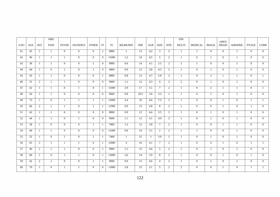
122
S.NO AGE SEX
ABD.
PAIN FEVER JAUNDICE OTHER IT TC BILIRUBIN INR ALB SIZE SITE
SIN/
MULTI MEDICAL IMAGE
OPEN
DRAIN AMOEBIC PYOGE COMP
41 45 1 1 0 0 0 1 8800 1 13 4.2 3 2 1 1 0 0 1 0 0
42 40 1 1 1 0 0 0 11000 1.2 14 4.5 5 2 1 0 1 0 1 0 0
43 38 1 1 0 0 1 0 8900 0.6 14 4.1 2.5 2 1 1 0 0 1 0 0
44 44 1 0 1 0 1 0 4800 0.9 12 3.8 4.5 2 1 0 1 0 1 0 0
45 26 1 1 0 0 0 1 8800 0.8 13 4.7 5.8 2 1 0 2 1 1 0 1
46 31 2 1 1 0 0 0 9600 1.1 12 4.3 4 2 2 0 1 0 1 0 0
47 42 1 1 0 1 0 1 11000 3.9 17 3.1 7 2 1 0 2 1 1 0 1
48 56 1 1 0 0 0 0 8600 0.8 18.5 3.8 3.5 1 1 0 1 0 1 0 0
49 73 1 0 1 1 1 1 12000 3.4 16 4.6 7.5 2 1 0 0 1 0 1 1
50 66 2 1 1 0 1 1 11900 0.9 13 4.8 8 2 1 0 0 1 0 1 0
51 42 1 1 0 0 0 0 8900 0.9 12 4.6 3.5 2 1 0 1 0 1 0 0
52 44 1 1 0 1 0 0 9600 1.1 13 4.1 4.9 2 1 0 1 0 1 0 0
53 38 1 0 0 0 1 1 7800 1.2 12 3.8 7 2 1 0 0 1 1 0 0
54 46 1 1 0 0 0 0 11000 0.8 14 3.1 2 2 1 1 0 0 1 0 0
55 52 1 0 1 0 1 1 7400 1 15 3 3.9 2 1 0 1 0 1 0 0
56 65 2 1 1 1 0 1 12000 4 16 4.1 7 2 1 0 0 1 0 1 1
57 46 1 1 1 0 0 1 9900 1.1 15 4.6 5 2 1 0 1 0 1 0 0
58 68 1 0 1 1 0 1 13000 3.6 14 3.9 8 2 1 0 0 1 0 1 0
59 42 2 1 0 0 1 1 9000 0.9 13 4.6 4 2 1 0 1 0 1 0 0
60 70 1 0 1 1 0 0 11800 3.8 15 3.1 5 2 2 0 0 1 0 1 1

123