Embed Size (px)
Citation preview

IP Journal of Nutrition, Metabolism and Health Science 2020;3(3):85–89
Content available at: https://www.ipinnovative.com/open-access-journals
IP Journal of Nutrition, Metabolism and Health Science
Journal homepage: www.ipinnovative.com
Review Article
A clinical case study and medical nutritional therapy in pediatricmeningioencephalitis
Edwina Raj1,*, C P Ravi Kumar2, Arunadevi J1, Rebecca Renee Joseph1,Chetan Ginigeri3
1Dept. of Clinical Nutrition and Dietetics, Aster CMI Hospital, Bengaluru, Karnataka, India2Dept. of Paediatric Neurology, Aster CMI Hospital, Bengaluru, Karnataka, India3Dept. of Paediatric, Aster CMI Hospital, Bengaluru, Karnataka, India
A R T I C L E I N F O
Article history:Received 13-08-2020Accepted 23-09-2020Available online 28-10-2020
Keywords:Pediatric MeningoencephalitisViral EncephalitisNasogastric feedingHuman herpesvirusMedical Nutrition Therapy inMeningoencephalitis
A B S T R A C T
Meningoencephalitis is an inflammation of the brain and its surrounding protective membranes. Meningitisis a life-threatening disease and can lead to significant sequelae. We were presented with a 6 year 5 monthsold female child on ventilation, retrieved by referral hospital on examination revealed human herpesvirus6 through cerebrospinal fluid analysis and diagnosed with meningoencephalitis on Magnetic ResonanceImaging(MRI) with three repeated failed extubation and hence planned tracheostomy. Child presented nomovements with suspected motor or sensory (vision, hearing) disability, generalized tonic-clonic seizure(GTCS), altered sensorium requiring a long term rehabilitation and nutrition support. On close monitoring,a continuous effort of three months and with a multidisciplinary clinical approach from different disciplinesincluding neurologist, intensivist, clinical dietitian and physiotherapy, the child successfully completedthe long term rehabilitation treatment with remarkable improvement observed within 12 weeks bothneurologically and nutritionally with a better clinical outcome.
© 2020 Published by Innovative Publication. This is an open access article under the CC BY-NC license(https://creativecommons.org/licenses/by-nc/4.0/)
1. Introduction
Meningoencephalitis can be caused by bacteria, viruses,fungi, and protozoan or as secondary sequel of otherinflammations like AIDS. The inflammation causesthe brain to swell leading to confusion, changes inalertness, and seizures. In general, meningoencephalitis andencephalitis represent uncommon responses to commoninfections. Most infected patients have a mild syndromeof meningoencephalitis rather than severe encephalitis.1
Viral encephalitis also includes slow viral and chronicdegenerative diseases of presumed viral origin.2 Correctimmediate diagnosis and introduction of symptomatic andspecific therapy has a dramatic influence upon survivaland reduces the extent of permanent brain injury. Virusesare predominant causes of infectious encephalitis. Viralencephalitis is a medical emergency and its prognosis
* Corresponding author.E-mail address: [email protected] (E. Raj).
depends on the kind of causative pathogen and immunestatus of the patient. Herpes simplex virus (HSV) is themost prevalent cause of viral encephalitis in the developedcountries with the annual incidence rate of 1/250,000-500,000 people. In viral encephalitis, the virus first entersthe body and then replicates in local or regional tissues,such as gastrointestinal tract, skin, urogenital, or respiratorysystem. Subsequent dissemination in the central nervoussystem occurs through hematogenic routes (enterovirus,arbovirus, HSV, HIV, mumps) or through retrogradetransportation via axons, such as in the case of herpesvirus, rabies virus, or prion protein. Most of the guidelinescited focal neurological signs, seizures, fever, altered levelsof consciousness (ALOC) and changes to personality orbehaviour as signs and symptoms of encephalitis in bothchildren and adults.3
https://doi.org/10.18231/j.ijnmhs.2020.0172582-6301/© 2020 Innovative Publication, All rights reserved. 85

86 Raj et al. / IP Journal of Nutrition, Metabolism and Health Science 2020;3(3):85–89
2. Materials and Methods
A 6yr 5m old female was retrieved from another hospitalwith ET tube insitu. Child developed fever 7days backwhich was high grade not associated with chills and rigors,decreasing on medication temporarily. Child complainedof headache more in the frontal region and no diurnalvariation. Child had decreased appetite since the beginningof fever and since last 2 days was taking only liquids.On 6th day of fever child became drowsy and hencewas taken to hospital and was admitted. Child developedconvulsions on the same day in the form of GTCSwhich was aborted with Midazolam. Child was startedon Levetiracetam. On the same day child was intubatedin v/o poor GCS. LP was done in the hospital whichshowed predominant polymorphs and so child was startedon Magnex (Cefoperazone + Sulbactam), Acyclovir. MRIwas done which was suggestive of meningoencephalitis andchild was started on steroids. HSV PCR, Blood cultureand CSF culture were sent and reports awaited. Child wasreferred to our hospital for further management.
2.1. Physical Examination
On physical examination, general physical findings shouldbe noted including heart rate, respiratory rate, bloodpressure, presence or absence of meningismus, and signs ofinvolvement of other systems, particularly skin rashes andlymph nodes, as well as signs of trauma. Fundoscopicalexamination including indirect ophthalmoloscopy mayelucidate vasculitis and also, the shaken baby syndrome incase of non-traumatic injury. Neurological findings dependon which part of the brain is primarily involved. Findingsmay predominate in any part of the brain or non - CNS sitesmay predominate.
2.2. Past History
Child had episode of fever in the first week of Februarywhich subsided after taking medications. Child had anotherepisode of fever two weeks back which lasted for 4 days andthen subsided. Child was active during the fever free period.
2.3. Birth History
Full term child of Normal delivery. Birth weight was 2.6kg.No History of NICU stay. Child has been vaccinated up - tothe age of 3 years.
2.4. Family History
Patient is the second child of non-consanguineous marriage.Child has one elder and one younger sibling. No history ofsimilar complaints in the family
2.5. Case details
2.5.1. VitalsHR-80/min
Spo2- 98% with 80% Fio2BP- 102/60 mm hgPP- well felt and warm
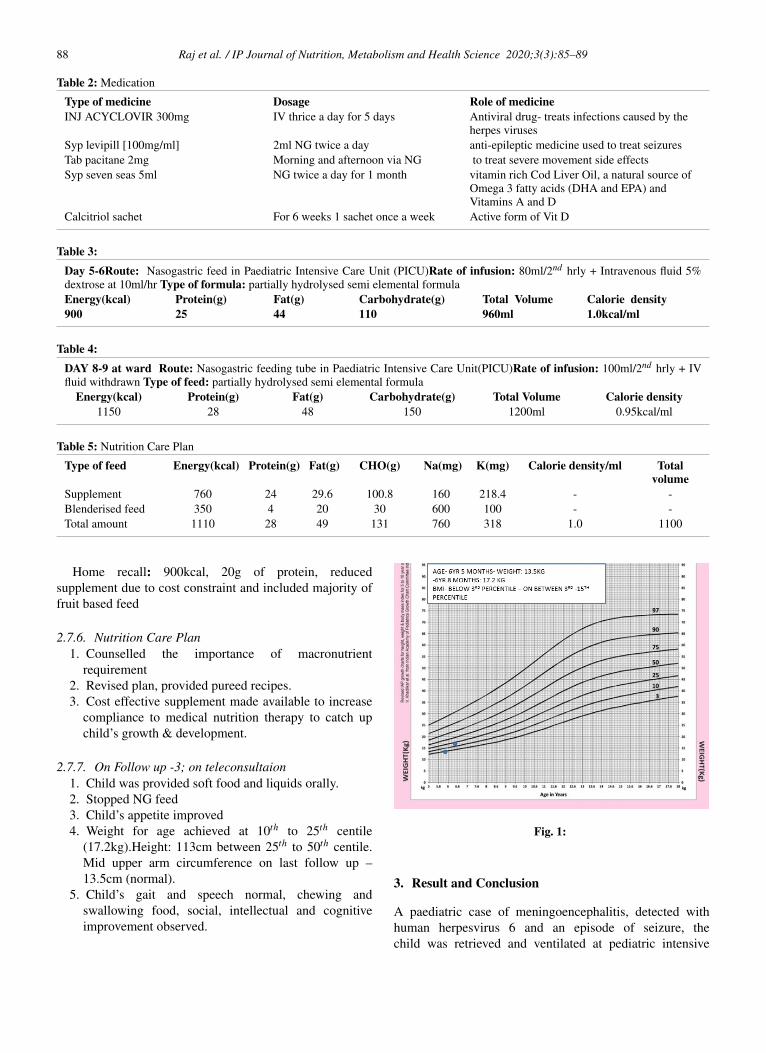
2.5.2. Physical Examination1. Height: Unable to stand (confined to bed & ventilated)2. Weight for Age on admission- 13.6kg (on 3rd centile)3. Weight for age on follow up after 3 weeks- 17.5kg
(between 10th to 25th centile)4. Mid upper arm circumference on last follow up –
13.5cm
2.6. Treatment Plan
On day one, child was on mechanical ventilation withICD insertion. EEG was conducted which showed slowingof waves (probable encephalopathy). Medications such asCeftriaxone, Acyclovir and azithromycin were introduced.Neuro-protection and anti-edema measures were taken.Sedation was optimised and neurologist opinion was given.Patient wasn’t given anything orally. ECHO procedure wasdone which showed normal
On day two, ceftiaxone drug was continued along withNG feeds and IV fluids were tapered down. Sodium levelswere monitored upto levels 135-145 and 3% Nacl wastapered down accordingly and NG feeds were initiated.Patient had altered sensorium
On day three, sedation was stopped and sensorium wasreassessed. She developed tonic posturing, hence sedationwas restarted. Polyuria was present so was decided to bereplaced with NS if hemodynamic instability was seen.
On day four, tonic posturing was still present withpolyuria, right sided pneumothorax was observed whichresolved. A trial of CPAP was given but the child didnot tolerate more than 15 min hence it was changed backto Control mode. CSF analysis was done which showednormal protein sugars, differential count of all lymphocytesand Human Herpes virus 6 was detected. Extubation wasattempted for the past 3 days, without success. Child hadneeded long term rehabilitation, hence Tracheostomy wasplanned. Child did not show any movements, which meantthat there would be a motor or sensory (vision, hearing)disability. Child’s parents were counselled regarding theneed for ventilation and tracheostomy. Child was openingeyes spontaneously with no eye contact. Ophthalmicexamination was done which showed normal.
On day five, child was seen to be holding on withoutventilation but however intubation and tracheostomy wasrequired to be considered. Child did need long termrehabilitation and support as she was critically ill. Child wasextubated with spontaneous breathing trial (SBT) Trial and

Raj et al. / IP Journal of Nutrition, Metabolism and Health Science 2020;3(3):85–89 87
Table 1: Clinical Parameters
Electrolytes 14.3.20 16.3.20 17.3.20Sodium (mg) 141 139 143Potassium(mg) 3.35 4.0 4.0Urea(mg) 21 -Heamoglobin(g) 9.3 - 9.7Magnesium (mg) Normal range (1.7-2.3) 1.6mg 1.7
Dexamethasone 4mg was given.On day six, sensorium still remained poor however,
polyurea was better and pneumothorax was resolved. RightICD was placed and on removal lungs showed goodexpansion on the right side. Child was on HFNC with stablehemodynamics, Airway maintained breathing pattern andshowed improvement, blood gas was normal with goodoxygenation but sensorium remained poor, with no focusand lifting limbs against gravity.
1. On day seven, Air mattress was used and the child’sposition was changed every four hours with help ofphysiotherapy. Child was maintaining saturation inroom air with no respiratory distress. Central NervousSystem showed better eye focusing, pacitane drugwas initiated and levetiracetam was given orally. Onday ten, the patient was mobilized, antibiotics andNG feed were continued along with multivitamins,calcium and patient was shifted to ward. HNFC wasdiscontinued with intermittent focusing and posturingepisodes appeared to be less. Child was still onNG feeds and was discharged after training motheron NG feeds and physiotherapy. On discharge shewas hemodynamically stable, but had neurologicalsequela meaning she was unable to recognize or makeeye contact, not able to move against gravity. Sherequired a long term physiotherapy and nutritionalrehabilitation. Child’s prognosis was unpredictable.However on OPD visit patient still had issues withposturing after a week but tolerated NG feeds.
2.7. Nutrition Care Plan
Day one the child was nil per oral, thereby nasogastric feedwas initiated within 24 hours and IV fluids were tapered. Apolymeric formula was provided as trickle feed but replacedwith partially hydrolysed formula after 3 days due to feedintolerance.4 A peptide based semi- elemental formula wasprescribed at the rate of 10 ml 2nd hrly thereby full fluidswas targeted by day 6 due to a potential risk of refeedingsince the child had history of poor oral intake since oneweek, underweight and low levels of potassium, magnesiumwas observed.
2.7.1. Nutrition assessment1. Subjective Global Assessment grade- C
2. Weight for age on admission 13.5kg – on 3rd centile(underweight).
2.7.2. Nutritional challenges1. Underweight for age with high risk of refeeding
syndrome:5 poor oral intake since 1 week, was onlyon oral liquid & hypokalemia on admission was noted.
2. Feed intolerance; on starting trickle feeds (standardpolymeric formula)
3. Route: nasogastric feeding tube until discharge withno improvements neurologically observed for 9 days.
4. Poor socioeconomic status and lack of awareness ofimportance of macro nutrients among the family.
5. Mother had the burden of taking care of twoother siblings and manage the family, therefore lessimportance was given to this child.
2.7.3. Nutrient requirement in PICU:Energy (kcal): 802 kcal/day (WHO equation5)
Protein(g): 24.3g /day (1.8g/kg/day)Fat(g): 40% of total energy prescribedCarbohydrate(g): 50% of total energy prescribedFluid requirement (ml):1175ml
2.7.4. Nutrient requirement when shifted to ward:Energy (kcal): 75kcal/kg/day (RDA) - modified RDA:85kcal/kg/day (1147kcal/day)
Protein(g): 1.1g/kg/day (RDA)-modified RDA 2g /day(27g/day)
Fat (g): 35% of total energy prescribedCarbohydrate (g): 55% of total energy prescribedFluid requirement (ml):1175mlOn follow up-1 at OPD – after 4 weeks of dischargeHome recall: followed as prescribed & requested for
blenderised home based feed preparationRoute- Nasogastric feeding tube + Oral feeds attempted
(pureed, calorie dense feed thrice /day)
2.7.5. Supplement: continued the same supplement astolerated earlier by the childComposition of home based feed: nuts, oilseeds, dry-fruit,meat broth, vegetables, fruit, turmeric, pepper, coconut oil,olive oil
On Follow up 2 at OPD: after 8 weeks of discharge
88 Raj et al. / IP Journal of Nutrition, Metabolism and Health Science 2020;3(3):85–89
Table 2: Medication
Type of medicine Dosage Role of medicineINJ ACYCLOVIR 300mg IV thrice a day for 5 days Antiviral drug- treats infections caused by the
herpes virusesSyp levipill [100mg/ml] 2ml NG twice a day anti-epileptic medicine used to treat seizuresTab pacitane 2mg Morning and afternoon via NG to treat severe movement side effectsSyp seven seas 5ml NG twice a day for 1 month vitamin rich Cod Liver Oil, a natural source of
Omega 3 fatty acids (DHA and EPA) andVitamins A and D
Calcitriol sachet For 6 weeks 1 sachet once a week Active form of Vit D
Table 3:Day 5-6Route: Nasogastric feed in Paediatric Intensive Care Unit (PICU)Rate of infusion: 80ml/2nd hrly + Intravenous fluid 5%dextrose at 10ml/hr Type of formula: partially hydrolysed semi elemental formulaEnergy(kcal) Protein(g) Fat(g) Carbohydrate(g) Total Volume Calorie density900 25 44 110 960ml 1.0kcal/ml
Table 4:DAY 8-9 at ward Route: Nasogastric feeding tube in Paediatric Intensive Care Unit(PICU)Rate of infusion: 100ml/2nd hrly + IVfluid withdrawn Type of feed: partially hydrolysed semi elemental formula
Energy(kcal) Protein(g) Fat(g) Carbohydrate(g) Total Volume Calorie density1150 28 48 150 1200ml 0.95kcal/ml
Table 5: Nutrition Care Plan
Type of feed Energy(kcal) Protein(g) Fat(g) CHO(g) Na(mg) K(mg) Calorie density/ml Totalvolume
Supplement 760 24 29.6 100.8 160 218.4 - -Blenderised feed 350 4 20 30 600 100 - -Total amount 1110 28 49 131 760 318 1.0 1100
Home recall: 900kcal, 20g of protein, reducedsupplement due to cost constraint and included majority offruit based feed
2.7.6. Nutrition Care Plan1. Counselled the importance of macronutrient
requirement2. Revised plan, provided pureed recipes.3. Cost effective supplement made available to increase
compliance to medical nutrition therapy to catch upchild’s growth & development.
2.7.7. On Follow up -3; on teleconsultaion1. Child was provided soft food and liquids orally.2. Stopped NG feed3. Child’s appetite improved4. Weight for age achieved at 10th to 25th centile
(17.2kg).Height: 113cm between 25th to 50th centile.Mid upper arm circumference on last follow up –13.5cm (normal).
5. Child’s gait and speech normal, chewing andswallowing food, social, intellectual and cognitiveimprovement observed.
Fig. 1:
3. Result and Conclusion
A paediatric case of meningoencephalitis, detected withhuman herpesvirus 6 and an episode of seizure, thechild was retrieved and ventilated at pediatric intensive
Raj et al. / IP Journal of Nutrition, Metabolism and Health Science 2020;3(3):85–89 89
care unit with repeated failed extubation, poor prognosis,underweight on admission, with high risk of refeedingsyndrome, altered sensorium. Initially there was no signs ofimprovement observed neurologically (unable to recognizeor make eye contact, not able to move against gravity)in first two weeks at the onset of disease and continuedon nasogastric tube on discharge. The child was regularlyfollowed up with pediatric neurologist, nutritionist andphysiotherapist to ensure the child’s catch up growth anddevelopment is attained, to prevent growth faltering andprotein energy wasting. The medical nutrition therapyinvolved fine tuning of child’s nutrition care plan basedon the nutritional challenges, with regular monitoring wewere able to observe successful clinical outcome in the childwith an improvement in weight , transition to oral feed andnormal neurological development was observed.
4. Source of Funding
None.
5. Conflict of Interest
None.
References1. Study of acute viral meningoencephalitis in children in sub-himalayan
tarai region: clinico-epidemiological, etiological, and imaging profile.Indian J Child Health. 2015;2(4):177–81.
2. Kennedy PGE. Viral encephalitis: causes, differential diagnosis, andmanagement. Journal of Neurology, Neurosurgery and Psychiatry;2015.
3. Steiner I, Budka H, Chaudhuri A. Viral meningoencephalitis: Areview of diagnostic methods and guidelines for management. Eur JNeurol;17(8):999–e57.
4. Goday PS, Mehta NM, Lee JH. Pediatric Critical Care Nutrition. AsiaPac J Clin Nutr. 2015;30(4):581.
5. Mehta NM, Skillman HE, Irving SY, Coss-Bu JA, Vermilyea S,Farrington EA, et al. Guidelines for the Provision and Assessment ofNutrition Support Therapy in the Pediatric Critically Ill Patient: Societyof Critical Care Medicine and American Society for Parenteral andEnteral Nutrition. JPEN J Parenter Enteral Nutr. 2017;41(5):706–42.
Author biography
Edwina Raj Senior Dietitian
C P Ravi Kumar Consultant
Arunadevi J Dietitian
Rebecca Renee Joseph Dietitian
Chetan Ginigeri Consultant
Cite this article: Raj E, Kumar CPR, Arunadevi J, Joseph RR, GinigeriC. A clinical case study and medical nutritional therapy in pediatricmeningioencephalitis. IP J Nutr Metab Health Sci 2020;3(3):85-89.