Embed Size (px)
Citation preview

BULLETIN OF MARINE SCIENCE.30(4):858-870, 1980
A CLARIFICATION OF THE GENUS PERNA (MYTILIDAE)
Scott E. Siddull
ABSTRACT
Many morphological features of adult mussels are variable and often of limited value in establishing taxonomic relationships. Synonymies in the Mytilidae are numerous, the genus Perna notwithstanding. The historical development and geographical distribution of the three species placed in the genus Pema are reviewed.
As a post-metamorphic character, the presence of Lateral hinge teeth which develop after metamorphosis are unique and consistent criteria for distinguishing juvenile mussels of the genus Perna. The development and fate of provincular, primary lateral, secondary lateral and dysodont hinge teeth is presented in a series of scanning electron micrographs. Dis- tinguishing features of soft-part anatomy and adult shells are summarized. Several anatomical features are discussed with respect to a possible trend in specialization among the genera
Perna, Choromyfilus, Aulacomya and Mytilus.
The Mytilidae, or true mussels, demonstrate a great deal of variation in mor- phological features which are taxonomically important in the Bivalvia. Thus the taxonomic status of species within the Mytilidae is often confused. For want of reliable morphological features by which to distinguish species and genera, the inconsistent or “plastic” character of gross adult shell morphology has, in the past, shaped the family’s hierarchy. The existence of physiological races and a wide range of eco-morphs in the Mytilidae has complicated the interpretation of experimental evidence and created a taxonomic challenge for the researcher working with mytilids. Only recently have warm water mussels of the genus Perna been studied extensively, primarily because of their value as a food resource. Nomenclatural errors have persisted in these studies which have separately treated synonymous species of mussels and reported on eco-physio- logical differences between two or more races of the same species. In this paper, I describe several larval and adult characteristics of the three living species cur- rently placed in the genus Perna (P. perna, P. viridis and P. canuliculus), briefly review the historical development and geographical distribution of the species, and present criteria for distinguishing larvae and adults of the genus Perna from those of the genus Myths.
HISTORICAL REVIEW
In 1932, the rosters of Index Animalium reported on the use of approximately 400 different species names in the genus’ Mytilus and another 71 names in the genus Perna. Unfortunately, the name Perna had been used to describe two genera of mytilids, Perna (Retzius, 1788) and Modiolus (H. and A. Adams, ISSS), and a genus of Pteriacea, Zsognomon (Bruguiere, 1792). When Retzius (1788) set forth the genus Perna, he listed the type specimen as Perna magellanica which according to Lamy (1936-1937) is synonymous with Linnaeus’ Mya perna and whose holotype was presumably collected from the Straits of Magellan. Both are properly referred to as Perna perna (L.) (Lamy, 1936-1937). P. magellanica as described by Retzius must not be confused with Mytilus magellanicus Chemnitz which, as Soot-Ryen (1955) details, probably belongs to the genus Aulacomya.
Linnaeus (1758) first described Mytilus viridis while Gmelin (1791) is credited for M. cunaliculus in spite of Martyn’s (1784) earlier work which has been de-
858

SIDDALL: CLARIFICATION OF THE GENUS PERNA 859
termined to possess no status in zoological nomenclature (I.C.Z.N. opinion 456). The several works of Born, Chemnitz, Dillwyn, Gmelin and Lamarck expanded the list of species in the genus Perna but Hanley (1843, 1855) temporarily reversed this trend by lumping together several species. One notable point of confusion was Lamarck’s use of M. latus (a synonym for Choromytilus chorus; Soot-Ryen, 1952), a name also employed by Chemnitz for the New Zealand green mussel, now referred to as Perna canaficulus (Fleming, 1959). In Hanley’s work (1843) the synonymy of M. viridis L. and M. smurugdinus Chemnitz was discussed as was the synonymy between M. cunuliculus (Gmelin) and M. lutus Chemnitz. March (1853) set aside the genus Chloromyu for those species belonging to Ret- zius’ earlier genus, Pernu. The use of the genus Chloromyu added to the con- fusion, persisting beyond Lamy’s later work.
Following the turn of the century, von Ihering (1901, 1907) and particularly Jukes-Browne (1905) discussed hinge and ligament structures and muscle scars as bases for establishing the taxonomic hierarchy of the Mytilidae. This work laid the foundation for Lamy’s (1936-1937) analysis of museum specimens involving both the genera Mytilus and Pernu. The confused taxonomic interrelationships involving Mytilus and Pernu which developed in the 19th century were simplified by Lamy’s comprehensive works followed by those of Soot-Ryen (1952, 1955). Dodge (1952) still regarded Chloromyu as a subgenus of Mytilus. In 1952, Soot- Ryen divided the genus Pernu (at the time, still referred to as Chloromyu) into two groups: 1) those having a pitted resilial ridge belonging to the genus Chlo- romyu, and 2) those having a compact resilial ridge forming a new genus, Cho- romytilus with the genotype C. chorus Molina 1782. Soot-Ryen (1955) showed Chloromyu to be an invalid synonym for Pernu and clarified much of the taxo- nomic nomenclature of the mytilids by retaining Pernu (Retzius) 1788 for those species of mytilids resembling Mytilus s. str. but which have a pitted resilial ridge and discontinuous posterior retractor muscle scars and lack an anterior adductor muscle. Dance (1974) placed C. chorus Molina in the genus Pernu but gave no new evidence supporting this change. The close taxonomic relationships among the genera Mytilus, Pernu, Choromytilus, and Aulucomyu are briefly described by Soot-Ryen (1952). In Beauperthuy’s (1967) analysis of the Venezuelan mytil- ids, including P. perna, the genus “Chloromytilus” appears most probably as a typographical error referring to Soot-Ryen’s valid genus Choromytilus.
Table 1 is not a comprehensive survey of the historical development of the genus Pernu, but it summarizes a great many synonymies involving the three species.
MATERIALSAND METHODS
Larvae and adults of the three species of Pemu were collected from 21 locations distributed throughout known geographical ranges including type localities. Specimens of P. perna were collected in Venezuela, Brazil and South Africa, P. vin’dis from Pakistan, India (both coasts), Singapore and the Philippines (two stations 600 km distant), and P. canaliculus from the north island of New Zealand. Observations of soft-part anatomy were made from adult specimens preserved in buffered 5% formalin. Live adults of P. pevna (Cumana, Venezuela) and P. viridis (Bacoor Bay, Philippines) were imported to Miami and quarantined. Current New Zealand governmental policy does not permit exportation of living specimens of P. cana/iculu~s.
Live P. perna and P. viridis adults were held in running seawater (28-32c/r0 at 22-26°C) while being conditioned for spawning. Adults and developing larvae were fed a mixed phytoplankton diet of Chaetoceros calcitrum, Dunaliellu tertiolecta, Isochrysis gulbana, Monochtysis lutheri, and Tetra-
selmis suecicn. Ripe adults spawned regularly within one half hour of being exposed to 35°C seawater for 10 min. Details of the culture procedures are outlined in Siddall (1979a). Larval cultures were maintained for 24 days while samples of larvae were withdrawn and preserved in Carriker’s (1950) solution every 24 h for later examination. The lack of suitable substrates for settlement within the

860 BULLETIN OF MARINE SCIENCE, VOL. 30, NO. 4, 1980
Table I. Important works describing P. perna, P. viridis and P. canaliculus and summary of syn- onymies
1758 1780 1785 1791 1817 1819 1843 1855 1901 1905 1905 1905 1907 1919 1931 1936 1952 1955 1964 1965 1965
Myu perna Linnaeus, Syst. Nat., Ed. X Mytilus pictus Born, Test. Mus. Caes. Vindob. Mytilus hfricanus Chemnitz, Conch. Cab. VIII Mytilus afer Gmelin, Svst. Nat., Ed. XIII Mytilus elongatus Lamarck <non Chemn.), Anim. s. Vert. VI Mytilus perna L. Lamarck, Anim. s. Vert. VI Mytilus perna L. Hanley, Cat. Rec. Biv. Sh. Mya perna L. Hanley, Ipsa Linn. Conch. Mytilus perna L. von Ihering, Proc. Malac. Sot. Lond. IV Mytilus (Chloromya) perna L. Jukes-Browne, Proc. Malac. Sot. Lond. VI Mytilus (Chloromya) pictus Born Jukes-Browne, Proc. Malac. Sot. Lond. VI Mytilus (Chloromya) afer Gmelin Jukes-Browne, Proc. Malac. Sot. Lond. VI Mytilus perna L. von Ihering, Anal. Mus. Nat. Buenos Aires XIV Mytilus (Chloromya) perna L. Lamy, Bull. Mus. Hist. Nat. XXV Mytilus (Chloromya) perna L. Lamy, Bull. Mus. Hist. Nat. III. 2e Ser. Mytilus (Chloromyu) perna L. Lamy, Rev. Mytilidae J. Conchyl. LXXX Chloromyu perna L. Dodge, Bull. Amer. Mus. Nat. Hist., 100 Perna perna L. Soot-Ryen, Allan Hancock Pac. Exped. 20(l) Perna perna L. Barnard, Ann. S. Afr. Mus. 47 Mytilus (Chloromya) venezolanus Andreu, Inst. Invest. Pesq. Barcelona 5 Chloromya perna L. Padilla, Rev. Inst. Invest. Pesq. Uruguay
l(4) 1967 Perna perna L. Beauperthuy, Bol. Inst. Oceanogr. Univ.
Oriente, VI 1969 Perna perna L. Nordsieck, Die Europaischen Meeresmuscheln
Perna picta picta Born Nordsieck, Die Europaischen Meeresmuscheln Pernu perna L. Lubet, F.A.O. Synopses sur les Peches, 1973
Vol. 88 1973 Perna perna L. Zaoualt, Rapp. Comm. Inst. Mer. Med. 22 1974 Perna perna L. Day, Guide Mar. Life. S. Afr. Shores 1975 Pernu perna L. Rios, Brazilian Mar. Mall. Iconography 1976 Perna (Perna) picta (Born) Buccheri and Palisano, Conchiglie 12
Pernu viridis (Linnaeus) 1758
1758 1785 1791 1819 1819 1843 1843 1855 1857 1905
Linnaeus, Syst. Nat., Ed. X Chemnitz. Conch. Cab. VIII Gmelin, Syst. Nat., Ed. XIII Lamarck, Anim. s. Vert. VI Lamarck, Anim. s. Vert. VI Hanley, Cat. Rec. Biv. Sh. Hanley, Cat. Rec. Biv. Sh. Hanley, Ipsa Linn. Conch. Reeve, Conch. Icon. Jukes-Browne, Proc. Malac. Sot.
Lond. VI 1906 1936 1937 1937 1950 1950 1952 1968 1974
Mytilus viridis Mytilus smaragdinus Mytilus smaragdinus Chemnitz Mytilus opalus Mytilus smaragdinus Gmelin Mytilus smarngdinus Mytilus viridis L. Mytilus viridis L. Mytilus smaragdinus Chemnitz M. (Chloromya) smaragdinus
Chemnitz Mytilus viridis L. Mytilus (Chloromya) viridis L. Mytilus smaragdinus Chemnitz Mytilus viridis L. Mytilus smaragdinus Chemnitz Mytilus viridis L. Chloromya viridis L. Mytilus viridis L. Perna viridis L.
Dautzenberg and Fischer, J. Conchyl. LIV Lamy, Rev. Mytilidae, J. Conchyl. LXXX Serene, Invent. Invert. Mar. Indochine Serene, Invent. Invert. Mar. Indochine Suvatti, Fauna Thailand Suvatti, Fauna Thailand Dodge, Amer. Mus. Nat. Hist., 100 Cheriyan, Symp. Mollusca 3 Ahmed, Bol. Inst. Oceanogr. Univ.
1974 Perna viridis L. Oriente XIII
Dance, Encyc. of Shells
Perna pernu (Linnaeus) 1758

SIDDALL: CLARIFICATION OF THE GENUS PERNA 861
Table 1. Continued
1784 178.5 1791 1843 1873 1873 1913
1924
1959
Perna canalicu/us (Gmelin) 1791
Mytilus canuliculus Mytilus lutus Mytilus cunaliculus Mytilus cunuliculus Martyn Mytilus lutus Chemnitz Mytilus smarugdinus M. (Chloromyu) cunuliculus
Martyn
Martyn, Univ. Conch. II Chemnitz (non Lamarck), Conch. Cab. VII Gmelin, Syst. Nat., Ed. XIII
-Hanley, Cat. Rec. Biv. Sh. Hutton (non Lamarck), Cat. Mar. Moll. Hutton (non Chemnitz), Cat. Mar. Mall. Suter, Man. N. Zealand Mall.
Mytilus cunuliculus Martyn
Pernu cunuliculus Martyn
Odhner, Vidensk, Medd. fra Dansk, Foren., Bd. 77
Fleming, Trans. Roy. Sot. N. Zealand 87
cultures forced a delay in completion of metamorphosis. Onset of metamorphosis was determined by presenting a suitable substrate to a subsample of larvae; if more than half of the larvae secreted a byssus, that culture was deemed capable of initiating metamorphosis. This procedure was carried out every 12 h beginning 6 days after fertilization. These metamorphosing pediveligers were cultured separately through completion of metamorphosis (secretion of the dissoconch shell; see Bayne, 1965).
Preserved samples were cleaned of organic matter in a buffered l-2% sodium hypochlorite solution for 30 set to 3 min, rinsed, air-dried and then examined with an AMR-900 scanning electron micro- scope. Structures associated with the hinge line, i.e., provincular and lateral hinge teeth and ligament pit, were examined in detail. Micrographs of periostracum cross sections were made from fractures of juvenile (0.5 cm) and small adult (l-2 cm) shells.
RESULTS
Morphogenesis of the hinge lines in larvae and juveniles of the three species is shown in Figure 1. At 26”C, the prodissoconch I stage (“D” stage larvae) is reached 14-18 h after fertilization by larvae of P. perna and P. viridis. At the prodissoconch II stage, larval shells of all three species have a variable number of provincular hinge teeth (Fig. lA-C). Prior to metamorphosis (upper two rows of Fig. l), the ends of the provinculum (as defined by Rees, 1950) widen as the outer hinge teeth enlarge. Throughout larval growth, the number of provincular hinge teeth increases.
At 26”C, both P. perna and P. viridis secrete the first byssus lo-20 days after fertilization. This indicates the onset of metamorphosis. In pediveligers preserved at this time, the ligament pit is apparent just below the central area of the pro- vinculum (li, Fig. 1G). Also a series of what will be referred to as primary lateral hinge teeth (1,) Fig. 1H) forms on the dorsal margin posterior and adjacent to the provincular teeth of both right and left valves. These primary lateral teeth, like the provincular teeth, interdigitate with those of the opposing valve. Between 10 and 18 primary lateral teeth are present in pediveligers which are allowed to complete metamorphosis (Fig. lJ-L). In contrast, metamorphosed postlarvae of Mytihs edulis do not develop primary lateral hinge teeth immediately adjacent to the provincular teeth (Cox, 1969).
However, following completion of metamorphosis, plantigrades of Perna (and Mytilus; Cox, 1969 and Le Pennec and Masson, 1976) develop two additional series of teeth. A series of hinge teeth (1, in Fig. 2) appears on the dorsal shell margin immediately posterior and adjacent to the primary lateral hinge teeth. These hinge teeth, herein referred to as secondary lateral teeth, are distinctly larger than the primary lateral teeth in Perna. The differences between primary and secondary lateral teeth are most clearly shown in the upper plate of Figure

X62 RUI I E 1 IN OF MARINF SCIF,N(‘F. VOI 70. NO 4. 1980
I R perna I I I? viridis I I R canaliculus 1
Figure 1. Morphogenesis of hinge lines in three species of Pcmtr; all views are of left valves with the exception of Ik and II. a-c, early prodissoconch II shells (1000x): shell lengths are: a = IO2 wrn, b = 105 Wm. c = 110 pm. d-f, late prodissoconch II shells, 6 to 8 days old (500x); shell lengths are: d = I98 pm, e = 190 pm, f = 180 +m. g-i, metamorphosing pediveligers, 10 to I2 days old (500x): shell lengths are: g = 230 wrn, h = 215 Frn, i = 235 pm. j-l, plantigrades, 18 to 24 days old (250x); maximum shell dimensions are: j = 315 pm, k = 310 pm. I = 2X5 Wm. m-o, juvenile mussels, 40 to 45 days old (50x); maximum shell dimensions are: m = 500 pm, n = 430 pm, o = 480 Wm. dy = dysodont teeth, I, = primary lateral hinge teeth, I, = secondary lateral hinge teeth. Ii = ligament pit, and p 7 provincular hinge teeth.
2. While provincular teeth are oriented perpendicularly to the provinculum, the secondary laterals are oriented longitudinally and more nearly parallel to the adjacent shell margin. In metamorphosed Mytilus gallr,provin~icr/i,v, Le Pennec and Masson (1976) described these posterior laterals as having the same longi- tudinal or fan-shaped orientation. In juvenile Porno, the definitive ligament ap- pears along the posterodorsal shell margin and subsequently expands obscuring the primary and secondary lateral hinge teeth.
The second series of teeth develops anterior to the beaks and somewhat distant from the provincular teeth (dy in Fig. 2). These anteroventral teeth are not part of the dorsal hinge structure, are not covered by the ligament and are the only hinge teeth which remain distinctly visible in the adult. As discussed by Cox (1969), these are the typical dysodont of mytilids. Le Pennec and Masson (1976) refer to these dysodont teeth as ventral cardinal teeth in juvenile Mytilus gullo- provinc’icrlis. In Pcrna, dysodont teeth are oriented perpendicularly to the an- teroventral shell margin in juveniles, yet oriented longitudinally in adults. In members of the genus Pcrtztl, dysodont teeth usually number two (Fig. 2, lower plate), interdigitate with one or two teeth present on the opposing valve, and may be formed by the radial ridges of the lunule (Soot-Ryen, 1955). The interspecific differences between P. pcrnn and P. viridis regarding the position and number of dysodont teeth as mentioned by Dodge (1952) were not consistent in the ma- terial examined in this study. As Jukes-Browne (1905), Lamy (1936), Beauperthuy (1967) and others have observed, the number of dysodont teeth (l-2) and position (right or left valve) vary.

\IDI)Al I. Cl AKIFICAIION OF IHF GFNIIS PhHNA 863
Figure 2. (upper) High magnification detail of primary (1,) and secondary (I,) lateral hinge teeth of left valve of a juvenile P. c-~rntr/ic~u/u,r (maximum shell dimension = 480 pm). (lower) Dysodont hinge teeth (dy) along anteroventral shell margin (left valve) of juvenile P. p~~‘n~tr (maximum shell dimen- sion = 3 mm).
Though the number and size of the provincular, primary lateral and secondary lateral teeth show considerable variation, there are no significant differences among the three species of Pernu studied. Examination of preserved larval and adult specimens of all three species confirmed Seed’s (1968) contention that shell

X64 IllI IXTIN OF MARINE S(‘IRN(‘I-‘., VOI.. 30, NO. 4, 19X0
cana liculus
Figure 3. Ultrastructural details of three species of Pcvnrr: a-c, provincular hinge teeth showing ridges along teeth CWOOx); d-f. primary lateral hinge teeth of dorsal shell margins demonstrating coarse ultrastructure (2500x): g-l, cross sections of three-layered periostracum as seen in juvenile shell fractures (500x).
shape and thickness are characters of little taxonomic value in the Mytilidae. Variations in shell coloration and patterns are considerable in all material; how- ever, light colored zigzag markings are most common in young P. cmncrliculm specimens. P, ~~rrz~ adults are typically brown to red-maroon with irregular areas of light brown and green. Brilliant green and blue-green predominate in P. viridis juveniles while adult shells are less brilliant and have a greater proportion of brown. In older specimens of all species, abrasion of the external anterior surface removes the periostracum exposing the white to pink outer shell surface.
Interspecific variations in soft-part anatomy are inconsistent and difficult to document. Bifurcating papillae or tentacles along the mantle edge are most pro- nounced in P. perncr, less co in P. I*iridi.s. The upper edges of the gill lamellae are more strongly attached to the mantle in all three species of Perntr than in Mytilrrs eclrrlis. Finally, in P. pcrnrr and P. Lliriciis, two separate gonoductc lead to the mantle cavity; distinct streams of gametes are visible in spawning adults. This is in contrast to M. cdulis which has a single gonoduct opening (Field, 1922).
Figure 3 presents ultrastructural details of the hinge and periostracum. Figures 3A-C reveal the ridged nature of the provincular hinge teeth. Where right and left valves remained articulated during examination, it was apparent that these ridges consistently interlock with their counterparts on the teeth of the opposing
SIDDALL: CLARIFICATION OF THE GENUS PERNA 865
R canaliculus Mytilus edulis
Figure 4. Muscle scar patterns on cleaned right valves of Prmcr and Myfilus. Contrast two-part retractor muscle scars of Prrnu with the single scar of Myfilus. Anterior adductor muscle of Mytilus is only partially visible in the anteroventral angle of the shell. No anterior adductor is present in the genus Perna. Drawn l/2 life size.
valve. Lutz and Hidu (1979) also observed these ridges in other mytilids. Pre- sumably the function of the ridges is to stabilize motions of larval valves which must gape widely to accommodate the relatively large velum (Stanley, 1978, and Wailer’s discussion following Stanley’s paper). Stanley (1978) refers to these ridges in the Trigoniidae as secondary dentition, a term which should not be confused with secondary lateral teeth defined above. Figures 3D-F depict the ultrastructure of the primary lateral teeth which contrasts with the smooth or amorphous character of the provincular teeth. Because this feature is consistent and restricted to the primary lateral teeth, it is not an artifact resulting from the brief exposure to hypochlorite during sample preparation. Carriker (1979) describes in detail the minor ultrastructural effects of cleaning molluscan shells with sodium hypochlorite. Figures 3g-i show the three layered structure of the juvenile periostracum resembling that described by Dunachie (1963) for M. edulis. Analysis of X-ray dispersion during scanning electron microscope examination confirmed the organic nature of the periostracum but also revealed an elevated calcium content in the middle layer 2.5 times greater than that of the surrounding layers of periostracum. Such levels of calcium are probably related to the pres- ence of calcified, spicule-like structures in the periostracum (Carter and Aller, 1975). Though the coloration of the adult mytilid is a feature of the periostracum, the presence or distribution of trace elements within the periostracum did not correlate with adult color patterns.
Variations in the pattern of muscle scars left on juvenile and adult shells are shown in Figure 4. On a macroscopic level, the shell layer associated with sites of muscle attachment, the myostracum, may be seen as muscle scars and the pallial line. The discontinuous nature of the retractor muscle scar is one of the definitive characters of the genus Perna. The anterior-most component of the retractor muscle complex is attached to the shell at a point removed from the

866 BULLETIN OF MARINE SCIENCE.VOL. 30. N0.4. 1980
1. ’ w \ 3 E perna
B vm B canaliculus
Figure 5. Geographical distribution of the three species of Prmrr.
point of attachment of posterior retractors. This results in a two-part or discon- tinuous muscle scar. In contrast, the middle and posterior retractor muscles in Mytilus are united and leave a continuous band of myostracum along the dorsal margin of the pallial line (see also Fig. 4). In Figure 4, the white resilial ridge, which is distinctly pitted or porous in both Perna and Mytilus, appears as the stippled band along the dorsoanterior shell margins. None of the three species of Perna have an anterior adductor muscle. Mytilus edulis, on the other hand, does have an anterior adductor muscle, although relatively small, which attaches along the anteroventral margin (partially seen in Fig. 4). Newell (1969) stated that an anterior adductor is present in P. perna juveniles but such was not the case for material examined in this study.
DISCUSSION
Geographic distributions of the species of Perna are shown in Figure 5. This figure was compiled primarily from the literature with minor geographical exten- sions resulting from personal communications with the many researchers and malacologists who assisted in the collection of material for this study. Reports in the literature of P. perna in cooler waters south of Rio de la Plata, Argentina, to the Straits of Magellan are open to question (Penchaszadeh, personal communi- cation). Beauperthuy (1967, p. 35) discusses the possibility that the type locality of P. perna, the Straits of Magellan, was erroneously assigned. Although Lamy (1920, 1936-1937) and Soot-Ryen (1955) do not rule out the presence of a mussel conspecific with P. perna in South America (M. achatinus, M. elongatus?), the preserved material examined in this study indicated a single species, P. perna, present in Venezuela, along the coast of Brazil at Recife, in the Straits of Ma- gellan, and along the African continent. Experimental evidence on the limited duration of planktonic stages in Perna (Siddall, 1979 and unpublished data) in- dicates that this distribution could not be a result of long distance transport of P. perna across the North or South Atlantic oceans. The possibility for widespread
SIDDALL: CLARIFICATION OF THE GENUS PERNA 867
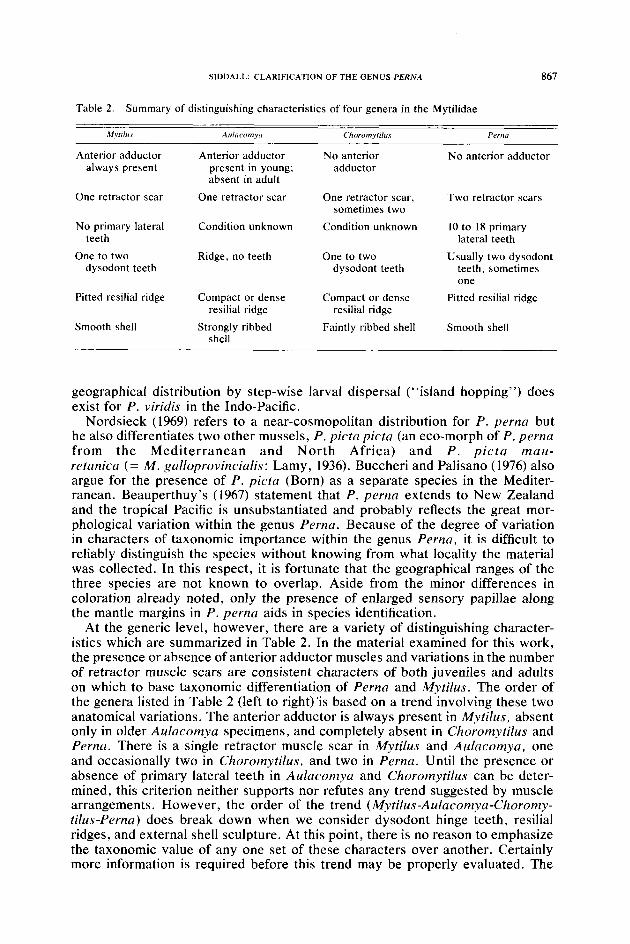
Table 2. Summary of distinguishing characteristics of four genera in the Mytilidae
Mpfr/r,.v A!ll~KO?7ly~l Choromyrilus PfWn
Anterior adductor always present
Anterior adductor present in young; absent in adult
No anterior adductor
No anterior adductor
One retractor scar One retractor scar One retractor scar, sometimes two
Two retractor scars
No primary lateral teeth
One to two dysodont teeth
Condition unknown
Ridge, no teeth
Condition unknown
One to two dysodont teeth
10 to 18 primary lateral teeth
Usually two dysodont teeth, sometimes one
Pitted resilial ridge
Smooth shell
Compact or dense resilial ridge
Strongly ribbed shell
Compact or dense resilial ridge
Faintly ribbed shell
Pitted resilial ridge
Smooth shell
geographical distribution by step-wise larval dispersal (“island hopping”) does exist for P. viridis in the Indo-Pacific.
Nordsieck (1969) refers to a near-cosmopolitan distribution for P. perna but he also differentiates two other mussels, P. picta picta (an eco-morph of P. perna from the Mediterranean and North Africa) and P. picta mau- retcrnica (= M. gnlloprovincialis: Lamy, 1936). Buccheri and Palisano (1976) also argue for the presence of P. picta (Born) as a separate species in the Mediter- ranean. Beauperthuy’s (1967) statement that P. perna extends to New Zealand and the tropical Pacific is unsubstantiated and probably reflects the great mor- phological variation within the genus Perna. Because of the degree of variation in characters of taxonomic importance within the genus Perna, it is difficult to reliably distinguish the species without knowing from what locality the material was collected. In this respect, it is fortunate that the geographical ranges of the three species are not known to overlap. Aside from the minor differences in coloration already noted, only the presence of enlarged sensory papillae along the mantle margins in P. perna aids in species identification.
At the generic level, however, there are a variety of distinguishing character- istics which are summarized in Table 2. In the material examined for this work, the presence or absence of anterior adductor muscles and variations in the number of retractor muscle scars are consistent characters of both juveniles and adults on which to base taxonomic differentiation of Perna and Mytilus. The order of the genera listed in Table 2 (left to right)‘is based on a trend involving these two anatomical variations. The anterior adductor is always present in Mytifus, absent only in older Aulacomya specimens, and completely absent in Choromytilus and Perna. There is a single retractor muscle scar in Mytifus and Aufacomya, one and occasionally two in Choromytilus, and two in Perna. Until the presence or absence of primary lateral teeth in Aulacomya and Choromytilus can be deter- mined, this criterion neither supports nor refutes any trend suggested by muscle arrangements. However, the order of the trend (Mytilus-Aulacomya-Choromy- tilus-Perna) does break down when we consider dysodont hinge teeth, resilial ridges, and external shell sculpture. At this point, there is no reason to emphasize the taxonomic value of any one set of these characters over another. Certainly more information is required before this trend may be properly evaluated. The

868 BULLETIN OF MARINE SCIENCE, VOL. 30, NO. 4, 1980
underlying significance of the suggested trend, if there is any, remains unclear but may relate to the degree of evolutionary “specialization” of each genus. From that standpoint, Perna, having lost the anterior adductor muscle, divided the retractor muscle complex, and developed primary lateral hinge teeth and branch- ing papillae on the mantle margin may be more specialized, yet the advantages of such morphological alterations are not obvious. Some support for this trend in specialization may be found in Ahmed (1974) and Ahmed and Sparks (1970), who showed that the diploid chromosome number in M. e&/is, M. californianus, and P. perna is 28 while in P. viridis it is 30. Based on the potential mechanisms for such numerical differences in karyotypes, Ahmed (1974) tentatively concluded that the genus Perna is more specialized than Mytilus and that P. viridis is more specialized than P. perna.
In his 1969 review, Cox points out that following metamorphosis, M. edulis develops hinge teeth “along the posterodorsal margin some distance beyond the row of crenulations persisting from the prodissoconch, which are obliterated as the ligament extends posteriorly. Similar teeth appear along the margin anterior to the beak, also beyond the row of crenulations, and some of these persist to form the dysodont teeth of the adult mussel.” I have termed Cox’s posterodorsal series of teeth secondary laterals. Primary lateral teeth immediately adjacent to the provincular teeth are not mentioned by Cox or by Lutz and Hidu (1979) who examined larvae and early post-larvae of M. edulis or by Le Pennec and Masson (1976) working with M. galloprovincialis. In Perna, both the primary lateral teeth and the ligament pit are present in stage 3 pediveligers (Bayne, 1965) which had not yet completed metamorphosis. Secondary lateral and dysodont teeth appear very soon after (less than 24 h) completion of metamorphosis as indicated by secretion of the dissoconch shell. However, because of the 24-h sampling interval used in this study, it is not possible to further resolve the timing of these events in Perna. Furthermore, one would expect significant differences in the timing of ontogenetic events between Mytilus, a temperate genus, and the subtropical to tropical species of Perna. Though larval hinge structure appears to be a relatively conservative taxonomic character (Rees, 1950; Cox, 1969), there are significant variations within the families Ostreidae (Ranson, 1948) and Mytilidae (present study).
The presence of primary lateral hinge teeth allows us to differentiate larvae of Perna from those of Mytilus. Adults of the genera are most reliably distinguished by patterns of muscle scars. Fewer criteria are available for distin- guishing among the three species of Perna ; geographic origin, coloration and some aspects of soft-part morphology are useful. Recognition of environmental variations which favor the development of ecomorphs of these mussels should minimize the current tendency to confuse the taxonomic status and nomenclature of the three species of Perna.
ACKNOWLEDGMENTS
Special thanks are due Drs. T. R. Wailer, R. A. Lutz and E. S. Iversen for critical review of the manuscript. I am also indebted to the great number of researchers and malacologists who collected and forwarded preserved material used in this effort. This is a contribution from the Rosenstiel School of Marine and Atmospheric Science, University of Miami.
LITERATURE CITED
Ahmed, M. 1974. Chromosomes of two species of the marine mussel Pernrr (Mytilidae:Pelecypoda). Bol. Inst. Oceanogr. Univ. Oriente 13: 17-22.

SIDDALL: CLARIFICATION OF THE GENUS PERNA 869
and A. K. Sparks. 1970. Chromosome number, structure and autosomal polymorphism in the marine mussels Mytilus edulis and Mytilus californianus. Biol. Bull. 138: I-13.
Bayne, B. L. 1965. Growth and delay of metamorphosis of the larvae of Mytilus edulis (L.). Ophelia 2: l-47.
Beauperthuy, I. 1967. Los mitilidos de Venezuela (Mollusca, Bivalvia). Bol. Inst. Oceanogr. Univ. Oriente 6: 7-l IS.
Buccheri, G., and G. Palisano. 1976. Nouvi dati sulla distribuzione geografica di Perna (Perna) picta (Born, 1780) e considerazioni sistematiche sulla specie. Conchiglie 12: 143-1.56.
Carriker, M. R. 1950. Killing and preservation of bivalve larvae in fluids. Nautilus 64: 14-17. -. 1979. Ultrastructural effect of cleaning molluscan shell with sodium hypochlorite (Clorox).
Nautilus 93: 47-49. Carter, J. G., and R. C. Aller. 1975. Calcification in the bivalve periostracum. Lethaia 8: 315-320. Cox, L. R. 1969. General features of Bivalvia. Pages N2-N129 in R. C. Moore, ed. Treatise on
invertebrate paleontology. Part N, Vol. 1. Dance, S. P. 1974. The encyclopedia of shells. Blanford Press, London. 288 pp. Dodge, H. 1952. A historical review of the mollusks of Linnaeus. Bull. Amer. Mus. Nat. Hist. 100:
Article 1. 263 pp. Dunachie, J. F. 1963. The periostracum of Mytilus edulis. Trans. Roy. Sot. Edinburgh 65: 383-411. Field, I. A. 1922. Biology and economic value of the sea mussel Mytilus edulis. Bull. U.S. Bureau
of Fisheries. Washington 38: 127-259. Fleming, C. A. 1959. Notes on New Zealand Recent and Tertiary Mussels (Mytilidae). Trans. Roy.
Sot. N. Z. 87: 165-178. Gmelin, J. F. 1791. Linnaeus, C., Systema Naturae per Regna Tria Naturae. Ed. 13, Aucta, Refor-
mata, Cura. J. F. Gmelin. Holmiae. Vol. 1. Part 6. Hanley, S. C. T. 1843. An illustrated and descriptive catalogue of Recent bivalve shells. William and
Northgate, London, XVIII. 392 pp. -. 1855. Ipsa Linnaei Conchylia. The shells of Linnaeus determined from his manuscripts and
collections-also an exact reprint of the Vermes Testacea of the “Systema Naturae” and Man- tissa. London. 556 pp.
Jukes-Browne, A. J. 1905. A review of the genera of the family Mytilidae. Proc. Malacol. Sot. London 6: 21 l-224.
Lamy, E. 1920. Notes sur les especes de Myrilus decrites par Lamarck. Bull. Mus. Nat. Hist. Nat., Paris 26: 330-335, 415-422, 520-526.
-. 1936-1937. Revision des Mytilidae vivantes du Museum National d’Histoire Naturelle de Paris. J. Conchyliogie 80: 66-102, 107-198, 229-295, 307-363; 81: 5-71, 99-132, 169-197.
Le Pennec, M., and M. Masson. 1976. Morphogenese de la coquilli de Mytilus galloprovinciulis (Lmk.) eleve en laboratoire. Cah. Biol. Mar. 17: 113-l 18.
Lutz, R. A., and H. Hidu. 1979. Hinge morphogenesis in the shells of larval and early post-larval mussles (Mytilus edulis L. and Modiolus modiolus L.). J. Mar. Biol. Ass., U.K. 59: 111-121.
Martyn, I. 1784. The Universal Conchologist II. London, 4 ~01s. March, 0. A. L. 1853. Catalogus Conchyliorum quae Reliquit D. Alphonso d’Aguirra and Gadea,
Comes de Yoldi. Fast. Secundus, Acephala, Hafniae. 74 pp. Newell, N. D. 1969. Classification of Bivalvia. Pages N205-N224 in R. C. Moore, ed. Treatise on
invertebrate paleontology. Part N, Vol. 1. Nordsieck, F. 1969. Die Europaischen Meeresmuscheln (Bivalvia). G. Fischer, Stuttgart. xiii,
231 pp. Ranson, G. 1948. Prodissoconques et classification des ostreides vivantes. Bull. Mus. Hist. Nat.,
Belg. 24: l-12. Rees, C. B. 1950. The identification and classification of lamellibranch larvae. Hull Bull. Mar. Ecol.
3: 73-104. Retzius, A. J. 1788. Dissertatio Historico-Naturalis Nova Testaceaorum Genera, Lundae, IV. 23 pp. Seed, R. 1968. Factors influencing shell shape in the mussel Mytilus edulis. J. Mar. Biol. Ass., U.K.
48: 561-584. Siddall, S. E. 1979a. Temporal changes in the salinity and temperature requirements of mussel larvae
of the genus Perna. Proc. World Mariculture Sot. 9: 549-566. -. 1979b. Effects of temperature and salinity on metamorphosis in two tropical mussels. Proc.
Nat. Shellfisheries Assoc. 69: 199. Soot-Ryen, T. 1952. Choromyfilus, a new genus in the Mytilidae. Sot. Malacol. “Carlos de la Torre”
Habana, Rev. 8: 121-122. -. 1955. A report on the family Mytilidae (Pelecypoda). Hancock Pacific Exped. 20: l-17.5. Stanley, S. M. 1978. Aspects of the adaptive morphology and evolution of the Trigoniidae. Phil.
Trans. Roy. Sot. London B 284: 247-258. von Ihering, H. 1901. On the South American species of Mytilidae. Proc. Malacol. Sot. London 4:
84-98.

870 BULLETIN OF MARINE SCIENCE,VOL.30,NO.4, 1980
-. 1907. Les mollusques fossiles du Tertiaire et du Cretace superieur de I’Argentine. Anal. Mus. Nat., Buenos Aires. Ser. 3, Vol. 7, XIII, 611 pp.
von Linnaeus, C. 1758. Systema Naturae per Regna Triae Naturae. Ed. 10, Holmiae. Vol. 1.
DATE ACCEPTED: October 10, 1979.
ADDRESS: Rosenstiel School of Marine rend Atmospheric Science, 4600 Rickenhrrcker Ctrrrsr~\~ry. Miami, Florida 33149.