Embed Size (px)
Citation preview

CASE REPORT Open Access
A case series of post COVID-19mucormycosis—a neurological prospectiveTamer Roushdy* and Eman Hamid
Abstract
Background: Direct neurological manifestations of coronavirus disease whether peripheral or central are reportedworldwide. Yet, along the 3rd wave of the pandemic especially in India, an associated angioinvasive opportunisticinfection with mucormycosis in COVID-19 cases is emerging.
Case presentation: The current case series which represents 4 patients with mucormycosis post COVID-19 is oneof a few if not the first case series that discusses post COVID-19 mucormycosis from a neurological prospective in atertiary hospital in Egypt.All cases but one presented with total ophthalmoplegia, and only one was diagnosed as a cavernous sinusthrombosis; meanwhile, orbital cellulitis and orbital apex syndrome were responsible of ophthalmoplegia in twocases.Mortality reached 25%, and the case that died suffered cutaneous as well as rhino-cerebral type with a delayedpresentation to hospital.
Conclusion: A rare but fatal fungal infection is ought to be nowadays kept in mind in COVID-19 active cases aswell as in recovered COVID-19 patients, especially those who have comorbid medical conditions as uncontrolleddiabetes and who were treated with large doses of corticosteroids.
Keywords: Coronavirus, COVID-19, Mucormycosis, Orbital cellulitis, Orbital apex syndrome, Cavernous sinusthrombosis
BackgroundSevere acute respiratory syndrome coronavirus 2(SARS-CoV-2) is besieging the world for more than ayear since its declaration by the World HealthOrganization as a pandemic in March 2020. Its effecton central nervous system has been reported alongmany studies and reviews either through affectingvascular system in different ways leading to strokes[1] or through retrograde extension to the brainthrough the olfactory nerve [2].Olfactory nerve affection in coronavirus disease of the
year 2019 (COVID-19) is well known. Anosmia andhyposmia have been reported by many COVID-19 pa-tients worldwide [3].
Yet, it seems that different cranial nerves are being af-fected by COVID-19 either directly in the context of theacute virus infection phase like the olfactory nerve or asa result of complications related to coronavirus.In this case series, different cranial nerves involved in
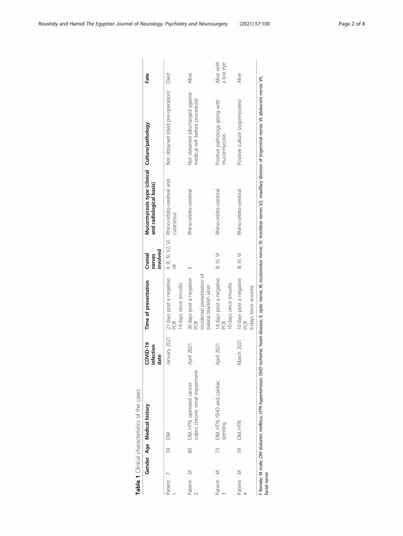
4 cases suffering mucormycosis as an opportunistic fun-gal infection post COVID-19 infection are presentedwith a highlight on different anatomical and pathologicalexplanations for such cranial nerve affection (Table 1).A formal written consent was obtained from all cases
to publish their medical history, laboratory results, andimaging for radiological as well as clinical lesions.
Case presentationPatient 1A 59-year-old female, with uncontrolled diabetes melli-tus (DM), suffered COVID-19 in January 2021 with a
© The Author(s). 2021 Open Access This article is licensed under a Creative Commons Attribution 4.0 International License,which permits use, sharing, adaptation, distribution and reproduction in any medium or format, as long as you giveappropriate credit to the original author(s) and the source, provide a link to the Creative Commons licence, and indicate ifchanges were made. The images or other third party material in this article are included in the article's Creative Commonslicence, unless indicated otherwise in a credit line to the material. If material is not included in the article's Creative Commonslicence and your intended use is not permitted by statutory regulation or exceeds the permitted use, you will need to obtainpermission directly from the copyright holder. To view a copy of this licence, visit http://creativecommons.org/licenses/by/4.0/.
* Correspondence: [email protected] Department, Faculty of Medicine, Ain Shams University, 38Abbasia, PO 11591, Cairo, Egypt
The Egyptian Journal of Neurology, Psychiatry and Neurosurgery
Roushdy and Hamid The Egyptian Journal of Neurology, Psychiatry and Neurosurgery (2021) 57:100 https://doi.org/10.1186/s41983-021-00355-8
Table
1Clinicalcharacteristicsof
thecases
Gen
der
Age
Med
ical
history
COVID-19
infection
date
Timeof
presentation
Cranial
nerves
invo
lved
Muc
ormycosistype(clin
ical
andradiological
basis)
Culture/patho
logy
Fate
Patient
1F
59DM
Janu
ary2021
21days
postane
gative
PCR
14days
sincesinu
sitis
II,III,IV,V2,VI,
VII
Rhino-orbito-cereb
raland
cutane
ous
Not
obtained
(diedpre-op
eration)
Died
Patient
2M
80DM,H
TN,ope
ratedcancer
colon,chronicrenalimpairm
ent
April2021
30days
postane
gative
PCR
Incide
ntalpresen
tatio
nof
palatalb
lackishulcer
IIRh
ino-orbito-cereb
ral
Not
obtained
(discharge
dagainst
med
icalwillbe
fore
proced
ure)
Alive
Patient
3M
73DM,H
TN,ISH
Dandcardiac
sten
ting
April2021
14days
postane
gative
PCR
10days
sincesinu
sitis
III,IV,VI
Rhino-orbito-cereb
ral
Positivepatholog
ygo
ingwith
mucormycosis
Alivewith
alosteye
Patient
4M
59DM,H
TNMarch
2021
10days
postane
gative
PCR
6days
sincesinu
sitis
III,IV,VI
Rhino-orbito-cereb
ral
Positivecultu
re(zygom
ycetes)
Alive
Ffemale;
Mmale;
DM
diab
etes
mellitus;H
TNhy
perten
sion
;ISH
Dischem
iche
artdisease;
II,op
ticne
rve;III,o
culomotor
nerve;IV,trochlear
nerve;V2
,maxillarydivision
oftrigem
inal
nerve;
VI,abd
ucen
tne
rve;
VII,
facial
nerve
Roushdy and Hamid The Egyptian Journal of Neurology, Psychiatry and Neurosurgery (2021) 57:100 Page 2 of 8

positive polymerase chain reaction (PCR) and was man-aged with broad spectrum antibiotics for bacterial pneu-monia on top of viral pneumonitis and corticosteroids ina dose exceeding 90 mg per day.The patient presented to the hospital 21 days post
COVID-19 with 2 weeks duration of unilateral right fa-cial swelling and deviation of angle of the mouth to theleft, complete ophthalmoplegia, no perception of light,and ptosis along the right eye.An elevated skin lesion with dark discoloration along
the forehead and right cheek as well as decreased sensa-tion along maxillary division of trigeminal nerve wasnoted (Fig. 1A).PCR for COVID-19 was negative; complete blood count
(CBC) showed moderate leukocytosis 17.4 × 10³/mm3
with relative neutrophilia 88% (range 35.0–80.0), absoluteneutrophilia 15.31 (range 1.8–7.7) and relative lymphope-nia 10.8% (range 18–44), C-reactive protein (CRP) 65(negative < 6.0), glycated hemoglobin (HbA1C) 12, andserum sodium (Na) 131.4mmol/L (range 136–145).
Magnetic resonance imaging (MRI) brain and mag-netic resonance venography (MRV) with cavernous viewas well as MRI orbit with contrast on a 1.5 Tesla superconductive system revealed diffuse enhancement muco-sal thickening, along the right maxillary, ethmoidal, andfrontal sinuses with occlusion of the right ostiomeatalcomplex, retro-orbital diffuse edema with faint enhance-ment, chronic small vessel disease, and intact venoussystem with no filling defects (Fig. 1B, C).Direct ophthalmoscope revealed right dilated unreact-
ive pupil, intact anterior segment, and pale disc with sus-pected retinal artery occlusion.Dermatology consultation for the forehead lesion pro-
visionally diagnosed it as cutaneous mucormycosis fortrue cut excision biopsy. Otolaryngology plan was toperform excision biopsy through endoscopic as well asexternal approach.The patient was placed on broad spectrum antibiotics,
and systemic amphotericin B was initiated. Yet, on thenext day prior to the scheduled surgery, the patient
Fig. 1 A Red star highlighting elevated dark colored forehead and right cheek that goes with cutaneous form of mucormycosis. B Red arrowalong axial MRI with obliteration of right ostiomeatal complex. C Red arrow along coronal MRI revealing obliteration of right ostiomeatal complexwith infiltration of infection to the right orbital cavity. Blue arrow showing maxillary, ethmoid and frontal sinusitis all around the rightorbital cavity
Roushdy and Hamid The Egyptian Journal of Neurology, Psychiatry and Neurosurgery (2021) 57:100 Page 3 of 8

suddenly suffered a disturbance in conscious level with aGlasgow coma scale (GCS) 4/15 and was transferred tothe intensive care unit (ICU) intubated and ventilated.Later on the same day, the patient died.
Patient 2Patient 2 was an 80-year-old male, diabetic, hyperten-sive, with history of operated cancer colon in 2011 andchronic renal impairment. COVID-19 for this patientwas in April 2021 for which he received corticosteroids.The patient presented 30 days later in May 2021 withhard palate sluggish ulcer 1 cm diameter, blackish, andpainless not communicating with nasal cavity, with ac-companied decrease in visual acuity in the right eye tothe degree of seeing objects at 2 m.PCR for COVID-19 was negative; CBC showed normal
leukocyte count 8.2 × 10³/mm3 with relative neutrophi-lia 84.1% (range 40.0–80.0) and relative lymphopenia6.7% (range 20–40), CRP 35 (negative < 6.0), HbA1C 9.5,serum creatinine 2.1, and Na 117 mmol/L (range 136–145).MRI paranasal sinuses performed prior admission at 3
Tesla high field MRI unit revealed near pan sinusitis es-pecially along bilateral frontal, ethmoidal, maxillary si-nuses, and left sphenoid. Both ethmoid sinuses showpredominantly fungal sinusitis with bacterial sinusitis,while the left maxillary show more bacterial but withfungal component. No abnormal extension of the in-flammatory process towards the cavernous sinus regionswas noted. Both orbits had normal appearance exceptfor mild form of the left optic neuritis that needed post
intravenous contrast study that was inapplicable second-ary to renal impairment (Figs. 2 and 3).The patient was planned for evacuation and drainage
of the sinuses under general anesthesia after correctinghis hyponatremia.Broad spectrum antibiotics and systemic amphotericin
B were administered. Steroid therapy for optic neuritiswas considered yet not initiated upon relative request,and the patient was discharged against medical will.
Patient 3A 73-year-old male patient, diabetic, hypertensive, withischemic heart with coronary stenting, had COVID-19infection in April 2021 for which he received dexa-methasone 8 mg/2 ml ampoule for 7 days and third-generation cephalosporin.Two weeks post COVID-19, the patient presented
with headache, nasal congestion, complete leftophthalmoplegia, complete ptosis, and conjunctivalchemosis (Fig. 4A).PCR for COVID-19 was negative; CBC was normal,
CRP 37.6 (negative < 6), HbA1C 9.5, and Na 127 mmol/L (136–145).Computed tomography (CT) of the paranasal sinuses
without intravenous contrast revealed pan sinusitismuch more evident along the left side with obliteratedostiomeatal complex, abnormal fatty stranding sur-rounding the distal left side optic nerve close to theoptic foramen suspicious of orbital cellulitis (Fig. 4).MRI orbit with contrast revealed diffuse enhancing
mucosal thickening with internal fluid level and true dif-fusion restriction denoting acute sinusitis in the left
Fig. 2 Axial MRI without contrast along the maxillary sinus showing bilateral sinusitis yet more on the left side with hypo intense margins thatgoes radiologically with fungal infection with heavy metals deposition and hyper intense center that represents bacterial nature
Roushdy and Hamid The Egyptian Journal of Neurology, Psychiatry and Neurosurgery (2021) 57:100 Page 4 of 8

frontal, ethmoidal, and maxillary sinuses, with left orbitalfat diffuse edema with faint enhancement confirming or-bital cellulitis.The patient was placed on systemic and topical eye an-
tibiotics and systemic liposomal amphotericin B in adose of 0.7 mg/kg intravenous once per day over 6 h for14 days; drainage and biopsy for the sinuses wasperformed.Postoperatively, the patient’s headache improved, yet
his eye motion was not regained.Specimen for an excised infected polypoid lesion was
sent for pathological assessment. Microscopic descrip-tion was in favor of mucormycosis in which there was apartially ulcerated and partially hyperplastic respiratoryepithelium. The subepithelial tissue was edematous, con-gested with dense mixed inflammatory cellular infiltraterich in neutrophils. Foci of suppuration were seen, withfew fungal hyphae, thick walled, non-septate and with ir-regular branching and occasionally at right angles.
Patient 4Patient 4 was a 59-year-old male, with uncontrolled DM,and hypertensive, with history of COVID-19 in March 2021.The patient presented with 6 days duration of right
total ophthalmoplegia, ptosis, conjunctival chemosis, andproptosis (Fig. 5) and blackish discoloration of hardpalate.
PCR for COVID-19 was negative; CBC showed moder-ate leukocytosis 15.3 × 10³/mm3 with relative neutrophilia87.9% (range 40.0–80.0), absolute neutrophilia 13.4 (range2.0–7.0), and relative lymphopenia 1.2% (range 1.0–3.0),erythrocyte sedimentation rate (ESR) 75mm/h (range 2–20), HbA1C 12.2, and Na 130mmol/L (136–145).CT paranasal sinuses revealed pan sinusitis along the
right maxillary, ethmoidal, and frontal sinuses with oblit-eration of the ostiomeatal complex.MRV cavernous view with contrast showed enhance-
ment and filling defect.A provisional diagnosis of rhino-cerebral-
mucormycosis and associated cavernous sinus throm-bosis was considered, and the patient underwentcomplete drainage of the sinuses; the culture revealedgrowth of zygomycetes fungi as well as Klebsiellamultiple-drug resistant (MDR).The patient initiated full dose anticoagulant, systemic
amphotericin B in a dose of 0.7 mg/kg intravenous onceper day over 6 h for a total of 14 days and amikacin anti-biotic based on maxillary sinus culture and sensitivitywith an excellent recovery and no residual eye motiondefect.
DiscussionCOVID-19 infection does not stand only at the acutephase. It has consequences in patients who are either
Fig. 3 Blue arrow along non contrast MRI T2 WIs highlighting mild form of left optic neuritis
Roushdy and Hamid The Egyptian Journal of Neurology, Psychiatry and Neurosurgery (2021) 57:100 Page 5 of 8

immunocompromised, with uncontrolled diabetes, andwho are treated for long periods with high doses of ste-roids [4].Opportunistic infection with fungi in nasal sinuses is
rare, yet it is being reported nowadays in the COVID-19pandemic [5]. Mucormycosis caused by mucormycetes
molds has five forms based on site of spread being si-nuses, orbital and brain (rhino-orbito-cerebral), pulmon-ary, gastrointestinal, cutaneous, and disseminated [4].Mucormycosis is diagnosed through different ap-
proaches. According to centers for disease control and pre-vention (CDC), facial swelling, headache, nasal or sinuscongestion, and blackish discoloration within nose or pal-ate are a clinical diagnostic approach. Radiological diagno-sis is reachable through CT or MRI, and laboratorydiagnosis is obtainable either by culture or pathology [6–8].Along this case series, we have collected 4 cases of
post COVID-19 mucormycosis that were presented forsuspicion of cavernous sinus thrombosis to neurologydepartment of Ain Shams University Specialized hospitalalong the first 5 months of 2021 along the third wave ofthe pandemic.Four cases in a 5-month duration encountered in a sin-
gle hospital are considered a relative breakthrough in inci-dence of such type of rare opportunistic disease. In aretrospective study conducted in children’s cancer hospital57357 in Egypt along a decade from 2007 to 2017, only 45cases developed mucormycosis [9]. Another study in 2010by Zaki and Colleagues that was conducted at the sametertiary hospital where the current case series took placereported 10 cases within 12months' duration [10].In the 2010 study, only 2 (20%) cases had a sinus
mucormycosis, while in the current case series, 100% of
Fig. 4 A Chemosis and total ophthalmoplegia of the left eye. B Red arrow showing proptosis of the left eye along paranasal CT. C Blue arrowpointing to infiltration of soft tissue retro orbit sharing in the presentation of orbital apex syndrome donating invasion from infected andinflamed sinuses. D Blue arrow representing infiltration of periantral fat donating invasion from nearby sinuses
Fig. 5 Chemosis of conjunctiva and total ophthalmoplegia withptosis elevated by the examiner’s finger
Roushdy and Hamid The Egyptian Journal of Neurology, Psychiatry and Neurosurgery (2021) 57:100 Page 6 of 8

the cases had a sinus infection besides one who had alsoa cutaneous forehead and cheek type on clinicalsuspicion.Laboratory wise, besides the uncontrolled diabetes in
the four patients which is considered a risk factor forcompromised immunity, the four patients had hypona-tremia as a baseline presentation. Low serum Na is acommon finding in viral and bacterial infections and isreported in COVID-19 cases. Yet, for fungal infection, itis rarely described [11, 12].Mucormycosis in the current study is represented
from neurological prospective rather than pathologicalor surgical ones.Three cases had total ophthalmoplegia, and only one
of them was proven to have cavernous sinus thrombosisas a responsible cause for ophthalmoplegia, whereas theother 2 cases total ophthalmoplegia was secondary todirect spread or indirect inflammatory process involvingthe orbital cavity and causing orbital cellulitis.Two cases had vision affection: one had a demyelinat-
ing signal along the optic nerve shown in non-contrastT2 WIs with a recommendation of post contrast studythat was inapplicable secondary to renal impairment.The second patient had dilated unreactive pupil with asuspected retinal artery occlusion in directophthalmoscope.Reduction in visual acuity in cases with mucormycosis
is explainable on the basis of infarctions in blood vesselssupplying retina or optic nerve, compression on thenerve along its course within cavernous sinus, or directinfection and necrosis [13, 14].Isolated optic neuritis or retro-bulbar optic neuropathy
is infrequently reported with mucormycosis. In thecurrent case series, patient 2 was a probable mucormy-cosis case based on the European Organization for Re-search and Treatment of Cancer and Mycoses StudyGroup (EORTC/MSC) with clinical and radiologicalsigns going with optic neuritis that was a result of in-flammatory response rather than direct invasion ratherthan the one reported by Kahloun and colleagues [15].Prognosis in mucormycosis is poor secondary to the
nature of such opportunistic angioinvasive fungal infec-tion that affects immunocompromised patients with un-controlled diabetes which is a common risk factor inmost cases, besides the delay in presentation to emer-gency units.Patient 1 in the current series presented to hospital
following 2 weeks duration with cutaneous as well assinus suspicion of post COVID-19 fungal infection andII, III, IV, V2, and VI cranial nerve affection representingwhat is termed orbital apex syndrome, besides lowermotor neuron facial on top of the swollen face and fore-head. The next day and prior to the scheduled operation,the patient suddenly deteriorated and died.
Delayed presentation and combined cutaneous as wellas rhino-orbito-cerebral types may rationalize aggres-siveness of infection and fatality in such case.This highlights the importance of time in seeking
medical care in such cases, as the other three casessought medical advice in an earlier course and only onecase was left with a lost eye as a lifelong consequence.A 25% mortality in this collected case series of 5
months is considered high if compared to 10% in 10cases along Zaki and colleagues’ 12months durationcase series [10].
ConclusionMucormycosis is a rare and occasionally fatal opportun-istic infection that affects immunocompromised patients.Most patients who encounter mucormycosis are diabeticwith uncontrolled diabetes.In the COVID-19 era, the rate of mucormycosis seems
to be increasing, and the earlier the presentation to hos-pitals, the better the outcome.A standard dose recommended by the World Health
Organization based on the RECOVERY trial that is 6 mgof dexamethasone once daily for no more than 7–10days should be strictly adhered to and lower dosesshould be considered in immunocompromised or dia-betic patients.
AbbreviationsSARS-CoV-2: Severe acute respiratory syndrome coronavirus 2; COVID-19: Coronavirus disease of the year 2019; DM: Diabetes mellitus;PCR: Polymerase chain reaction; CBC: Complete blood count; CRP: C-reactiveprotein; HbA1C: Glycated hemoglobin; Na: Sodium; MRI: Magnetic resonanceimaging; MRV: Magnetic resonance venography; GCS: Glasgow coma scale;ICU: Intensive care unit; CT: Computed tomography; ESR: Erythrocytesedimentation rate; MDR: Multiple drug resistant; CDC: Centers for DiseaseControl and Prevention; EORTC/MSC: European organization for research andtreatment of cancer and mycoses study group
AcknowledgementsNot applicable
Authors’ contributionsAll authors have read and approved the manuscript. TR: concept behind thework, drafting the manuscript, collecting the cases, analyzing cases’investigations. EH: concept behind the work, revision of the manuscript.
FundingNo funds were received to fulfill this work.
Availability of data and materialsThe corresponding author takes full responsibility for the data, has full accessto all of the data, and has the right to publish any and all data separate andapart from any sponsor.
Declarations
Competing interestThe authors declare that there is no competing interest.
Ethics approval and consent to participateAll procedures performed in the study were in accordance with the ethicalstandards of the Faculty of Medicine, Ain Shams University Research andEthical Committee, and with the 1964 Helsinki declaration and its later
Roushdy and Hamid The Egyptian Journal of Neurology, Psychiatry and Neurosurgery (2021) 57:100 Page 7 of 8

amendments or comparable ethical standards. An approval from the localethical committee of faculty of medicine Ain Shams University wasapproved.
Consent for publicationWritten informed consent was obtained from participants or next of kin forpublication of this case series and accompanying images. We obtainedapproval from research ethics committee on April 2020.
Received: 1 June 2021 Accepted: 13 July 2021
References1. Roushdy T, Hamid E. A review on SARS-CoV-2 and stroke pathogenesis and
outcome. Egypt J Neurol Psychiatr Neurosurg. 2021;57(1):63. https://doi.org/10.1186/s41983-021-00319-y.
2. Meinhardt J, Radke J, Dittmayer C, Franz J, Thomas C, Mothes R, et al.Olfactory transmucosal SARS-CoV-2 invasion as a port of central nervoussystem entry in individuals with COVID-19. Nat Neurosci. 2021;24(2):168–75.https://doi.org/10.1038/s41593-020-00758-5.
3. Aziz M, Goyal H, Haghbin H, Lee-Smith WM, Gajendran M, Perisetti A. Theassociation of “loss of smell” to COVID-19: a systematic review and meta-analysis. Am J Med Sci. 2021;361(2):216–25. https://doi.org/10.1016/j.amjms.2020.09.017.
4. Ribes JA, Vanover-Sams CL, Baker DJ. Zygomycetes in human disease. ClinMicrobiol Rev. 2000;13(2):236–301. https://doi.org/10.1128/CMR.13.2.236.
5. Dyer O. Covid-19: India sees record deaths as “black fungus” spreads fear.BMJ. 2021;373:n1238. https://doi.org/10.1136/bmj.n1238.
6. Centers for Disease Control and Prevention, National Center for Emergingand Zoonotic Infectious Diseases (NCEZID), Division of Foodborne,Waterborne, and Environmental Diseases (DFWED): symptoms ofmucormycosis, January 14, 2021. https://www.cdc.gov/fungal/diseases/mucormycosis/symptoms.html.
7. Herrera DA, Dublin AB, Ormsby EL, Aminpour S, Howell LP. Imaging findingsof rhinocerebral mucormycosis. Skull Base. 2009;19(2):117–25. https://doi.org/10.1055/s-0028-1096209.
8. Therakathu J, Prabhu S, Irodi A, Sudhakar SV, Yadav VK, Rupa V. Imagingfeatures of rhinocerebral mucormycosis: a study of 43 patients. Egypt JRadiol Nucl Med. 2018;49(2):447-52. https://doi.org/10.1016/j.ejrnm.2018.01.001.
9. Madney Y, Khedr R, Ahmed N, El-Mahallawy H, Youssef A, Taha H, et al.Overview and outcome of mucormycosis among children with cancer:report from the Children’s Cancer Hospital Egypt. Mycoses. 2019;62(11):984–9. https://doi.org/10.1111/myc.12915.
10. Zaki SM, Elkholy IM, Elkady NA, Abdel-Ghany K. Mucormycosis in Cairo,Egypt: review of 10 reported cases. Med Mycol. 2014;52(1):73–80. https://doi.org/10.3109/13693786.2013.809629.
11. Kashef Hamadani BH, Franco-Paredes C, McCollister B, Shapiro L, BeckhamJD, Henao-Martínez AF. Cryptococcosis and cryptococcal meningitis: newpredictors and clinical outcomes at a United States academic medicalcentre. Mycoses. 2018;61(5):314–20. https://doi.org/10.1111/myc.12742.
12. Królicka AL, Kruczkowska A, Krajewska M, Kusztal MA. Hyponatremia ininfectious diseases-a literature review. Int J Environ Res Public Health. 2020;17(15):5320. https://doi.org/10.3390/ijerph17155320.
13. Sano T, Kobayashi Z, Takaoka K, Ota K, Onishi I, Iizuka M, et al. Retrobulbaroptic neuropathy associated with sphenoid sinus mucormycosis. NeurolClin Neurosci. 2018 Sep;6(5):146–7. https://doi.org/10.1111/ncn3.12216.
14. Alsuhaibani AH, Al-Thubaiti G, Al Badr FB. Optic nerve thickening andinfarction as the first evidence of orbital involvement with mucormycosis.Middle East Afr J Ophthalmol. 2012;19(3):340–2. https://doi.org/10.4103/0974-9233.97957.
15. Kahloun R, Abroug N, Ksiaa I, Mahmoud A, Zeghidi H, Zaouali S, et al.Infectious optic neuropathies: a clinical update. Eye Brain. 2015;7:59–81.https://doi.org/10.2147/EB.S69173.
Publisher’s NoteSpringer Nature remains neutral with regard to jurisdictional claims inpublished maps and institutional affiliations.
Roushdy and Hamid The Egyptian Journal of Neurology, Psychiatry and Neurosurgery (2021) 57:100 Page 8 of 8