Embed Size (px)
Citation preview

Copyright © 2014 by The Korean Association for the Study of the LiverThis is an Open Access article distributed under the terms of the Creative Commons Attribution Non-Commercial License (http://creativecommons.org/licenses/by-nc/3.0/) which permits unrestricted non-commercial use, distribution, and reproduction in any medium, provided the original work is properly cited.
pISSN 2287-2728 eISSN 2287-285X
http://dx.doi.org/10.3350/cmh.2014.20.2.208Clinical and Molecular Hepatology 2014;20:208-213Case Report
INTRODUCTION
Tuberculosis (TB) is an infectious disease that is prevalent
worldwide, but obstructive jaundice secondary to abdominal TB
remains rare.1 Patients with bile duct involvement of TB causing
obstructive jaundice have protracted symptoms such as malaise,
jaundice, and weight loss, which are indistinguishable from those
of cholangiocarcinoma.2 Obstructive jaundice can be caused by
tuberculous enlargement of the head of the pancreas, tuberculous
lymphadenitis, tuberculous stricture of the biliary tree, or a tuber-
culous mass of the retroperitoneum.1 Fifteen cases of pericholedo-
cal tuberculous lymphadenitis were reported in Korea.3-17 There
were two cases of pericholedocal tuberculous lymphadenitis with
duodenal TB10,14 and two cases of pericholedocal tuberculous
lymphadenitis with portal hypertension.11,13 This is the first case re-
port of pericholedocal tuberculous lymphadenitis with portal hy-
pertension concomitant with duodenal TB in Korea. Here we re-
port a case of obstructive jaundice with portal hypertension
caused by pericholedochal tuberculous lymphadenitis with duode-
nal TB in addition to a review of tuberculous lymphadenitis in Ko-
rea.
CASE
A 30-year-old man admitted our hospital due to jaundice. One
year ago, he had been diagnosed with pulmonary TB that he had
completed a six-month regimen of anti-TB medication (isoniazid,
A case of obstructive jaundice caused by tuberculous lymphadenitis: A literature reviewSu Jung Baik1, Kwon Yoo2, Tae Hun Kim2, Il Hwan Moon2, and Min-Sun Cho3
1Department of Internal Medicine, Health Promotion Center, Yonsei University Gangnam Severance Hospital, Seoul, 2Department of Internal Medicine and 3Department of Pathology, Ewha Woman’s University School of Medicine, Seoul, Korea
Corresponding author : Kwon YooDepartment of Internal Medicine, Ewha Medical Research Institute, Ewha Womans University School of Medicine, Ewha Womans University Mokdong Hospital, 911-1 Mok-dong, Yangcheon-gu, Seoul 158-710, KoreaTel. +82-2-2650-2632, Fax. +82-2-2655-2076E-mail; [email protected]
Abbreviations: AFB, acid-fast bacillus; AST, aspartate aminotransferase; ALT, alanine aminotransferase; BUN, blood urea nitrogen; CBD, common bile duct; CT, computed tomography; PCR, polymerase chain reaction; M. tuberculosis, Mycobacterium tuberculosis; PTBD, percutaneous transhepatic biliary drainage; TB, Tuberculosis
Received : Oct. 2, 2012 / Revised : Nov. 7, 2012 / Accepted : Nov. 19, 2012
Obstructive jaundice caused by tuberculous lymphadenitis is a rare manifestation of tuberculosis (TB), with 15 cases having been reported in Korea. We experienced a case of obstructive jaundice caused by pericholedochal tuberculous lymphadenitis in a 30-year-old man. The patient’s initial serum total bilirubin level was 21.1 mg/dL. Abdominal computed tomography revealed narrowing of the bile duct by a conglomerated soft-tissue mass involving the main portal vein. Abrupt obstruction of the common bile duct was observed on cholangiography. Pathologic analysis of a ultrasonography-guided biopsy sample revealed chronic granulomatous inflammation, and an endoscopic examination revealed esophageal varices and active duodenal ulceration, the pathology of which was chronic noncaseating granulomatous inflammation. Hepaticojejunostomy was performed and pathologic analysis of the conglomerated soft-tissue mass revealed chronic granulomatous inflammation with caseation of the lymph nodes. Tuberculous lymphadenitis should be considered in patients presenting with obstructive jaundice in an endemic area. (Clin Mol Hepatol 2014;20:208-213)Keywords: Tuberculosis; Lymphadenitis; Portal hypertension

209
Su Jung Baik, et al. Obstructive jaundice by tuberculous lymphadenitis
http://www.e-cmh.org http://dx.doi.org/10.3350/cmh.2014.20.2.208
rifampicin, etambutol and pyrazinamide for 2 months, then con-
tinuing with isoniazid, rifampicin and ethambutol for the remain-
ing 4 months) at local clinic. After treatment, he had no other
problems until the development of jaundice. Abdominal ultraso-
nography performed at a local clinic which showed bile duct dila-
tation. The patient was referred to our hospital for evaluation of
the biliary obstruction.
On physical examination, the patient’s sclera was icteric. There
was no hepatomegaly, splenomegaly or ascites. Hemoglobin was
10.7 g/dL, platelets were 227,000/μL, and white blood cell count
was 9300/μL. The serum total bilirubin was 21.1 mg/dL and direct
bilirubin was 12.4 mg/dL. AST was 160 IU/L and ALT was 147 IU/L.
Serum BUN, creatinine, amylase and lipase levels were within nor-
mal range. Viral marker assays were negative for hepatitis B sur-
face antigen, IgM anti-hepatitis A and anti-hepatitis C virus. Dy-
namic computed tomography (CT) showed both intrahepatic duct
and extrahepatic bile duct dilation with abrupt narrowing of the
proximal common bile duct (CBD). The proximal CBD was encased
by a soft tissue mass (Fig. 1A, 1B). This lesion spread from the he-
patic hilum to the hepatoduodenal ligament and pancreatic head.
Figure 1. Liver and chest computed tomography (CT) images and cholangiogram via percutaneous transhepatic biliary drainage catheter. (A, B) Liver dynamic CT showing the main portal vein encased by a soft-tissue mass (arrow), which had spread from the hepatic hilum (A) to the pancreatic head (B). Calcifications were observed within the soft-tissue mass (arrow in A). (C) High-resolution chest CT showing multiple nodules in both upper lobes with calcification and fibrotic bands. Traction bronchiectasis is also seen. (D) Cholangiogram showing abrupt common bile duct (CBD) obstruction with a dilated intrahepatic duct.
A
C
B
D

210
Clin Mol HepatolVolume_20 Number_2 June 2014
http://www.e-cmh.orghttp://dx.doi.org/10.3350/cmh.2014.20.2.208
Central calcification was observed in the lesion and the main por-
tal vein was encased by soft tissue mass. The patient’s chest X-ray
showed patchy and fibrotic opacities in both upper lungs with
volume decrease. High-resolution chest CT showed multiple nod-
ules with calcification and fibrotic bands in both upper lobes and
the superior segment of both lower lobes considered stable TB
(Fig. 1C). The cholangiogram from percutaneous transhepatic bili-
ary drainage (PTBD) showed abrupt proximal CBD obstruction
with dilated intrahepatic ducts (Fig. 1D). The guide wire and cath-
eter were not passed through the narrowed segment.
Upper gastrointestinal endoscopy revealed grade 1 esophageal
varices (Fig. 2A) and active duodenal ulceration was noted at the
bulb (Fig. 2B). Pathologic examination of the duodenal ulceration
showed chronic non-caseating granulomatous inflammation (Fig.
2C, 2D). TB polymerase chain reaction (TB-PCR) and acid fast ba-
cillus (AFB) stain were all negative.
Figure 2. Upper gastrointestinal endoscopic findings and pathology of the duodenal ulcer. (A, B) Endoscopic examination showing esophageal varices (A) and a duodenal ulcer at the bulb (B). (C, D) Pathologic examination of the duodenal ulcer showing noncaseating granulomatous inflammation with a multinucleated giant cell [arrow; hematoxylin and eosin (H&E) stain; C, ×100; D, ×200].
A B
C D

211
Su Jung Baik, et al. Obstructive jaundice by tuberculous lymphadenitis
http://www.e-cmh.org http://dx.doi.org/10.3350/cmh.2014.20.2.208
Percutaneous ultrasonography-guided biopsy of the soft tissue
mass was performed and pathologic examination showed chronic
granulomatous inflammation with fibrosis (Fig. 3A). TB-PCR was
negative and AFB and periodic acid Schiff stains did not demon-
strate acid-fast bacilli or fungal organisms. Bacteria and Mycobac-terium tuberculosis (M. tuberculosis) were not identified in blood
and bile fluid from PTBD. The soft tissue mass was considered to
be conglomerated lymph nodes or a true mass lesion. Drug sensi-
tivity test for M. tuberculosis could not be performed because of
no growth of the organism.
An explorative laparotomy was performed to relieve the biliary
obstruction and to exclude malignancy. Several conglomerated
lymph nodes encasing CBD and portal vein were observed during
surgery. The pancreas and liver appeared grossly normal and the
gallbladder was not distended. Examination of frozen sections of
the conglomerated lymph nodes showed chronic ill-defined granu-
lomatous inflammation and fibrosis. Cholecystectomy and a Roux-
en-Y bypass hepaticojejunostomy were performed. Final patholog-
ic examination showed chronic granulomatous inflammation of
the lymph nodes with caseation (Fig. 3B, 3C). AFB staining did not
identify acid-fast bacilli in the gallbladder, bile duct or lymph
nodes. TB-PCR showed positive band in lymph nodes. After the
operation, total bilirubin level decreased to 3.2 mg/dL. The patient
was treated with anti-TB medication and bilirubin level had de-
creased to normal six weeks after surgery.
DISCUSSION
TB of the biliary system is rare and difficult to diagnose.2 Ob-
structive jaundice caused by tuberculous lymphadenitis is most of-
ten attributed to mechanical obstruction of the biliary tract by
lymph nodes or mass lesions.1 Patients with tuberculous lymphad-
enitis usually present with obstructive jaundice, which may be
confused with hepatobiliary malignancies.2 The annual incidence
of hepatobiliary TB is reported as 1.05% of all TB infections.18
Hepatobiliary TB is caused by two mechanisms.19 The first
mechanism is the direct spread of caseous materials from the por-
tal tracts into the bile duct and the second is secondary inflamma-
tion related to tuberculous periportal adenitis.19
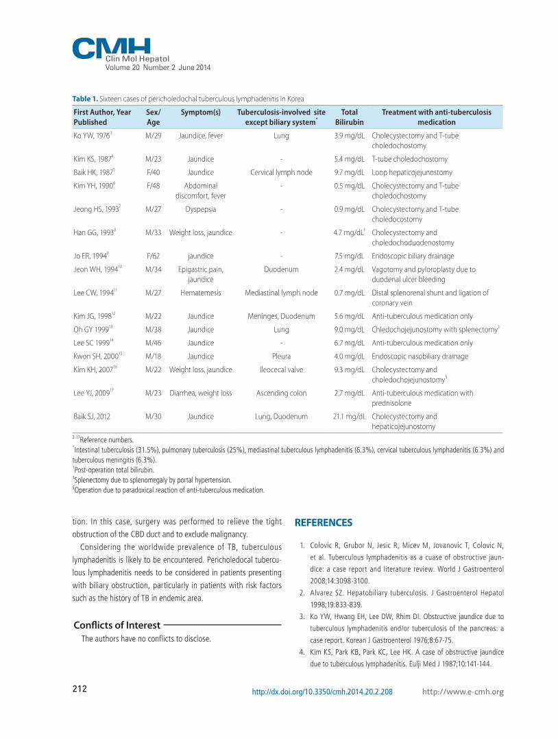
Table 1 lists pericholedocal tuberculous lymphadenitis reported
in Korea including this case.3-17 Pericholedocal tuberculous lymph-
adenitis in Korea showed a 81.3% male preponderance. The initial
total bilirubin level ranged from 0.5 to 21.1 mg/dL with a mean of
5.9 mg/dL. Including the present case, 11 cases were treated by
surgery with anti-TB medication and five cases were treated by
anti-TB medication alone or anti-TB medication with endoscopic
nasobiliary drainage or prednisolone.9,12,14,15,17 In Korea, pericho-
ledocal tuberculous lymphadenitis has been associated with intes-
tinal TB (31.3%), pulmonary TB (25%), mediastinal tuberculous
lymphadenitis (6.3%), cervical tuberculous lymphadenitis (6.3%)
and tuberculous meningitis (6.3%) (Table 1).
Anti-TB medication without surgical intervention is desirable,
but there are two emerging problems.2 First, multi-drug resistant
strains of M. tuberculosis are becoming increasingly prevalent.
Second, the bile duct can be severely damaged by repeated in-
flammatory reactions and may thus be irreversibly scarred.20 In this
case, the conglomerated lymph nodes encased the main portal
vein and this resulted in portal vein hypertension, thus causing the
esophageal varices. In Korea, there were two previous reports of
portal hypertension associated with portal vein obstruction by
pericholedocal tuberculous lymphadenitis.11,13 Including the pres-
ent case, all three cases of pericholedocal tuberculous lymphade-
nitis with portal hypertension were treated by surgical interven-
Figure 3. Pathology of a percutaneous ultrasonography-guided biopsy sample of the soft-tissue mass encasing the proximal CBD. Pathologic findings of a percutaneous ultrasonography-guided biopsy sample of a soft-tissue mass encasing the proximal CBD showing chronic granulomatous inflammation with fibrosis (H&E stain; A, ×200), and a caseating granuloma (arrow; H&E stain; B, ×40; C, ×200).
A B C
212
Clin Mol HepatolVolume_20 Number_2 June 2014
http://www.e-cmh.orghttp://dx.doi.org/10.3350/cmh.2014.20.2.208
tion. In this case, surgery was performed to relieve the tight
obstruction of the CBD duct and to exclude malignancy.
Considering the worldwide prevalence of TB, tuberculous
lymphadenitis is likely to be encountered. Pericholedocal tubercu-
lous lymphadenitis needs to be considered in patients presenting
with biliary obstruction, particularly in patients with risk factors
such as the history of TB in endemic area.
Conflicts of InterestThe authors have no conflicts to disclose.
REFERENCES
1. Colovic R, Grubor N, Jesic R, Micev M, Jovanovic T, Colovic N,
et al. Tuberculous lymphadenitis as a cuase of obstructive jaun-
dice: a case report and literature review. World J Gastroenterol
2008;14:3098-3100.
2. Alvarez SZ. Hepatobiliary tuberculosis. J Gastroenterol Hepatol
1998;19:833-839.
3. Ko YW, Hwang EH, Lee DW, Rhim DI. Obstructive jaundice due to
tuberculous lymphadenitis and/or tuberculosis of the pancreas: a
case report. Korean J Gastroenterol 1976;8:67-75.
4. Kim KS, Park KB, Park KC, Lee HK. A case of obstructive jaundice
due to tuberculous lymphadenitis. Eulji Med J 1987;10:141-144.
Table 1. Sixteen cases of pericholedochal tuberculous lymphadenitis in Korea
First Author, Year Published
Sex/Age
Symptom(s) Tuberculosis-involved site except biliary system*
TotalBilirubin
Treatment with anti-tuberculosis medication
Ko YW, 19763 M/29 Jaundice, fever Lung 3.9 mg/dL Cholecystectomy and T-tube choledochostomy
Kim KS, 19874 M/23 Jaundice - 5.4 mg/dL T-tube choledochostomy
Baik HK, 19875 F/40 Jaundice Cervical lymph node 9.7 mg/dL Loop hepaticojejunostomy
Kim YH, 19906 F/48 Abdominal discomfort, fever
- 0.5 mg/dL Cholecystectomy and T-tube choledochostomy
Jeong HS, 19937 M/27 Dyspepsia - 0.9 mg/dL Cholecystectomy and T-tube choledocostomy
Han GG, 19938 M/33 Weight loss, jaundice - 4.7 mg/dL† Cholecystectomy and choledochoduodenostomy
Jo ER, 19949 F/62 jaundice - 7.5 mg/dL Endoscopic biliary drainage
Jeon WH, 199410 M/34 Epigastric pain, jaundice
Duodenum 2.4 mg/dL Vagotomy and pyloroplasty due to duodenal ulcer bleeding
Lee CW, 199411 M/27 Hematemesis Mediastinal lymph node 0.7 mg/dL Distal splenorenal shunt and ligation of coronary vein
Kim JG, 199812 M/22 Jaundice Meninges, Duodenum 5.6 mg/dL Anti-tuberculous medication only
Oh GY 199913 M/38 Jaundice Lung 9.0 mg/dL Chledochojejunostomy with splenectomy‡
Lee SC 199914 M/46 Jaundice - 6.7 mg/dL Anti-tuberculous medication only
Kwon SH, 200015 M/18 Jaundice Pleura 4.0 mg/dL Endoscopic nasobiliary drainage
Kim KH, 200716 M/22 Weight loss, jaundice Ileocecal valve 9.3 mg/dL Cholecystectomy and choledochojejunostomy§
Lee YJ, 200917 M/23 Diarrhea, weight loss Ascending colon 2.7 mg/dL Anti-tuberculous medication with prednisolone
Baik SJ, 2012 M/30 Jaundice Lung, Duodenum 21.1 mg/dL Cholecystectomy and hepaticojejunostomy
2-17Reference numbers.*Intestinal tuberculosis (31.5%), pulmonary tuberculosis (25%), mediastinal tuberculous lymphadenitis (6.3%), cervical tuberculous lymphadenitis (6.3%) and tuberculous meningitis (6.3%).†Post-operation total bilirubin.‡Splenectomy due to splenomegaly by portal hypertension.§Operation due to paradoxical reaction of anti-tuberculous medication.
213
Su Jung Baik, et al. Obstructive jaundice by tuberculous lymphadenitis
http://www.e-cmh.org http://dx.doi.org/10.3350/cmh.2014.20.2.208
5. Baik HK, Kim YI. Common bile duct obstruction due to tuberculous
lymphadenitis. Tuber Respir Dis 1987;34:246-249.
6. Kim YH, Lee DK, Kwon SO, Jang WI, Kim SY, Cho HY, et al. One
case of biliary tract obstruction caused by tuberculous adenitis. Ko-
rean J Gastrointest Endosc 1990;10:351-354.
7. Jeong HS, Ko YG, Hong SW. Biliary tract obstruction due to tuber-
culous lymphadenopathy. J Korean Surg Soc 1993:44;772-776.
8. Han KK, Kim HS, Park YH, Kwon KH, Lee DY, Lee JS. A case of
obstructive jaundice due to tuberculous lymphadenitis. Korean J
Gastroenterol 1993;25:1070-1074.
9. Lee CH, Kim JS, Lee G, Bak YT, Kim JH, Kim JG, et al. A case of
tuberculous lymphadenitis causing obstructive jaundice. Korean J
Gastrointest Endosc 1994;14:115-120.
10. Jeon WH, Sohn HJ, Yoon YB, Choi ES, Kim HS. A case of duodenal
tuberculosis presenting ulcer bleeding and obstructive jaundice.
Korean J Gastroenterol 1994;26:573-578.
11. Lee CW, Lee YS, Cho GY, Kim JY, Min YI. A case of extra-hepatic
portal hypertension caused by periportal tuberculous lymphadeni-
tis. J Korean Med Sci 1994;9:264-267.
12. Kim JG, Kim KS, Woo ST, Kim YJ, Lim KC, Lee SA, et al. A case of
obstructive jaundice due to tuberculous lymphadenitis with duode-
nal tuberculosis. Korean J Gastroenterol 1998;31:398-403.
13. Oh GY, Song DS, Kim SM, Kwun NK, Kim YS, Lee JM, et al. Bili-
ary tract obstruction and portal hypertension due to tuberculous
lymphadenitis. Korean J Gastroenterol 1999;33:443-448.
14. Lee SC, Koo BS, Park HL, Ahn SY, Lee SU, Han BH, et al. A case of
isolated-organ tuberculosis causing common bile duct obstruction:
tuberculous periductal lymphadenitis. Korean J Gastrointest Endosc
1999;19:143-147.
15. Kwon SH, Kwak SJ, Oh HY, Yeo MA, Lee KW, Kim HG, et al. A case
of obstructive jaundice caused by tuberculous portal lymphade-
nopathy. Korean J Gastroenterol 2000;35:820-825.
16. Kim KH, Ku YS, Kim KK, Kim HO, Kim GH, Ko KI, et al. A case of
surgical treatment of tuberculous cholangitis and lymphadenitis
with obstructive jaundice due to progressive stricture of bile duct.
Korean J Gatrointest Endosc 2007;35:287-291.
17. Lee YJ, Jung SH, Hyun WJ, Kim SH, Lee Hle, Yang HW, et al. A case
of obstructive jaundice caused by paradoxial reaction during an-
tituberculous chemotherapy for abdominal tuberculosis. Gut Liver
2009;3:338-342.
18. Chong VH. Hepatobiliary tuberculosis: a review of presentations
and outcomes. South Med J 2008;101:356-361.
19. Kok KY, Yapp SK. Tuberculosis of the bile duct: a rare cause of ob-
structive jaundice. J Clin Gastroenterol 1999;29:161-164.
20. Yeh TS, Chen NH, Jan YY, Hwang TL, Jeng LB, Chen MF. Obstructive
jaundice caused by biliary tuberculosis: spectrum of the diagnosis
and management. Gasteointest Endosc 1999;50:105-108.