Embed Size (px)
Citation preview
A 1.2-Å snapshot of the final step of bacterial cellwall biosynthesisWenlin Lee*, Michael A. McDonough†, Lakshmi P. Kotra*, Zhi-Hong Li*, Nicholas R. Silvaggi†, Yoshifumi Takeda*,Judith A. Kelly†‡, and Shahriar Mobashery*‡
†Department of Molecular and Cell Biology and Institute of Materials Science, University of Connecticut, Storrs, CT 06269-3125; and *Institute for DrugDesign and the Department of Chemistry, Wayne State University, Detroit, MI 48202-3489
Edited by Gregory A. Petsko, Brandeis University, Waltham, MA, and approved November 14, 2000 (received for review August 11, 2000)
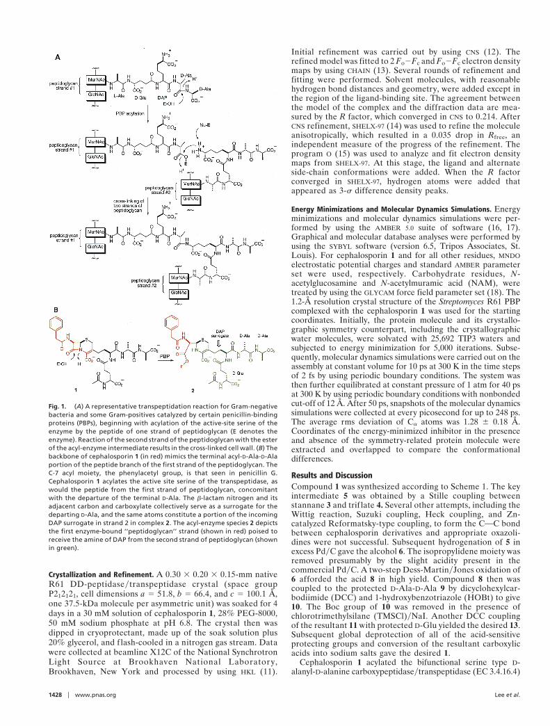
The cell wall imparts structural strength and shape to bacteria. It ismade up of polymeric glycan chains with peptide branches that arecross-linked to form the cell wall. The cross-linking reaction, cat-alyzed by transpeptidases, is the last step in cell wall biosynthesis.These enzymes are members of the family of penicillin-bindingproteins, the targets of b-lactam antibiotics. We report herein thestructure of a penicillin-binding protein complexed with a cepha-losporin designed to probe the mechanism of the cross-linkingreaction catalyzed by transpeptidases. The 1.2-Å resolution x-raystructure of this cephalosporin bound to the active site of thebifunctional serine type D-alanyl-D-alanine carboxypeptidaseytranspeptidase (EC 3.4.16.4) from Streptomyces sp. strain R61reveals how the two peptide strands from the polymeric substratesare sequestered in the active site of a transpeptidase. The structureof this complex provides a snapshot of the enzyme and the boundcell wall components poised for the final and critical cross-linkingstep of cell wall biosynthesis.
S ince their clinical introduction more than 50 years ago,b-lactam antibiotics have been essential drugs in combating
bacterial infections. To date, b-lactams constitute approximately60% of all clinically used antibiotics. These antibiotics interferewith cell wall biosynthesis. All bacteria, with very few exceptions,have a cross-linked cell wall (1, 2). The cross-linking takes placeas the last step in biosynthesis of cell wall, a reaction that iscatalyzed by bacterial transpeptidases (1, 3–6), enzymes that relyon an active-site serine for their reactions. The activated serineof transpeptidase attacks the carbonyl of the penultimate D-Alaof the peptidoglycan precursor, with concomitant departure ofthe terminal D-Ala, to give rise to an acyl-enzyme intermediate(Fig. 1A). An amine from the side chain of another peptidogly-can (diaminopimelate, modified lysine, or ornithine derivative)reacts with the ester of the acyl-enzyme intermediate to give thecross-linked species.
It was suggested by Tipper and Strominger (7) that a b-lactamantibiotic, such as a penicillin or a cephalosporin, mimics theconformation of the acyl-D-Ala-D-Ala portion of the bacterialpeptidoglycan. It also was found that b-lactam antibiotics co-valently modify the active-site serine of transpeptidase via theb-lactam carbonyl group, which spatially corresponds to thecarbonyl of the penultimate D-Ala of the pentapeptide. Thepenicillin structure in its entirety remains tethered to the enzymeas an integral part of the long-lived acyl-enzyme species, pre-senting a steric barrier to the approach of the second strand ofpeptidoglycan at the ester carbonyl (8). With the active sitecovalently modified by the antibiotic, the enzyme is no longeravailable for its normal function, transpeptidation is prevented,and bacterial death ensues.
We designed and synthesized cephalosporin 1, which incor-porates components of the cell wall in its structure (Fig. 1B). Weenvisioned that cephalosporin 1 would modify the active-siteserine of the transpeptidase, as would other b-lactams, bymimicking the acyl-D-Ala-D-Ala portion of the peptidoglycan(the portions of 1 in red in Fig. 1B). But, the portion to the right
of the acylated species with our cephalosporin (in green; com-plex 2, Fig. 1B) would mimic the approaching nucleophile,diaminopimelate, from the second strand of the peptidoglycan.Hence, cephalosporin 1 was expected to be an inhibitor oftranspeptidases, albeit a unique one with structural character-istics of the two strands of peptidoglycan. Complex 2 representsa snapshot of the two peptidoglycan strands just before thecross-linking reaction, when diaminopimelate from the secondstrand approaches the ester of the acyl-enzyme intermediateformed by the transpeptidase bound to the first strand ofpeptidoglycan. Although there are some variations in the struc-tures for the cross-links of peptidoglycans in different types ofbacteria, the structure of peptidoglycan that is incorporated intocephalosporin 1 is seen for all Gram-negative bacteria and occursin many Gram-positive organisms (1). Therefore, the mechanis-tic conclusions drawn from complex 2 would be valid for themajority of bacteria. We hasten to add that the two ‘‘strands ofpeptidoglycan’’ would be tethered to each other in complex 2. Itturns out that this reduction in the degrees of freedom for thetwo strands is absolutely necessary for the structure determina-tion, in light of the fact that numerous attempts at bindingderivatives of peptidoglycan into the PBP active sites have so farfailed to reveal any structural information. We report herein thesynthesis of cephalosporin 1, the complex of a transpeptidasemodified by 1 and its mechanistic implications for the cross-linking of the bacterial cell wall.
MethodsThe synthetic procedures for the preparation of cephalosporin 1and each of the intermediates, and their characterization byhigh-field 13C and 1H NMR, IR, and high-resolution massspectra can be found in the supplemental material and Tables 2and 3, which are published on the PNAS web site, www.pnas.org.All compounds containing the oxazolidine moiety exhibited twosets or broadening of NMR signals at room temperature, due tothe presence of a dynamic equilibrium between two conformersin solution in the absence of enzyme (9, 10). Additionally,compounds with peptide chains attached to the cephalosporinmoiety also exhibited additional conformers. These conforma-tional states were studied either by recording the spectra athigher temperatures or by reducing the concentration to mini-mize such conformational states that may be induced in anintermolecular set of interactions.
This paper was submitted directly (Track II) to the PNAS office.
Abbreviations: PBP, penicillin binding protein; DAP, diaminopimelate; NAM, N-acetylmu-ramic acid.
Data deposition: The coordinates of the complex reported in this paper have been depos-ited in the Protein Data Bank, www.rcsb.org (PDB ID code 1HVB).
See commentary on page 1319.
‡To whom reprint requests should be addressed. E-mail: [email protected] [email protected].
The publication costs of this article were defrayed in part by page charge payment. Thisarticle must therefore be hereby marked “advertisement” in accordance with 18 U.S.C.§1734 solely to indicate this fact.
PNAS u February 13, 2001 u vol. 98 u no. 4 u 1427–1431
BIO
CHEM
ISTR
Y
Crystallization and Refinement. A 0.30 3 0.20 3 0.15-mm nativeR61 DD-peptidaseytranspeptidase crystal (space groupP212121, cell dimensions a 5 51.8, b 5 66.4, and c 5 100.1 Å,one 37.5-kDa molecule per asymmetric unit) was soaked for 4days in a 30 mM solution of cephalosporin 1, 28% PEG-8000,50 mM sodium phosphate at pH 6.8. The crystal then wasdipped in cryoprotectant, made up of the soak solution plus20% glycerol, and f lash-cooled in a nitrogen gas stream. Datawere collected at beamline X12C of the National SynchrotronLight Source at Brookhaven National Laborator y,Brookhaven, New York and processed by using HKL (11).
Initial refinement was carried out by using CNS (12). Therefined model was fitted to 2 Fo2Fc and Fo2Fc electron densitymaps by using CHAIN (13). Several rounds of refinement andfitting were performed. Solvent molecules, with reasonablehydrogen bond distances and geometry, were added except inthe region of the ligand-binding site. The agreement betweenthe model of the complex and the diffraction data are mea-sured by the R factor, which converged in CNS to 0.214. AfterCNS refinement, SHELX-97 (14) was used to refine the moleculeanisotropically, which resulted in a 0.035 drop in Rfree, anindependent measure of the progress of the refinement. Theprogram O (15) was used to analyze and fit electron densitymaps from SHELX-97. At this stage, the ligand and alternateside-chain conformations were added. When the R factorconverged in SHELX-97, hydrogen atoms were added thatappeared as 3-s difference density peaks.
Energy Minimizations and Molecular Dynamics Simulations. Energyminimizations and molecular dynamics simulations were per-formed by using the AMBER 5.0 suite of software (16, 17).Graphical and molecular database analyses were performed byusing the SYBYL software (version 6.5, Tripos Associates, St.Louis). For cephalosporin 1 and for all other residues, MNDOelectrostatic potential charges and standard AMBER parameterset were used, respectively. Carbohydrate residues, N-acetylglucosamine and N-acetylmuramic acid (NAM), weretreated by using the GLYCAM force field parameter set (18). The1.2-Å resolution crystal structure of the Streptomyces R61 PBPcomplexed with the cephalosporin 1 was used for the startingcoordinates. Initially, the protein molecule and its crystallo-graphic symmetry counterpart, including the crystallographicwater molecules, were solvated with 25,692 TIP3 waters andsubjected to energy minimization for 5,000 iterations. Subse-quently, molecular dynamics simulations were carried out on theassembly at constant volume for 10 ps at 300 K in the time stepsof 2 fs by using periodic boundary conditions. The system wasthen further equilibrated at constant pressure of 1 atm for 40 psat 300 K by using periodic boundary conditions with nonbondedcut-off of 12 Å. After 50 ps, snapshots of the molecular dynamicssimulations were collected at every picosecond for up to 248 ps.The average rms deviation of Ca atoms was 1.28 6 0.18 Å.Coordinates of the energy-minimized inhibitor in the presenceand absence of the symmetry-related protein molecule wereextracted and overlapped to compare the conformationaldifferences.
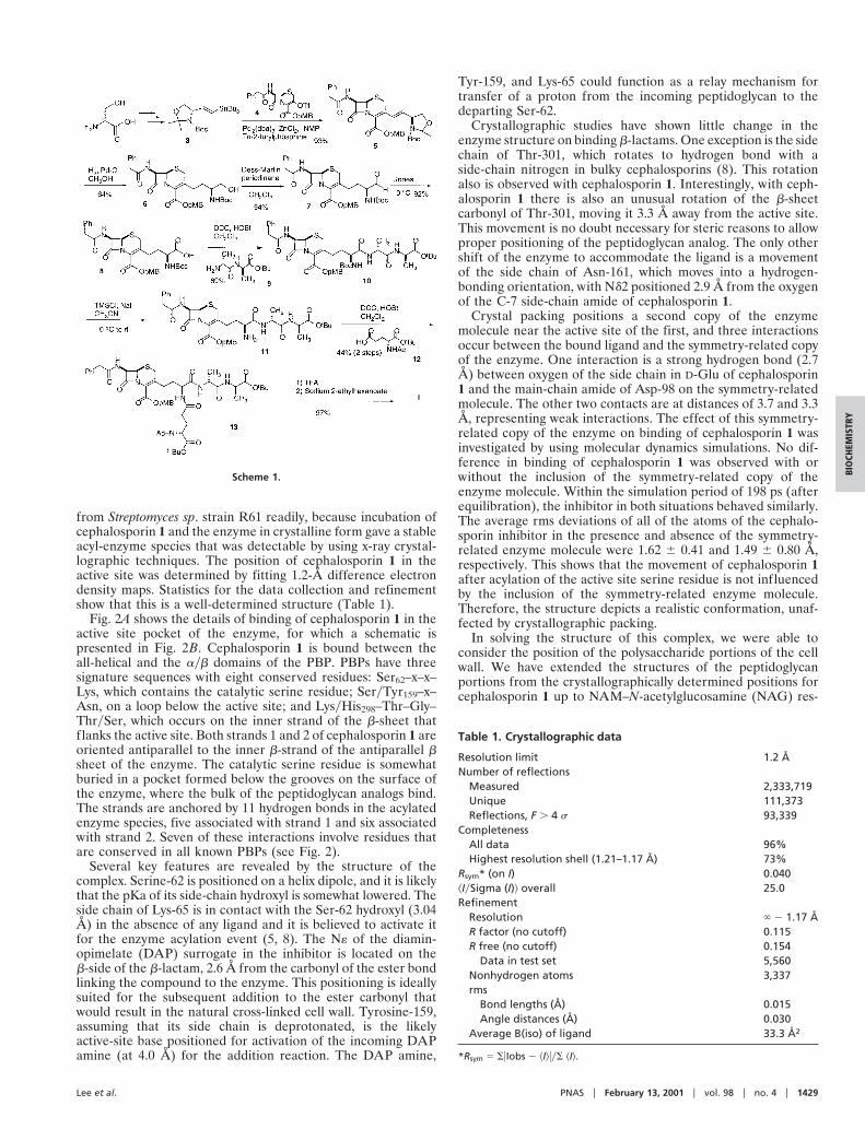
Results and DiscussionCompound 1 was synthesized according to Scheme 1. The keyintermediate 5 was obtained by a Stille coupling betweenstannane 3 and trif late 4. Several other attempts, including theWittig reaction, Suzuki coupling, Heck coupling, and Zn-catalyzed Reformatsky-type coupling, to form the COC bondbetween cephalosporin derivatives and appropriate oxazoli-dines were not successful. Subsequent hydrogenation of 5 inexcess PdyC gave the alcohol 6. The isopropylidene moiety wasremoved presumably by the slight acidity present in thecommercial PdyC. A two-step Dess-MartinyJones oxidation of6 afforded the acid 8 in high yield. Compound 8 then wascoupled to the protected D-Ala-D-Ala 9 by dicyclohexylcar-bodiimide (DCC) and 1-hydroxybenzotriazole (HOBt) to give10. The Boc group of 10 was removed in the presence ofchlorotrimethylsilane (TMSCl)yNaI. Another DCC couplingof the resultant 11 with protected D-Glu yielded the desired 13.Subsequent global deprotection of all of the acid-sensitiveprotecting groups and conversion of the resultant carboxylicacids into sodium salts gave the desired 1.
Cephalosporin 1 acylated the bifunctional serine type D-alanyl-D-alanine carboxypeptidaseytranspeptidase (EC 3.4.16.4)
Fig. 1. (A) A representative transpeptidation reaction for Gram-negativebacteria and some Gram-positives catalyzed by certain penicillin-bindingproteins (PBPs), beginning with acylation of the active-site serine of theenzyme by the peptide of one strand of peptidoglycan (E denotes theenzyme). Reaction of the second strand of the peptidoglycan with the esterof the acyl-enzyme intermediate results in the cross-linked cell wall. (B) Thebackbone of cephalosporin 1 (in red) mimics the terminal acyl-D-Ala-D-Alaportion of the peptide branch of the first strand of the peptidoglycan. TheC-7 acyl moiety, the phenylacetyl group, is that seen in penicillin G.Cephalosporin 1 acylates the active site serine of the transpeptidase, aswould the peptide from the first strand of peptidoglycan, concomitantwith the departure of the terminal D-Ala. The b-lactam nitrogen and itsadjacent carbon and carboxylate collectively serve as a surrogate for thedeparting D-Ala, and the same atoms constitute a portion of the incomingDAP surrogate in strand 2 in complex 2. The acyl-enzyme species 2 depictsthe first enzyme-bound ‘‘peptidoglycan’’ strand (shown in red) poised toreceive the amine of DAP from the second strand of peptidoglycan (shownin green).
1428 u www.pnas.org Lee et al.
from Streptomyces sp. strain R61 readily, because incubation ofcephalosporin 1 and the enzyme in crystalline form gave a stableacyl-enzyme species that was detectable by using x-ray crystal-lographic techniques. The position of cephalosporin 1 in theactive site was determined by fitting 1.2-Å difference electrondensity maps. Statistics for the data collection and refinementshow that this is a well-determined structure (Table 1).
Fig. 2A shows the details of binding of cephalosporin 1 in theactive site pocket of the enzyme, for which a schematic ispresented in Fig. 2B. Cephalosporin 1 is bound between theall-helical and the ayb domains of the PBP. PBPs have threesignature sequences with eight conserved residues: Ser62–x–x–Lys, which contains the catalytic serine residue; SeryTyr159–x–Asn, on a loop below the active site; and LysyHis298–Thr–Gly–ThrySer, which occurs on the inner strand of the b-sheet thatflanks the active site. Both strands 1 and 2 of cephalosporin 1 areoriented antiparallel to the inner b-strand of the antiparallel bsheet of the enzyme. The catalytic serine residue is somewhatburied in a pocket formed below the grooves on the surface ofthe enzyme, where the bulk of the peptidoglycan analogs bind.The strands are anchored by 11 hydrogen bonds in the acylatedenzyme species, five associated with strand 1 and six associatedwith strand 2. Seven of these interactions involve residues thatare conserved in all known PBPs (see Fig. 2).
Several key features are revealed by the structure of thecomplex. Serine-62 is positioned on a helix dipole, and it is likelythat the pKa of its side-chain hydroxyl is somewhat lowered. Theside chain of Lys-65 is in contact with the Ser-62 hydroxyl (3.04Å) in the absence of any ligand and it is believed to activate itfor the enzyme acylation event (5, 8). The N« of the diamin-opimelate (DAP) surrogate in the inhibitor is located on theb-side of the b-lactam, 2.6 Å from the carbonyl of the ester bondlinking the compound to the enzyme. This positioning is ideallysuited for the subsequent addition to the ester carbonyl thatwould result in the natural cross-linked cell wall. Tyrosine-159,assuming that its side chain is deprotonated, is the likelyactive-site base positioned for activation of the incoming DAPamine (at 4.0 Å) for the addition reaction. The DAP amine,
Tyr-159, and Lys-65 could function as a relay mechanism fortransfer of a proton from the incoming peptidoglycan to thedeparting Ser-62.
Crystallographic studies have shown little change in theenzyme structure on binding b-lactams. One exception is the sidechain of Thr-301, which rotates to hydrogen bond with aside-chain nitrogen in bulky cephalosporins (8). This rotationalso is observed with cephalosporin 1. Interestingly, with ceph-alosporin 1 there is also an unusual rotation of the b-sheetcarbonyl of Thr-301, moving it 3.3 Å away from the active site.This movement is no doubt necessary for steric reasons to allowproper positioning of the peptidoglycan analog. The only othershift of the enzyme to accommodate the ligand is a movementof the side chain of Asn-161, which moves into a hydrogen-bonding orientation, with Nd2 positioned 2.9 Å from the oxygenof the C-7 side-chain amide of cephalosporin 1.
Crystal packing positions a second copy of the enzymemolecule near the active site of the first, and three interactionsoccur between the bound ligand and the symmetry-related copyof the enzyme. One interaction is a strong hydrogen bond (2.7Å) between oxygen of the side chain in D-Glu of cephalosporin1 and the main-chain amide of Asp-98 on the symmetry-relatedmolecule. The other two contacts are at distances of 3.7 and 3.3Å, representing weak interactions. The effect of this symmetry-related copy of the enzyme on binding of cephalosporin 1 wasinvestigated by using molecular dynamics simulations. No dif-ference in binding of cephalosporin 1 was observed with orwithout the inclusion of the symmetry-related copy of theenzyme molecule. Within the simulation period of 198 ps (afterequilibration), the inhibitor in both situations behaved similarly.The average rms deviations of all of the atoms of the cephalo-sporin inhibitor in the presence and absence of the symmetry-related enzyme molecule were 1.62 6 0.41 and 1.49 6 0.80 Å,respectively. This shows that the movement of cephalosporin 1after acylation of the active site serine residue is not influencedby the inclusion of the symmetry-related enzyme molecule.Therefore, the structure depicts a realistic conformation, unaf-fected by crystallographic packing.
In solving the structure of this complex, we were able toconsider the position of the polysaccharide portions of the cellwall. We have extended the structures of the peptidoglycanportions from the crystallographically determined positions forcephalosporin 1 up to NAM–N-acetylglucosamine (NAG) res-
Table 1. Crystallographic data
Resolution limit 1.2 ÅNumber of reflections
Measured 2,333,719Unique 111,373Reflections, F . 4 s 93,339
CompletenessAll data 96%Highest resolution shell (1.21–1.17 Å) 73%
Rsym* (on I) 0.040^IySigma (I)& overall 25.0Refinement
Resolution ` 2 1.17 ÅR factor (no cutoff) 0.115R free (no cutoff) 0.154
Data in test set 5,560Nonhydrogen atoms 3,337rms
Bond lengths (Å) 0.015Angle distances (Å) 0.030
Average B(iso) of ligand 33.3 Å2
*Rsym 5 SuIobs 2 ^I&uyS ^I&.
Scheme 1.
Lee et al. PNAS u February 13, 2001 u vol. 98 u no. 4 u 1429
BIO
CHEM
ISTR
Y
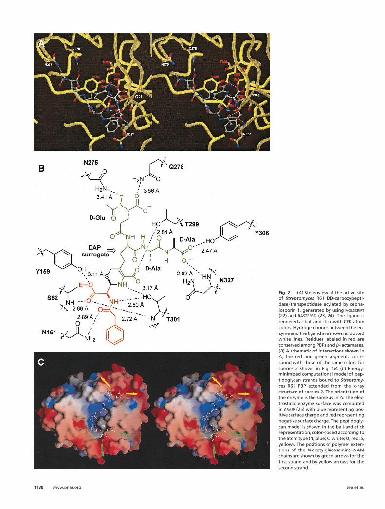
Fig. 2. (A) Stereoview of the active siteof Streptomyces R61 DD-carboxypepti-daseytranspeptidase acylated by cepha-losporin 1, generated by using MOLSCRIPT
(22) and RASTER3D (23, 24). The ligand isrendered as ball and stick with CPK atomcolors. Hydrogen bonds between the en-zyme and the ligand are shown as dottedwhite lines. Residues labeled in red areconserved among PBPs and b-lactamases.(B) A schematic of interactions shown inA; the red and green segments corre-spond with those of the same colors forspecies 2 shown in Fig. 1B. (C) Energy-minimized computational model of pep-tidoglycan strands bound to Streptomy-ces R61 PBP extended from the x-raystructure of species 2. The orientation ofthe enzyme is the same as in A. The elec-trostatic enzyme surface was computedin GRASP (25) with blue representing pos-itive surface charge and red representingnegative surface charge. The peptidogly-can model is shown in the ball-and-stickrepresentation, color-coded according tothe atom type (N, blue; C, white; O, red; S,yellow). The positions of polymer exten-sions of the N-acetylglucosamine–NAMchains are shown by green arrows for thefirst strand and by yellow arrows for thesecond strand.
1430 u www.pnas.org Lee et al.
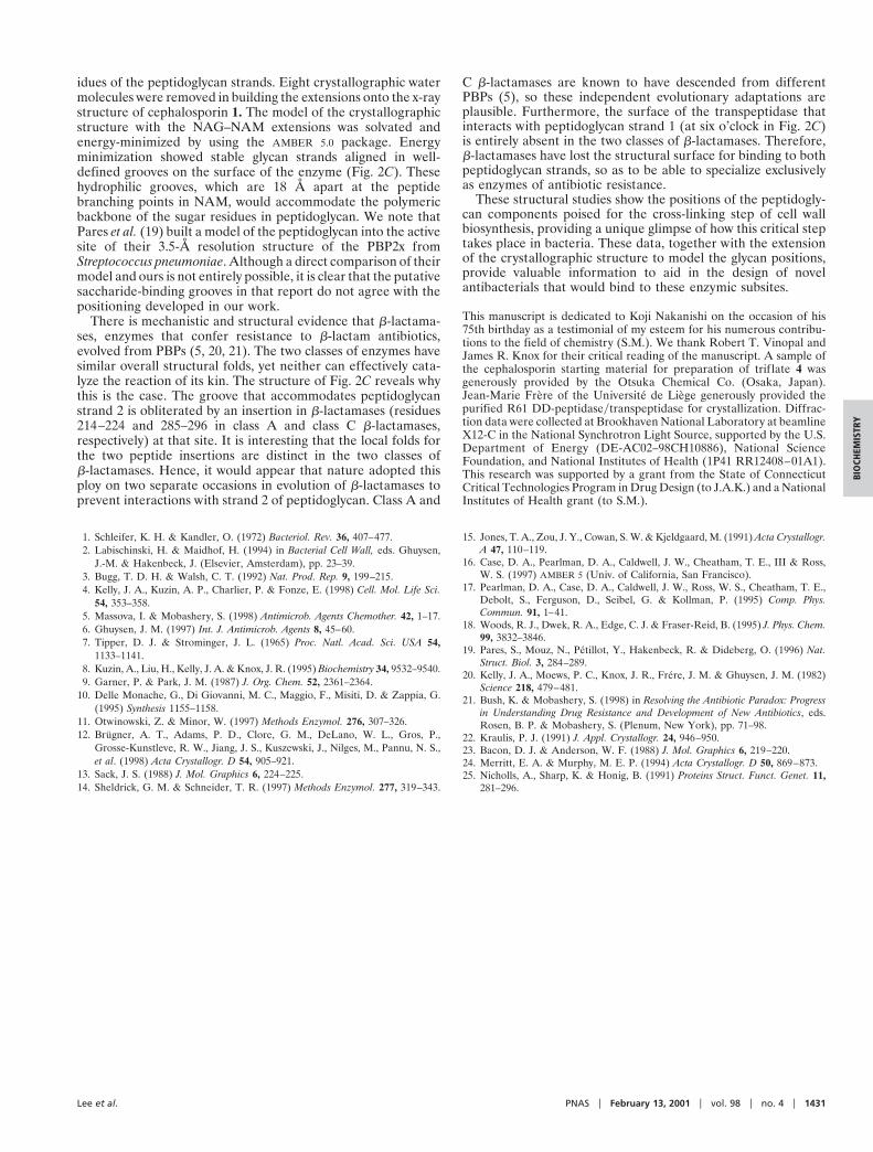
idues of the peptidoglycan strands. Eight crystallographic watermolecules were removed in building the extensions onto the x-raystructure of cephalosporin 1. The model of the crystallographicstructure with the NAG–NAM extensions was solvated andenergy-minimized by using the AMBER 5.0 package. Energyminimization showed stable glycan strands aligned in well-defined grooves on the surface of the enzyme (Fig. 2C). Thesehydrophilic grooves, which are 18 Å apart at the peptidebranching points in NAM, would accommodate the polymericbackbone of the sugar residues in peptidoglycan. We note thatPares et al. (19) built a model of the peptidoglycan into the activesite of their 3.5-Å resolution structure of the PBP2x fromStreptococcus pneumoniae. Although a direct comparison of theirmodel and ours is not entirely possible, it is clear that the putativesaccharide-binding grooves in that report do not agree with thepositioning developed in our work.
There is mechanistic and structural evidence that b-lactama-ses, enzymes that confer resistance to b-lactam antibiotics,evolved from PBPs (5, 20, 21). The two classes of enzymes havesimilar overall structural folds, yet neither can effectively cata-lyze the reaction of its kin. The structure of Fig. 2C reveals whythis is the case. The groove that accommodates peptidoglycanstrand 2 is obliterated by an insertion in b-lactamases (residues214–224 and 285–296 in class A and class C b-lactamases,respectively) at that site. It is interesting that the local folds forthe two peptide insertions are distinct in the two classes ofb-lactamases. Hence, it would appear that nature adopted thisploy on two separate occasions in evolution of b-lactamases toprevent interactions with strand 2 of peptidoglycan. Class A and
C b-lactamases are known to have descended from differentPBPs (5), so these independent evolutionary adaptations areplausible. Furthermore, the surface of the transpeptidase thatinteracts with peptidoglycan strand 1 (at six o’clock in Fig. 2C)is entirely absent in the two classes of b-lactamases. Therefore,b-lactamases have lost the structural surface for binding to bothpeptidoglycan strands, so as to be able to specialize exclusivelyas enzymes of antibiotic resistance.
These structural studies show the positions of the peptidogly-can components poised for the cross-linking step of cell wallbiosynthesis, providing a unique glimpse of how this critical steptakes place in bacteria. These data, together with the extensionof the crystallographic structure to model the glycan positions,provide valuable information to aid in the design of novelantibacterials that would bind to these enzymic subsites.
This manuscript is dedicated to Koji Nakanishi on the occasion of his75th birthday as a testimonial of my esteem for his numerous contribu-tions to the field of chemistry (S.M.). We thank Robert T. Vinopal andJames R. Knox for their critical reading of the manuscript. A sample ofthe cephalosporin starting material for preparation of trif late 4 wasgenerously provided by the Otsuka Chemical Co. (Osaka, Japan).Jean-Marie Frere of the Universite de Liege generously provided thepurified R61 DD-peptidaseytranspeptidase for crystallization. Diffrac-tion data were collected at Brookhaven National Laboratory at beamlineX12-C in the National Synchrotron Light Source, supported by the U.S.Department of Energy (DE-AC02–98CH10886), National ScienceFoundation, and National Institutes of Health (1P41 RR12408–01A1).This research was supported by a grant from the State of ConnecticutCritical Technologies Program in Drug Design (to J.A.K.) and a NationalInstitutes of Health grant (to S.M.).
1. Schleifer, K. H. & Kandler, O. (1972) Bacteriol. Rev. 36, 407–477.2. Labischinski, H. & Maidhof, H. (1994) in Bacterial Cell Wall, eds. Ghuysen,
J.-M. & Hakenbeck, J. (Elsevier, Amsterdam), pp. 23–39.3. Bugg, T. D. H. & Walsh, C. T. (1992) Nat. Prod. Rep. 9, 199–215.4. Kelly, J. A., Kuzin, A. P., Charlier, P. & Fonze, E. (1998) Cell. Mol. Life Sci.
54, 353–358.5. Massova, I. & Mobashery, S. (1998) Antimicrob. Agents Chemother. 42, 1–17.6. Ghuysen, J. M. (1997) Int. J. Antimicrob. Agents 8, 45–60.7. Tipper, D. J. & Strominger, J. L. (1965) Proc. Natl. Acad. Sci. USA 54,
1133–1141.8. Kuzin, A., Liu, H., Kelly, J. A. & Knox, J. R. (1995) Biochemistry 34, 9532–9540.9. Garner, P. & Park, J. M. (1987) J. Org. Chem. 52, 2361–2364.
10. Delle Monache, G., Di Giovanni, M. C., Maggio, F., Misiti, D. & Zappia, G.(1995) Synthesis 1155–1158.
11. Otwinowski, Z. & Minor, W. (1997) Methods Enzymol. 276, 307–326.12. Brugner, A. T., Adams, P. D., Clore, G. M., DeLano, W. L., Gros, P.,
Grosse-Kunstleve, R. W., Jiang, J. S., Kuszewski, J., Nilges, M., Pannu, N. S.,et al. (1998) Acta Crystallogr. D 54, 905–921.
13. Sack, J. S. (1988) J. Mol. Graphics 6, 224–225.14. Sheldrick, G. M. & Schneider, T. R. (1997) Methods Enzymol. 277, 319–343.
15. Jones, T. A., Zou, J. Y., Cowan, S. W. & Kjeldgaard, M. (1991) Acta Crystallogr.A 47, 110–119.
16. Case, D. A., Pearlman, D. A., Caldwell, J. W., Cheatham, T. E., III & Ross,W. S. (1997) AMBER 5 (Univ. of California, San Francisco).
17. Pearlman, D. A., Case, D. A., Caldwell, J. W., Ross, W. S., Cheatham, T. E.,Debolt, S., Ferguson, D., Seibel, G. & Kollman, P. (1995) Comp. Phys.Commun. 91, 1–41.
18. Woods, R. J., Dwek, R. A., Edge, C. J. & Fraser-Reid, B. (1995) J. Phys. Chem.99, 3832–3846.
19. Pares, S., Mouz, N., Petillot, Y., Hakenbeck, R. & Dideberg, O. (1996) Nat.Struct. Biol. 3, 284–289.
20. Kelly, J. A., Moews, P. C., Knox, J. R., Frere, J. M. & Ghuysen, J. M. (1982)Science 218, 479–481.
21. Bush, K. & Mobashery, S. (1998) in Resolving the Antibiotic Paradox: Progressin Understanding Drug Resistance and Development of New Antibiotics, eds.Rosen, B. P. & Mobashery, S. (Plenum, New York), pp. 71–98.
22. Kraulis, P. J. (1991) J. Appl. Crystallogr. 24, 946–950.23. Bacon, D. J. & Anderson, W. F. (1988) J. Mol. Graphics 6, 219–220.24. Merritt, E. A. & Murphy, M. E. P. (1994) Acta Crystallogr. D 50, 869–873.25. Nicholls, A., Sharp, K. & Honig, B. (1991) Proteins Struct. Funct. Genet. 11,
281–296.
Lee et al. PNAS u February 13, 2001 u vol. 98 u no. 4 u 1431
BIO
CHEM
ISTR
Y