Embed Size (px)
Citation preview

99mTc-Interleukin-8 for Imaging AcuteOsteomyelitisStefan Gratz, Huub J.J.M. Rennen, Otto C. Boerman, Wim J.G. Oyen, Pieter Burma, and Frans H.M. Corstens
Department of Nuclear Medicine and Section of Histomorphology, Orthopaedic Research Laboratory,University Medical Center Nijmegen, Nijmegen, The Netherlands
Early and accurate diagnosis of osteomyelitis remains a clinicalproblem. Acute osteomyelitis often occurs in infants and mostoften is located in the long bones. Radiologic images showchanges only in advanced stages of disease. Scintigraphic im-aging with 99mTc-methylene diphosphonate (MDP), or bonescanning, is much more sensitive in detecting acute osteomy-elitis but lacks specificity. We evaluated the performance of99mTc-interleukin-8 (IL-8) in an experimental model of acuteosteomyelitis. Methods: Acute pyogenic osteomyelitis was in-duced in 10 rabbits by inserting sodium morrhuate and Staph-ylococcus aureus into the medullary cavity of the right femur.The cavity was closed with liquid cement. A sham operation wasperformed on the left femur. Routine radiographs were obtainedjust before scintigraphy. Ten days after surgery, the rabbitswere divided into 2 groups of 5 animals, received an injection ofeither 18.5 MBq 111In-granulocytes or 18.5 MBq 67Ga-citrate,and were imaged both 24 h after injection and 48 h after injec-tion. On day 12, the rabbits received either 18.5 MBq 99mTc-MDP or 18.5 MBq 99mTc-IL-8, and serial images were acquiredat 0, 1, 2, 4, 8, 12, and 24 h after injection. Uptake in the infectedfemur was determined by drawing regions of interest. Ratios ofinfected femur (target) to sham-operated femur (background)(T/Bs) were calculated. After the final images were obtained, therabbits were killed and the right femur was dissected and ana-lyzed for microbiologic and histopathologic evidence of osteo-myelitis. Results: Acute osteomyelitis developed in 8 of 10rabbits. All imaging agents correctly detected the acute osteo-myelitis in these animals. The extent of infection was optimallyvisualized with 67Ga-citrate and delayed bone scanning,whereas diaphyseal photopenia was noted with both 99mTc-IL-8and 111In-granulocytes. In 1 rabbit with osteomyelitis, imagingresults were falsely negative with 111In-granulocytes and falselypositive with 99mTc-MDP. Quantitative analysis of the imagesrevealed that the uptake in the infected region was highest with67Ga-citrate (4.9 6 0.8 percentage injected dose [%ID]) and99mTc-MDP (4.7 6 0.7 %ID), whereas the uptake in the infectedarea was significantly lower with 99mTc-IL-8 (2.2 6 0.2 %ID) and111In-granulocytes (0.8 6 0.2 %ID) (P , 0.0042). In contrast, theT/Bs were significantly higher for 99mTc-IL-8 (T/B, 6.2 6 0.3 at4 h after injection) than for 67Ga-citrate, 99mTc-MDP, and 111In-granulocytes, which had ratios of 1.5 6 0.4, 1.9 6 0.2, and 1.4 60.1, respectively (P , 0.0001). Radiography correctly revealedacute osteomyelitis in only 2 of 8 rabbits. Conclusion: In this
rabbit model of osteomyelitis, 99mTc-IL-8 clearly revealed theosteomyelitic lesion. Although the absolute uptake in the osteo-myelitic area was significantly lower than that obtained with99mTc-MDP and 67Ga-citrate, the T/Bs were significantly higherfor 99mTc-IL-8 because of fast background clearance. The easeof preparation, good image quality, and lower radiation burdensuggest that 99mTc-IL-8 may be a suitable imaging agent for thescintigraphic evaluation of acute osteomyelitis.
Key Words: acute osteomyelitis; interleukin-8; imaging infec-tion; technetium
J Nucl Med 2001; 42:1257–1264
I n cases of delayed therapy, acute osteomyelitis can,within several days, cause severe malformations of the bonethat can become disabling and substantially affect the qual-ity of life (1). Acute osteomyelitis usually is diagnosed onthe basis of imaging, laboratory tests, and clinical exami-nations (2–5). In nuclear medicine,99mTc-methylenediphosphonate (MDP) and67Ga-citrate are sensitive agentsfor the detection of osteomyelitis. Their specificity, how-ever, is low because both agents accumulate in any areawith increased bone turnover (6). Radiolabeled white bloodcells (7,8) as imaging agents have a much higher specificityfor scintigraphic imaging of osteomyelitis. The preparationof radiolabeled autologous white blood cells, however, islaborious and time consuming and carries a small but def-inite risk of personal contamination by the patients’ bloodand inadvertent cross-contamination between patients(9,10).
New radiopharmaceuticals, such as radiolabeled mono-clonal antigranulocyte antibody preparations, have beenproposed for easy and fast imaging of infection (11,12).These new agents displayed high sensitivity and specificityin patients with acute osteomyelitis (13–15), presumablybecause they act through cell-specific labeling of surfaceantigens as present on granulocytes.
Granulocytes are known to express interleukin-8 (IL-8)receptors abundantly (16–19). Therefore, the proinflamma-tory chemotactic cytokine IL-8 may be an interesting alter-native to the current methods for imaging acute osteomy-elitis.
Received Jan. 16, 2001; revision accepted Apr. 9, 2001.For correspondence or reprints contact: Stefan Gratz, MD, Department of
Nuclear Medicine, University Medical Center Nijmegen, P.O. Box 9101, 6500HB Nijmegen, The Netherlands.
99MTC-IL-8 FOR IMAGING ACUTE OSTEOMYELITIS • Gratz et al. 1257
by on April 9, 2019. For personal use only. jnm.snmjournals.org Downloaded from

Recently, we introduced IL-8 as a new scintigraphicimaging agent. In a rabbit model withEscherichia colisoft-tissue infection,99mTc-hydrazinonicotinamide-IL-8 al-lowed rapid visualization of the infectious foci as early as1 h after injection, with high and rapid accretion of theradiolabel in the abscess (20). This study was performedprimarily to gain our first preclinical experience with the useof this new 99mTc-IL-8 tracer to detect experimentally in-duced osteomyelitis. Therefore, we evaluated99mTc-IL-8 asan imaging agent in a rabbit model of acute osteomyelitis.The results were compared with those obtained using theconventional and well-established agents99mTc-MDP,67Ga-citrate, and111In-granulocytes.
MATERIALS AND METHODS
Animal Osteomyelitis ModelThis study was performed in accordance with the guidelines of
the local animal welfare committee. Adult female New ZealandWhite rabbits (weight range, 2.5–3.0 kg) were obtained from thecentral animal laboratory, University of Nijmegen (Nijmegen, TheNetherlands), caged individually, and fed regular rabbit diet andwater ad libitum. In 10 rabbits, acute osteomyelitis was induced asdescribed previously, with minor modifications (21–23). The rab-bits were anesthetized with a mixture of halothane, nitrous oxide,and oxygen and placed prone on the operation table. Both hind legswere shaved, disinfected with a 2% tincture of iodine, and isolatedby sterile drapes. The trochanter tertius was exposed bilaterally,and the cortex was penetrated gently using a hand drill. A smallsyringe with a 2-mm-long silicone tube (outer diameter, 3.0 mm)was inserted into the femoral canal, and 0.5 mL 5% sodiummorrhuate (QUAD Pharmaceuticals Inc., Indianapolis, IN) wasinoculated in the canal. Morrhuate, a complex of fatty acids rich inarachidonic acid (24,25), was used to induce a local irritation.Subsequently, 0.5 mL of 53 106 colony-forming units ofStaph-ylococcus aureus(ATCC 25923; American Type Culture Collec-tion, Manassas, VA) was inoculated. For the sham procedure onthe right femur, the canal was washed with 1 mL saline and leftwithout a local irritation/infection procedure. Finally, both holes inthe trochanter were sealed with a small amount of liquid carbox-ylate cement (Durelon; ESPE Dental AG, Seefeld, Germany).After polymerization of the cement, the wounds at both sides werecleaned with sterile saline solution and closed. The animals wereexamined regularly with special attention to wound healing, bodytemperature, and body weight.
Radiopharmaceuticals99mTc-IL-8. The 99mTc-labeled IL-8 was prepared as described
previously (20), with minor modifications. Briefly, to a 1.5-mLvial containing 6mg IL-8 was added 0.4 mL tricine solution(N-[Tris(hydroxymethyl)methyl]glycine) (Fluka, Buchs, Switzer-land; 100 mg/mL in 25 mmol/L succinate buffer, pH 5.0) and 0.1mL isonicotinic acid solution (Sigma, St. Louis, MO; 20 mg/mL in25 mmol/L succinate buffer, pH 5.0) (26). After the reactionmixture had been purged with a gentle stream of nitrogen, 25mLSnSO4 solution (1.0 mg/mL in 0.1N HCl) and 350 MBq99mTcO4
2
were added. After having been heated to 70°C for 30 min, thereaction mixture was cooled to room temperature and the radio-chemical purity was determined by instant thin-layer chromatog-raphy (ITLC) on ITLC-SG strips (Gelman Laboratories,
Ann Arbor, MI) with 0.1 mol/L citrate, pH 6.0, as the mobilephase.
The 99mTc-IL-8 was purified on a Sephadex G-25 column (PD-10; Pharmacia, Uppsala, Sweden) eluted with 0.5% bovine serumalbumin in phosphate-buffered saline. The labeling efficiency ofthe 99mTc-IL-8 preparation exceeded 90%. After Sephadex G-25purification, the radiochemical purity of the radiopharmaceuticalexceeded 98% as determined by ITLC. The specific activity of thepurified preparation was 50 MBq/mg (425 GBq/mmol).
111In-Granulocytes.Carotid artery cannulation was performedon 1 anesthetized donor rabbit. A total of 100 mL blood wascarefully drawn into acid citrate dextrose tubes (containing 7 mLacid citrate dextrose per 35 mL blood). The total leukocyte countof the donor rabbits was 6.83 109 cells/L, with approximately50% granulocytes. The granulocytes were purified according to themethod described by Lillevang et al. (27), with minor modifica-tions (28). Briefly, the blood was mixed with 0.1 volume of 6%dextran (Dextran 500; Pharmacia) solution in 0.9% NaCl andallowed to settle for 1 h at room temperature. The leukocyte-richsupernatant was layered carefully on 0.3 volume of Nycoprepdensity medium (Nycomed, Oslo, Norway; 14.1% Nycodenz [Ny-comed], 0.3% NaCl, 5 mmol/L tricine/NaOH, pH 7.2, 1.077 g/mLdensity, 265 mOsm) and centrifuged for 15 min at 600g. Theplasma above the mononuclear cells, the mononuclear band, andthe density medium above the granulocyte pellet were carefullyremoved. The pellet was washed with 5 mL Hanks’ balanced saltsolution (HBSS) with 10% autologous plasma and centrifuged for10 min at 50g. The cell pellet was resuspended in 1.5 mL HBSSwith 10% rabbit plasma. After this purification procedure, thegranulocyte purity was.90%. Subsequently, 185 MBq111In-oxine were added to the cell suspension. The cells were incubatedat room temperature for 30 min and centrifuged for 10 min at 50g.The pellet was resuspended in 5 mL cell-free autologous plasma.Labeling efficiency (cell-associated activity/total activity added)exceeded 80%. The functional integrity of the labeled granulocyteswas evaluated by their in vivo performance, including transitthrough the lungs, as well as uptake in the liver and spleen. A doseof 18 MBq 111In-oxine–labeled granulocytes was administeredintravenously to each rabbit.
67Ga-Citrate. 67Ga-citrate (DRN 3103) was purchased fromMallinckrodt, Inc. (St. Louis, MO). A dose of approximately 18MBq 67Ga-citrate per rabbit was injected intravenously.
99mTc-MDP. A kit containing MDP and stannous chloride waslabeled with99mTc with a labeling efficiency. 95% as determinedby ITLC. A dose of 18 MBq99mTc-MDP was administered intra-venously.
Receptor-Binding AssaysThe receptor-binding fraction of the99mTc-IL-8 preparation was
determined in receptor-binding assays essentially as described byLindmo et al. (29). For receptor-binding assays, Jurkat cells trans-fected with CXCR1 or CXCR2 were used (30). The cells werecultured at 37°C in a humidified atmosphere of air and CO2 (95:5)in Roswell Park Memorial Institute (RPMI) 1640 medium(GIBCO, Gaithersburg, MD) containing 10% fetal calf serum, 1%glutamine, penicillin and streptomycin, 53 1025 mol/L b-mer-captoethanol, and 1.5mg/mL puromycin. Series of serially dilutedcell suspension (0.25–43 108 cells/mL) were incubated with10,000 cpm of99mTc-IL-8 in assay buffer (RPMI 1640, 0.5%bovine serum albumin, and 0.05% NaN3). Duplicates of the lowestcell concentration were incubated in the presence of at least a
1258 THE JOURNAL OF NUCLEAR MEDICINE • Vol. 42 • No. 8 • August 2001
by on April 9, 2019. For personal use only. jnm.snmjournals.org Downloaded from

100-fold molar excess of unlabeled IL-8 to correct for nonspecificbinding. After 30 min of incubation at 37°C, the cells werecentrifuged (5 min, 2000g) and the radioactivity in the pellet (totalbound radioactivity) was measured in a shielded well scintillationg-counter (Wizard; Pharmacia). The data were graphically ana-lyzed in a modified Lineweaver–Burk plot: a double-inverse plotof the conventional binding plot (specifically bound fraction vs.cell concentration). The receptor-binding fraction at infinite cellexcess was calculated by linear extrapolation to the ordinate. The99mTc-IL-8 preparation showed a CXCR1 receptor-binding frac-tion of 55% and a CXCR2 receptor-binding fraction of 45%.
Study DesignThe rabbits were randomized into 2 groups: A (n 5 5) and B
(n 5 5). The scintigraphic studies were started 10 d after inductionof osteomyelitis. The radiopharmaceuticals were injected in a fixedorder. On the first day of the imaging experiment, the rabbits ofgroup A received 18.5 MBq111In-granulocytes intravenously andthe rabbits of group B received 18.5 MBq67Ga-citrate intrave-nously. The scintigraphic images were acquired 24 and 48 h afterinjection of each of the radiotracers. On the third day of theimaging experiment (12 d after induction of osteomyelitis), therabbits of group A received 18.5 MBq99mTc-MDP intravenouslyand the rabbits of group B received 18.5 MBq99mTc-IL-8 intra-venously. Crossover of remaining67Ga and111In activity in the99mTc channel was,5% as determined before and after injectionof the 99mTc-labelled compounds. To exclude pharmaceutical in-terference with the cell-binding capacity of the radiotracer, we didnot sedate the rabbits. The rabbits were immobilized in a mold andplaced prone on a gamma camera equipped with a medium-energycollimator (Orbiter; Siemens Inc., Hoffman Estates, IL) for the67Ga and111In studies and with a parallel-hole, low-energy colli-mator for the99mTc-MDP and99mTc-IL-8 studies. Imaging wasperformed at 24 and 48 h (for111In and 67Ga, respectively) afterinjection. The imaging sessions for99mTc-MDP and99mTc-IL-8were at 3 min (blood-pool image) and 4 h (delayed image) afterinjection for the bone scans and at 2 min and 1, 2, 4, 8, 12, and 24 hafter injection for the serial IL-8 images. Images (300,000 countsper image) were obtained and stored in a 2563 256 matrix.
The scintigraphic results were analyzed both qualitatively andquantitatively. All scans were evaluated qualitatively without knowl-edge of the histopathologic outcome. Findings were considered pos-itive if focal uptake of radioactivity at the osteomyelitic site exceededuptake at the sham-operated site. For quantitative analysis, regions of
interest were drawn over the infected right femur and the sham-operated left femur, as well as over the whole body. Additionalregions of interest (at 0, 1, 4, 10, and 20 h after injection) over thelungs, liver, and spleen were drawn on the scintigrams of the rabbitsthat received111In-granulocytes. Counts were corrected for differ-ences in the number of pixels before ratios and percentages werecalculated. The ratios of infected femur (target) to sham-operatedfemur (background) (T/Bs) were calculated. Residual activity at theosteomyelitic site was determined and measured as fraction of theinjected dose (whole body set tot 5 0).
Radiologic and Histologic AnalysesThe results of the scintigraphic studies were compared with the
results of radiologic, microbiologic, and histopathologic examina-tion. Conventional radiographs were obtained after the imagingsessions. While unaware of the results of all other procedures, weevaluated the radiograms with respect to 3 parameters (periostealthinning, increased radiolucency, and inclusion of air) (31). Im-mediately after completion of the scintigraphic studies, the rabbitswere killed with an overdose of sodium phenobarbital. The rightfemur of each animal was excised and tissue debris was removed.The femur was halved longitudinally using a high-speed dentaldrill with a circular metal saw. The bone cement was carefullyremoved. One bone specimen of each femur was sent for micro-biologic examination. For histopathologic examination, the otherbone specimens were fixed in 4% buffered formalin and decalci-fied in 10% ethylenediaminetetraacetic acid. Longitudinal sectionswere made, mounted on slides, and stained with hematoxylin andeosin. All sections were reviewed microscopically with respect to3 parameters: purulent inflammation with infiltration of the bonemarrow by polymorphonuclear leukocytes, necrotic bone, andbone fistulas. The presence of osteomyelitis was confirmed on thebasis of these histopathologic findings.
Statistical AnalysisMean uptake values are given as percentage injected dose
(%ID) 6 1 SEM. For paired data, 1-way ANOVA using InStatsoftware (version 3.00; GraphPad Software, Inc., San Diego, CA)was performed to compare uptake at the osteomyelitic and sham-surgery sites for the different agents. In addition, repeated-mea-sures ANOVA was used to evaluate differences between theimages acquired at different times for each agent. The level ofsignificance was set at 0.05.
TABLE 1Results for Group A
Rabbitno.
99mTc-MDP
111In-granulocytes
Radiology Macroscopy Histology MicrobiologyBlood-pool
image Delayed image 24 h 48 h
1 11 111 1 11 1 111 1 12 1 11 1 1 2 1 1 13 2 11 6 6 2 — 2 24 1 1 6 1 2 1 1 15 11 11 6 1 2 1 1 1
1 5 positive (111 5 complete; 11 5 extensive; 1 5 part only); 2 5 negative; 6 5 equivocal; macroscopy 5 extension ofosteomyelitis.
99MTC-IL-8 FOR IMAGING ACUTE OSTEOMYELITIS • Gratz et al. 1259
by on April 9, 2019. For personal use only. jnm.snmjournals.org Downloaded from

RESULTS
Tables 1 and 2 summarize the observations for groups Aand B, respectively. Eight of the 10 rabbits had histopatho-logic and microbiologic evidence of acute osteomyelitis inthe right femur. Macroscopically, in 3 of 8 rabbits theosteomyelitis in the right femur had affected the completefemur, whereas in 5 of 8 rabbits the infection was apparentonly in the diaphyseal and distal parts of the femur. Theproximal part of the osteomyelitis-negative rabbit (rabbit 3)showed some minimal leukocytic infiltration, which wasmost likely caused by a local reaction to loosening of thecement. No evidence of infection was found in rabbit 6. Themicrobiologic studies of the bone specimens were concor-
dant with the histopathologic findings in all infected rabbitsand confirmed the presence ofS. aureusinfection in 8 of 10rabbits. Histopathologically, the infected medullar tissuewas characterized by large accumulations of polymorpho-nuclear leucocytes (PMNs) and some lymphocytes. Abun-dant areas of debris and of necrosis were seen. The trabec-ular bone near the growth plate was, in most cases, necroticand infiltrated by many PMNs. The cortical bone showednecrosis and infiltration of PMNs, osteoclastic resorption,and a strong periosteal callous formation (Fig. 1). In con-trast, radiologic findings were abnormal in only 2 rabbits,showing discrete periosteal thinning and increased radiolu-cency.
FIGURE 1. (A) Medullar tissue of controlanimal containing numerous fat cells (v)and red marrow (3150). (B) Medullar tissueof low-grade infection: fibrous tissue andosteoclastic erosion on endosteal surfaceof femoral cortex (3150). (C) Large accu-mulation of PMNs in medullar tissue ofheavily infected bone specimen (350). (D)Enlargement of central part because ofPMN accumulation (3250). (E) Erosionchannels and osteoclasts (arrows) in corti-cal infected bone (3100). (F) Eroded areasin cortical bone filled with leukocytes(380).
TABLE 2Results for Group B
Rabbitno.
99mTc-IL-8 67Ga-citrate
Radiology Macroscopy Histology Microbiology2 h 4 h 24 h 48 h
6 2 2 2 2 2 — 2 27 1 11 1 11 1 111 1 18 1 11 1 11 6 111 1 19 1 11 11 11 2 1 1 1
10 11 11 11 11 2 11 1 1
1 5 positive (111 5 complete; 11 5 extensive; 1 5 part only); 2 5 negative; 6 5 equivocal; macroscopy 5 extension ofosteomyelitis.
1260 THE JOURNAL OF NUCLEAR MEDICINE • Vol. 42 • No. 8 • August 2001
by on April 9, 2019. For personal use only. jnm.snmjournals.org Downloaded from
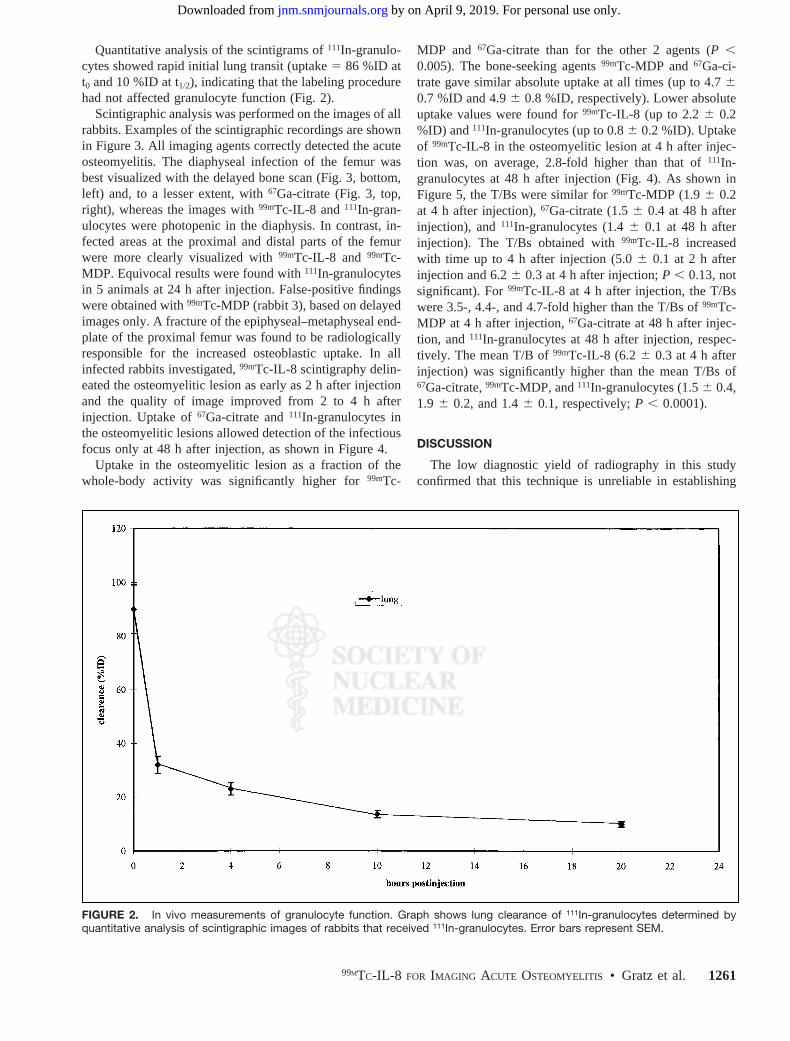
Quantitative analysis of the scintigrams of111In-granulo-cytes showed rapid initial lung transit (uptake5 86 %ID att0 and 10 %ID at t1/2), indicating that the labeling procedurehad not affected granulocyte function (Fig. 2).
Scintigraphic analysis was performed on the images of allrabbits. Examples of the scintigraphic recordings are shownin Figure 3. All imaging agents correctly detected the acuteosteomyelitis. The diaphyseal infection of the femur wasbest visualized with the delayed bone scan (Fig. 3, bottom,left) and, to a lesser extent, with67Ga-citrate (Fig. 3, top,right), whereas the images with99mTc-IL-8 and 111In-gran-ulocytes were photopenic in the diaphysis. In contrast, in-fected areas at the proximal and distal parts of the femurwere more clearly visualized with99mTc-IL-8 and 99mTc-MDP. Equivocal results were found with111In-granulocytesin 5 animals at 24 h after injection. False-positive findingswere obtained with99mTc-MDP (rabbit 3), based on delayedimages only. A fracture of the epiphyseal–metaphyseal end-plate of the proximal femur was found to be radiologicallyresponsible for the increased osteoblastic uptake. In allinfected rabbits investigated,99mTc-IL-8 scintigraphy delin-eated the osteomyelitic lesion as early as 2 h after injectionand the quality of image improved from 2 to 4 h afterinjection. Uptake of67Ga-citrate and111In-granulocytes inthe osteomyelitic lesions allowed detection of the infectiousfocus only at 48 h after injection, as shown in Figure 4.
Uptake in the osteomyelitic lesion as a fraction of thewhole-body activity was significantly higher for99mTc-
MDP and 67Ga-citrate than for the other 2 agents (P ,0.005). The bone-seeking agents99mTc-MDP and67Ga-ci-trate gave similar absolute uptake at all times (up to 4.760.7 %ID and 4.96 0.8 %ID, respectively). Lower absoluteuptake values were found for99mTc-IL-8 (up to 2.26 0.2%ID) and111In-granulocytes (up to 0.86 0.2 %ID). Uptakeof 99mTc-IL-8 in the osteomyelitic lesion at 4 h after injec-tion was, on average, 2.8-fold higher than that of111In-granulocytes at 48 h after injection (Fig. 4). As shown inFigure 5, the T/Bs were similar for99mTc-MDP (1.96 0.2at 4 h after injection),67Ga-citrate (1.56 0.4 at 48 h afterinjection), and111In-granulocytes (1.46 0.1 at 48 h afterinjection). The T/Bs obtained with99mTc-IL-8 increasedwith time up to 4 h after injection (5.06 0.1 at 2 h afterinjection and 6.26 0.3 at 4 h after injection;P , 0.13, notsignificant). For99mTc-IL-8 at 4 h after injection, the T/Bswere 3.5-, 4.4-, and 4.7-fold higher than the T/Bs of99mTc-MDP at 4 h after injection,67Ga-citrate at 48 h after injec-tion, and111In-granulocytes at 48 h after injection, respec-tively. The mean T/B of99mTc-IL-8 (6.2 6 0.3 at 4 h afterinjection) was significantly higher than the mean T/Bs of67Ga-citrate,99mTc-MDP, and111In-granulocytes (1.56 0.4,1.9 6 0.2, and 1.46 0.1, respectively;P , 0.0001).
DISCUSSION
The low diagnostic yield of radiography in this studyconfirmed that this technique is unreliable in establishing
FIGURE 2. In vivo measurements of granulocyte function. Graph shows lung clearance of 111In-granulocytes determined byquantitative analysis of scintigraphic images of rabbits that received 111In-granulocytes. Error bars represent SEM.
99MTC-IL-8 FOR IMAGING ACUTE OSTEOMYELITIS • Gratz et al. 1261
by on April 9, 2019. For personal use only. jnm.snmjournals.org Downloaded from

acute bone infection when an additional pathologic condi-tion is present. In osteomyelitis, the bone destruction causedby vascular ischemia, by enzymes of disintegrated poly-morphs, and by increased intramedullary pressure (32) canbe visualized with bone-seeking agents such as67Ga-citrateand99mTc-MDP. Both radiopharmaceuticals have proven tobe sensitive in detecting pathologically increased bone turn-over, although the specificity of both agents is limited: otherconditions may also cause increased uptake, such as tumors,activated osteoarthritis, and noninfectious inflammatory le-sions (33). An increase in specificity can be achieved usingagents targeting the neutrophils that have infiltrated thebone marrow. Up to now, these neutrophilic infiltrates havebeen visualized with radiolabeled leukocytes (34) or withradiolabeled antibodies directed against epitopes as presenton granulocytes (35).
With 99mTc-IL-8, most infections in the rabbits weredetected as early as 2 h after injection, whereas delayed 48-hpostinjection images were necessary with111In-granulocytes(36). With 99mTc-IL-8 as well as with67Ga-citrate, all in-fected sites were detected, whereas delayed bone scanningalone was falsely positive in 1 rabbit and111In-granulocytesmissed 1 case of osteomyelitis.
This study showed that99mTc-IL-8, a proinflammatorychemotactic cytokine, allows visualization of osteomyeliticlesions as evidenced by infiltrated neutrophils, presumablyby targeting the surface receptors on granulocytes. Further-more,99mTc-IL-8 performed at least as well as99mTc-MDPand67Ga-citrate in the localization of acute bone infection.
FIGURE 3. Scintigraphic images of rabbits with complete infec-tion of femur. (Top) 99mTc-IL-8 image 4 h after injection (left) and67Ga-citrate image 48 h after injection (right) for rabbit 8 of group B.(Bottom) 99mTc-MDP image 4 h after injection (left) and 111In-gran-ulocyte image 48 h after injection (right) for rabbit 1 of group A.
FIGURE 4. Uptake at osteomyelitic site as determined by quantitative analysis of scintigraphic images of group A rabbits administered99mTc-MDP and 111In-granulocytes and group B rabbits administered 99mTc-IL-8 and 67Ga-citrate. Error bars represent SEM.
1262 THE JOURNAL OF NUCLEAR MEDICINE • Vol. 42 • No. 8 • August 2001
by on April 9, 2019. For personal use only. jnm.snmjournals.org Downloaded from
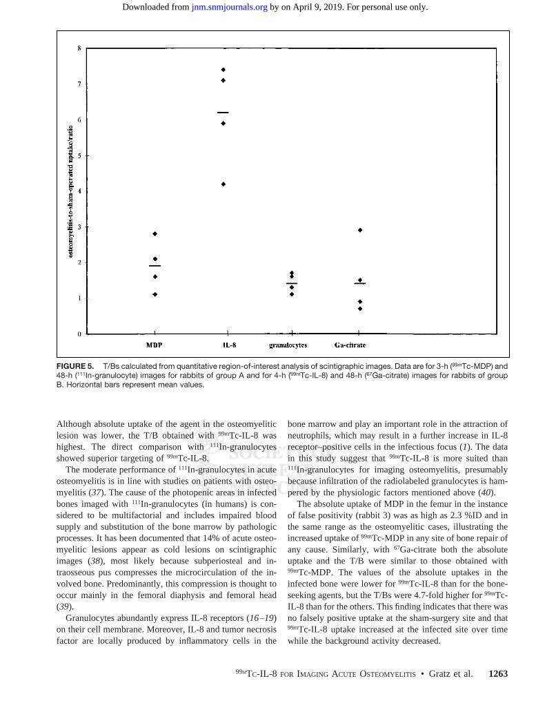
Although absolute uptake of the agent in the osteomyeliticlesion was lower, the T/B obtained with99mTc-IL-8 washighest. The direct comparison with111In-granulocytesshowed superior targeting of99mTc-IL-8.
The moderate performance of111In-granulocytes in acuteosteomyelitis is in line with studies on patients with osteo-myelitis (37). The cause of the photopenic areas in infectedbones imaged with111In-granulocytes (in humans) is con-sidered to be multifactorial and includes impaired bloodsupply and substitution of the bone marrow by pathologicprocesses. It has been documented that 14% of acute osteo-myelitic lesions appear as cold lesions on scintigraphicimages (38), most likely because subperiosteal and in-traosseous pus compresses the microcirculation of the in-volved bone. Predominantly, this compression is thought tooccur mainly in the femoral diaphysis and femoral head(39).
Granulocytes abundantly express IL-8 receptors (16–19)on their cell membrane. Moreover, IL-8 and tumor necrosisfactor are locally produced by inflammatory cells in the
bone marrow and play an important role in the attraction ofneutrophils, which may result in a further increase in IL-8receptor–positive cells in the infectious focus (1). The datain this study suggest that99mTc-IL-8 is more suited than111In-granulocytes for imaging osteomyelitis, presumablybecause infiltration of the radiolabeled granulocytes is ham-pered by the physiologic factors mentioned above (40).
The absolute uptake of MDP in the femur in the instanceof false positivity (rabbit 3) was as high as 2.3 %ID and inthe same range as the osteomyelitic cases, illustrating theincreased uptake of99mTc-MDP in any site of bone repair ofany cause. Similarly, with67Ga-citrate both the absoluteuptake and the T/B were similar to those obtained with99mTc-MDP. The values of the absolute uptakes in theinfected bone were lower for99mTc-IL-8 than for the bone-seeking agents, but the T/Bs were 4.7-fold higher for99mTc-IL-8 than for the others. This finding indicates that there wasno falsely positive uptake at the sham-surgery site and that99mTc-IL-8 uptake increased at the infected site over timewhile the background activity decreased.
FIGURE 5. T/Bs calculated from quantitative region-of-interest analysis of scintigraphic images. Data are for 3-h (99mTc-MDP) and48-h (111In-granulocyte) images for rabbits of group A and for 4-h (99mTc-IL-8) and 48-h (67Ga-citrate) images for rabbits of groupB. Horizontal bars represent mean values.
99MTC-IL-8 FOR IMAGING ACUTE OSTEOMYELITIS • Gratz et al. 1263
by on April 9, 2019. For personal use only. jnm.snmjournals.org Downloaded from

CONCLUSION
In this study,99mTc-IL-8 accurately revealed acute osteo-myelitis in a rabbit model.99mTc-IL-8 correctly identified allrabbits with acute osteomyelitis within 4 h after injection.The ease of preparation, early good image quality, andlower radiation burden suggest that99mTc-IL-8 may be asuitable imaging agent for the scintigraphic evaluation ofacute osteomyelitis.
ACKNOWLEDGMENTS
The authors thank Gerry Grutters and Henny Eikholt(Central Animal Laboratory, University of Nijmegen) forexpert assistance with the animals, Emile Koenders (De-partment of Nuclear Medicine, University Medical CenterNijmegen) for excellent technical assistance, and Drs. PiusLoetscher and Marco Baggiolini (Theodor Kocher Institute,University of Bern, Bern, Switzerland) for the kind gift ofthe cell lines. This study was supported in part by theDeutsche Forschungs Gesellschaft.
REFERENCES
1. Lew DP, Waldvogel FA. Osteomyelitis.N Engl J Med.1997;336:999–1007.2. David R, Barron BJ, Madewell JE. Osteomyelitis, acute and chronic.Radiol Clin
North Am.1987;25:1171–1201.3. Ram PC, Martinez S, Korobkin M, Breiman RS, Gallis HR, Harrelson JM. CT
detection of intraosseous gas: a new sign of osteomyelitis.AJR.1981;137:721–723.
4. Morrison WB, Schweitzer ME, Batte WG, Radack DP, Russel KM. Osteomy-elitis of the foot: relative importance of primary and secondary MR imagingsigns.Radiology.1998;207:625–632.
5. Rifai A, Nyman R. Scintigraphy and ultrasonography in differentiating osteomy-elitis from bone infarction in sickle cell disease.Acta Radiol.1997;38:139–143.
6. Gratz S, Doerner J, Oestmann JW, et al.67Ga-citrate and 99Tcm-MDP forestimating the severity of vertebral osteomyelitis.Nucl Med Commun.2000;21:111–120.
7. Joseph K, Damann V, Engeroff G, Gruner KR. Labeling of leukocytes with99mTc-HMPAO: first clinical results [in German].Nucl Compact.1986;17:277–283.
8. Schauwecker DS. Osteomyelitis: diagnosis with In-111-labeled leukocytes.Ra-diology. 1989;171:141–146.
9. Rojas-Burke J. Health officials reacting to infection mishaps.J Nucl Med.1992;33:13N–14N, 27N.
10. Kaim A, Maurer T, Ochsner P, Jundt G, Kirsch E, Mueller-Brand J. Chroniccomplicated osteomyelitis of the appendicular skeleton: diagnosis with techne-tium-99m labelled monoclonal antigranulocyte antibody-immunoscintigraphy.Eur J Nucl Med.1997;24:732–738.
11. Becker W, Goldenberg DM, Wolf F. The use of monoclonal antibodies andantibody fragments in the imaging of infectious lesions.Semin Nucl Med.1994;24:142–153.
12. Becker W, Borst U, Fischbach W, Pasurka B, Schafer R, Borner W. Kinetic dataof in-vivo labeled granulocytes in humans with a murine Tc-99m-labelled mono-clonal antibody.Eur J Nucl Med. 1989;15:361–366.
13. Becker WS, Saptogino A, Wolf FG. The single late 99Tcm granulocyte antibodyscan in inflammatory diseases.Nucl Med Commun. 1992;13:186–92.
14. Becker W, Bair J, Behr T, et al. Detection of soft-tissue infections and osteomy-elitis using a technetium-99m-labeled anti-granulocyte monoclonal antibodyfragment.J Nucl Med. 1994;35:1436–1443.
15. Holmes WE, Lee J, Kuang WJ, Rice GC, Wood WI. Structure and functionalexpression of a human interleukin-8 receptor.Science.1991;253:1278–1280.
16. Lange JM, Boucher CA, Hollak CE, et al. Failure of zidovudine prophylaxis afteraccidental exposure to HIV-1.N Engl J Med.1990:10;322:1375–1377.
17. Murphy PM, Tiffany HL. Cloning of complementary DNA encoding a functionalhuman interleukin-8 receptor.Science.1991;253:1280–1283.
18. Lee J, Horuk R, Rice GC, Bennett GL, Camerato T, Wood WI. Characterizationof two high affinity human interleukin-8 receptors.J Biol Chem.1992;267:16283–16287.
19. Cerretti DP, Kozlosky CJ, Vanden Bos T, Nelson N, Gearing DP, Beckmann MP.Molecular characterization of receptors for human interleukin-8, GRO/melanomagrowth-stimulatory activity and neutrophil activating peptide-2.Mol Immunol.1993;30:359–367.
20. Rennen HJJM, Boerman OC, Oyen WJG, van der Meer JWM, Corstens FHM.Specific and rapid scintigraphic detection of infection with Tc-99m-labeledinterleukin-8.J Nucl Med.2000;42:117–123.
21. O’Reilly T, Kunz S, Sande E, Zak O, Sande MA, Tauber MG. Relationshipbetween antibiotic concentration in bone and efficacy of treatment of staphylo-coccal osteomyelitis in rats: azithromycin compared with clindamycin and ri-fampin.Antimicrob Agents Chemother. 1992;36:2693–2697.
22. Nelson DR, Buxton TB, Luu QN, Rissing JP. An antibiotic resistant experimentalmodel of Pseudomonas osteomyelitis.Infection. 1990;18:246–248.
23. Mader JT, Wilson KJ. Models of osteomyelitis. In: Zak O, Sande MA, eds.Experimental Models in Antimicrobial Chemotherapy. Vol 2. London, U.K.:Academic Press; 1986:155–173.
24. Rissing JP, Buxton TB, Weinstein RS, Shockley RK. Model of experimentalchronic osteomyelitis in rats.Infect Immun. 1985;47:581–586.
25. Henry NK, Rouse MS, Whitesell AL, McConnell ME, Wilson WR. Treatment ofmethicillin-resistant Staphylococcus aureus experimental osteomyelitis with cip-rofloxacin or vancomycin alone or in combination with rifampin.Am J Med.1987;82:73–75.
26. Liu S, Edwards DS, Harris AR. A novel ternary ligand system for99mTc-labelingof hydrazino-nicotinamide-modified biologically active molecules using imine-N-containing heterocycles as coligands.Bioconjug Chem.1998;9:583–595.
27. Lillevang ST, Toft P, Nilsen B. A method for isolating granulocytes from rabbitblood without causing activation.J Immunol Methods. 1994;28;169:137–138.
28. Boyum A, Lovhaug D, Tresland L, Nordlie EM. Separation of leucocytes:improved cell purity by fine adjustments of gradient medium density and osmo-lality. Scand J Immunol.1991;34:697–712.
29. Lindmo T, Boven E, Cuttitta F, Fedorko J, Bunn PA Jr. Determination of theimmunoreactive fraction of radiolabeled monoclonal antibodies by linear extrap-olation to binding at infinite antigen excess.J Immunol Methods. 1984;72:77–89.
30. Alvarez-Gonzalez R, Eichenberger R, Loetscher P, Althaus FR. A new highlyselective physicochemical assay to measure NAD1in intact cells.Anal Biochem.1986;156:473–480.
31. David R, Barron BJ, Madewell JE. Osteomyelitis, acute and chronic.Radiol ClinNorth Am. 1987;25:1171–1201.
32. Kahn DS, Pritzker KP. The pathophysiology of bone infection.Clin Orthop.1973;96:12–19.
33. Modic MT, Feiglin DH, Piraino DW, et al. Vertebral osteomyelitis: assessmentusing MR.Radiology.1985;157:157–166.
34. Peters AM, Saverymuttu SH, Reavy HJ, Danpure HJ, Osman S, Lavender JP.Imaging of inflammation with indium-111 tropolonate labeled leukocytes.J NuclMed. 1983;24:39–44.
35. Joseph K, Hoffken H, Bosslet K, Schorlemmer HU. Imaging of inflammationwith granulocytes labelled in vivo.Nucl Med Commun. 1988;9:763–769.
36. Oyen WJ, Claessens RA, van-Horn JR, van-der-Meer JW, Corstens FH. Scinti-graphic detection of bone and joint infections with indium-111-labeled nonspe-cific polyclonal human immunoglobulin G.J Nucl Med.1990;31:403–412.
37. Mok YP, Carney WH, Fernandez-Ulloa M. Skeletal photopenic lesions in111In-WBC imaging.J Nucl Med.1984;25:1322–1326.
38. Datz LF, Thorne DA. Cause and significance of cold bone defects on indium-111-labeled leukocyte imaging.J Nucl Med.1987,28:820–823.
39. Athens JW, Haab OP, Raab SO. Leukokinetic studies: the total blood, circulatingand marginal granulocyte pools and granulocyte turnover rate in normal subjects.J Clin Invest.1961;40:989–995.
40. Weiblein BJ, Forstrom LA, McCullough J. Kinetics of In-111-labelled granulo-cytes.J Lab Clin Med.1979;94:246–255.
1264 THE JOURNAL OF NUCLEAR MEDICINE • Vol. 42 • No. 8 • August 2001
by on April 9, 2019. For personal use only. jnm.snmjournals.org Downloaded from
2001;42:1257-1264.J Nucl Med. Stefan Gratz, Huub J.J.M. Rennen, Otto C. Boerman, Wim J.G. Oyen, Pieter Burma and Frans H.M. Corstens
Tc-Interleukin-8 for Imaging Acute Osteomyelitis99m
http://jnm.snmjournals.org/content/42/8/1257This article and updated information are available at:
http://jnm.snmjournals.org/site/subscriptions/online.xhtml
Information about subscriptions to JNM can be found at:
http://jnm.snmjournals.org/site/misc/permission.xhtmlInformation about reproducing figures, tables, or other portions of this article can be found online at:
(Print ISSN: 0161-5505, Online ISSN: 2159-662X)1850 Samuel Morse Drive, Reston, VA 20190.SNMMI | Society of Nuclear Medicine and Molecular Imaging
is published monthly.The Journal of Nuclear Medicine
© Copyright 2001 SNMMI; all rights reserved.
by on April 9, 2019. For personal use only. jnm.snmjournals.org Downloaded from
![[99mTc]-labelled interleukin-8 as a diagnostic tool compared to … · Carl Schønheyder4,5, Annemarie Eek 8, Otto Boerman , Ole Lerberg Nielsen9 1 Department of Nuclear Medicine,](https://img.dokumen.tips/doc/110x75/5fc06cc2848582774511b6c8/99mtc-labelled-interleukin-8-as-a-diagnostic-tool-compared-to-carl-schnheyder45.jpg)