Embed Size (px)
Citation preview

9. Diseases of Groundnut
Tikka leaf spots
Early leaf spot: Cercopora arachidicola (Sexual Stage: Mycosphaerella arachidis)
Late leaf spot: Phaeoisariopsis personata (Syn : Cercospora personata)
(Sexual stage : Mycosphaerella berkeleyii)
Symptoms
The disease occurs on all above ground parts of the plant, more severely on the leaves.
The leaf symptoms produced by the two pathogens can be easily distinguished by appearance,
spot colour and shapes. Both the fungi produce lesions also on petiole, stem and pegs. The
lesions caused by both species coalesce as infection develops and severely spotted leaves shed
prematurely. The quality and yield of nuts are drastically reduced in severe infections.
Pathogen C. arachidicola (Sexual stage: M. arachidis) Symptoms

The pathogen is intercellular and do not produce haustoria and become intracellular when
host cells die. The fungus produces abundant sporulation on the upper surface of the leaves.
Conidiophores are olivaceous brown or yellowish brown in colour, short, 1 or 2 septate,
unbranched and geniculate and arise in clusters.
Conidia are sub hyaline or pale yellow, obclavate, often curved 3-12 septate, 35- 110 x
2.5 - 5.4 m in size with rounded to distinctly truncate base and sub-acute tip. The perfect stage
of the fungus produces perithecia as ascostromata. They are globose with papillate ostiole. Asci
are cylindrical to clavate and contain 8 ascospores. Ascospores are hyaline, slightly curved and
two celled, apical cell larger than the lower cell.
P. personata (C. personata) (Sexual stage: M. berkeleyii)
The fungus produces internal and intercellular mycelium with the production of
haustoria. The conidiphores are long, continuous, 1-2 septate, geniculate, arise inclusters and
olive brown in colour. The conidia are cylindrical or obclavate, short, measure
18-60 x 6-10 m, hyaline to olive brown, usually straight or curved slightly with 1-9 septa, not
constricted but mostly 3-4 septate. The fungus in its perfect stage produces perithecia as
ascostromata which are globose or broadly ovate with papillate ostiole. Asci are cylindrical to
ovate, contain 8 ascospores. Ascospores are 2 celled and constricted at septum and hyaline.
Favourable Conditions
• Prolonged high relative humidity for 3 days.
• Low temperature (20 C) with dew on leaf surface.
• Heavy doses of nitrogen and phosporus fertilizers
• Deficiency of magesium in soil.
Disease cycle
The pathogen survives for a long period in the infected plant debris through conidia,
dormant mycelium and perithecia in soil. The volunteer groundnut plants also harbour the
pathogen. The primary infection is by ascospores or conidia from infected plant debris or infectd
seeds. The secondary spread is by wind blown conidia. Rain splash also helps in the spread of
conidia.
Management
• Remove and destory the infected plant debris.

• Eradicate the volunteer groundnut plants.
• Keep weeds under control.
• Treat the seeds with Carbendazim or Thiram at 2g/kg.
• Spray Carbendazim 500g or mancozeb 2 kg or Chlorothalonil 2 kg/ha and if necessary,
repeat after 15 days.
• Grow moderately resistant varieties like ALR 1.
Rust - Puccinia arachidis
Symptoms
The disease attacks all aerial parts of the plant. The disease is usually found when the
plants are about 6 weeks old. Small brown to chestnut dusty pustules (uredosori) appear on the
lower surface of leaves. The epidermis ruptures and exposes a powdery mass of uredospores.
Corresponding to the sori, small, necrotic, brown spots appear on the upper surface of leaves.
The rust pustules may be seen on petioles and stem. Late in the season, brown teliosori, as dark
pustules, appear among the necrotic patches. In severe infection lower leaves dry and drop
prematurely. The severe infection leads to production of small and shriveled seeds.
Pathogen
The pathogen produces both uredial and telial stages. Uredial stages are produced
abundant in groundnut and production of telia is limited. Uredospores are pedicellate,
Symptoms

unicellular, yellow, oval or round and echinulated with 2 or 3 germpores. Teliospores are dark
brown with two cells. Pycnial and aecial stages have not been recorded and there is no
information available about the role of alternate host.
Teliospores
Uredospores
Favourable Conditions
• High relative humidity (above 85 per cent).
• Heavy rainfall.
• Low temperature (20-25˚C).
Disease cycle
The pathogen survives as uredospores on volunter groundnut plants. The fungus also
survives in infected plant debris in soil. The spread is mainly through wind borne inoculum of
uredospores. The uredospores also spread as contamination of seeds and pods. Rainsplash and
implements also help in dissemination. The fungus also survives on the collateral hosts like
Arachis marginata, A. nambyquarae and A. prostrate.
Management
• Avoid monoculturing of groundnut.
• Remove volunteer groundnut plants and reservoir hosts.
• Spray mancozeb 2 kg or Wettable Sulphur 3 kg or Tridemorph 500ml or Chlorothalonil 2
kg/ha.
• Grow moderatelyresistant varieties like ALR 1.
Collar rot or seedling blight or crown rot - Aspergillus niger and A. pulverulentum
Symptoms
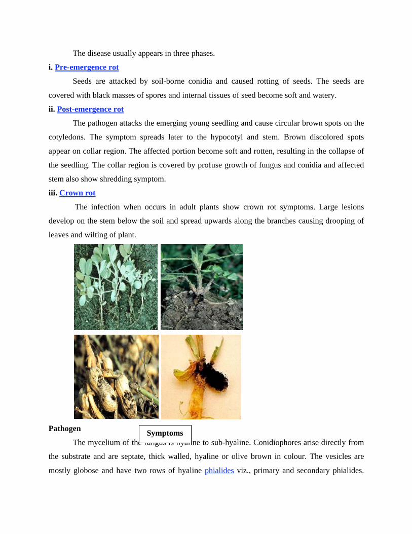
The disease usually appears in three phases.
i. Pre-emergence rot
Seeds are attacked by soil-borne conidia and caused rotting of seeds. The seeds are
covered with black masses of spores and internal tissues of seed become soft and watery.
ii. Post-emergence rot
The pathogen attacks the emerging young seedling and cause circular brown spots on the
cotyledons. The symptom spreads later to the hypocotyl and stem. Brown discolored spots
appear on collar region. The affected portion become soft and rotten, resulting in the collapse of
the seedling. The collar region is covered by profuse growth of fungus and conidia and affected
stem also show shredding symptom.
iii. Crown rot
The infection when occurs in adult plants show crown rot symptoms. Large lesions
develop on the stem below the soil and spread upwards along the branches causing drooping of
leaves and wilting of plant.
Pathogen
The mycelium of the fungus is hyaline to sub-hyaline. Conidiophores arise directly from
the substrate and are septate, thick walled, hyaline or olive brown in colour. The vesicles are
mostly globose and have two rows of hyaline phialides viz., primary and secondary phialides.
Symptoms

The conidial head are dark brown to black. The conidia are globose, dark brown in colour and
produce in long chains.
Favourable Conditions
• Deep sowing of seeds.
• High soil temperature (30-35˚ C).
• Low soil moisture.
Disease cycle
The pathogen survive in plant debris in the soil, not necessarily from a groundnut
crop. Soil-borne conidia cause disease carry over from season to season. The other primary
source is the infeced seeds. The pathogen is also seedborne in nature.
Management
• Crop rotation.
• Destruction of plant debris.
• Remove and destroy previous season's infested crop debris in the field
• Seed treatment with Trichoderma viride / T.harzianum @ 4 g/kg of seeds and soil
application of Trichoderma viride / T.harzianum at 2.5kg/ha, preferably with organic
amendments such as castor cake or neem cake or mustard cake @ 500 kg/ ha.
Root rot - Macrophomina phaseolina
Symptoms
In the early stages of infection, reddish brown lesion appears on the stem just above the
soil level. The leaves and branches show drooping, leading to death of the whole plant. The
decaying stems are covered with whitish mycelial growth. The death of the plant results in
shredding of bark. The rotten tissues contain large number of black or dark brown, thick walled
sclerotia. When infection spreads to underground roots, the sclerotia are formed externally as
well as internally in the rotten tissue. Pod infection leads to blackening of the shells and sclerotia
can be seen inside the shells.
Pathogen
The fungus produces hyaline to dull brown mycelium. The sclerotia are thick walled and
dark brown in colour.
Favourable Conditions

• Prolonged rainy season at seedling stage and low lying areas.
Disease cycle
The fungus remains dormant as sclerotia for a long period in the soil and in infected plant
debris. The primary infection is through soil-borne and seed-borne sclerotia. The secondary
spread of sclerotia is aided by irrigation water, human agency, implements and cattle etc.
Management
• Treat the seeds with thiram or carbendazim 2g/kg or Trichoderma viride at 4g/kg.
• Spot drench with Carbendazim at 0.5 g/lit.
Rossette - Groundnut rosette assistor virus (GRAV), Groundnut rosette virus and Groundnut
rosette satellites
Symptoms
The affected plants are characterized by the appearance of dense clump or dwarf shoots
with tuft of small leaves forming in a rosette fashion. The plant exhibits chlorosis and mosaic
mottling. The infected plants remain stunted and produce flowers, but only a few of the pegs may
develop further to nuts but no seed formation.
Pathogen
The disease is caused by a complex mixture of viruses viz.,Groundnut rosette assistor
virus (GRAV), Ground nut rosette virus and Groundnut rosette satellites is an isometric, not
enveloped and 28nm diameter (reported from India) and it gives no overt symptom in groundnut.
Groundnut rosette virus is with ssRNA genome, which becomes packaged in GRAV virious and
thus depends on it for aphid transmission, but produces no overt symptoms in groundnut. The
Symptoms

groundnut rosette satellites are satellite RNAs that control the symptoms and cause the different
types of rosette (chlorotic, green and mosaic).
Disease Cycle
The primary source of spread by aphid vector, Aphis craccivora and A. gossipii in a
persistent manner, retained by vector but not transmitted congenitally. The virus is not
transmitted by any other means like mechanical or seed or pollen. The virus can survive on the
volunteer plants of groundnut and other weed hosts.
Management
• Practice clean cultivation.
• Use heavy seed rate and rogue out the infected plants periodically.
• Spray Monocrotophos or Methyl demeton at 500 ml/ha.
Groundnut bud necrosis disease - Groundnut bud necrosis virus (GBNV- Tospo virus)
Symptoms
First symptoms are visible 2-6 weeks after infection as ring spots on leaves. The newly
emerging leaves are small, rounded or pinched inwards and rugose with varying patterns of
mottling and minute ring spots. Necrotic spots and irregularly shaped lesions develop on leaves
and petioles. Stem also exhibits necrotic streaks.
Symptoms

Plant becomes stunted with short internodes and short auxillary shoots. Leaflets show
reduction in size, distortion of the lamina, mosaic mottling and general chlorosis. In advanced
conditions, the necrosis of buds occurs. Top bud is killed and necrosis spreads downwards.
Drastic reduction in flowering and seeds produced are abnormally small and wrinkled with the
dark black lesions on the testa.
Pathogen
It is caused by Groundnut bud necrosis virus (GBNV). The virus particles are spherical,
30 nm in diameter, enveloped, ssRNA with multipartite genome.
Disease cycle
The virus perpetuates in the weed hosts viz., Bidens pilosa, Erigon bonariensis,
Tagetes minuta and Trifolium subterraneum. The virus is transmitted by thrips viz., Thrips palmi,
T. tabaci and Frankliniella sp.
Management
• Adopt plant spacing of 15x15 cm.
• Remove and destory infected plants up to 6 weeks after sowing.
• Application of Monocrotophos 500 ml/ha, 30 days after sowing either alone or in
combination with AVP (Anti Viral Principle) extracted from sorghum or coconut leaves.
Spray the crop with 10 per cent AVP at 500 lit/ha, ten and twenty days after sowing.
Minor diseases
Stem rot - Sclerotium rolfsii
Symptoms
The first symptom is the sudden drying of a branch which is completely or partially in
contact with the soil. The leaves turn brown and dry but remain attached to the plant. Near soil
on stems white growth of fungus mycelium is appeared. As the disease advances white
mycelium web spreads over the soil and the basal canopy of the plant. The sclerotia, the size and
colour of mustard seeds, appear on the infected areas as the disease develops and spreads. The
entire plant may be killed or only two or three branches may be affected. Lesions on the
developing pegs can retard pod development. Infected pods are usually rotted.

Management
• Cultural practices such as deep' covering or burial of organic matter before planting, non-
dirting cultivation by avoiding movement of soil up around the base of plants and
preventing accumulation of organic debris are extremely useful in reducing the disease.
• Crop rotation with wheat, corn and soyabean may minimize the incidence of stem rot.
• Seed treatment with Carbendazim / Thiram / Captan @ 2-3 g/kg seed.
• Seed treatment with Trichoderma viride formulation (4g/kg) followed by application of
2.5kg Trichoderma viride formulation mixed with 50kg farm yard manure before sowing.
Wilt - Fusarium oxysporum and F. solani
Symptoms
Germinating seeds are attacked by the pathogens shortly before emergence. There is
general tissue disintegration and the surface of the seedling is covered with sporulating
mycelium. Damping off symptoms characterized by brown to dark brown Water soaked sunken
lesions on the hypocotyl which later encircle the stem and extend above the soil level. Roots are
also attacked, especially the apical portions. The affected seedlings become yellow and wilted.
The leaves turn greyish green and the plants dry up and die. The roots and stems show internal
vascular browning and discolouration. These fungi are also commonly associated with pod rot.
Management
• Seed treatment with systemic fungicides like Carbendazim at 2g/kg seed.
Anthracnose - Colletotrichum dematium and C. capsici
Symptoms
Symptoms
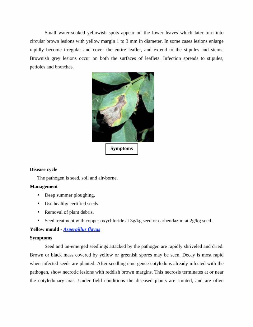
Small water-soaked yellowish spots appear on the lower leaves which later turn into
circular brown lesions with yellow margin 1 to 3 mm in diameter. In some cases lesions enlarge
rapidly become irregular and cover the entire leaflet, and extend to the stipules and stems.
Brownish grey lesions occur on both the surfaces of leaflets. Infection spreads to stipules,
petioles and branches.
Disease cycle
The pathogen is seed, soil and air-borne.
Management
• Deep summer ploughing.
• Use healthy certified seeds.
• Removal of plant debris.
• Seed treatment with copper oxychloride at 3g/kg seed or carbendazim at 2g/kg seed.
Yellow mould - Aspergillus flavus
Symptoms
Seed and un-emerged seedlings attacked by the pathogen are rapidly shriveled and dried.
Brown or black mass covered by yellow or greenish spores may be seen. Decay is most rapid
when infected seeds are planted. After seedling emergence cotyledons already infected with the
pathogen, show necrotic lesions with reddish brown margins. This necrosis terminates at or near
the cotyledonary axis. Under field conditions the diseased plants are stunted, and are often
Symptoms

chlorotic. The leaflets are reduced in size with pointed tips, widely varied in shape and
sometimes with veinal clearing.
Management
• Since the fungus is a weak parasite, agronomic practices which favour rapid germination
and vigorous growth of seedling will reduce the chance of A. flavus infection.
• Seed treatment with carbendazim or captan or thiram at 2g/kg seed.
Grey mould - Botrytis cinerea
Infection is seen on leaves, stem and underground parts of the groundnut. Initially
infection occurs at ground level by a light grey fungal rot which causes death of the plants.
Bacterial wilt - Pseudomonas solanacearum
Infected plants appear unhealthy, chlorotic and wilt under water stress. Dark brown
discolouration of xylem is seen. Grey slimy liquid ooze out of the vascular bundles.
Leaf spot - Alternaria arachidis and A. tenuissima
Symptoms
Lesions produced by A. arachidis are brown in colour and irregular in shape surrounded
by yellowish halos. Symptoms produced by A. tenuissima are characterized by blighting of
apical portions of leaflets which turn light to dark brown colour. Lesions produced by A.
alternata are small, chlorotic, water soaked, that spread over the surface of the leaf. The lesions
become necrotic and brown and are round to irregular in shape. Veins and veinlets adjacent to
the lesions become necrotic. Lesions increase in area and their central portions become pale,
rapidly dry out, and disintegrate. Affected leaves show chlorosis and in severe attacks become
prematurely senescent. Lesions can coalesce, give the leaf a ragged and blighted appearance.
Symptoms

Management
• Foliar application of Mancozeb (2kg/ha) or Copper oxychloride (2kg/ha) or Carbendazim
(500g/ha).
Indian Peanut Clump Disease - Peanut Clump virus
Earlier this disease was confused with groundnut rosstte. Now it is recognized as a
distinct virus causing clump disease. The leaves turn very dark and plants become severely
stunted. The disease is soil borne and transmitted by a fungus, Polymyxa graminis. The pH of the
soil affects transmission. It is also transmitted by seed. The virus is rod shaped, 190-245nm long
x 21nm wide, not enveloped, ssRNA genome.
Other virus diseases of minor importance occurring on groundnut are:
Peanut chlorotic streak (caused by Caulimovirus, occurs only in India), Peanut green
mosaic and mottle (caused by a Potyvirus), peanut stunt (caused by Cucumovirus), groundnut
chlorotic spot (caused by a Potexvirus), groundnut eye spot (caused by Potyvirus) and groundnut
ringspot.