Embed Size (px)
Citation preview

868
Direct Autocrine Regulation of Colon Cancer Cell Growth by VEGF IsMediated via Activation of VEGF-R1/2 and Transactivation of EGF-R: DirectEffect of VEGF on CRC Cell Proliferation and Reduced ApoptosisAmrita Ahluwalia, Michael K. Jones, Andrzej S. Tarnawski
Background & Aims: VEGF is a key stimulator of angiogenesis and promotes the growthcolon tumors via increased nutrient and oxygen supply. A recent study demonstrated thatcolorectal cancer (CRC) cells have increased expression of VEGF and its receptors VEGF-R1 & VEGF-R2, which indicates a possibility of direct stimulation of CRC cell growth byVEGF. However, the role of VEGF in CRC cell proliferation by an autocrine mechanismhas not been fully explored in depth. We hypothesized that VEGF secreted by CRC cellsbinds to VEGF-R1 & VEGF-R2 on CRC cells and: 1) promotes proliferation of these cellsthrough an autocrine pathway, 2) inhibits CRC apoptosis. Methods: We used: (1) humanCRC cell lines - HCT116 & HT29 and (2) normal colonic epithelial cells - NCM356 &NCM460 cultured in nutrient media. Studies: 1) VEGF, VEGF-R1 & VEGF-R2 mRNA andprotein expression of by Real-Time RT-PCR and immunoblotting; 2) VEGF165 secretioninto the culture media by ELISA; 3) cell proliferation by BrdU assay; 4) apoptosis by TUNELstaining and assessment of activated caspases 3 and 9; 5) Inhibition of VEGF receptorfunction using AAL993 inhibitor, which blocks function of VEGF-R1 and VEGF-R2; 6)assessment of phosphorylation of EGF receptor (EGF-R) to test its transactivation by VEGF.Results: 1) Normal colonic cells do not secrete VEGF (< 35 pg/ml culture media) whileCRC cells express and secrete high levels of VEGF (840 - 1500 pg/ml culture media); 2)Normal colonic epithelial cell lines expressed no or minimal VEGF-R1 & VEGF-R2 mRNAand protein while CRC cell lines exhibit strong expression of VEGF-R1 & VEGF-R2 vs.normal colonic epithelial cells by 14- and 24-fold, respectively (all p < 0.001); 3) CRC celllines have significantly increased cell proliferation by 31% (p < 0.01) and decreased apoptosisvs. normal colonic epithelial cells; 3) Inhibition of both VEGF-R1 & VEGF-R2 receptorsusing AAL-993 significantly decreased CRC cell proliferation by (2.2-fold) ; 4) Treatmentof CRC cells with AAL-993 decreased phosphorylation of EGF-R by 1.9-fold (p < 0.01).Conclusions: 1) VEGF and its VEGF-R1 & VEGF-R2 receptors are increased in CRC celllines; 2) CRC cells secrete VEGF, which stimulates CRC cell proliferation via an autocrinemechanism by its VEGF-R1 & VEGF-R2 receptors and also inhibits apoptosis; 3) VEGFreceptor signaling pathway is critical for activating EGF-R in CRC cells; 4) These studiessuggest a cross-talk between VEGF receptor and EGF-R signaling in CRC cells, whichpromotes these cells' proliferation.
869
Discovery and Therapeutic Potential of FGFR2 Gene Amplification in DiffuseGastric CancerWen Min Lau, Zhi Jiang Zang, Eileen Teng, Kakoli Das, Wei-Peng Yong, Ming Teh, JinWei Tan, Li Ming Tania Chia, Amy Tay, Asim Shabbir, Koji Kono, Shing Leng Chan,Patrick Tan, Jimmy B. So
Background: Diffuse gastric cancer (DGC) is a poorly differentiated adenocarcinoma whichclassically infiltrates into the stomach wall with "signet ring" cells without forming a discretemass. DGC is linked to poor prognosis, and targeted therapy is not available. Molecularanalysis of DGC has been hampered by the difficulty in obtaining primary tumors of sufficienttumor cellularity without contaminating normal fibroblasts. In addition preclinical modelsthat reliably predict clinical activity of novel compounds are also lacking. Methods: Toovercome the scarcity of cancer cells and abundance of stromal tissue, we isolated cancercells from the malignant ascites of DGC patients. Red blood cells were removed using Ficolland CD45+ hematopoietic cells depleted using antibody-conjugated magnetic beads. Weperformed exome sequencing on the cancer cells and peripheral white blood cells from thesame patient to identify somatic alterations in DGC. In addition, xenografts were generatedusing the isolated cancer cells to establish preclinical cancer models for DGC. Results: Exomesequencing revealed multiple point mutations in DGC including p53 mutation in one sample.We also found amplification of FGFR2 and KRAS genes. Cancer cells isolated from theascites formed subcutaneous tumors that recapitulate the histology of primary DGC tumorswith characteristic signet ring cells and intracellular mucin. Xenograft tumors with amplifica-tion of FGFR2 gene were confirmed by Fluorescence in situ hybridization (FISH) and thesecells have increased FGFR2 transcripts and protein. We found that FGFR2 amplificationalone was sufficient to predict the sensitivity of xenograft tumors to treatment with FGFRinhibitor, AZD4547. In contrast, these tumors regardless of amplification status of FGFR2gene responded poorly to cisplatin, a standard drug for DGC. Conclusion: We found highprevalence of FGFR2 gene amplification in DGC and established reliable xenograft modelsthat could predict tumor response to targeted therapy.
870
IL-6 Signaling Drives hMSH3 Mismatch Repair Dysfunction to GenerateEMAST Formation in Colon CancerStephanie Tseng-Rogenski, Yashushi Hamaya, Daniel Y. Choi, John M. Carethers
Background and Aims: Elevated Microsatellite Alterations at Selected Tetranucleotide repeats(EMAST) is a genetic signature found in up to 60% of colorectal cancers (CRCs), and iscaused by an acquired, somatic dysfunction of the DNA mismatch repair (MMR) proteinhMSH3. EMAST cancers are associated with inflammation, and provide patients a poorsurvival prognosis. The molecular mechanisms for hMSH3 dysfunction are unclear; we havepreviously shown that human CRC cells under oxidative stress (simulating inflammation)induce a nuclear-to-cytosol shift for hMSH3, potentially generating a location "loss-offunction" phenotype. Here, we evaluated the role of IL-6 as the inducer of oxidative stressand hMSH3 subcellular shift, as it stood out from a number of pro-inflammatory cytokinesthat we tested. Methods: We examined 4 hMSH3-expressing cell lines for IL-6 and IL-6Rexpression (by ELISA and flow cytometry), and IL-6-generated reactive oxygen species (ROS).We determined subcellular localization of MMR proteins by immunohistochemistry andWestern blotting after whole cell lysates were separated into nuclear and cytosolic fractions.
S-151 AGA Abstracts
To examine the IL-6/IL-6R/STAT3 pathway, we inhibited signaling via anti-IL-6 antibody,recombinant soluble gp130Fc, and NSC74859 (blocks STAT3 function). We utilized aconstitutively activated STAT3 (S3c) to demonstrate hyperactivation of the pathway. Nativetetranucleotide frameshift mutations were detected by DNA fragment analysis and sequenc-ing. Human colon cancers were immunostained with anti-IL-6 antibody to examine intrinsicIL-6 levels. Results: IL-6 (0-1 ng/mL) induced a dose-dependent hMSH3 nuclear-to-cytosolshift in all cell lines, and intracellular ROS generation correlated simultaneously with thehMSH3 subcellular shift. No other MMR proteins (hMLH1, hMSH2, hMSH6) shifted withIL-6 treatment. All cell lines expressed soluble IL-6R (sIL-6R) but not membranous IL-6R,and IL-6 treatment activated STAT3 (p-STAT3Tyr705). Blocking sIL-6R or inhibiting STAT3activation abrogated the hMSH3 shift. Forced expression of S3c resulted in increased cytosolichMSH3 in absence of IL-6 treatment. IL-6-treated cells demonstrated ~8-fold increase intetranucleotide mutations over controls, indicating EMAST formation. EMAST-positivehuman colon cancers strongly associated with IL-6 expression (P<0.05). Conclusions: Thepro-inflammatory cytokine IL-6 through its IL-6/sIL-6R/STAT3 pathway is responsible forthe hMSH3 location "loss-of-function" phenotype that generates intrinsic DNA frameshiftmutations and EMAST. This is the first demonstration of a cytokine-induced DNA repairdefect that may further the progression of cancer and worsen patient prognosis. Inhibitionof IL-6 signaling and/or reducing inflammation might be targeted to reduce EMAST formationand improve prognosis for patients with this common finding in cancers.
871
Identification and Characterization of Nuclear Export and Import SignalsWithin the Mismatch Repair Protein hMSH3 That Control Its SubcellularLocation in Response to InflammationDaniel Y. Choi, Paul Martin, Stephanie Tseng-Rogenski, John M. Carethers
Background and Aims: The DNA mismatch repair (MMR) system is critical for correctingerrors during DNA replication and its proteins are frequent targets for inactivation (germlineor somatic), driving cancer progression. We recently discovered that the MMR proteinhMSH3, whose nuclear expression is reduced in colorectal tumors that show intraepithelialinflammation and is associated with poor colorectal cancer patient prognosis, reversiblytranslocates in vitro from the nucleus to the cytosol in response to inflammation, demonstrat-ing that hMSH3 may be a shuttling protein. To test this hypothesis, we identified twoputative nuclear export signals (NES1, NES2) and a putative nuclear localization signal(NLS) within hMSH3 through bioinformatic approaches, none of which are present in otherMMR proteins. Our aim was to (a) assess if the NESs and NLS are functional, (b) determineif the two NESs work together or act independently, and (c) create point mutations withinthe signals to determine critical functioning amino acids. Methods: We generated hMSH3-FLAG, allowing forced expression of hMSH3. The corresponding NESs and NLS withinhMSH3-FLAG was mutated to assess each signal's capability for directing hMSH3's subcellularlocalization. We created constructs based on pRev(1.4) export system to examine the func-tionality of the NESs: (a) constructs containing one and/or both NESs fused with GFP (NES1-GFP, NES2-GFP, NES1-NES2-GFP), (b) construct containing both NESs with all hydrophobicresidues mutated (complete mutant NES1-NES2-GFP), and (c) constructs containing singlemutation of each of the hydrophobic residues. For the NLS, we created: (a) a constructcontaining the NLS fused with GFP (NLS-GFP), and (b) NLS-GFP mutated at all arginineand lysine residues. Constructs were transfected into SW480 and A549 MMR-proficientcells and observed using a fluorescent microscope. Results: hMSH3-FLAG was detected inthe nucleus of the transfected cells. NES1-GFP and/or NES2-GFP individually showed nuclear> cytosolic GFP signals; NES1-NES2-GFP demonstrated increased cytoplasmic GFP, whilecomplete mutant NES1-NES2-GFP reverted to nuclear GFP. Amino acid F169 was the mostcritical position for nuclear export, followed by I172 and L174 residues. Other individualmutant hydrophobic residues did not have detectable impact on GFP localization. Cellstransfected with NLS-GFP show predominant nuclear signal, and mutant NLS-GFP demon-strated increased cytoplasmic GFP, indicating that the mutant was incapable of directing asmuch GFP into the nucleus. Conclusion: NES1 and NES2 work in conjunction to maximizehMSH3's nuclear export function, and hMSH3 NLS possesses detectable nuclear importfunction. These sites likely allow inflammation to shift hMSH3 to the cytoplasm, with reversalback to the nucleus when inflammation has abated.
872
HSP70 Is Required for Loss of Heterozygosity in the ApcMin/+ Model ofColorectal CancerYun Tao, Jeannette S. Messer, Kathleen H. Goss, John Hart, Marc Bissonnette, Eugene B.Chang
Heat shock protein 70 (Hsp70) is a stress-induced molecular chaperone protein that isexpressed at high levels in many cancers, including human colorectal cancer (CRC). Theinduction of Hsp70 in cancer cells is thought to be a cell survival mechanism activated bymalignant transformation, rather than a mechanism of transformation. However, the role ofHsp70 in cancer has primarily been studied in vitro. This led us to study ApcMin/+ micedeficient in Hsp70 to investigate the role of Hsp70 in intestinal tumor development underphysiologic conditions, ApcMin/+Hsp70-/- mice developed fewer and smaller intestinaltumors characterized by increased cell death and decreased proliferation versus controls. Alarge body of evidence from experimental models and humans with CRC has implicatedincreased β-catenin transcriptional activity in the aberrant growth seen in CRC. Tumors fromApcMin/+Hsp70-/- mice had decreased nuclear β-catenin localization as well as decreased β-catenin transcriptional activity. Nuclear localization of non-degradable β-catenin was alsocompromised, suggesting that the localization defect was at the level of nuclear transport.Since neither Hsp70 nor β-catenin contains a nuclear localization signal (NLS), we searchedfor an Hsp70 interacting protein containing one. Bcl2-associated anthanogene (Bag-1) con-tains an NLS and was found in physical association with Hsp70 and β-catenin. In addition,Hsp70 regulated Bag-1 expression and siRNA knockdown of Bag-1 decreased nuclear localiza-tion of β-catenin. Therefore, Hsp70 forms a complex with β-catenin and Bag-1 in order totransport β-catenin into the nucleus. We also investigated β-catenin degradation in ApcMin/+Hsp70-/- cells versus controls. In the ApcMin/+ model of intestinal tumorigenesis mice
AG
AA
bst
ract
s
AG
AA
bst
ract
shave one truncated, non-functional copy of Apc and one wild-type copy. Apc is essentialfor constitutive cytosolic degradation of β-catenin and in the ApcMin/+ model somaticmutation inactivates the second copy of Apc, leading to tumorigenesis. In ApcMin/+Hsp70-/-tumors, β-catenin mRNA was comparable to controls, but cytosolic protein was decreasedwhile ubiquitination was increased. This suggests that β-catenin degradation is increased inApcMin/+Hsp70-/- tumors. Since β-catenin degradation requires Apc, we examined Apcexpression in Hsp70-/- versus Hsp70+/+ tumors. Tumors from ApcMin/+Hsp70+/+ micehad the expected loss of Apc expression, whereas tumors from ApcMin/+Hsp70-/- micewere positive for Apc. Thus, Hsp70 is required for the loss of heterozygosity seen in theApcMin/+ model of intestinal tumorigenesis. In conclusion, Hsp70 overexpression plays akey role in tumor initiation through promotion of gene editing and tumor proliferationthrough nuclear transport of β-catenin. Hence, Hsp70 presents an attractive target for tumortherapy and prevention in genetically predisposed individuals.
873
The "Trojan Horse" Strategy: Selective Eradication of Cancer Cells byAdenovirus-Based Delivery of Cytotoxic Agents: An Alternative Method forTargeting Pancreatic (PC) and Colorectal Cancers (CRC)Shiran Shapira, Assaf Shapira, Diana Kazanov, Sarah Kraus, Moshe Oren, Nadir Arber
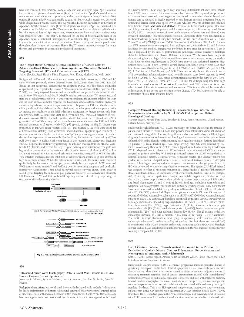
Background: K-Ras and p53 mutations are present in a high percentage of CRC and PCcases. We have previously shown (Biomed Pharm, 2005,Cancer Gene Ther,2012, Exp CellRes,2012) that adenovirus, carrying the pro-apoptotic PUMA (p53-upregulated modulatorof apoptosis) gene, regulated by Ets and AP1/Ras-responsive elements (RREs; Py2/4/5-SV40-PUMA), selectively targeted Ras-mutated tumor cells and suppressed their growth in vitroand in vivo. We used a MazE-MazF (MazEF) unique toxin-antitoxin (TA) system encodedfrom the E. coli chromosome (Fig.1). Under silent conditions the antitoxin inhibits the toxinand the toxin-antidote complex represses the TA operon, whereas after activation, proteolyticantitoxin degradation outpaces its synthesis. Aim: 1) Improve the RRE and the therapeuticefficacy and specificity of the vectors by substituting the lethal gene with more potent toxins2) Increase the regulation of the RRE-lethal gene expression 3) Protect normal cells fromany adverse effects. Methods: The MazF-mCherry fusion gene, truncated derivative of Pseu-domonas exotoxin (PE38), the full regulated MazEF TA cassette were cloned into a "firstgeneration" ΔE1/ΔE3 human type-5 adenoviral-vector under the control of RRE, and SV40-minimal promoter or 17 repeats of the RCG p53-specific binding site (Fig.2). Viruses werepropagated in HEK293 toxin-resistant cells. WT and mutated K-Ras cells were tested forcell proliferation, viability, toxin-expression, and induction of apoptosis upon treatment. Toincrease selectivity and further protection, a WT p53-responsive region was used to inducethe antidote expression in normal cells. Results: First, Ras activity was assessed by GTPasepull-down assay to evaluate cell compliance to our recombinant adenovirus. ResistantHEK293 helper cells constitutively expressing the antitoxin encoded from the pIRES2-MazE-ires-EGFP plasmid, and vectors for targeted gene delivery were established. The yield washigher after propagation in the resistant cells. Specific massive cell death (>50%) at lowMOIs was induced in K-Ras activated cells upon treatment, compared to WT-K-Ras cells.Viral infection induced a marked inhibition of cell growth and apoptosis in cells expressinghigh Ras activity whereas WT-K-Ras cells remained unaffected. The results were measuredqualitatively by fluorescent microscopy, quantitatively by the enzymatic MTT assay andFACS analysis using annexinV and RedDot2 dyes for measuring apoptosis and dead cells,respectively. Conclusions: These novel adenoviral vectors carrying either, PE38, MazF orMazEF genes targeting the K-Ras and p53 pathways can serve to selectively and efficientlykill Ras-mutated PC and CRC cells while sparing normal cells, thereby improving theoutcome of these devastating diseases.
874
Ultrasound Shear Wave Elastography Detects Bowel Wall Fibrosis in Ex VivoHuman Crohn's Disease SpecimensJonathan R. Dillman, Ryan W. Stidham, Laura A. Johnson, Jonathan M. Rubin, Peter D.Higgins
Background and Aims: Narrowed small bowel with thickened walls in Crohn's disease canbe due to inflammation or fibrosis. Ultrasound-generated shear waves travel through tissueat differential rates, with increased speed in stiffer, more fibrotic tissue. While this technologyhas been applied to breast masses and liver fibrosis, it has not been applied to the bowel
S-152AGA Abstracts
in Crohn's disease. Shear wave speed may accurately differentiate inflamed from fibroticbowel. SWS can be measured transcutaneously, but prior to FDA approval, we performedex vivo studies of resected bowel segments. We aimed to determine whether bowel wallfibrosis can be detected in freshly-resected ex vivo human intestinal specimens based onultrasound-derived shear wave speed (SWS), and whether SWS can differentiate inflamedfrom fibrotic bowel. Materials and Methods: 17 intact (>3 cm) bowel segments (15 smalland 2 large intestine) from 12 patients with known or suspected inflammatory bowel disease(8 CD, 3 UC, 1 carcinoid tumor of bowel with adjacent inflammation and fibrosis) wereprocured immediately following surgical resection. Ultrasound shear wave elastography ofthe bowel wall was performed using two methods (Virtual Touch Quantification [VTQ] andVirtual Touch-IQ [VT-IQ]; Acuson S3000, Siemens Medical Solutions USA, Inc.). 18 short-axis SWS measurements were acquired from each specimen; 3 from the 9, 12, and 3 o'clocklocations for each method. Imaging was performed in two areas for specimens >10 cm inlength (separated by ≥5 cm). A gastrointestinal pathologist scored correlative histologicslides for inflammation and fibrosis. Differences in mean SWS between bowel segmentsdemonstrating low and high inflammation/fibrosis scores were assessed using the studentt-test. Receiver operating characteristic (ROC) curve analysis was performed. Results: Highfibrosis score (N=11) bowel segments demonstrated significantly greater mean SWS thanlow fibrosis score (N=6) bowel segments (VTQ: 1.59±0.37 vs. 1.18±0.08 m/s, p=0.004; VT-IQ: 1.87±0.44 vs. 1.50±0.26 m/s; p=0.049). There was no significant difference in meanSWS between high inflammation score and low inflammation score bowel segments (p >0.05for both VTQ and VT-IQ). ROC curves demonstrated areas under the curve of 0.91 [95%,0.67-0.99] (VTQ) and 0.77 [95%, 0.51-0.94] (VT-IQ) for distinguishing low from highfibrosis score bowel segments. Conclusion: Ex vivo bowel wall SWS measurements increasewhen intestinal fibrosis is extensive and transmural. This is not affected by coincidentinflammation. In the ex vivo samples from severe disease, VTQ SWS appears to be able todifferentiate fibrotic from inflamed bowel.
875
Complete Mucosal Healing Defined by Endoscopic Mayo Subscore StillDemonstrates Abnormalities by Novel iSCAN Endoscopic and RefinedHistological GradingsMarietta Iacucci, Miriam Fort Gasia, Jonathan R. Love, Remo Panaccione, Gilaad Kaplan,Subrata Ghosh, Xianyong Gui
Background: High definition(HD)- iSCAN endoscopy can better characterize the mucosa inpatients with ulcerative colitis (UC) and may provide more information about inflammationand mucosal healing(MH). However, the gold standard of mucosal healing is still histologicaldiagnosis. More sensitive endoscopic and histological assessment is needed in order to betterdefine the MH and to identify subtle remaining histologic abnormalities. Patients & methods:78 patients (40 male, median age= 42y, range=19-90y) with UC were assessed by HD-iSCAN colonoscopy (Pentax EC-3490Fi; Pentax, Japan) as well as by white light endoscopy(WLE). Mayo endoscopic subscore and UC endoscopic index of severity (UCEIS) score wereassigned to patients according WLE findings. Mucosal pattern on iSCAN was graded as 1=normal, 2=mosaic pattern, 3=tubular-gyrus, 4=nodular rosette. The vascular pattern wasgraded as 1= normal, 2=spiral isolated vessels, 3=crowded tortuous vessels, 4=Irregularvessels. A histological grading and scoring system that assesses all changes possibly seen inIBD was developed for a more comprehensive evaluation. This system (GUI-ECAP system)was designed to reflect all histologic changes in IBD categorized as 1) Extent of inflammation(focal, multifocal, diffuse), 2) Chronicity (crypt architectural alteration, Paneth cell metapla-sia), 3) Activity (surface epithelium changes, neutrophilic cryptitis, crypt abscess, cryptdestruction, lamina propria mononuclear cellularity, lamina propria neutrophil infiltration,and basal plasmacytosis), and 4) Plus additional findings, including eosinophilia andlymphoid follicles/aggregates. An established histologic grading system, New York MountSinai score was used to validate the grading of inflammation. Results: Of the 78 patientswith UC, 23 (29%) patients had Mayo endoscopic subscore of 0. Of these 23 patients, 18patients (78%) had abnormal vascular pattern on iSCAN and 7 (30%) had abnormal mucosalpattern on iSCAN. By using ECAP histologic scoring all 23 patients (100%) showed varioushistologic abnormalities including crypt architectural alteration [19, (83%)], surface epithe-lium abnormality [16, (70%)], crypt destruction [3, (13%)], increase in lamina propriamononuclear cells [15, (65%)], basal plasmacytosis [11, (48%)], lamina propria neutrophilicinfiltration [5, (21%)] and other additional findings [19, (83%)]. The 23 patients with Mayoendoscopic subscore of 0 had a median UCEIS score of 10 (range 10-14). Conclusion:The subtle histologic abnormalities underlying the apparently healed mucosa with Mayoendoscopic subscore of 0 can be detected by using refined histological scoring system (ECAP)in combination with iSCAN. Sensitive endoscopic techniques such as iSCAN and histologicscoring such as ECAP can detect residual abnormalities in the vast majority of patients withseemingly complete MH in UC.
876
Use of Contrast Enhanced Transabdominal Ultrasound in the ProspectiveEvaluation of Crohn's Disease: Contrast Enhancement Responsiveness andNonresponse to Treatment With AdalimumabKerri L. Novak, Gilaad Kaplan, Amelia Kellar, Alexandra Wilson, Remo Panaccione, ElnazEhteshami Afshar, Stephanie R. Wilson
Background: Crohn's disease (CD) is a chronic, progressive immune-mediated disease ingenetically predisposed individuals. Clinical symptoms do not necessarily correlate withdisease activity, thus there is increasing attention given to accurate, objective means ofmeasuring treatment response. Use of contrast enhancement (CEUS) with transabdominalultrasound correlates with disease activity, and is objective and safe, with improved accuracybeyond baseline sonography. The aim of this study was to prospectively evaluate sonographiccontrast response to induction with adalimumab, correlated with endoscopy as a goldstandard. Methods: This is an IRB-approved, single-center, prospective study, evaluatingpatients with active CD induced with adalimumab (ADA). Baseline clinical score (HarveyBradshaw/ HBI), C-reactive protein (CRP), ileocolonoscopy and transabdominal ultrasoundwith CEUS were completed within 2 weeks at time zero and 6 months if indicated, with