Embed Size (px)
Citation preview

175 5
Endovascular Procedures for the Treatment of Peripheral Vascular
DiseaseKathryn G. Dougherty and Zvonimir Krajcer
Key Points
• Endovascular intervention is the fastest growing area of vascular medicine.
• Duplex ultrasound with color fl ow imaging has become a common technique for both arterial and venous imaging.
• The easiest evaluation for peripheral arterial disease is the ankle-brachial index (ABI). The risk of cardiovascular events increases by 10% for every 0.1 decrease in ABI.
• Magnetic resonance imaging (MRI) and gadolinium-based magnetic resonance angiography (MRA) are pre-ferred alternatives to x-ray imaging with contrast in providing information about arterial anatomy and fl ow.
• Major goals of deep vein thrombosis are (1) to prevent pulmonary emboli, (2) to diminish the severity and dura-tion of lower extremity symptoms, (3) to prevent the postthrombotic syndrome, and (4) to minimize the risk of recurrent venous thrombosis.
• Low molecular weight heparin appears to be as effective as unfractionated heparin in the treatment of lower extremity venous thrombosis.
• Patients with a large, proximal deep vein thrombosis are likely to benefi t from early recanalization of obstructed iliac veins with extraction or dissolution of the thrombus.
• Percutaneous rheolytic thrombectomy has emerged as a therapeutic option for the treatment of deep vein throm-bosis (DVI).
• The best results of peripheral transluminal angioplasty are achieved when addressing stenotic lesions that are focal <3 cm, concentric, and noncalcifi c.
• Percutaneous transluminal angioplasty (PTA) has been somewhat disappointing in the treatment of femoropop-liteal arterial stenoses with a high rate of restenosis. Stents in this position have also carried a high rate of restenosis and are prone to compression, deformity, and fractures that lead to restenosis. Sirolimus-coated stents have reduced the frequency of restenosis.
• Atherectomy is used to treat peripheral vascular disease.Endoluminal stent grafts are indicated primarily in the treatment of aneurysms, fi stulas, and sealing vessel perforations.
• Angioplasty and stenting are the treatments of choice for atherosclerotic renal artery stenosis.
• All endovascular carotid interventions currently per-formed are stent-based with an embolic protection device.
Treatment of Vascular Diseases
Background and History
Historically, atherosclerosis has been implicated as the main cause of peripheral vascular disease; however, other patholo-gies causing vascular disease include infl ammatory, aneu-rysmal, degenerative, dysplastic, vasospastic, and embolic diseases. There are also certain disorders that may alter peripheral vascular function, such as outlet or entrapment syndromes that affect the arterial or venous circulation. Clinical manifestations of occlusive disease depend on the vascular pathophysiology and the regional blood fl ow that is
82
Treatment of Vascular Diseases . . . . . . . . . . . . . . . . . . . 1755Venous Obstructions: Iliofemoral Venous Disease. . . . 1758Lower Extremity Arterial Ischemia . . . . . . . . . . . . . . . . 1761Aortoiliac Disease . . . . . . . . . . . . . . . . . . . . . . . . . . . . . . 1762Femoropopliteal and Infrapopliteal Disease . . . . . . . . . 1763Management of Acute Limb Ischemia. . . . . . . . . . . . . . 1763
Abdominal Aortic Aneurysms . . . . . . . . . . . . . . . . . . . . 1771Renovascular Disease . . . . . . . . . . . . . . . . . . . . . . . . . . . 1774Fibromuscular Dysplasia. . . . . . . . . . . . . . . . . . . . . . . . . 1774Atherosclerotic Renal Artery Stenosis. . . . . . . . . . . . . . 1775Extracranial Carotid Artery Disease . . . . . . . . . . . . . . . 1776Summary. . . . . . . . . . . . . . . . . . . . . . . . . . . . . . . . . . . . . . 1778
CAR082.indd 1755CAR082.indd 1755 12/1/2006 3:37:43 PM12/1/2006 3:37:43 PM

175 6 c h a p t e r 82
affected. Treatment modalities, therefore, can be dramati-cally different depending on pathogenesis and morphology of the disease.
The type and nature of symptoms including the location, mode of onset, duration, and aggravating and relieving factors should be carefully considered and will offer a clue to a diag-nosis. Likewise, a complete past medical and personal history, including concurrent disease, risk factors, occupation, medi-cation usage, and family history, is crucial to the diagnosis and treatment of a patient with vascular disease, whether in the arterial or venous circulation.
Endovascular intervention is the fastest growing area of vascular medicine and requires dedication on the part of practitioners. Endovascular procedures have already changed the way arterial and venous diseases are managed. It is likely these techniques will have an even greater infl uence in the future. Transluminal angioplasty has proven to be a helpful adjunct to arterial reconstructive surgery rather than a com-petitive modality. It can be used to treat lesions that produce symptoms insuffi cient to justify open surgery and to sim-plify or help manage complicated or diffi cult cases. Endovas-cular techniques require specialized skills and training in peripheral vascular diseases, diagnostic angiography, inter-ventional techniques, and therapeutic alternatives.
Noninvasive Testing
To identify functionally limiting cases of venous insuffi -ciency or claudication, it is necessary to perform a thorough and comprehensive evaluation including an historical review of symptoms, risk factors, physical examination, and the use of noninvasive vascular testing. Duplex ultrasound with color fl ow imaging has become a common technique for both arterial and venous imaging.
Venous Noninvasive Testing
Venous imaging is composed of two parts. First, the deep veins of the legs are imaged uncompressed and then compressed starting at the inguinal ligament and down the thigh at 2- to 3-cm intervals. The second part of the Doppler venous exami-nation involves testing four separate sites: the posterior tibial vein, popliteal vein, superfi cial femoral vein, and common femoral vein. The character and direction of the venous fl ow is documented during separate compression maneuvers, prox-imal and distal to the site being imaged. Proximal site com-pression should close the unidirectional valves of the vein, completely arresting the venous fl ow. An incompetent vein will permit augmented velocity in the reverse direction.
When evaluating a patient with suspected venous insuf-fi ciency several criteria are used, including compressibility of the vein, appearance of an echogenic band, color fl ow patency, and change in the vessel diameter during Valsalva. Duplex ultrasound is highly accurate for the diagnosis of above-the-knee venous thrombosis; however, duplex ultra-sound is less sensitive and specifi c in asymptomatic patients and for diagnosis of calf vein thrombosis.
Arterial Noninvasive Testing
The easiest evaluation for peripheral artery disease (PAD) is the ankle-brachial index (ABI), which is the simplest test
that can determine if a patient has PAD. The test, in addition, has a predictive value to determine if the patient has an ele-vated risk for heart attack, stroke, or death in the next 5 years. The clopidogrel versus aspirin in patients at risk of ischemic events (CAPRIE) study demonstrated that over a 3-year period, the risk of cardiovascular events increased by 10.2% for every 0.1 decrease in ABI.1
The ABI is conducted with the patient in the supine posi-tion. The ABI is a ratio of the systolic pressure at the ankle and the brachial artery. The systolic pressure at the ankle should be equal to or higher than that of the arms. The increased resistance imposed by the greater length of passage to the legs creates a relatively higher systolic pressure even though the cardiac output remains constant. The normal ratio is therefore 1.0 or higher (Table 82.1). Doppler wave-forms are also used to substantiate ABIs and segmental pres-sure measurement data. Doppler signals are documented at the tibial, popliteal, superfi cial femoral, and common femoral artery sites. The ABI ratio is then correlated with analysis of the Doppler arterial waveform. The normal peripheral arte-rial waveform demonstrates a bi- or triphasic pattern. The Doppler waveform degrades to a monophasic state at or distal to pressure depletion (stenosis). Further broadening of the waveform suggests an increasingly severe degree of stenosis, or occlusion.
Ultrasonography is a very useful noninvasive imaging modality in patients with renal artery stenosis (Table 82.2). It can predict the presence or absence of renal artery stenosis, and can monitor the size of the kidney. This technique can identify patients likely to achieve a benefi cial response after revascularization, and can follow patients for the presence of restenosis after stenting (Fig. 82.1). Duplex ultrasound, unfor-tunately, may yield inadequate images in obese patients and in those with excess bowel gas, calcifi cation shadows, or near or complete occlusions.
Duplex ultrasonography is also the diagnostic tool of choice for initial assessment of carotid artery disease. Duplex ultrasonography allows morphologic and functional assess-ment of the carotid bifurcation and is highly accurate in detecting severe carotid stenosis (Table 82.3).
TABLE 82.1. Severity of lower extremity peripheral arterial disease (PAD) based on ankle/brachial index
Severity of disease Resting ankle-brachial index (ABI)
Normal >0.95Mild 0.8–0.95Moderate 0.5–0.79Severe <0.5
TABLE 82.2. Duplex velocity and ratio criteria to assess hemodynamic signifi cance
Renal artery diameter Renal artery peakreduction systolic velocity Renal-aortic ratio
Normal <180 cm/sec <3.5<60% ≥180 cm/sec with <3.5≥60% Turbulence < or ≥3.5 ≥200 cm/secOcclusion No signal No signal
CAR082.indd 1756CAR082.indd 1756 12/1/2006 3:37:43 PM12/1/2006 3:37:43 PM

e n d ova s c u l a r p ro c e du r e s f or t h e t r e at m e n t of p e r i p h e r a l va s c u l a r di s e a s e 1757
Endovascular Procedure
Detecting the extent and location of the disease prior to the intervention is crucial. Catheter-based digital subtraction angiography is the gold standard for evaluation of vascular disease. Noninvasive testing with magnetic resonance angiography (MRA), however, has shown sensitivities and specifi cities comparable to invasive modalities. Magnetic resonance imaging (MRI) technology and gadolinium-based MRA are the preferred alternatives to x-ray imaging with contrast as they provide information on perivascular anatomy and assessment of blood fl ow.
Magnetic resonance angiography is a useful imaging method to evaluate the abdominal aorta and all of the major branches (Fig. 82.2). It can evaluate renal artery anatomy and concurrently assess renal function as well. Rapid acquisition MRA can be performed to assess peripheral arteries and veins of the upper and lower extremities in a variety of disease processes. Magnetic resonance angiography provides reliable imaging from the aortic arch to the intracranial branches. It provides concomitant brain imaging to assess
the extent of ischemic events and to exclude intracranial pathologies (e.g., vascular malformations or tumors).
Magnetic resonance angiography can image both the sys-temic and portal venous systems. The inferior vena cava and branches can be evaluated for occlusion, tumor encasement, and the presence of thrombus. In renal failure, patients are unable to tolerate large contrast boluses used in traditional angiography and computed tomography (CT) angiography. Magnetic resonance angiography is the modality of choice for these patients.
The principal role of diagnostic MRA is screening for disease in at-risk individuals and provision of a roadmap for planning further therapy.
The Endovascular Suite
The establishment of a modern endovascular suite arranged in an ergonomically devised fashion is crucial to remaining in the forefront of developments in the treatment of patients with arterial and venous disorders.
The endovascular suite should offer full operative sterile conditions, particularly for procedures like endoluminal grafting that incorporate prosthetic materials. It must be large enough to accommodate the equipment and staff needed if it were necessary to convert from endovascular to a surgi-cal procedure.
TABLE 82.3. Duplex ultrasound criteria for internal carotid artery stenosis
Stenosis Peak systolic End-diastolic(%) velocity velocity Flow characteristics
0–19 <105 cm/sec – Normal20–39 <105 cm/sec – Spectral broadening40–59 105–150 cm/sec – Increased spectral
broadening60–79* 151–240 cm/sec – Marked spectral
broadening80–99 >240 cm/sec >135 cm/sec Marked spectral
broadeningOccluded – – No fl ow;
externalized common carotid artery signal
* Peak systolic velocity, internal carotid artery/common carotid artery.
FIGURE 82.1. Duplex ultrasonography shows a patent covered stent in the left popliteal artery (arrow).
FIGURE 82.2. Magnetic resonance angiography of the abdominal aorta using gadolinium contrast medium shows aneurysmal dila-tion of the abdominal aorta, severe bilateral renal artery stenosis (straight arrows), and severe bilateral iliac artery disease (curved arrows).
CAR082.indd 1757CAR082.indd 1757 12/1/2006 3:37:43 PM12/1/2006 3:37:43 PM

175 8 c h a p t e r 82
The key component to the endovascular suite is high-quality imaging equipment. Image-intensifi er technology has been the gold standard; however, digital fl at panel detec-tor technology has evolved and it is clear that there are sig-nifi cant advantages of improved image capture, fi eld of view, dynamic range, and dose management. A fi xed fl at panel detector system uses less radiation and provides approxi-mately 40% more coverage with a larger fi eld of view and a dynamic range fi ve to 10 times greater than image-intensifi er systems. Digital unsubtracted and subtracted angiography is a very useful modality when evaluating intracranial vas-cular problems. In addition, this digital feature is extremely valuable when using other contrast agents such as CO2 or gadolinium in patients who might be at increased risk for “traditional” iodinated contrast angiography.
“Road-mapping” allows superimposition of a real-life fl uoroscopy on a previously recorded angiographic image. These images are retained as a road map and are used to facili-tate the positioning of interventional devices. They are also helpful for comparing anatomy before and after intervention, and for performing on-line measurement of severity of ste-nosis. This technique is extremely benefi cial when negotiat-ing wires and catheters through tortuous vessels and when there is concern of contrast-induced renal dysfunction.
Venous Obstructions: Iliofemoral Venous Disease
Thromboembolism is a crucial but often overlooked area of endovascular care. It is estimated that 27% of the American population has a detectable form of lower extremity venous abnormality (mainly varicose veins), whereas 1% to 3% may have ongoing symptoms of chronic venous insuffi ciency.2 Venous thromboembolism is the third most common vascu-lar disorder in the United States, with only stroke syndromes having a higher rank.3
Management of venous disease has been conservative, and percutaneous interventions are rarely applied. Most physicians recognize the early complication of acute deep vein thrombosis (DVT), which is pulmonary embolism; however, the frequency and severity of the postthrombotic syndrome is generally not appreciated. Deep vein thrombo-sis represents a spectrum of clinical events ranging from asymptomatic calf vein thrombosis to the painful, blue, swollen limb of phlegmasia cerulean dolens resulting from extensive iliofemoral DVT.2 Obstructive venous problems can be lifelong, leading to limb conditions that cause sub-stantial morbidity and the need for chronic medical care (Fig. 82.3).
Deep vein thrombosis often occurs in the setting of an underlying venous pathology. Most signifi cant is left iliac vein stenosis, located where the vein crosses beneath the right common iliac artery, and it can also involve the inferior vena cava (May-Thurner syndrome). A variety of idiopathic and identifi able risk factors may cause iliofemoral DVT (Table 82.4), but the most common is the compression of the left iliac vein by the right iliac artery.
Management of DVT has evolved in recent years to encompass the use of low molecular weight heparin as well as pharmacologic thrombolysis and mechanical thrombec-
tomy.4,5 The major goals of therapy for treating DVT include the following:
1. Prevent pulmonary embolism 2. Diminish the severity and duration of lower extremity
symptoms 3. Prevent the postthrombotic syndrome 4. Minimize the risk of recurrent venous thrombosis
Anticoagulation is recommended fi rst for the manage-ment of patients with DVT to prevent pulmonary embolism as well as to prevent propagation of thrombus. Recent studies
FIGURE 82.3. A 57-year-old man with right iliac vein thrombosis.
TABLE 82.4. Causes of deep vein thrombosis
Known thrombophiliaCongenital venous anomaliesTrauma (including fractures of the pelvis, femur, or tibia)Retroperitoneal fi brosisMalignancy-tissue factor, cancer procoagulantProlonged immobilizationPregnancyOral contraceptives—hormone replacement therapyMay-Thurner syndrome
CAR082.indd 1758CAR082.indd 1758 12/1/2006 3:37:44 PM12/1/2006 3:37:44 PM

e n d ova s c u l a r p ro c e du r e s f or t h e t r e at m e n t of p e r i p h e r a l va s c u l a r di s e a s e 175 9
suggest that low molecular weight heparin is as effective as, if not superior to, unfractionated heparin.6 Anticoagulation, however, is often ineffective when there is large clot burden associated with iliofemoral disease. Anticoagulation has no thrombolytic activity and therefore does not dissolve the existing clot; it simply prevents the propagation of thrombo-sis. Patients with large, proximal DVT are likely to benefi t from early recanalization of the obstructed iliac veins with extraction or dissolution of the thrombus.6
Catheter-Directed Thrombolysis
Systemic administration of thrombolytic agents is associated with increased risk of bleeding complications. Contraindica-tions for thrombolysis include recent surgery, stroke, or gas-trointestinal bleeding. Early trials of systemic thrombolytic therapy for DVT demonstrated that only 45% of patients had signifi cant or complete lysis.7,8 Those who had successful clot lysis had a signifi cant reduction on postthrombotic mor-bidity and preservation of venous valve function.9 In addi-tion, clinical evidence suggested that systemic administration of thrombolytic agents was not effective treatment of DVT because of ineffi cient diffusion of the agents into the large venous thrombi.10,11 To reach a concentration in the area of thrombosis that is high enough to achieve lysis, large doses of thrombolytic must be administered. Catheter-directed thrombolysis was proposed as a means to gain the benefi ts of thrombolytics while minimizing the potential systemic side effects by focusing delivery of the agent directly to the thrombus, thereby reducing the total drug dose as well as the amount of drug that enters the systemic circulation.12 In this technique, a catheter (end hole or multi–side hole) is posi-tioned with its tip in the thrombus, and heparin and the lytic agent is administered. Lysis of the thrombus can be moni-tored by repeated injection of contrast while repositioning the catheter as needed.
A large number of reports have emerged supporting favor-able outcomes with catheter-directed thrombolysis.13–15 The largest published results with catheter-based therapy have come from the National Venous Thrombolysis Registry.14 Complete or partial thrombus dissolution was achieved in 83% of the patients and primary patency at 1 year was 60%. There were, however, bleeding complications associated with catheter-directed thrombolysis. The National Venous Thrombolysis Registry reported bleeding severe enough to require blood transfusion in 11% of patients and an intracra-nial bleeding rate of 0.2%. Initial success rates ranged from 80% to 85%, and may have been higher had the treatment
been restricted to patients with acute DVT. Treatment of chronic DVT leads to a higher failure rate.
Percutaneous Mechanical Thrombectomy
Percutaneous rheolytic thrombectomy has emerged as an advantageous option for the treatment of DVT. Several per-cutaneous mechanical thrombectomy devices have been approved for treatment of hemodialysis graft thrombosis. They have been subsequently been applied to the treatment of DVT (Table 82.5). The AngioJet® rheolytic thrombectomy system (Possis® Medical, Minneapolis, MN) is designed to produce an area of extremely low pressure at the catheter tip by controlled high-velocity saline jets. Via this mechanism, thrombus surrounding the catheter tip is macerated and rapidly evacuated via effl uent lumen into a collection chamber.
The system consists of three components: a single-use catheter, a single-use pump set, and a drive unit. The catheter (6F Xpeedior, AngioJet, Possis Medical) is a dual lumen cath-eter that operates over a 0.035-inch guidewire. The drive unit generates a high-pressure (10,000 psi) pulsatile fl ow for the saline as it exits the distal catheter tip through multiple retrograde-directed jets. The rheolytic thrombectomy system works in an isovolumetric manner: the saline infusion fl ow rate (60 cc/minute) is in balance with the evacuation rate of the thrombus.
The AngioJet system and other such devices are particu-larly useful in patients with contraindications to pharmaco-logic thrombolysis. Mechanical thrombectomy has greater success in patients with acute and subacute DVT.
Power-Pulse Spray
More recently an off-label technique has been described using rheolytic thrombectomy in conjunction with throm-bolytics [i.e., 10 to 20 mg tissue-type plasminogen activator (t-PA), Genetech, San Francisco, CA; or 250,000 to 500,000 units of urokinase, Abbott Laboratories, Abbott Park, IL] in 50 to 100 cc of saline depending on the length of thrombus being treated) to rapidly facilitate thrombus extraction.16,17 The infusion of saline is replaced with a thrombolytic agent, and a stopcock is placed on the catheter outfl ow lumen between the catheter and the aspiration tubing, converting the thrombectomy catheter into a high-pressure infusion-labeled “power pulsing.” High-pressure lysis is delivered at 0.6 mL per pump/pulse while advancing the catheter in incre-ments of 0.5 to 1.0 mm/second. After lysing, clot dissolution is allowed by waiting approximately 20 minutes. The
TABLE 82.5. Catheters that are used for clot extraction
Catheter Mechanism of clot extraction Clot suctioning capability
AngioJet (Possis)* Venturi + microfragmentation YesHydrolyser (Cordis) Venturi YesOasis (Boston Scientifi c/MediTech) Venturi YesAmplatz (Microvena) Microfragmentation NoHelix (EV3) Microfragmentation NoTreretola (Arrow International) Microfragmentation NoCasteneda and Cragg Brush (Micro Therapeutics) Microfragmentation No
CAR082.indd 1759CAR082.indd 1759 12/1/2006 3:37:44 PM12/1/2006 3:37:44 PM

176 0 c h a p t e r 82
stopcock is then turned to the open position and aspiration is performed. There are several advantages of this technique over traditional catheter direct thrombolysis, including reduction of treatment time and thrombolytic dose exposure, which in turn reduces bleeding complications, the need for repeated venography, and intensive care monitoring. The power-pulse technique followed by the standard use of the AngioJet is advocated as a rapid and effective means to treat DVT, reducing the long-term sequelae of chronic DVT. This technique produces rapid thrombolysis resulting in clinical and venographic improvement with complications and improves the patient’s quality if life.
Venous Angioplasty and Stenting
Once the thrombolysis is complete and the clot burden is reduced, venography should be performed to determine the underlying pathology and to treat the culprit lesion. Iliac vein stenoses require stent placement in addition to balloon angioplasty (Fig. 82.4). The most common problem is left iliac vein compression by the right iliac artery (May-Thurner syndrome18). Angioplasty and stenting have been quite successful with acceptable patency rates.19–21 A variety of stents are available, ranging from balloon-expandable to self-expanding stents. The key criteria for choosing a stent for venous insertion include large diameter (10 to 20 mm) to accommodate the large iliac veins as well as the inferior vena cava. Self-expanding stents are the most suitable for the venous implantation. In the case of severe stenosis, however, when self-expanding stent radial force is insuffi cient, a
balloon-expandable stent in conjunctions with a self-expanding stent can be used.
Upper Extremity Deep Vein Thrombosis
Upper extremity DVT in the axillary, subclavian, and bra-chiocephalic veins is increasingly common secondary to an increased number of patients on prolonged central venous access. Occasionally patients may present with venous thrombosis because of thoracic outlet syndrome due to com-pression of the neurovascular bundle involving the brachial plexus, subclavian artery, and subclavian vein. Other causes of upper extremity DVT are listed in Table 82.4. Long-term effects of upper extremity DVT are similar to lower extrem-ity DVT including severe postphlebitic syndrome and func-tional disability in the affected extremity. Defi nitive management is similar to lower extremity DVT with under-lying anatomic abnormalities addressed after acute throm-bus removal. Secondary causes of central venous obstruction can be treated with percutaneous transluminal angioplasty (PTA) and stent implantation. Those patients who suffer from thoracic outlet syndrome and external compression may benefi t from resection of the fi rst rib.
Superior Vena Cava Thrombosis Syndrome
Patients with superior vena cava (SVC) syndrome typically present with fullness in the head and neck that worsens in the recumbent position or physical exertion. Superior vena cava syndrome is also associated with severe throbbing head-aches, dizziness, confusion, and orthopnea.
AFIGURE 82.4. (A) After rheolytic thrombectomy in a patient with May-Thurner syndrome, percutaneous transluminal angioplasty (PTA) of the left iliac stenosis (arrow) is performed from the contra-
lateral iliac vein. (B) After stent placement, the follow-up venogram shows good fl ow with minimal residual stenosis (arrow).
B
CAR082.indd 1760CAR082.indd 1760 12/1/2006 3:37:44 PM12/1/2006 3:37:44 PM

e n d ova s c u l a r p ro c e du r e s f or t h e t r e at m e n t of p e r i p h e r a l va s c u l a r di s e a s e 1761
Diagnosis of SVC syndrome is generally made by CT scan or magnetic resonance venography (MRV) of the chest with contrast and should include evaluation to rule out malig-nancy. Cross-sectional imaging is useful to evaluate for the presence of central venous thrombus and to rule out extrin-sic compression.
Thrombolysis should be considered for acute SVC syn-drome. Thrombolytics, however, are contraindicated in patients with metastatic disease. Venography is necessary prior to any intervention and should be performed bilaterally via the basilic veins. Angioplasty and stenting (either balloon expandable or self-expanding stents) should be performed in cases of stenosis and may be especially benefi cial for patients with advanced malignancy. In cases of superimposed throm-bus, thrombolysis or rheolytic thrombectomy can be per-formed prior to stenting.
Lower Extremity Arterial Ischemia
Background and History
It is well established that atherosclerosis is a common sys-temic disease involving the coronary and peripheral circula-tion. Manifestations of coronary artery disease and PAD frequently coexist because they share the same risk factors, which include age, diabetes, hypertension, hyperlipidemia, and tobacco abuse. Essentially all patients with coronary artery disease should be screened for PAD since more often than not PAD may be clinically silent until life-altering symptoms occur.
The most common clinical symptom of PAD is claudica-tion. Current management of patients with claudication is targeted at improvement in walking capacity, symptomatic relief, and improvement in quality of life.22 Therapeutic strat-egies include aggressive risk factor modifi cation, smoking cessation and exercise training.
Many primary care physicians, however, ascribe symp-toms of claudication to old age or osteoarthritis. Therefore, some patients may present with critical limb ischemia or nonhealing ulcers. Amputation and mortality rates from critical lower limb ischemia are closely associated with delay of treatment.23,24
The United States National Institutes of Health suggests that PAD results in over 60,000 hospitalizations annually, each lasting an average of 11 days.3 As population ages, the prevalence of claudication increases. Half of the patients with PAD either have no symptoms or believe the symptoms they have are unrelated to PAD. These patients, however, are at markedly increased risk of early mortality and cardiovas-cular morbidity.
Interventional Approach
The appropriate access site is crucial to the success of the procedure. Location of the lesion determines the choice of access site. Ideally, the access site should be as close to the intended target lesion as possible.
There are three main arterial regions—iliac arteries, femoropopliteal arteries, and infrapopliteal arteries—in lower extremity peripheral intervention. Each region is dis-tinguished based on differences in pathophysiology, inter-ventional approach, and interventional results.
There are two standard approaches for femoral, popliteal, and tibial peroneal interventions: the contralateral retro-grade approach and the antegrade approach. A retrograde puncture is technically easier, and there is also a tendency for fewer complications compared to the antegrade approach. The vascular sheath used for contralateral retrograde access should be of a nonkinking design. Retrograde access should be used in patients with large body mass index and in those at high risk of hematoma.
Other vascular access sites that may be utilized in special circumstances include antegrade femoral, popliteal, and brachial access.25 Antegrade femoral artery access offers improved wire control, although this approach may carry a higher risk of vascular complications than the standard con-tralateral retrograde approach.26 Brachial artery access is con-sidered in individuals with bilateral iliac or distal aortic occlusions. In addition, when selective cannulation of the visceral vessels (i.e., superior mesenteric, celiac, or renal arteries) is necessary, the brachial approach may make nego-tiating the angulated origin of these branches easier. The left brachial artery, however, is preferable to minimize the risk of stroke, as entry from the right exposes both the carotid and vertebral artery to embolization.
The popliteal access is useful for proximal occlusions of the superfi cial femoral artery (SFA) that cannot be accessed from above. The popliteal artery lies deep in the popliteal fossa in close proximity to both the nerve and venous struc-tures. Limitations to this access site include the requirement that the patient lie prone during the procedure and the diffi -culty localizing the artery. The popliteal artery is best accessed using the Doppler needle approach.
Medical Management and Pharmacologic Approaches
In addition to consideration of revascularization, patients with PAD should be targeted for appropriate secondary pre-vention for cardiovascular events as they are at high risk. Cardiovascular risk factor reduction therapies should be instituted aggressively. This includes pre-interventional treat-ment with antiplatelet agents. There are no controlled data to guide antithrombotic management with PAD. A common practice is to front-load patients undergoing interventions with 300 mg of clopidogrel at least 12 hours prior to endovas-cular intervention. Adjunctive glycoprotein IIb/IIIa use can help prevent distal embolization in the setting of poor fl ow status or angiographic evidence of thrombus, or in diabetics.
Procedurally, heparin anticoagulation should be main-tained to achieve and activated clotting of >250 seconds. For patients who are at high risk of bleeding or thrombotic com-plications, Bivalirudin (0.75 mg/kg bolus followed by 1.75 mg/kg/h) may be used instead of heparin. Upon completion of the intervention the Bivalirudin can be discontinued. Post-procedure, patients should take aspirin lifelong, and after stent implantation they should take clopidogrel for 1 to 6 months.
Percutaneous Transluminal Angioplasty
Percutaneous transluminal angioplasty has been success-fully used for treatment of renal, iliac, femoral, tibial-peroneal, subclavian, carotid, and other arterial stenosis. The
CAR082.indd 1761CAR082.indd 1761 12/1/2006 3:37:45 PM12/1/2006 3:37:45 PM

176 2 c h a p t e r 82
best results of PTA are achieved when addressing stenotic lesions that are focal, <3 cm, concentric, and noncalcifi c. In spite of the signifi cant improvement of balloon and catheter technology, PTA often remains plagued with unacceptable restenosis rates for treatment of complex lesions, requiring reintervention. This is particularly common when treating complex, multifocal stenosis or occlusions in the iliac, femoral-popliteal, and tibial-peroneal arteries.
Stent Implantation
Vascular stents were developed to deal with the major limita-tions of balloon angioplasty: unsuitability of some lesions, residual stenosis and failure due to dissection or recoil, and late failure due to restenosis. None of these problems, though, has been completely solved by using stents.
Balloon expandable stents are generally premounted on a balloon and can be placed accurately, using less metal and greater radial strength. Because of a risk of deformation or “crush,” balloon expandable stents are not recommended for use in femoropopliteal locations. Balloon expandable stents are best suited for accurate placement at the site of ostial lesions, such as in the renal, celiac, and mesenteric arteries.
They are also ideal for treatment of ostial stenosis of aortic arch vessels. Self-expanding stents, conversely, behave elasti-cally and do not become deformed; however, they have less radial strength than balloon expandable stents. The latest developments in self-expanding stents include laser cut nitinol stents. The thermal memory of this alloy results in expansion to a predefi ned shape when in contact with blood at body temperature. Although nitinol stents exert a constant pres-sure on the arterial wall with greater radial strength, exact placement is not as accurate as balloon expandable stents.
Aortoiliac Disease
Bilateral aortoiliac lesions are typically approached via bilat-eral retrograde common femoral artery access. Recon-structing the bifurcation with balloon expandable stents is recommended and requires simultaneous infl ation and defl a-tion (“kissing technique”) of the balloons to ensure optimal luminal gain. Balloon expandable stents allow more accurate placement of the stent and ensures adequate dilation of ostial lesions. In addition, balloon expandable stents offer more radial strength and better visualization (Fig. 82.5).
AB
FIGURE 82.5. (A) Abdominal aortogram in a 57-year-old woman shows severe right common iliac artery disease (straight arrows) and complete interruption at the aortic bifurcation (curved arrows).
(B) Follow-up angiography after complete reconstruction of the aor-toiliac bifurcation using bilateral femoral artery access and “kissing” stents (arrows).
CAR082.indd 1762CAR082.indd 1762 12/1/2006 3:37:45 PM12/1/2006 3:37:45 PM

e n d ova s c u l a r p ro c e du r e s f or t h e t r e at m e n t of p e r i p h e r a l va s c u l a r di s e a s e 176 3
Lesions in the common and ostial internal iliac arteries can be treated with a variety of stents (either balloon expand-able or self-expanding). However, balloon expandable stents offer superior radial strength and visualization, which is crucial when positioning at the ostium of the internal iliac artery.
When treating lesions in the external iliac artery, self-expanding stents are preferred. The external iliac and common femoral arteries have a notoriously high rate of dis-section. Predilation with an undersized balloon followed by placement of the self-expanding stent across the entire lesion allows postdilation to the reference vessel size. Care should always be taken not to dilate outside the stent’s margins. In addition, stents in the common femoral artery should be avoided if at all possible. But if stenting is mandated due to dissection or signifi cant plaque burden, a self-expanding stent with a coil confi guration should be used. The coil con-fi guration has an open design that allows fl exibility, and, if needed, subsequent access at a later date.
The TransAtlantic Inter-Society Consensus (TASC) has developed a set of guidelines to direct appropriate interven-tion in lesions of the iliofemoral vasculature based on lesion type (Table 82.6).27 The TASC guidelines are recommenda-tions and serve as a reference point to highlight current data and outcomes in lesion subsets and outline the preferred approach between endovascular and surgical options. Newer technologies, however, have been developed and allow endo-vascular approaches to be considered in the vast majority of all types of lesions including type D lesions (diffuse disease involving the aorta and both iliac arteries) where previously the procedure of choice was surgery.
Femoropopliteal and Infrapopliteal Disease
The femoral artery represents the most targeted region in the treatment of PAD. More than 50% of all PAD cases involve the femoral artery. The femoral artery is one of the longest
vessels in the body and is surrounded by two major fl exion points. Lesions in the femoropopliteal region are often long and ulcerated, and total occlusions outnumber stenoses. The forces exerted on the SFA include torsion, compression, exten-sion, and fl exion. These forces exert signifi cant stress on the SFA and result in challenges for endovascular devices.
The application of angioplasty in the treatment of femo-ropopliteal disease has been somewhat disappointing when compared to bypass surgery. Angioplasty is associated with high rates of restenosis due to elastic recoil and barotrau-mas.28–31 Irregular mural dissections and multiple infl ow and outfl ow lesions further reduce the procedural success rate. Attempts with the use of stents have also been disappointing and also carry a high restenosis rate due to reactive intimal hyperplasia.32–36 Stents are also prone to compression, defor-mity, and fractures that frequently lead to restenosis.
Tibial and peroneal disease usually occurs in patients with advanced and diffuse PAD. Many patients with tibiope-roneal disease are diabetic with peripheral neuropathy. Patients with this disease generally present with severe isch-emic symptoms or ulcerative or gangrenous changes. If left untreated, limb ischemia results in incipient limb loss. The principal goal of the treatment of critical limb ischemia is to restore straight-line, pulsatile blood fl ow, which relieves pain and achieves wound healing.
Management of Acute Limb Ischemia
The most common etiology of acute limb ischemia is throm-bosis or embolization. Patients with acute limb ischemia present with sudden and more severe symptoms than patients with chronic lower limb ischemia because collateral vessel formation may not present. Acute limb ischemia is an emer-gency situation, and minimizing the delay in treatment is paramount. The treatment goal is rapid restoration of blood fl ow to the ischemic region before irreversible metabolic changes occur.
TABLE 82.6. Transatlantic Inter-Society Consensus (TASC) classifi cation of lesions in the aortoiliac and femoral-popliteal lesions for guiding intervention
Iliac disease Femoral lesions
TASC A Single <3 cm of CIA or EIA (unilateral/bilateral) Single <3 cm in length, not involving SFATASC B 1. Single 3–10 cm, not extending into CFA 1. Single stenoses/occlusion 3–5 cm long, not involving 2. 2 stenoses <5 cm long in CIA and/or EIA not distal popliteal extending to CFA 2. Heavily calcifi ed ≤3 cm or multiple stenoses/occlusions, <3 cm 3. Unilateral CIA occlusion 3. Single or multiple lesions in the absence of continuous tibial
runoffTASC C 1. Bilateral 5–10 cm stenosis of CIA and/or EIA, not 1. Single stenosis/occlusion >5 cm extending into CFA 2. Multiple stenosis/occlusions 3–5 cm, with or without heavy 2. Unilateral EIA occlusion/stenosis not extending calcium into CFA 3. Bilateral CIA occlusionTASC D 1. >10 cm, lesions or diffuse, multiple unilateral Complete CFA or SFA occlusions stenoses involving the CIA, EIA, CFA 2. Unilateral occlusion involving both CIA and EIA 3. Bilateral EIA occlusions 4. Diffuse disease involving the aorta and both iliacs or lesions in a patient requiring aortic and or iliac surgery (AAA)
CIA, common iliac artery; EIA, external iliac artery; CFA, common femoral artery; AAA, abdominal aortic aneurysm; SFA, superfi cial femoral artery.
CAR082.indd 1763CAR082.indd 1763 12/1/2006 3:37:45 PM12/1/2006 3:37:45 PM

176 4 c h a p t e r 82
A history and physical examination should be rapidly obtained with a thorough arterial pulse examination. The physical examination should also include the assessment of motor and sensory function. Patients with acute limb isch-emia typically present pain at rest, pallor, poikilothermia, paresthesia, or paralysis and absent arterial pulses.
Therapy for acute limb ischemia should include immedi-ate anticoagulation (unless contraindicated), intravenous fl uid, and supplemental oxygenation. Patients should also receive oral aspirin. A full bolus dose of intravenous heparin (60 IU/kg) should be given with a maintenance infusion started to maintain the activated partial thromboplastin time 2 to 2.5 times the baseline. If the patient has a known history of heparin-induced thrombocytopenia or antithrom-bin III defi ciency, Bivalirudin (1/mg/kg intravenous bolus followed by a continuous infusion of 2.5 mg/kg per hour) can be used. Hyperkalemia, metabolic acidosis, and myoglobin-uria are some of the principal metabolic parameters that need to be monitored and treated.
Angiography should be considered next to document thrombi and arterial lesions. Arteriography is usually per-formed from the contralateral unaffected limb. The choice of the appropriate therapy depends on the amount and the age of the thrombus, the surface area, the degree of ischemia, and whether there is an underlying correctable anatomic cause. Therapeutic thrombolysis with agents such as t-PA or urokinase can be used to lyse the thrombus using a multi–side-hole catheter (Table 82.7). Catheter directed thromboly-sis has some drawbacks, because it usually requires 12 to 24 hours of treatment. Fibrinogen and hematocrit must be checked every 6 hours and frequent lower extremity exami-nation is required. In addition, thrombolytics have several well-known contraindications, including recent surgery or active bleeding, recent head trauma, intracranial neoplasms, and severe hypertension.
Rheolytic thrombectomy, however, can potentially mini-mize some of these drawbacks. The “power pulse” spray technique previously described has been used in patients with acute arterial limb ischemia (Fig. 82.6). Allie and co-workers37 reported their experience with chemical and mechanical thrombolysis with either tenecteplase or uroki-nase power pulsing. Their limb salvage at 30-days was 91%.
Alternative Techniques
Since many of the patients with critical limb ischemia are poor surgical candidates, alternative techniques have been explored to address complex lesion morphology where angio-plasty has been limited. New recanalization tools and tech-niques have evolved with a high degree of success. Coronary techniques have been applied in the treatment of small infra-genicular vessels. With the introduction of hydrophilic guide
wires, excimer laser, atherectomy, and cryoplasty, the endo-vascular approach is usually the fi rst treatment.
Cryoplasty
The technique of cryoplasty, which is approved for use in the peripheral vasculature, allows for simultaneous balloon dila-tation and freezing of the stenotic plaque. The PolarCathTM peripheral balloon catheter (CryoVascular Systems, Los Gatos, CA) is infl ated with liquid nitrous oxide, where it expands into a gas and infl ates the balloon. The evaporation of the liquid draws energy and the surface of the balloon rapidly cools to −10°C. The drop in temperature causes inter-stitial saline to freeze and expand as ice forms, generating high radial, longitudinal, and circumferential forces. Because the cold reaction alters the morphology of elastin fi bers, the acute result in femoropopliteal vessels is stent-like in appear-ance without the implantation of a foreign body (Fig. 82.7).
The PolarCath system consists of three components: a disposable, hand-held microprocessor-controlled infl ation unit; a coaxial balloon dilation catheter; and a cartridge of liquid nitrous oxide (Fig. 82.8). The vessel wall is exposed to a predetermined algorithm of temperature, pressure, and dwell time.
The late impact of cryoplasty is to induce surrounding smooth muscle cells into an apoptotic life cycle (programmed cell death), which appears to favorably affect restenosis rates. Cryoplasty has reduced arterial dissection rates to less than 10% (from greater than 40% with PTA), and thus limited the need for stenting.38 In a series of 129 patients treated with the PolarCath cryoplasty device in the iliac and femoral arteries, 65% were free of symptoms 6 months after the intervention.38 Target vessel revascularization was <15% at 9 months. It is simple and easy to use and may be useful in longer lesions and in areas in which stenting is not possible. This is a major improvement over expected outcomes with conventional therapies in the femoropopliteal lesions.
Cutting Balloon Angioplasty
Cutting balloonTM (Boston Scientifi c, Natick, MA) angio-plasty provides another potential alternative to conventional balloon angioplasty. Four microsurgical atherectomy blades are equipped longitudinally on the outer surface of a non-compliant angioplasty balloon and designed to treat balloon-resistant stenoses (Fig. 82.9). The Cutting balloon is available in 10-, 15-, and 20-mm lengths, and is 2.0 to 8.0 mm in diam-eter. During balloon infl ation, the microblades are exposed, and they cut and dilate the atherosclerotic segment and provide controlled fault lines for more precise crack propaga-tion. Through controlled dissection, these localized expan-sion planes theoretically reduce stretch and mural injury.39,40 In popliteal and infrapopliteal arteries, Cutting balloon angioplasty has been performed successfully with improved 1-year patency rates compared to conventional balloon angio-plasty (Fig. 82.10).41,42
Excimer Laser Assisted Angioplasty
The pulsed excimer laser (Spectranetics, Colorado Springs, CO) has been extensively evaluated in debulking atheroscle-rotic material, demonstrating that the photoablative effect of
TABLE 82.7. Dosage for intraarterial thrombolysis
Retaplase 0.25–1.0 U/hAlteplase 0.2–1.0 mg/hTenecteplase 0.25–0.5 mg/hUrokinase 80–200,000 U/h
CAR082.indd 1764CAR082.indd 1764 12/1/2006 3:37:45 PM12/1/2006 3:37:45 PM

e n d ova s c u l a r p ro c e du r e s f or t h e t r e at m e n t of p e r i p h e r a l va s c u l a r di s e a s e 176 5
A
D E
B,C
FIGURE 82.6. (A) A 36-year-old man was admitted with acute right buttock and right leg claudication. The patient had a cardiac cathe-terization 3 days prior to admission at a different hospital. Angiog-raphy was performed from the contralateral femoral artery and shows an occluded right external iliac (straight arrow) and common femoral artery (curved arrow). (B) After crossing the occlusion with a 0.035-inch angled Glidewire® (Terumo®, Boston Scientifi c, Medi-Tech®, Natick, MA) an angiogram shows extensive clot formation in the right external iliac and common femoral arteries (arrows). (C) “Power-pulse spray” using the rheolytic thrombectomy system
(6F Xpeedior, AngioJet®, Possis Medical, Minneapolis, MN) was performed throughout the entire length of the thrombosed area. Ten minutes was allowed for lysis, and AngioJet thrombus extraction was performed. Repeat angiography shows some residual thrombus in the distal right external iliac artery (arrow). (D) One more pass using the power-pulse technique was performed using the same technique. The outfl ow lumen is blocked by using a stopcock, con-verting the thrombectomy catheter into a high-pressure infuser (arrow). Lysis is delivered with a pulsatile fl ow at 10,000 psi. (E) Final angiography shows complete thrombus dissolution.
laser light can be used to recanalize occlusions not amenable to balloon angioplasty.43 The excimer laser delivers short pulses of monochromatic light through a fi beroptic catheter to the atherosclerotic plaque and thrombotic material, de -bulking the lesions and facilitating subsequent balloon dila-tation. The short pulse duration disrupts molecular bonds by photochemical processes using the lytic potential of ultravi-olet light.
In the randomized, multicenter Peripheral Laser Angio-plasty (PELA) study, excimer laser–assisted angioplasty was compared to angioplasty alone in 251 patients with total
occlusions of the superfi cial femoral artery. There were no signifi cant differences in the two groups with respect to procedural success rates, complications, and 12-month patency rates.43 In the laser group, however, the use of stents was signifi cantly lower. In the Laser Angioplasty for Critical Limb Ischemia (LACI-2) trial, 145 patients with 423 lesions and 155 critically ischemic limbs were included and under-went laser-assisted angioplasty. Procedural success and long-term patency rates appear to be comparable to historical angioplasty data. There is no additional long-term benefi t from the use of laser.44
CAR082.indd 1765CAR082.indd 1765 12/1/2006 3:37:45 PM12/1/2006 3:37:45 PM

176 6 c h a p t e r 82
Brachytherapy
Another method of counteracting restenosis that proved effective in coronary arteries is intravascular radiation (brachytherapy). Vascular brachytherapy has been a promis-ing technology with the potential to reduce restenosis in the periphery. Several trials have investigated the use of thera-
peutic radiotherapy to both prevent and treat PAD resteno-sis.45–48 Three-year follow-up data on intracoronary radiation patients have raised further questions regarding dosimetry, the incidence of edge effect, and the late thrombosis phenom-enon. The late thrombosis is probably due to the delay in healing associated with radiation and can be remedied by prolonged antiplatelet therapy.
Radiation systems under development for the peripheral vasculature include external beam radiation, radioactive stents, and catheter-based systems. Reports of external beam radiation to femoral arteries, however, failed to reduce reste-nosis rates and radioactive stents lack homo geneity across the entire length of the stent. There are several catheter-based systems, although only one system (MicroSelectron HDR, Nucletron-Odelft, Veenendaal, the Netherlands) has been used in clinical trials. It uses a high-dose rate after-loader, which delivers iridium 192 into a segmented balloon radiation catheter. The computerized afterloading system drives the radiation source quickly to the treatment site, avoiding radiation exposure to the untreated arteries.
The Vienna-2, phase III trial, which tested the effective-ness of the Microseltron HDR system, was the fi rst prospec-tive and randomized study indicating signifi cant reduction in restenosis 6 months after PTA combined with brachy-therapy compared to sham radiation (28% versus 54%) in de novo or restenotic lesions in the peripheral arteries.48 Cur-rently, the application of brachytherapy in conjunction with stenting is under investigation (Vienna-5 trial). The PARIS
FIGURE 82.7. A 76-year-old man with diffuse atherosclerotic disease in the left superfi cial femoral artery underwent cryoplasty with a 5 mm × 60 mm PolarCath balloon. Multiple infl ations were performed leaving a minimal residual stenosis.
FIGURE 82.8. The PolarCath system contains a disposable, microprocessor-controlled, infl ation unit, a coaxial balloon dilata-tion catheter, and a liquid nitrous oxide infl ation cartridge.
CAR082.indd 1766CAR082.indd 1766 12/1/2006 3:37:46 PM12/1/2006 3:37:46 PM

e n d ova s c u l a r p ro c e du r e s f or t h e t r e at m e n t of p e r i p h e r a l va s c u l a r di s e a s e 1767
A
B
trial, which was the fi rst Food and Drug Administration (FDA)-approved randomized study, involving 300 patients, also failed to reveal long-term benefi t of gamma radiation of the femoral and popliteal arteries.49
Although intravascular brachytherapy has demonstrated encouraging results, gamma radiation is logistically diffi cult to perform. The high-activity iridium source that is required to treat long lesions in larger peripheral arteries cannot be safely performed in the catheterization laboratory or endo-vascular suite where there is inadequate shielding. Beta-radiation emitters must be in proximity with the vessel wall, which may be more diffi cult in the larger peripheral vessels.
Atherectomy
Another method of treating peripheral artery stenoses without causing barotrauma and plaque displacement is per-cutaneous atherectomy. The SilverHawkTM plaque excision system (Fox-Hollow Technologies, Redwood City, CA) is an atherectomy catheter that can effectively debulk lesions and improve lumen dimensions. The SilverHawk system consists of two components: a low profi le catheter and a palm sized drive unit. All device functionality is controlled by a single on/off thumb-switch that resides on the drive unit. A tiny blade on the tip of the catheter rotates when activated and
FIGURE 82.9. (A) Cutting balloon angioplasty of the right tibial peroneal trunk (arrows) is performed in a 63-year-old diabetic man who presented with a nonhealing ulcer on his right foot. (B) The
Cutting balloonTM has four microsurgical atherectomy blades that are exposed during balloon infl ation that cut and dilate the athero-sclerotic segment (arrow).
CAR082.indd 1767CAR082.indd 1767 12/1/2006 3:37:46 PM12/1/2006 3:37:46 PM
176 8 c h a p t e r 82
removes the plaque from the arterial wall (Fig. 82.11). After each pass, the cutter extends through the nose cone to pack the excised plaque and maximize the storage capacity of the collection chamber. The cutter blade is exposed at a fi xed height during the procedure. The fi xed height ensures thin shavings, and the housing and torque system provides clear visualization of the cutter position. The mechanical design allows excision of large amounts of plaque without over-stretching and injuring the vessel wall. When the nose cone is full, the device can be removed, cleaned, and reinserted if needed.
Several reports have indicated that the SilverHawk plaque excision system is safe and effective in the treatment of severe femoral, popliteal, tibial, and peroneal arterial steno-ses (Fig. 82.12). Revascularization can be accomplished with a high degree of success in both above and below the knee lesions with low complication rates.50–53 It appears to have a favorable patency rate superior to angioplasty alone. However, larger experience with a much longer follow-up is needed for better evaluation.
Drug-Eluting Stents
Neointimal hyperplasia is a key component of the general vascular response to injury after PTA and stenting.54,55 Numerous pharmacologic agents administered systemically have failed to eliminate restenosis after stenting.56 The short-term effi cacy of drug-eluting stents has been validated in the
coronary circulation.57 Early studies with drug-eluting (Sirolimus) stents in the femoropopliteal arteries demon-strated a reduction in intimal hyperplasia compared to non-coated stents.58 This reduction, however, was not statistically signifi cant. In addition, stent strut fractures were reported in 18% of the patients. Therefore, stent design modifi cations were required to accommodate the external forces the femoral artery is subjected to. Planned and ongoing clinical trials testing of a variety of different agents, in different vascular beds, using different stents designs will determine the full potential of drug-eluting stents.
Endografts
Endoluminal stent grafts are primarily indicated for use in treatment of aneurysms, fi stulas, and sealing vessel perfora-tions (Fig. 82.13).59 Because of the smooth inner surface of stent grafts, it has been suggested that restenosis may be reduced more effectively than with bare metal stents. The polytetrafl uoroethylene (PTFE) bypass graft material used to cover a nitinol self-expanding stent (Viabahn, W.L. Gore & Associates, Flagstaff, AZ) may limit the ingrowth of intimal hyperplasia and improve the long-term patency compared to balloon angioplasty and stenting. In two separate studies, the 2-year primary patency rates were reported to be 87% and 74%.60,61
Indications for using covered stents include the following:
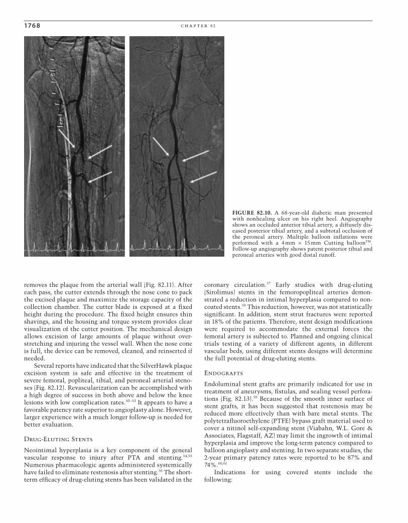
FIGURE 82.10. A 68-year-old diabetic man presented with nonhealing ulcer on his right heel. Angiography shows an occluded anterior tibial artery, a diffusely dis-eased posterior tibial artery, and a subtotal occlusion of the peroneal artery. Multiple balloon infl ations were performed with a 4 mm × 15 mm Cutting balloonTM. Follow-up angiography shows patent posterior tibial and peroneal arteries with good distal runoff.
CAR082.indd 1768CAR082.indd 1768 12/1/2006 3:37:47 PM12/1/2006 3:37:47 PM

e n d ova s c u l a r p ro c e du r e s f or t h e t r e at m e n t of p e r i p h e r a l va s c u l a r di s e a s e 176 9
FIGURE 82.11. The SilverHawkTM atherectomy catheter has a cobalt chromium cutter at the tip of the catheter that rotates when activated to remove the plaque from the arterial wall. The plaque is then pushed into the nose cone of the catheter (arrow).
FIGURE 82.12. SilverHawkTM atherectomy was performed after crossing a totally occluded (arrow on the left) right superfi cial femoral artery. Postatherectomy angiography (arrow on the right) shows widely patent right superfi cial femoral artery with good distal runoff.
• Arterial rupture• Arteriovenous fi stulas• Arterial trauma• Aneurysms
Possible indications for using covered stents are the following:
• Long occlusions or stenosis• Long dissections
For aneurysmal disease stent grafts have been success-fully used to exclude the aneurysm from the native cir-culation.59,62,63 All but one of the currently available endoluminal grafts used in the periphery are made of self-expanding nitinol. The Jostent® GraftMaster (Abbott Laboratories, Abbott Park, IL) is a balloon expandable system. This system uses double thin stainless steel with expand able PTFE coating between the two layers and is mounted on a semicompliant low-profi le balloon dilation catheter. It ranges from 3 to 5 mm in diameter and is 12, 19, and 26 mm in length. Its indications for use include treatment of free per-forations in native coronary arteries or saphenous vein grafts. It can also been used to exclude renal artery aneurysms (Fig. 82.14) or repair guidewire perforations in the arteries.
The WallgraftTM Endoprosthesis (Boston Scientifi c, Natick, MA), which is still in clinical trials for vascular use in the United States, has been used successfully to treat iliac, femoral, and popliteal aneurysms and iliac artery occlusive disease (Fig. 82.15).59,61–63 It is composed of polyethylene tere-phthalate (PET) graft material covering a Wallstent. The device is mounted on a UnistepTMPlus delivery system that has a working length of 90 cm. WallgraftTM is available in diameters from 6 to 14 mm and lengths of 20, 30, 50, and 70 mm (Fig. 82.16). The system accommodates a 0.035-inch guide wire.
CAR082.indd 1769CAR082.indd 1769 12/1/2006 3:37:47 PM12/1/2006 3:37:47 PM

17 70 c h a p t e r 82
A BFIGURE 82.13. (A) A 57-year-old man with a known history of pre-vious aorto-bi-iliac bypass presents with bilateral pulsatile masses. Aortogram shows bilateral iliac anatomic aneurysms. A 14 mm × 50 mm Wallgraft® was deployed in the right common and external
iliac artery and a 12 mm × 70 mm Wallgraft® was deployed in the left common and external iliac artery. (B) Follow-up angiography shows complete exclusion of both iliac aneurysms without evidence of endoleak.
A BFIGURE 82.14. (A) A 50-year-old woman with refractory hypertension was found to have a 30-mm right renal artery aneurysm (arrow). (B) Follow-up angiography after the aneurysm was excluded using a 16-mm and a 26-mm Jostent® (arrow).
CAR082.indd 1770CAR082.indd 1770 12/1/2006 3:37:48 PM12/1/2006 3:37:48 PM

e n d ova s c u l a r p ro c e du r e s f or t h e t r e at m e n t of p e r i p h e r a l va s c u l a r di s e a s e 17 71
Abdominal Aortic Aneurysms
Background and History
Abdominal aortic aneurysm (AAA) is a common disease that is associated with a signifi cant mortality if left untreated. This disease predominantly affects men who are 60 years or older. Men are fi ve times more frequently affected than women.64,65 More than 90% of these aneurysms are second-ary to atherosclerosis, and 89% are located in the infrarenal aorta. Surveillance studies indicate infrarenal aortic aneu-rysms 5.5 cm and 6.5 cm in size have annual rupture rates of 11% and 26%, respectively.66
There is a 90% mortality rate associated with an out-of-hospital rupture, with the mortality rate decreasing to 50% for those who undergo emergency surgery.64–66 To prevent this devastating event, more than 40,000 surgical AAA repairs are performed annually in the United States. The current mortality rate is less than 5% for elective repair of AAA and has an acceptable long-term survival. However, patients with associated comorbidities such as cardiovascu-lar or pulmonary conditions are at an increased risk with surgical repair. Unfortunately, if these high-risk patients are left untreated, they usually die from aneurysm rupture.
Endovascular stent grafts have been developed to avoid major abdominal surgery and to avoid the related morbidity and mortality. The less invasiveness of endovascular place-ment of stent grafts is particularly important because of coexisting morbid conditions in many of the patients presenting with AAA, and because it provides a therapeutic alternative for those who are not surgical candidates.
Designs Characteristics of Stent Grafts
A variety of graft designs and confi gurations have been tested in the laboratory and in clinical trials. The degree to which endovascular grafts differ from the characteristics of a surgi-cally placed graft vary widely with respect to method and mechanism of fi xation, mechanical properties of the stent graft, graft material, anatomic confi guration, and delivery systems.
Stainless steel or nitinol stent technology has been used in conjunction with various graft materials, such as polyester (Dacron), PTFE, or urethane polycarbonate. All endovascular
FIGURE 82.15. A 77-year-old man who had recent coronary inter-vention presents with painful pulsatile mass in the right groin. Angiography shows a large pseudoaneurysm in the right common iliac artery. A 10 mm × 50 mm Wallgraft® was placed in the right
external iliac and right common femoral artery. Follow-up angiog-raphy shows exclusion of the right common femoral artery pseudoaneurysm.
FIGURE 82.16. The Wallgraft® is available in diameters from 6 to 14 mm and has various lengths ranging from 20 to 70 mm.
CAR082.indd 1771CAR082.indd 1771 12/1/2006 3:37:49 PM12/1/2006 3:37:49 PM

17 7 2 c h a p t e r 82
stent graft devices, however, fall within one of three catego-ries: unibody in bifurcated or tubular design, modular (mul-ticomponent), and aorto-monoiliac. The latter is combined with the surgical placement of a femoral-femoral crossover prosthesis and occlusion of the contralateral iliac artery.
The search for an ideal endovascular stent graft is in an ongoing state of evolution. The advantages and disadvantages of current endoluminal grafts are under debate and will prob-ably continue to be debated for a number of years. While the long-term success of endoluminal stent grafts has yet to be demonstrated, a growing number of manufacturers and clini-cians continue to evaluate the ever-expanding number of stent grafts.
The development of endovascular aortic aneurysm repair (EVAR) is now in its second decade, and fi ve endografts have emerged from clinical trials and gained approval from the FDA: AncureTM (Guidant, Menlo Park, CA), AneuRxTM (Medtronic, Sunnyvale, CA), ZenithTM graft (Cook, Bloom-ington, IN), Excluder (W.L. Gore), and PowerLinkTM System (Endologix, Irvine, CA). Currently there is one other endo-graft that has an ongoing pivotal trial in the United States: Quantum (Cordis, Miami, FL).
Ancure Device
The AncureTM device is a unibody, unsupported graft, meaning that the entire device is loaded on a delivery system and that there is no structural support of the woven polyester graft fabric. The attachment system includes four indepen-dent V-shaped hooks that penetrate the arterial wall for fi xa-tion. There are separate attachments at the proximal and distal ends of the graft. The hooks are positioned transmu-rally and are affi xed by low-pressure balloon dilatation that
is incorporated in the delivery device. The use of the unibody design eliminates the endoleaks that can originate in the “overlaps” between various components of modular endo-grafts. Tortuous anatomy and narrowed, calcifi ed iliac arter-ies, however, make the deployment of this stiff, one-piece system very diffi cult. Several investigators reported that fewer than 30% of patients who were referred to their institu-tions for endoluminal AAA repair were candidates for Ancure device. This diffi cult deployment added to the complexity and the duration of the procedure. Because of the increasing number of clinical complications with Ancure, in July 2003 the manufacturer voluntarily halted production of the stent graft system and withdrew it from the market because of increased competition after the release of two new endograft systems.
AneuRx
The AneuRxTM device is a bifurcated, modular, fully sup-ported stent graft with a nitinol exoskeleton that is lined with a thin-walled polyester fabric. The self-expanding nitinol exoskeleton provides fl exibility and strength and is easily visible under fl uoroscopy. The two-piece system con-sists of a main bifurcation segment that is housed in a 21-French (F) delivery device and a contralateral limb that is housed in a 16F delivery device. Aortic fi xation of this endo-graft is accomplished by radial force at the attachment sites, which causes a frictional seal. Additional modular compo-nents include aortic and iliac extender cuffs. The iliac and aortic extender cuffs allow the user to make the appropriate length adjustments by overlapping these components with already deployed segments until the AAA is completely excluded from the arterial circulation (Fig. 82.17).67–69
FIGURE 82.17. Angi-ography before and after AneuRxTM stent-graft insertion in a patient with a 5.1-cm abdominal aortic aneurysm.
CAR082.indd 1772CAR082.indd 1772 12/1/2006 3:37:50 PM12/1/2006 3:37:50 PM

e n d ova s c u l a r p ro c e du r e s f or t h e t r e at m e n t of p e r i p h e r a l va s c u l a r di s e a s e 17 7 3
FIGURE 82.18. The ZenithTM is a modular bifurcated stent-graft system that utilizes a series a suprarenal barbs designed to mimic a surgical anastomosis.
FIGURE 82.19. The Gore Excluder® is a fully, nitinol-supported, modular system that is premounted, and constrained with a lacing suture that when pulled self-expands immediately.
Zenith
The ZenithTM stent-graft is a fully supported modular bifur-cated system that incorporates a polyester fabric on a network of Z-stents. The current delivery system utilizes a series of suprarenal barbs, which, when released, are designed to mimic a surgical anastomosis (Fig. 82.18).
Excluder
The Excluder® endograft was approved for use in November 2002 and is a self-expanding modular component system (Fig. 82.19). The Excluder consists of expanded PTFE on the luminal surface and a nitinol-supporting frame on the outer surface. The components are premounted on separate deliv-ery catheters and are constrained using lacing suture, which courses the length of the catheter and is attached to a deploy-ment knob at the operator end. When the knob is loosened and pulled back it, releases the suture and the stent-graft component expands immediately. An 18F sheath is used to advance the bifurcated aortic and a 12F is used for the con-tralateral limb.
The PowerLink System
The PowerLink® System is a self-expanding, unibody, bifur-cated graft. The cage is made of a single wire stainless steel that is interconnected without the use of sutures. The endo-skeleton is covered by extremely strong PTFE material and is delivered through a 20F or 21F catheter. The device is available for infrarenal and suprarenal fi xation, allowing treatment of a wide variety of neck and limb diameters (Fig. 82.20).
Patient Selection
When considering endovascular treatment, the patient’s life expectancy and the risk associated with conventional repair should be taken into account. Anatomic factors such as the length, shape, and angulation of the infrarenal neck of the aneurysm, involvement of the iliac arteries or occlusive disease, and tortuosity of the iliofemoral vessels should be taken into account when considering a patient.
Proper preoperative preparation for endovascular AAA repair is crucial to the success of the procedure. Correct selection of the diameter and length of the endograft is a key factor in minimizing the most common complications related to endovascular repair. Endograft-related endoleaks and stent graft migration can be avoided by proper patient selection and accurate stent graft placement. The successful implantation of endografts is predicated on sophisticated preoperative evaluation utilizing a combination of spiral CT scanning with three-dimensional reconstruction and contrast arteriography with calibrated marker catheter. Computed tomography scanning produces axial “cuts” or slices, allowing accurate measurement of the diameter of
CAR082.indd 1773CAR082.indd 1773 12/1/2006 3:37:50 PM12/1/2006 3:37:50 PM

17 74 c h a p t e r 82
but also presents as “fl ash” pulmonary edema because of increased afterload (hypertension) and preload (volume expansion).
Diagnosis and Management
Renal artery stenosis is the most common correctable cause of hypertension. More than 90% of cases of RAS are athero-sclerotic in nature and involve the ostium and the proximal portion of the renal artery, with plaque extending into the perirenal aorta.71 Therefore, the treatment of choice for ath-erosclerotic RAS is PTA and stenting.
Many noninvasive techniques demonstrate the presence of renovascular disease. Ultrasonography is the most ideal imaging modality for detecting RAS. Duplex velocity and ratio criteria to assess hemodynamic signifi cance are listed in Table 82.2. It is important to visualize the entire renal artery from the origin to the kidney parenchyma so that you can be assured of detecting RAS from both atherosclerosis and fi bromuscular dysplasia (FMD). Renal artery stenosis may cause renal insuffi ciency, uncontrolled hypertension, and recurrent congestive heart failure.
Fibromuscular Dysplasia
In contrast to atherosclerotic RAS, FMD often affects the distal two thirds of the renal artery and its branches. It accounts for approximately 10% of cases of RAS and is typi-cally seen in young or middle-aged females.72
Fibromuscular dysplasia is a noninfl ammatory, non-atherosclerotic vascular disease affecting medium-size to small vessels of unknown etiology. It results in fi bromus-cular alterations in the intimal or medial layers of the renal artery.72,73 It primarily affects women between the third and sixth decade and has a marked predilection for Caucasians.73
Fibromuscular dysplasia can be divided into three major pathologic subtypes: medial dysplasia, intimal fi broplasias, and adventitial fi broplasias. Of the medial dysplasia, medial fi broplasia is the most common type of FMD. The lesions are commonly seen bilaterally, and other vessels in the body, such as the carotid, vertebral, subclavian, mesenteric, and iliac arteries, may be involved.73
Angiography reveals the telltale “string of beads” appear-ance with the beads larger than the normal caliber of the artery (Fig. 82.21). In perimedial fi broplasias the beads are less numerous and smaller than the normal caliber of the artery, and intimal disease is noted for concentric band-like stenoses or smooth long stenoses. Spontaneous dissection is an associated complication of FMD. Symptoms include fl ank pain, hematuria, and accelerated hypertension. Early angiog-raphy is indicated in the appropriate clinical setting for diag-nosis and treatment.
Percutaneous transluminal angioplasty is recommended for patients with FMD, and stent implantation is generally not required because restenosis rates in this population are <15%. Surgical revascularization is now rarely used.72 This is due in a large part to the high technical success that can be achieved with PTA and the low morbidity.
FIGURE 82.20. The PowerLink® system is a unibody stainless steel endoskeleton system that is available in suprarenal or infrare-nal fi xation.
the infrarenal neck, and the maximal diameter of the AAA, the distal abdominal aorta, and the iliac arteries. The CT scanning should start above the origin of the superior mesenteric artery and extend to the inguinal area, with the use of 2- or 3-mm cuts. Computed tomography scanning, however, is less accurate for length sizing, because it often underestimates the distance from the renal arteries to the origin of internal iliac arteries. Therefore, length sizing of the aorta and iliac arteries is most accurately per-formed using a calibrated marker catheter and contrast arteriography.
Renovascular Disease
Background
The overall incidence of hypertension due to renal artery stenosis (RAS) is less than 5%.70 It is estimated that 2 to 4 million people in the United States have renovascular disease; however, the incidence appears to be rising because of increased atherosclerosis in an aging population. The risk factors and the epidemiology are the same as those for atherosclerosis elsewhere in the body. The clinical pre-sentation may be that of new-onset hypertension, abrupt worsening of blood pressure in a previously hypertensive patient, and progressive renal insuffi ciency. Severe bilateral renal artery disease can share all of the clinical features
CAR082.indd 1774CAR082.indd 1774 12/1/2006 3:37:51 PM12/1/2006 3:37:51 PM

e n d ova s c u l a r p ro c e du r e s f or t h e t r e at m e n t of p e r i p h e r a l va s c u l a r di s e a s e 17 75
Atherosclerotic Renal Artery Stenosis
Unlike FMD, atherosclerotic RAS does require stent implan-tation. Surgical revascularization is rarely performed except in the presence of AAAs or dissections. Surgical revascular-ization is associated with the morbidity and the hospital stay of a major operation, as well as complications including bypass graft thrombosis and nephrectomy in up to 4% and operative mortalities in up to 3%.74,75 Angioplasty and stenting have become the modalities of choice for atheroscle-rotic RAS. Multiple trials with stenting have demonstrated improvements in blood pressure control, attenuation of renal failure progression, and preservation of renal size.76–78
Not all patients demonstrate improvement in blood pres-sure control and renal failure after renal artery stenting. The renal artery resistance index and renal artery end-diastolic velocity have been used to help identify patients that may benefi t from renal artery stenting. A resistance index above 80 was found to be a strong predictor for a lack of benefi t after revascularization.79,80
Angioplasty and stent implantation is usually performed from the femoral approach using a 6F guiding catheter, a low-profi le balloon, and a stent that accommodates a 0.014-inch guidewire. The use of the low-profi le systems may potentially reduce renal artery embolic complications or perinephric hematomas related to branch vessel perforation caused by larger, bulkier systems. The RAS is usually predi-lated with a 4- to 5-mm balloon followed by a 5.5- to 7-mm stent. The stent should be balloon expandable to provide maximum radial strength and prevent recoil at the ostium.
Since the majority of atherosclerotic RAS is ostial in nature, stent deployment should allow 1 to 2 mm of stent to extend into the aorta (Fig. 82.22). The ostium is then postdilated with the stent balloon partially in the aorta using a higher infl ation pressure.
Some patients may experience worsening of renal func-tion after PTA and stenting. Atheromatous embolization has been indicated as a precipitating factor for this complica-tion.81,82 To eliminate the risk of atheroembolic material entering into the parenchyma, Henry and colleagues83 reported the use of distal protection to prevent this complica-tion. The true incidence of atheroembolism is uncertain because many patients can have a silent course due to the large functional kidney reserve, which allows for normal serum creatinine values despite a signifi cant decline of total glomerular fi ltration. Renal atheroembolism defi nitely poses a risk of renal function deterioration, and the use of distal
FIGURE 82.21. Selective right renal angiography showing the classic “string of beads” in a 46-year-old woman with fi bromuscular dysplasia and uncontrolled hypertension.
A
BFIGURE 82.22. (A) A 67-year-old man status post–left renal artery stenting now with right renal artery stenosis. (B) After PTA and stenting with a 6.0 mm by 15 mm stent.
CAR082.indd 1775CAR082.indd 1775 12/1/2006 3:37:52 PM12/1/2006 3:37:52 PM

17 76 c h a p t e r 82
protection may become the standard of care when treating atherosclerotic RAS.
Extracranial Carotid Artery Disease
Epidemiology
Extracranial carotid artery occlusive disease causes 25% of the 500,000 cerebrovascular accidents (CVAS) that occur each year in the United States.84 Stroke is the third most common cause of death in the United States. More than 2 million CVA survivors remain with varying degrees of dis-ability. One third of the patients suffering an ischemic stroke die and another third remain permanently disabled.
Several studies revealed that those patients with carotid artery stenosis greater than 75% have a 2% to 5% risk of suf-fering from ischemic CVA during the fi rst year.85,86 Following a transient ischemic attack (TIA), the risk of ischemic stroke is 12% to 13% in the fi rst year, and 30% to 37% in the 5 years following the initial event. For patients who developed CVA, the risk of a subsequent CVA is 5% to 9% per year, and approximately 24% to 45% of them have another CVA within the next 5 years. The presence of an ulcerated lesion also increases the risk of an ischemic CVA.87
The majority of patients with carotid stenosis, however, are asymptomatic and diagnosis is made following auscultation of a carotid bruit or routine ultrasound screening.
Carotid Endarterectomy
Carotid endarterectomy (CEA) was introduced in 1954 as a surgical therapy for the prevention of CVA for the extracra-nial carotid artery stenosis.88 With the goal of minimizing the mortality and morbidity, surgeons have refi ned CEA techniques over the past four decades to acceptable periop-erative complication rates for the great majority of patients.89 The number of CEAs has dramatically increased in the United States from 15,000 in 1971 to 130,000 in the last decade.89,90 Medical therapy with antiplatelet agents, however, still remains the standard of care for symptomatic patients with less than 50% stenosis.85
Prospective, randomized studies such as the North American Symptomatic Carotid Endarterectomy Trial (NASCET),91 the European Carotid Surgery Trial (ECST),92
and the Asymptomatic Carotid Atherosclerosis Study (ACAS)93 have proven the superiority of CEA over medical therapy for a selected group of patients with symptomatic and asymptomatic extracranial carotid artery stenosis. In NASCET, for patients with ≥70% carotid stenosis the risk of ipsilateral stroke was reduced by 65% in the surgical group.91 In ACAS, for patients with 60% carotid stenosis the risk of ipsilateral stroke was reduced by 53%.93 From NASCET and ACAS studies a clear benefi t from CEA was demonstrated in spite a perioperative stroke/death rate of 5.8% in symptomatic patients and 2.7% in asymptomatic patients.
Thus the prospective randomized clinical trials have established that CEA with skilled surgeons who have low perioperative stroke rates is the gold standard for treatment
of patients with symptomatic and asymptomatic severe carotid artery stenosis.94 However, CEA is not without its risks. There is a signifi cant discrepancy in complications after CEA among reported studies. The clinical and ana-tomic diversity of patients with carotid disease could par-tially lead to differences in outcomes with CEA among different studies. Rothwell et al.94 reported in meta-analysis of 50 CEA studies the risk of CVA to be 7.7% in the studies followed by a neurologist, and 2.3% in the studies followed-up only by surgeons. The mortality and morbidity of CEA was particularly high (18%) for patients who also had signifi -cant coronary artery disease.
Other complications of CEA are cranial nerve palsies (7.6% to 27%), hematoma (5.5% to 11%), and restenosis (5% to 9%).95 McCrory et al.95 retrospectively analyzed clinical data of 1160 patients who had CEA to determine the predic-tors of postoperative complications. They discovered that predictors of postoperative myocardial infarction, nonfatal strokes, and death were age over 75 years old, endarterectomy being performed in preparation for bypass surgery, internal carotid artery thrombus, intracranial carotid artery disease, and perioperative neurologic instability.
Restenosis After Carotid Endarterectomy
Another complication of CEA is restenosis. Although studies with complete angiographic follow-up have not been done after CEA, restenosis appears to occur in 8% to 19% of patients.87,89,96–98 Myointimal hyperplasia is a primary cause of restenosis within the fi rst 24 months after CEA.99 Progres-sive atherosclerosis plays a greater role in more delayed reste-nosis. Controversy remains as to whether vein or prosthetic patch repair results in a lower incidence of restenosis as compared to primary closure.85,88,89,97–99
Endarterectomy for recurrent carotid artery stenosis after endarterectomy is more diffi cult, due to scarring, and carries a signifi cantly higher complication rate than the original operation.88,97,98 The scarring external to the artery lengthens the duration of the procedure and requires more manipula-tion of the carotid bifurcation, which increases the risk of cerebral embolization and a recurrent laryngeal nerve injury.95–97
In the two largest series the major complication rate for repeat CEA ranges from 4.6% to 10.9%.97,99 From previous studies it is diffi cult to establish the frequency and severity of symptoms leading to the second CEA. In the Cleveland Clinic series of carotid reoperations, 5.1% of the patients had neurologic symptoms leading to the operation.99 In several retrospective studies, 22% to 90% of patients with restenosis had neurologic symptoms prior to second CEA.95–99
Carotid Angioplasty and Stenting
Virtually all endovascular carotid interventions currently performed are stent-based with an embolic protection device, which is a safer and more dependable procedure. Numerous reports have shown encouraging outcomes following pro-tected carotid stenting.100–108
The technique of carotid angioplasty and stenting is still evolving with the advances in technology and with the increased experience of the interventionists.
CAR082.indd 1776CAR082.indd 1776 12/1/2006 3:37:53 PM12/1/2006 3:37:53 PM

e n d ova s c u l a r p ro c e du r e s f or t h e t r e at m e n t of p e r i p h e r a l va s c u l a r di s e a s e 17 7 7
Several factors are essential to achieve the optimal result of carotid angioplasty and stenting:
• Detailed preprocedural clinical, noninvasive, and angio-graphic cerebrovascular evaluation
• Appropriate choice of arterial access site• Appropriate choice of guiding catheters, protection
device, and PTA balloons• Appropriate choice of stents (balloon expandable or
self-expandable)• Essential pharmacologic therapy• Adequate knowledge or support in performing intracra-
nial vascular rescue• Adequate postprocedural neurologic, invasive, and non-
invasive evaluation
An independent neurologic evaluation is essential to objectively assess the patient’s condition before and after the interventional procedure. Carotid duplex ultrasound should be performed before the procedure and as a routine noninva-sive follow-up. Aortic arch and four-vessel cerebral angiogra-phy with intracranial images are necessary to identify the extent of the disease and possible associated intracranial lesions. The patients are usually given antiplatelet agents 8 days before the procedure.
The femoral access is the most commonly used access. Cerebral angiography is performed with a 5F cerebral angio-graphic catheter. The patient is given intravenously heparin to achieve an activated clotting time greater than 275 seconds. After the lesion is identifi ed in multiple orthogonal views, a 0.035-inch extra support guidewire is advanced to the
common or external carotid artery and the diagnostic cath-eter is replaced with a 6F or 7F 90-cm-long sheath or an 8F or 9F guiding catheter. The 0.035-inch wire is removed and a 0.014-inch cerebral protection guidewire is advanced through the stenosis and deployed in the distal internal carotid just below the siphon. Predilation with a 4.0-mm balloon is performed. Hypotension and bradycardia are common at the time of PTA or stenting of the internal carotid bulb. These conditions are transient and respond well to atropine. Intravenous vasopressors for profound hypotension should be readily available.
After completion of the PTA, the balloon is removed and a self-expanding stent (for internal carotid lesions) or the balloon expandable stent (for common carotid lesions) is advanced to the lesion and deployed. It is advisable to perform PTA after deploying the self-expandable stent to achieve adequate apposition of the stent to the arterial wall (Fig. 82.23). During the procedure the patient’s neurologic status is frequently evaluated using a contralateral hand grip to detect any possible complications. Patients with lesions located in the carotid bulb may require intravenous vasopres-sor infusion overnight. Independent neurologic evaluation is performed prior to discharge the following day. The anti-platelet regimen is continued for at least 30 days if not indefi nitely.
Choice of Stent
The size of the internal carotid artery varies from 5 to 8 mm. The common carotid artery measures from 7 to 10 mm.
FIGURE 82.23. (A) Carotid angiography shows restenosis at the site of previous right carotid endarterectomy in a 58-year-old man. (B) After achieving an activated clotting time >275 seconds, an 0.014-inch, 6-mm AngioguardTMXP (Cordis, Miami, FL) was deployed in the distal right internal carotid artery and the restenosed area was predilated with a 4 mm × 20 mm coronary balloon. (C) A 7 mm ×
30 mm PreciseTM stent (Cordis) was deployed in the right common and internal carotid artery. (D) The stenosis was then postdilated with a 5.5 mm × 20 mm balloon. (E) Final angiography shows minimal residual stenosis with good distal fl ow passed the distal protection fi lter.
CAR082.indd 1777CAR082.indd 1777 12/1/2006 3:37:53 PM12/1/2006 3:37:53 PM

17 7 8 c h a p t e r 82
FIGURE 82.24. A tapered AcculinkTM.
FIGURE 82.25. The AccunetTM nitinol distal protection basket is used in conjunction with the AcculinkTM.
When the stent is placed across the bifurcation of the common carotid artery, it must adapt itself to arteries of different diameters. The AcculinkTM carotid stent system (Guidant, Santa Clara, CA) is the fi rst stent system approved for carotid angioplasty and stenting. The AccunetTM nitinol distal pro-tection basket is used in conjunction with the Acculink, which comes in diameters ranging from 5 to 10 mm and lengths of 20, 30, and 40 mm. A tapered Acculink (Fig. 82.24) is available in diameter tapering from 8 to 6 mm and 10 to 7 mm for stenting across the bifurcation of the common carotid and internal carotid. The Accunet has a 150-μm fi lter basket on a 0.014-inch wire (Fig. 82.25).
Several other carotid stent systems have been tested clin-ically and are awaiting approval from the FDA. Covered stents are also available; however, they will require several refi nements to be of benefi t for carotid angioplasty and stent-ing. It is possible that the covered stents will decrease the risk of embolization, although the risk of occlusion of exter-nal carotid artery should be taken into consideration. Patients for whom carotid angioplasty and stenting should be consid-ered include those with the following fi ndings:
• High cervical carotid (supramandibular) lesions that are surgically inaccessible
• Tandem lesions with proximal and distal internal carotid artery lesions
• Postoperative recurrent carotid artery stenosis• Nonatherosclerotic carotid artery stenosis (fi bromuscu-
lar dysplasia, Takayasu arteritis)• Prior radiation therapy to the neck with ipsilateral
stenosis
• Prior radical neck surgery stenosis• Common carotid artery origin lesions with an associated
internal carotid artery (ICA) lesion• Increased operative risk due to comorbid illnesses such
as coronary artery disease, in anticipation of coronary artery bypass surgery
Several other embolic protection devices have been inves-tigated and include fi lter devices, distal balloon occlusion with aspiration, and proximal balloon occlusion with fl ow reversal.99–108
Summary
Over the past 5 years endovascular strategies have revolu-tionized the treatment of PAD. Endovascular therapies, however, are still in their infancy, with the potential for growth in the treatment for venous disorders, renal artery stenting, endografts for abdominal and thoracic aneurysms, and carotid artery stenting. Endografts for thoracic aneu-rysms and carotid artery stenting with protection will accel-erate rapidly with their recent FDA approval. The widespread acceptance of endovascular techniques brings new initia-tives, devices, and more randomized trials.
It is quite clear that there are no true contraindications to endovascular intervention. Instead it is the risk–benefi t ratio of each possible treatment option that must be consid-ered for individual patients. Finally, the most important future aspect of endovascular techniques is axial cross-sectional imaging and the accurate interpretation of these images. Computed tomography angiography and MR angiog-raphy will remain the preferred physiologic modality for diagnosis and treatment of PAD. Therefore, the endovascular interventionalist must become profi cient at image interpre-tation to ensure decisive endovascular treatment planning.
References
1. Dormany JA. CAPRIE study: ankle-brachial index predictor of ischemic events. Cerebrovasc Dis 1999;9(suppl 1):1–28.
2. U.S. Department of Health and Human Services. A Chartbook on Cardiovascular, Lung and Blood Diseases: Morbidity and Mortality. Bethesda: National Institutes of Health, National Heart, Lung, and Blood Institute, 1994.
3. Comerota AJ. Venous thromboembolism. In: Rutherford RB, ed. Vascular Surgery, 4th ed. WB Saunders, Philadelphia: 1995:1785–1814.
4. Arcelus JI, Caprini JA, Monreal M, et al. The management and outcome of acute venous thromboembolism: a prospective reg-istry including 4011 patients. J Vasc Surg 2003;38:916–922.
5. Lensing AWA, Prins MH, Davidson BL, et al. Treatment of deep venous thrombosis with low-molecular-weight heparins: a meta-analysis. Arch Intern Med 1995;155:601–607.
6. Borst-Krafek B, Fink AM, Lipp C, et al. Proximal extent of pelvic vein and its association with pulmonary embolism. J Vasc Surg 2003;37:518–522.
7. Meyerovitz MF, Polak JF, Goldhaber SZ. Short-term response to thrombolytic therapy in deep venous thrombosis: predictive value of venographic appearance. Radiology 1992;184:345–348.
8. Ott P, Eldrup E, Oxholm P, et al. Streptokinase therapy in the routine management of deep venous thrombosis in the lower extremities. A retrospective study of phlebographic results and therapeutic complications. Acta Med Scand 1986;219:295–300.
CAR082.indd 1778CAR082.indd 1778 12/1/2006 3:37:53 PM12/1/2006 3:37:53 PM

e n d ova s c u l a r p ro c e du r e s f or t h e t r e at m e n t of p e r i p h e r a l va s c u l a r di s e a s e 17 7 9
9. Comerota AJ, Aldridge SA. Thrombolytic therapy for acute deep vein thrombosis. Semin Vasc Surg 1992;5(2):76–84.
10. Blinc A, Francis CW. Transport processes in fi brinolysis and fi brinolytic therapy. Thromb Haemost 1996;76:481–491.
11. Francis CW, Totterman S. Magnetic resonance imaging of deep vein thrombi correlates with response to thrombolytic therapy. Thromb Haemost 1995;73:386–391.
12. Semba CP, Dake MD. Iliofemoral deep venous thrombosis: aggressive therapy with catheter-directed thrombolysis. Radi-ology 1994;191:487–494.
13. Bjarnason H, Kruse JR, Asinger DA, et al. Iliofemoral deep venous thrombosis: safety and effi cacy outcome during 5 years of catheter-directed thrombolytic therapy. J Vasc Interv Radiol 1997;8:405–418.
14. Mewissen MW, Seabrook GR, Meissner MH, et al. Catheter-directed thrombolysis for lower extremity deep venous throm-bosis: report of a national multi-center registry. Radiology 1999;211:39–49.
15. Comerota AJ, Kagan SA. Catheter-directed thrombolysis for the treatment of acute iliofemoral deep venous thrombosis. Phlebology 2001;15:149–155.
16. Allie DE, Hebert CJ, Khan MH, et al. The power-pulse spray technique. Endovasc Today 2003;2(2):25–30.
17. Bush RL, Lin PH, Bates JT, et al. Pharmacomechanical throm-bectomy for the treatment of symptomatic lower extremity deep venous thrombosis: safety and feasibility study. J Vasc Surg 2004;40:965–970.
18. Ferris EJ, Lim Wn, Smith PL, et al. May-Thurner syndrome. Radiology 1983;147:29–31.
19. Patel NH, Stookey KR, Ketcham DB, et al. Endovascular man-agement of acute extensive iliofemoral deep venous thrombosis caused by May-Thurner syndrome J Vasc Intervent Radiol 2000;11:1297–1302.
20. Lamont JP, Pearl GJ, Patetsios P, et al. Prospective evaluation of endoluminal venous stents in the treatment of May-Thurner syndrome. Ann Vasc Surg 2002;16:61–64.
21. O’Sullivan GJ, Semba CP, Bittner CA, et al. Endovascular man-agement of iliac vein compression (May-Thurner syndrome). J Vasc Intervent Radiol 2000;1:823–836.
22. Hiatt WR. Medical treatment of peripheral vascular disease and claudication. N Engl J Med 2001;344:1608–1621.
23. Baxter-Smith D, Ashton F, Slaney G. Peripheral arterial embo-lism: a 20 year review. J Cardiovasc Surg 1988;29:453.
24. Linton RR. Peripheral arterial embolism: a discussion of the post-embolic vascular changes and their relation to the restora-tion of circulation in peripheral embolism. N Engl J Med 1941;341–354.
25. Nice C, Timmons G, Bartholemew, et al. Retrograde versus antegrade puncture for infra-inguinal angioplasty. Cardiovasc Intervent Radiol 2003;26(4):370–374.
26. Muller W, Fisher M, Bublitz G et al. Alternative vascular approaches in transluminal angioplasty of infra-inguinal arter-ies. Vasa Suppl 1991;33:200–201.
27. Dormandy JA, Rutherford RB. Management of peripheral arte-rial disease (PAD). TransAtlantic Inter-Society Consensus (TASC). J Vasc Surg 2000;31(1 pt 2):S1–S296.
28. Matsi PJ, Manninen HI, Ritva LV, et al. Femoropopliteal angio-plasty in patients with claudication: primary and secondary patency in 140 limbs with 1–3–year follow-up. Radiology 1994;191:727–733.
29. London NJM, Srinivasan R, Naylor AR, et al. Subintimal angio-plasty of femoropopliteal artery occlusions: the long-term results. Eur J Vasc Surg 1994;8:148–155.
30. Gordon IL, Conroy RM, Tobis JM, et al. Determinants of patency after percutaneous angioplasty and atherectomy of occluded superfi cial femoral arteries. Am J Surg 1994;168:115–119.
31. Murray JG, Apthorp LA, Wilkins RA. Long-segment (>10 cm) femoropopliteal angioplasty: improved technical success and long-term patency. Radiology 1995;195:158–162.
32. Henry M, Armor M, Ethevenot G, et al. Palmaz stent place-ment in iliac and femoropopliteal arteries: primary and second-ary patency in 310 patients with 2–4 year follow up. Intervent Radiol 1995;197:167–174.
33. Strecker EPK, Boos IBL, Gottmann D. Femoropopliteal artery stent placement: evaluation of long-term success. Radiology 1997;205:375–383.
34. Damaraju S, Cuasay L, Le D, et al. Predictors of primary patency failure in Wall-stent self-expanding endovascular prostheses for iliofemoral occlusive disease. Tex Heart Inst J 1997;l24:173–178.
35. Gray BH, Sullivan TM, Chills MB, et al. High incidence of restenosis/reocclusion of stents in the percutaneous treatment of long-segment superfi cial femoral artery disease after subop-timal angioplasty. Ann Vasc Surg 1997;25:74–80.
36. Gordon IL, Conroy RM, Arefi M, et al. Three-year outcome of endovascular treatment of superfi cial femoral artery occlusion. Arch Surg 2001;136:221–228.
37. Allie DE, Herbert CJ, Lirtzman MD, et al. Novel simultaneous combination chemical thrombolysis/rheolytic thrombectomy therapy for acute critical limb ischemia: the power pulse spray technique. Cathete Cardiovasc Interv 2004;63(4):512–522.
38. Brambillal N, Laudisa ML, Llambro M, et al. Cryoplasty in the treatment of iliac and superfi cial femoral artery disease. J Cardiovasc Surg 2004;45(suppl 1–3):12(abstr).
39. Unterberg C, Buchwald AB, Barath P, et al. Cutting balloon coronary angioplasty: initial clinical experience. Clin Cardiol 1993;16:660–664.
40. Barath P, Fishbein MC, Vari S, Forrester JS. Cutting balloon: a novel approach to percutaneous angioplasty. Am J Cardiol 1991;68:1249–1252.
41. Ansel GM, Sample NS, Botti III CF Jr, et al. Cutting balloon angioplasty of the popliteal and infrapopliteal vessels for symp-tomatic limb ischemia. Cathet Cardiovasc Interv 2004;61:1–4.
42. Dhar A. Cutting balloon angioplasty of in infrapopliteal arter-ies for better immediate and long-term patency. J Cardiovasc Surg 2004;45(suppl 1–3):12(abstr).
43. Laird JR, Reiser C, Biamino G, et al. Excimer laser assisted angioplasty for the treatment of critical limb ischemia. J Cardiovasc Surg 2004;45:239–248.
44. Steinkamp HJ, Rademaker J, Wissgott C, et al. Percutaneous transluminal laser angioplasty versus balloon dilatation for the treatment of popliteal artery occlusions. J Endovasc Ther 2002;9:882–888.
45. Bottcher HD, Schopohl B, Liermann D, et al. Endovascular irradiation—a new method to avoid recurrent stenosis after stent implantation in peripheral arteries: technique and preliminary results. Int Radiat Oncol Biol Phys 1994;29:183–186.
46. Liermann D, Kirchner J, Schopohl B, et al. Brachytherapy with iridium-192 HDR to prevent restenosis in peripheral arteries: an update. Herz 1998;23:394–400.
47. Minar E, Pokrajc B, Ahmadi R, et al. Brachytherapy for prophy-laxis against stent restenosis after long-segment femoropopli-teal angioplasty: pilot study. Radiology 1998;208:173–179.
48. Minar E, Pokrajac B, Maca T, et al. Endovascular brachytherapy for prophylaxis of restenosis after femoropopliteal angioplasty: results of a prospective randomized study. Circulation 2000;102:2694–2699.
49. Waksman R, Laird JR, Jurkovitz CT, et al. Intravascular radia-tion therapy after balloon angioplasty of narrowed femoropop-liteal arteries to prevent restenosis: results of the PARIS feasibility clinical trial. J Vasc Interv Radiol 2001;12(8):915–921.
CAR082.indd 1779CAR082.indd 1779 12/1/2006 3:37:54 PM12/1/2006 3:37:54 PM

17 8 0 c h a p t e r 82
50. Allie DE, Lirtzman CH, Wyatt CH, et al. Plaque excision: a novel treatment strategy for “real world” superfi cial femoral artery disease. A 10–month clinical experience. J Invasive Cardiol 2004(suppl).
51. Kanfzari DE, Chopra A, Gammon R. Early procedural and clinical outcomes for a novel method of revascularization in critical limb ischemia: limb salvage with SilverHawk Plaque excision. J Invasive Cardiol 2004(suppl).
52. Lorenz DP, Skolnick AA, Garcia LA. Early experience with the FoxHollow SilverHawk plaque excision system in the treat-ment of infrainguinal peripheral arterial disease. J Invasive Cardiol 2004(suppl).
53. Motarjeme A. Plaque excision of infrapopliteal arteries for limb salvage. J Invasive Cardiol 2004(suppl).
54. Kearny M, Pieczek A, Haley L, et al. Histopathology of in-stent restenosis in patients with peripheral artery disease. Circula-tion 1997;95:1998–2000.
55. Kuntz RE, Gibson CM, Nobuyoshi M, et al. Generalized model of restenosis after conventional angioplasty, stenting and direc-tional atherectomy. J Am Coll Cardiol 1993;21:12–25.
56. Lefkovits J, Topol EJ. Pharmacological approaches for the pre-vention of restenosis after percutaneous coronary intervention. Prog Cardiovasc Rep 2000;2:326–331.
57. Eisenberg MJ, Konnyu KJ. Review of randomized clinical trials of drug-eluting stents for the prevention of in-stent restenosis. Am J Cardiol 2006;98(3):362–366.
58. Duda SH, Bosiers M, Lammer J, et al. Sirolimus-eluding versus bare nitinol stent for obstructive superfi cial femoral artery disease: The SIROCCO II Trial. J Vasc Intervent Radiol 2005;16:331–338.
59. Kudelko PE, Alfaro-Franco C, Diethrich EB, Krajcer Z. Successful endoluminal repair of a popliteal artery aneurysm using the Wallgraft endoprosthesis. J Endovasc Surg 1998;373:377.
60. Saxon RR, Coffman JM, Gooding JM, et al. Long-term results of ePTFE stent graft versus angioplasty in the femoro-popliteal artery: Single center experience from a prospective, random-ized trial. Vasc Intervent Radiol 2003;14:303–311.
61. Jahnke T, Andresen R, Müller-Hülsbeck S, et al. Hemobahn stent grafts for the treatment of femoropopliteal arterial obstructions: midterm results of a prospective trial. J Vasc Intervent Radiol 2003;14:41–51.
62. Gupta K, Dougherty K, Hermman H, Krajcer Z. Endovas-cular repair of giant carotid pseudoaneurysms with Viabahn stent graft. Catheterization Cardiovasc Intervent 2004;62(1):64–68.
63. Howell M, Krajcer Z, Diethrich EB, et al. Wallgraft endopros-thesis for percutaneous treatment of femoral and popliteal artery aneurysms. J Endovasc Ther 2002;9:76–81.
64. Johnston WK. Multicenter prospective study of non-ruptured abdominal aortic aneurysms. Part II: variables predicting mor-bidity and mortality. J Vasc Surg 1989;9:437–447.
65. Chen JC, Hildebrand HD, Salvian AJ, et al. Predictors of death in ruptured and nonruptured aortic aneurysms. J Vasc Surg 1996;24:614–620.
66. The UK Small Aneurysm Trial Participants. Mortality results for randomized controlled trial of early elective surgery or ultrasonographic surveillance for small abdominal aortic aneu-rysms. The UK Aneurysm Trial Participants. Lancet 1998;352:1649–1655.
67. Beebe HG. Imaging modalities for aortic endografting. J Endo-vasc Surg 1997;4:111–123.
68. May J, White GH, Yu W, et al. Concurrent comparison of endoluminal repair versus no treatment for small abdominal aortic aneurysms. Eur J Vasc Endovasc Surg 1997;13:472–476.
69. Moore WS, for the EVT Investigators. The EVT tube and bifur-cated endograft systems: Technical considerations and clinical summary. J Endovasc Surg 1997;4:182–194.
70. Kaplan NM. Systemic hypertension: mechanisms and diagno-sis. In: Braunwald E, ed. Heart Disease: A Textbook of Cardio-vascular Medicine, 3rd ed. Philadelphia: WB Saunders, 1988:819–861.
71. Schreiber MJ, Pohl MA, Novick AC. The natural history of atherosclerotic and fi brous renal artery disease. Urol Clin North Am 1984;11:383–392.
72. Luscher TF, Lie JT, Stanson AW, et al. Arterial fi bromuscular dysplasia. Mayo Clin Proc 1987;62:931–952.
73. Begelman SM, Olin JW. Fibromuscular dysplasia. Curr Opin Rheumatol 2000;12:41–47.
74. Hansen KJ, Starr SM, Sands E, et al. Contemporary surgical management of renovascular disease. J Vasc Surg 1992;16:319–331.
75. Novick AC, Ziegelbaum M, Vidt DG, et al. Trends in surgical revascularization for renal artery disease: ten years experience. JAMA 1987;257:498–501.
76. Blum U, Krumme B, Flugel P, et al. Treatment of ostial renal-artery stenoses with vascular prostheses after unsuccessful balloon angioplasty. N Engl J Med 1997;336:459–465.
77. Tuttle KR, Chouinard RF, Webber JT, et al. Treatment of atherosclerotic ostial renal artery stenosis with intravascular stent. Am J Kidney Dis 1998;32:611–622.
78. Dorros G, Prine C, Mathiak L. Stenting of renal artery stenosis achieves better relief of obstructive lesion than balloon angioplasty. Cathet Cardiovasc Diagn 1993;29:191–198.
79. Radermacher J, Chavan A, Bleck J, et al. Use of Doppler ultra-sonography to predict the outcome of therapy for renal-artery stenosis. N Engl J Med 2001;344:410–417.
80. Mukherjee D, Bhatt DL, Robbins M, et al. Renal artery end-diastolic and renal artery resistance index as predictors of outcome after renal artery stenting. Am J Cardiol 2001;88:1064–1066.
81. Van de Ven PJ, Beutler JJ, Kaatee R, et al. Transluminal vascular stent for ostial atherosclerotic renal artery stenosis. Lancet 1995;346:672–674.
82. Wilms G, Staessen J, Baert AL, et al. Percutaneous transluminal angioplasty and renal function. Radiology 1989;29:195–200.
83. Henry M, Klonaris C, Henry I, et al. Protected renal stenting with the PercuSurge GuardWire device: a pilot study. J Endovasc Ther 2001;8:227–237.
84. McGovern PG, Burke GL, Sprafka JM, et al. Trends in mortal-ity, morbidity, and risk factor levels for stroke from 1960 through 1990. JAMA 1992;268:753–759.
85. Cohen M, Biller J, Saver JL. Advances in management of carotid disease. Curr Prob Cardiol 1994;19:473–532.
86. Roederer GO, Langois YE, Jager KA, et al. The natural history of carotid artery disease in asymptomatic patients with cervi-cal bruits. Stroke 1984;15:605–613.
87. Autet A, Pourcelot l, Saudeau D, et al. Stroke risk in patients with carotid stenosis. Lancet 1987;1:888–890.
88. De Bakey M. Carotid endarterectomy revisited. J Endovasc Surg 1996;3:4.
89. Pokras R, Dyken ML. Dramatic changes in the performance of endarterectomy for diseases of the extracranial arteries of the head. Stroke 1988;19:1289–1290.
90. Zarins CK. Carotid endarterectomy: the gold standard. J Endovasc Surg 1996;3:10–15.
91. North American Symptomatic Carotid Endarterectomy Trial Collaborators. Benefi cial effects of carotid endarterectomy in symptomatic patients with high grade carotid stenosis. N Engl J Med 1991;325:445–453.
CAR082.indd 1780CAR082.indd 1780 12/1/2006 3:37:54 PM12/1/2006 3:37:54 PM

e n d ova s c u l a r p ro c e du r e s f or t h e t r e at m e n t of p e r i p h e r a l va s c u l a r di s e a s e 17 81
92. European Carotid Surgery Trialists’ Collaborative group. MRC European Carotid Surgery Trial: interim results of symptom-atic patients with severe (70–99%) or with mild (0–29%) carotid stenosis. Lancet 1991;337:1235–1243.
93. Executive Committee for the Asymptomatic Carotid Athero-sclerotic Study. Endarterectomy for asymptomatic carotid artery stenosis. JAMA 1995;273:1421–1428.
94. Rothwell PM, Slattery J, Warlow CP. A systemic review of the risks of stroke and death due to endarterectomy for symptom-atic carotid stenosis. Stroke 1996;27(2):260–265.
95. McCrory DC, Goldstein LB, Samsa GP, et al. Predicting com plications of carotid endarterectomy. Stroke 1993;24:1285–1291.
96. Edwards WH Jr, Edward WH Sr, Mulherin JL, et al. Recurrent carotid artery stenosis. Ann Surg 1989;209:662–669.
97. Mattos MA, Hogson KJ, Londrey GL. Carotid endarterec-tomy: operative risks, recurrent stenosis, long term stroke rates in modern series. J Cardiovasc Surg 1992;33:387–400.
98. Das MV, Hertzer NR, Ratcliffe NB et al. Recurrent carotid stenosis: a fi ve year series of 65 reoperations. Ann Surg 1985;202:28–35.
99. Riccotta JJ, O’Brian MS, DeWeesse JA. Natural history of recur-rent residual stenosis after carotid endarterectomy: implica-tions for postoperative surveillance and surgical management. Surgery 1992;112:656–663.
100. Whitlow PL, Lylyk P, Parodi P. Protected carotid stenting: preliminary results of a multicenter trial. Circulation 1999;100:1–436(abstr).
101. Das S, Bendok BR, Getch CC, et al. Update on current registries and trials of carotid artery angioplasty and stent placement. Neurosurg Focus 2005;18(1):e2.
102. Yadav JS, Wholey MH, Kuntz RE, et al. Protected carotid artery stenting versus endarterectomy for high risk patients. N Engl J Med 2004;351(15):1493–1501.
103. Kachel R. Results of balloon angioplasty in carotid arteries, J Endovasc Surg 1996;3:22–30.
104. Oesterle S, Baim D, Hayasse M, et al. A coaxial catheter system for prevention of distal embolization. J Am Coll Cardiol 1998:31(suppl 2A):236.
105. Henry M, Amor M, Masson I, et al. Angioplasty and Stenting of the extracranial carotid arteries. J Endovasc Surg 1998;5:293–304.
106. Ohki T, Veith FJ. The potential of the PercuSurge GuardWire to prevent embolic events in endovascular interventions. Endovasc Multimedia Mag 1998;2:33–38.
107. Henry M, Amor M. The safety and effi cacy of the PercuSurge GuardWire in the treatment of carotid stenosis: European mul-ticenter trial. Presented at the 11th Transcatheter Cardiovascular Therapeutics meeting, Washington DC, October 22–25, 1999.
108. Henry M, Amor M, Klonaris, et al. Angioplasty and stenting of the extracranial carotid arteries. Tex Heart J 2000;27:150–158.
CAR082.indd 1781CAR082.indd 1781 12/1/2006 3:37:54 PM12/1/2006 3:37:54 PM

CAR082.indd 1782CAR082.indd 1782 12/1/2006 3:37:54 PM12/1/2006 3:37:54 PM