Embed Size (px)
Citation preview

1
Be sure to convert to your own time zone at www.worldhealthwebinars.com.au
PREVIEW ONLY
These notes are a preview.
Slides are limited.
Full notes available after purchase from
www.worldhealthwebinars.com.au
Taso Lambridis BSc (Physiotherapy) MSc (Sports Medicine)
4
What and how of myofascial release?
Function and role of fascia in the human body
The world of Myofascial Release
Why this 3-dimensional body wide matrix is of importance to manual therapists & clinicians
Stimulate further interest in myofascial system
5
Suite 3, 104 Spofforth Street Tel: (02) 8969 6300 ‘[email protected]
Cremorne, 2090, Sydney, NSW ‘www.spinalsynergy.com.au
6
2
Previously fascial tissue has been ignored
- not generally portrayed in
anatomy books
- not often a subject of study in the fields of anatomy or physiology
And fascia has been considered an inert structure
7
A dramatic shift in the scope of medical research to focus on the study of fascia
It now has its own identity within medical research
8
Fascia is the soft tissue component of the connective tissue system:
1. It forms a whole-body continuous three-dimensional matrix of structural
support
2. It interpenetrates and surrounds all organs, muscles, bones and nerve
fibres
9
PREVIEW ONLY
These notes are a preview.
Slides are limited.
Full notes available after purchase from
www.worldhealthwebinars.com.au
Fascia extends to all fibrous connective tissues:
Ligaments Tendons Aponeuroses Retinaculae Joint capsules The epineurium The meninges Periosteum
And all the endomysium and intermuscular fibres of the myofasciae
What is fascia? A review of different
nomenclatures. Schleip et al 2012
Journal of Bodywork and Movement Therapies
10
The traditional muscle-bone concept gives purely a mechanical model of movement
This is a reductionist approach
A one dimensional idea when considering human movement
The reductionist method fails to give us a picture of the fully integrated body when it comes to movement
11
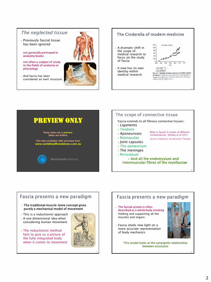
The fascial system is often described as a whole body stocking
- holding and supporting all the muscles and organs
Fascia sheds new light on a more accurate representation of body mechanics
This model looks at the synergistic relationship between structures
12
3
A myofascial approach is a more general system approach applied to posture and movement
It is used to describe and explain whole body
wide connections, linkages and function “When one part moves, the body as a whole
responds
Functionally the only tissue that can mediate such responsiveness is the connective tissue
13
PREVIEW ONLY
These notes are a preview.
Slides are limited.
Full notes available after purchase from
www.worldhealthwebinars.com.au
Suite 3, 104 Spofforth Street Tel: (02) 8969 6300 ‘[email protected]
Cremorne, 2090, Sydney, NSW ‘www.spinalsynergy.com.au
14
The word myofascia describes:
The bundled together inseparable nature of muscle tissue (myo)
And its accompanying web of connective tissue (fascia)
15
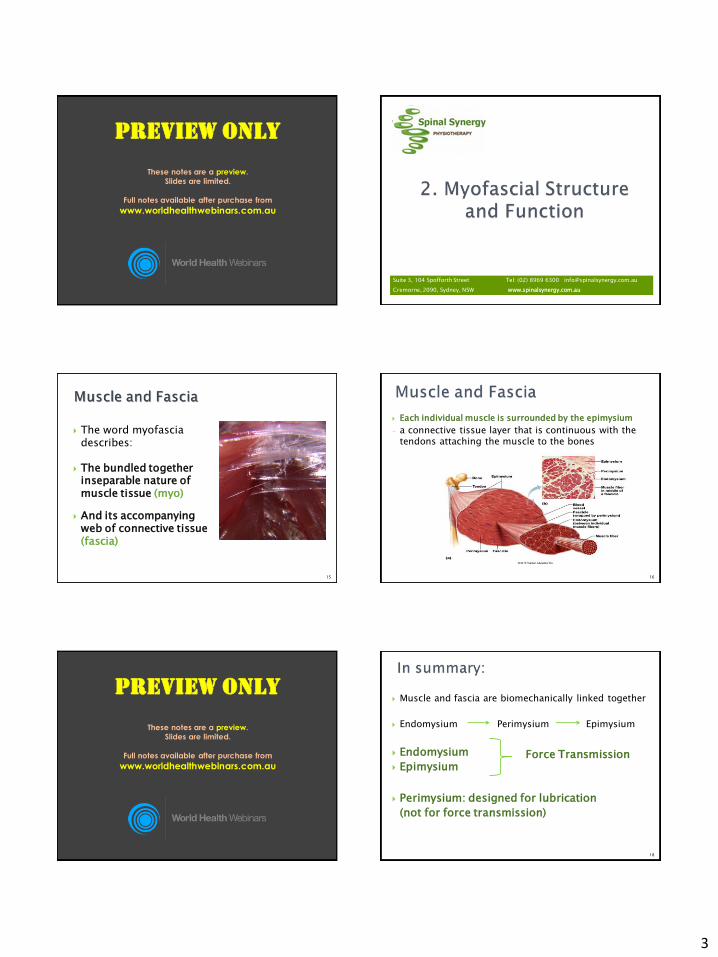
Each individual muscle is surrounded by the epimysium
- a connective tissue layer that is continuous with the tendons attaching the muscle to the bones
16
The perimysium is a continuous network of connective tissue which divides the muscle up into fascicles or muscle fibre bundles
Within each fascicle or muscle fibre bundle, the endomysium is a continuous network of connective tissue that separates individual muscle fibres
17
PREVIEW ONLY
These notes are a preview.
Slides are limited.
Full notes available after purchase from
www.worldhealthwebinars.com.au
Muscle and fascia are biomechanically linked together
Endomysium Perimysium Epimysium
Endomysium
Epimysium
Perimysium: designed for lubrication
(not for force transmission)
Force Transmission
18
4
These connective tissue layers are composed of collagen fibres and elastin fibres
A matrix of hydrated proteoglycans mechanically links the collagen fibre networks in these structures
19
The collagen fibres are mechanically stabilized by the formation of cross-links
The cross-links are essential for the mechanical strength and stiffness of the collagen fibres
Without them the collagen molecules would slide past each other under load and the fibres would have no strength
20
In real bodies, muscles hardly ever transmit their full force directly via tendons into the skeleton
Myofascial continuity does not begin or end with insertions, origins, or bones
A standard muscle-tendon depiction of muscle anatomy
21
PREVIEW ONLY
These notes are a preview.
Slides are limited.
Full notes available after purchase from
www.worldhealthwebinars.com.au
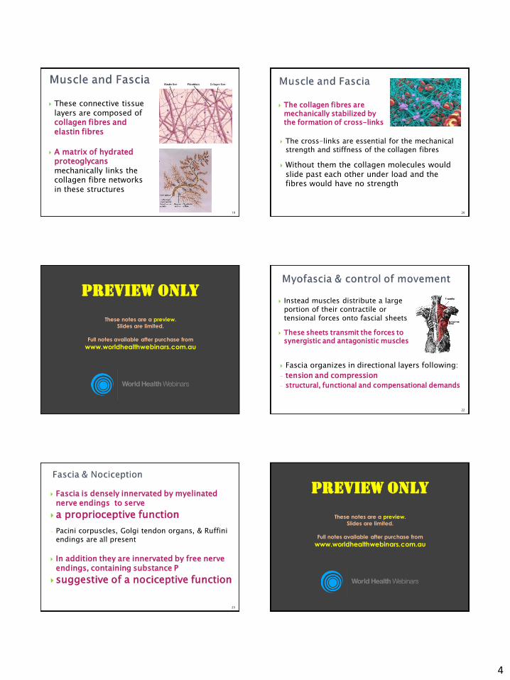
Instead muscles distribute a large portion of their contractile or tensional forces onto fascial sheets
These sheets transmit the forces to synergistic and antagonistic muscles
Fascia organizes in directional layers following:
- tension and compression
- structural, functional and compensational demands
22
Fascia is densely innervated by myelinated nerve endings to serve
a proprioceptive function
- Pacini corpuscles, Golgi tendon organs, & Ruffini endings are all present
In addition they are innervated by free nerve endings, containing substance P
suggestive of a nociceptive function
23
Micro-tearing and/or inflammation of fascia can be a direct source of musculoskeletal pain
New findings suggest that nociceptive activity of epimysial fasciae play a major role in delayed onset muscle soreness (DOMS) subsequent to repetitive eccentric exercise
Gibson et al, 2009
Antonio Stecco from the Fascial Manipulation © method proposes that:
Stretching fascia activates specific patterns of proprioceptors resulting in a perception of motor direction
24
PREVIEW ONLY
These notes are a preview.
Slides are limited.
Full notes available after purchase from
www.worldhealthwebinars.com.au

5
Patients with FM show evidence of inflammatory mediators in the intramuscular connective tissue
- Inflammatory markers were primarily found in the interstitial tissue between the muscle fibres
- Similar to muscles strained by eccentric muscle action
The connective tissue surrounding the muscles, not the muscle itself, may be the source of peripheral nociceptive input
There may be a dysfunctional healing process of the fascia in fibromyalgia
Liptan, 2010 25
Suite 3, 104 Spofforth Street Tel: (02) 8969 6300 ‘[email protected]
Cremorne, 2090, Sydney, NSW ‘www.spinalsynergy.com.au
26
Draws on work from several sources each with its own method:
Thomas Myers (author of the Anatomy Trains®)
Antonio Stecco, Julie Ann Day (Fascial Manipulation©)
Robert Schleip (Clinical researcher & Rolfer)
George Kousaleous (CORE Myofascial Therapy)
Leon Chaitow (Osteopath)
Jean-Pierre Barral (Visceral Manipulation)
27
PREVIEW ONLY
These notes are a preview.
Slides are limited.
Full notes available after purchase from
www.worldhealthwebinars.com.au
Many of the myofascial release approaches originate from the ideas of Dr Ida Rolf
- Structural Integration (mid-1940’s)
Rather than simply working on symptoms she focused on the relationship of parts to the whole
Ida Rolf gave new insight into the role of connective tissue and considered how this related to:
Structure and function of the human body
28
Rolf evolved a series and sequence of manipulations known as the 10-series
The treatment became “Structural Integration”
More commonly Rolfing practitioners are known as Rolfer’s
Differing schools of Structural Integration all have roots directly or indirectly in Rolfing
29
PREVIEW ONLY
These notes are a preview.
Slides are limited.
Full notes available after purchase from
www.worldhealthwebinars.com.au
Thomas Myers presents a model of:
Functional Interconnectedness The Anatomy Trains
present a 'longitudinal' view of myofascial anatomy
The Anatomy Trains maps the major structural and functional continuities in the body's fascial net
30
6
Superficial Back Line
Superficial Front Line
Lateral Line
Spiral Line
The Arm Lines
The Functional Lines
The Deep Front Line
Thomas Myers describes 12 sets of distinct lines of dissectible myofascial connections
The Myofascial Meridians or Anatomy Trains
31
The myofascial meridian lines are not acupuncture meridians
They are lines of pull based on standard Western anatomy
Each meridian describes one very precise line of pull through the body
32
The myofascial meridians follow the body’s connective tissue fabric
Each myofascial meridian has a function beyond each individual muscle within it
The Spiral Line Key principle of this method is:
Isolation versus Integration
33
PREVIEW ONLY
These notes are a preview.
Slides are limited.
Full notes available after purchase from
www.worldhealthwebinars.com.au
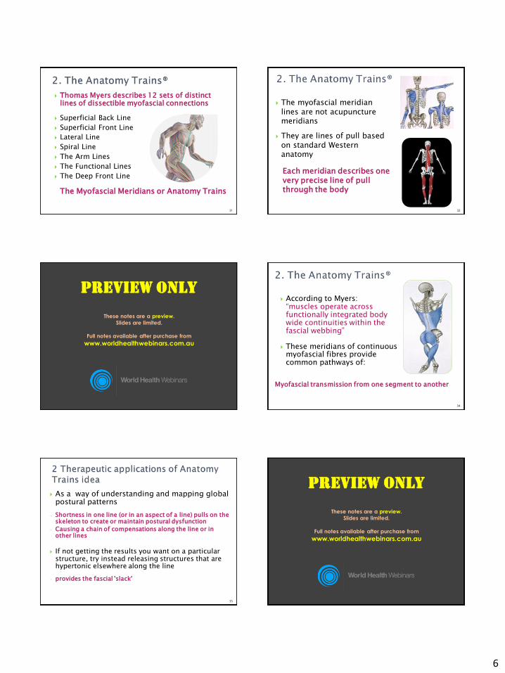
According to Myers: “muscles operate across functionally integrated body wide continuities within the fascial webbing”
These meridians of continuous myofascial fibres provide common pathways of:
Myofascial transmission from one segment to another
34
As a way of understanding and mapping global postural patterns
- Shortness in one line (or in an aspect of a line) pulls on the skeleton to create or maintain postural dysfunction
- Causing a chain of compensations along the line or in other lines
If not getting the results you want on a particular structure, try instead releasing structures that are hypertonic elsewhere along the line
- provides the fascial 'slack'
35
Fascial Manipulation© was developed an Italian physiotherapist Luigi Stecco
The method has evolved over the last 30 years
It presents a biomechanical model
Looks at the role of fascia in musculoskeletal disorders
Extensive dissection by Dr. Antonio Stecco and Dr. Carla Stecco have given a new insight into the histology and anatomy of fascia
36
PREVIEW ONLY
These notes are a preview.
Slides are limited.
Full notes available after purchase from
www.worldhealthwebinars.com.au
7
Histological study of the deep fasciae of the limbs
Expansions of the pectoral girdle muscles onto the brachial fascia
Application of FM® technique in chronic shoulder pain
The ankle retinacula: morphological evidence of the proprioceptive role of the fascial system
The anatomical & functional relation between gluteus maximus and fascia lata
Conservative treatment of carpal tunnel syndrome: comparison between laser therapy and FM®
Mathematical analysis of the flow of hyaluronic acid around fascia during manual therapy motions
Fascial components of the myofascial pain syndrome
22 citations in Pub Med alone
37
A whole series of dissection studies have verified that :
- pectoralis major - biceps brachii and
- palmaris longus muscles
insert expansions into the brachial and antebrachial fascia
and they follow a constant pattern
Stecco et al 2006, 2007, 2008, 2009
38
According to the Fascial Manipulation Method:
- The body can be divided into 14 segments
- Each body segment is served by six myofascial units
- In the Functional Manipulation© the therapist identifies specific points within a fascial sequence
- Deep massage on specific points aims at restoring tensional balance
The mainstay of this manual method lies in the identification of a specific, localised area of the fascia
in connection with a specific limited movement
39
It’s not just about myofascial release
It’s also about training an effective fascial body
1. Fascial Release
2. Fascial Stretch
3. Rebound Elasticity 4. Proprioceptive Refinement
Training of elastic recoil Focused training of the fascia could be
of great importance to athletes, dancers
and other movement advocates
40
PREVIEW ONLY
These notes are a preview.
Slides are limited.
Full notes available after purchase from
www.worldhealthwebinars.com.au
Most sports-related overload injuries occur within elements of the fascia loaded beyond their prepared capacity
If one's fascial body is well trained optimally elastic and resilient
1. then it may be relied on to perform effectively and
2. to offer a high degree of injury prevention
The intention of the fascia oriented training is to influence the matrix renewal via specific training activities which result in a more injury-resistant and resilient ‘body suit’
Movement practices: plyometrics, gyrokinesis, chi running, yoga or martial arts contain elements congruent with Fascial Fitness©
Schleip & Muller, 2013 41
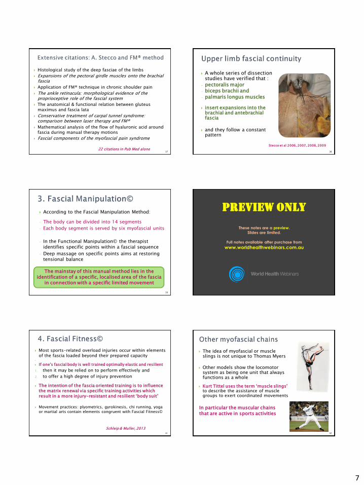
The idea of myofascial or muscle slings is not unique to Thomas Myers
Other models show the locomotor system as being one unit that always functions as a whole
Kurt Tittel uses the term ‘muscle slings’ to describe the assistance of muscle groups to exert coordinated movements
In particular the muscular chains that are active in sports activities
42

8
Suite 3, 104 Spofforth Street Tel: (02) 8969 6300 ‘[email protected]
Cremorne, 2090, Sydney, NSW ‘www.spinalsynergy.com.au
43
Myofascial release (MFR) is the application of a low load, long duration stretch into the myofascial complex
Intended to: 1. restore the optimal length
of this complex 2. decrease pain 3. and improve function
44
PREVIEW ONLY
These notes are a preview.
Slides are limited.
Full notes available after purchase from
www.worldhealthwebinars.com.au
Myofascial Release Other STW
Requires understanding of body wide fascial matrix, functional anatomy
Cultural basis: TCM, Thai-massage, Swedish style, Aromatherapy
Based on fascial anatomy Non-specific, ‘massage by numbers’ -Remedial, relaxation massage
Analysis of postural & movement patterns within a fascial web
General postural holding i.e. Upper cross syndrome (Janda)
Often treat away from pain areas Less emphasis on ‘knots’, TPs
Focuses on tender/tight area (local) Overemphasis on TPs
Lubricant: little to none Oils, ointments
Application: Deep, firm, slow Variable speed, depth
Direction: follows lines of fascia Variable strokes & direction
Involves some dynamic components while applying release technique
Patient, client passive Limited patient interaction
Fully integrated with rehabilitation, functional loading & strengthening fascia
Part of rehab but primarily a passive treatment intervention
45
An accurate analysis of the myofascial connections based on an understanding of fascial anatomy can provide indications as to where it is best to intervene
The localisation of precise points or key areas can render manipulation more effective
46
By restoring the length and health of restricted connective tissue
Pressure can be relieved on pain sensitive structures such as nerves and blood vessels
The rationale for these techniques can be traced to studies that looked at:
- the plastic
- viscoelastic and
- piezoelectric properties of connective tissue
47
PREVIEW ONLY
These notes are a preview.
Slides are limited.
Full notes available after purchase from
www.worldhealthwebinars.com.au
A recent study looked at repetitive motion strain as a modelled injury and demonstrated:
- enhanced apoptosis activity - and loss of intercellular integrity
However during treatment with MFR following repetitive strain injury resulted in:
1. normalization in apoptotic rate 2. cell morphology changes 3. and reorientation of fibroblasts
(Meltzer et al., 2010) 48
9
Under normal conditions, fascia tends to move with minimal restrictions
Injuries resulting from physical trauma, repetitive strain injury, and inflammation are thought to:
1. decrease fascia tissue length
2. and elasticity
3. resulting in fascial restriction
It is possible that pain relief due to myofascial release is secondary to returning the fascial tissue to its normal length
Schleip (2003) 49
PREVIEW ONLY
These notes are a preview.
Slides are limited.
Full notes available after purchase from
www.worldhealthwebinars.com.au
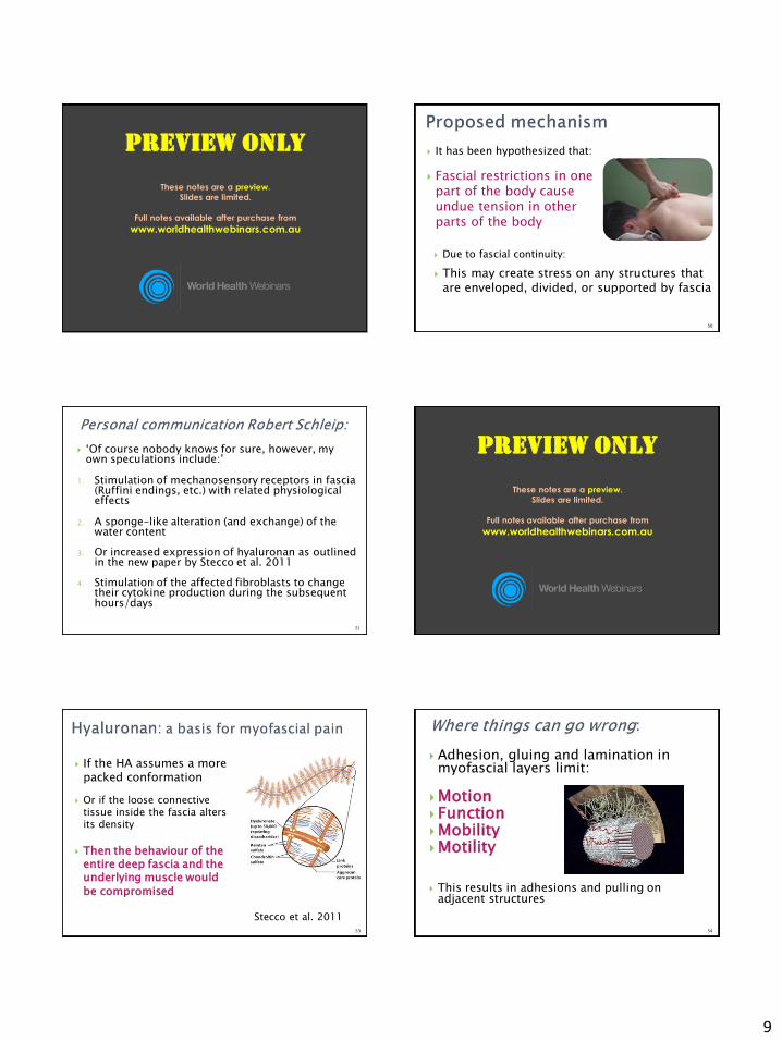
It has been hypothesized that:
Fascial restrictions in one part of the body cause undue tension in other parts of the body
Due to fascial continuity:
This may create stress on any structures that are enveloped, divided, or supported by fascia
50
‘Of course nobody knows for sure, however, my own speculations include:’
1. Stimulation of mechanosensory receptors in fascia (Ruffini endings, etc.) with related physiological effects
2. A sponge-like alteration (and exchange) of the water content
3. Or increased expression of hyaluronan as outlined in the new paper by Stecco et al. 2011
4. Stimulation of the affected fibroblasts to change their cytokine production during the subsequent hours/days
51
A study of fresh non-embalmed cadavers showed that:
1. The deep fascia presented a layer of hyaluronan (HA) between fascia & muscle and
2. Within the loose connective tissue it divided different fibrous sub-layers of the deep fascia
The hyaluronan (HA) within the deep fascia facilitates:
1. the free sliding of two adjacent fibrous fascial layers
2. promotes normal function associated with the deep fascia
Stecco et al. 2011
52
PREVIEW ONLY
These notes are a preview.
Slides are limited.
Full notes available after purchase from
www.worldhealthwebinars.com.au
If the HA assumes a more packed conformation
Or if the loose connective tissue inside the fascia alters its density
Then the behaviour of the entire deep fascia and the underlying muscle would
be compromised
Stecco et al. 2011 53
Adhesion, gluing and lamination in myofascial layers limit:
Motion Function Mobility Motility This results in adhesions and pulling on
adjacent structures
54
10
Non-physiological alteration of deep fascia could cause tensional changes along a related fascial sequence
Resulting in
- incorrect activation of nerve receptors
- uncoordinated movements
- and consequent nociceptive afferents
Myofascial release is aimed at: 1. Restoring the length and health of restricted
connective tissue
2. Altering proprioception & nociceptive outflow 3. Facilitate normal movement patterns
55
PREVIEW ONLY
These notes are a preview.
Slides are limited.
Full notes available after purchase from
www.worldhealthwebinars.com.au
Suite 3, 104 Spofforth Street Tel: (02) 8969 6300 ‘[email protected]
Cremorne, 2090, Sydney, NSW Fax: (02) 8969 6311 ‘www.spinalsynergy.com.au 56
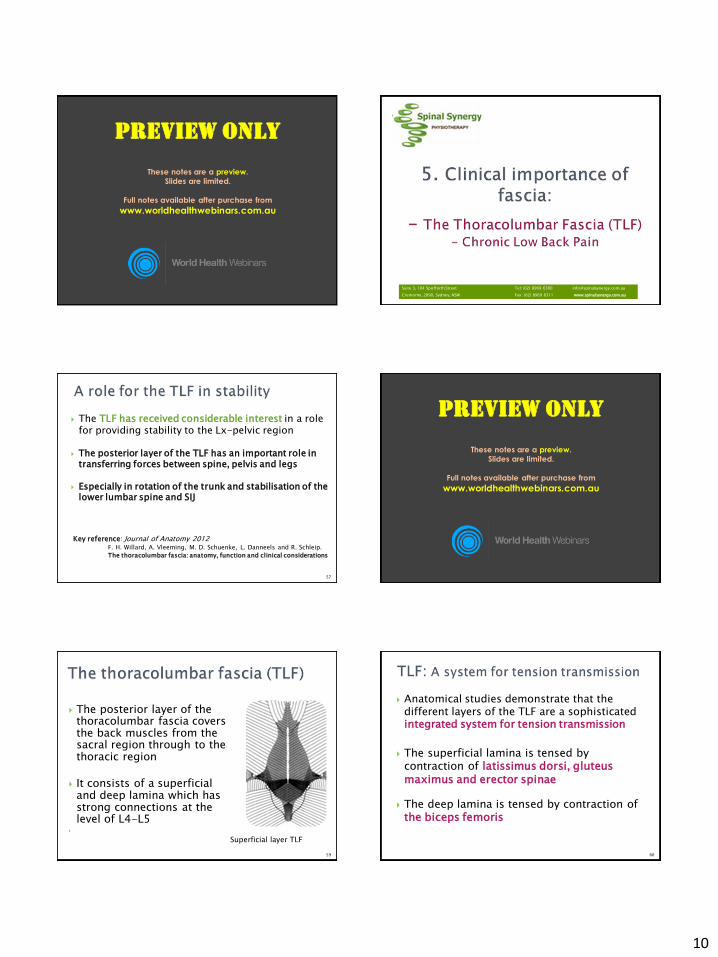
The TLF has received considerable interest in a role for providing stability to the Lx-pelvic region
The posterior layer of the TLF has an important role in transferring forces between spine, pelvis and legs
Especially in rotation of the trunk and stabilisation of the lower lumbar spine and SIJ
Key reference: Journal of Anatomy 2012 F. H. Willard, A. Vleeming, M. D. Schuenke, L. Danneels and R. Schleip.
The thoracolumbar fascia: anatomy, function and clinical considerations
57
Gluteus maximus, latissimus dorsi and biceps femoris are functionally coupled via the TLF
This allows for effective load transfer between spine, pelvis, legs and arms as a so-called integrated system
In dissection studies traction to these muscles caused a displacement of the posterior layer
Vleeming et al 1995: The posterior layer of the thoracolumbar fascia; function in load transfer from spine to legs. Spine
58
PREVIEW ONLY
These notes are a preview.
Slides are limited.
Full notes available after purchase from
www.worldhealthwebinars.com.au
The posterior layer of the thoracolumbar fascia covers the back muscles from the sacral region through to the thoracic region
It consists of a superficial and deep lamina which has strong connections at the level of L4-L5
Superficial layer TLF
59
Anatomical studies demonstrate that the different layers of the TLF are a sophisticated integrated system for tension transmission
The superficial lamina is tensed by contraction of latissimus dorsi, gluteus maximus and erector spinae
The deep lamina is tensed by contraction of the biceps femoris
60
11
Energizes the posterior oblique muscular sling
Connects the trunk to the pelvis and to the lower limbs
61
It has been hypothesized that in chronic LBP the connective tissue of the back is thicker and more disorganized as a result of:
- Remodelling
- Chronic inflammation
- Fibrosis - &/or fatty infiltration
Fascia displays visco-elastic properties and has been reported to stiffen with successive loading
And adaptive fascial thickening is possible
Barker & Briggs (1999)
62
PREVIEW ONLY
These notes are a preview.
Slides are limited.
Full notes available after purchase from
www.worldhealthwebinars.com.au
Ultra-sound study: evidence of altered lumbar connective tissue structure in patients with CLBP
Thickness and echogenicity of the combined subcutaneous and perimuscular zone were significantly greater in the LBP group
Patients with LBP had on average 25% greater perimuscular connective tissue thickening in the lumbar region than subjects with no-LBP after adjusting for BMI
Langevin et al 2009
63
A further study showed that the TLF shear strain was 20% lower in subjects with CLBP
There was no evidence that this difference was sex-specific although overall males had significantly lower shear strain than females
Reduced shear plane motion may be due abnormal trunk movement patterns &/or intrinsic connective tissue pathology
Langevin et al 2011
64
Single trauma or cumulative microtrauma causes sub-failure injuries of ligaments & mechanoreceptors
1. Injured mechanoreceptors generate corrupted transducer signals
2. This leads to corrupted muscle response patterns by the neuromuscular control unit
3. This leads to inefficient posture control
4. Poor balance & proprioception deficits
Panjabi M, European Spine Journal 2006
65
PREVIEW ONLY
These notes are a preview.
Slides are limited.
Full notes available after purchase from
www.worldhealthwebinars.com.au
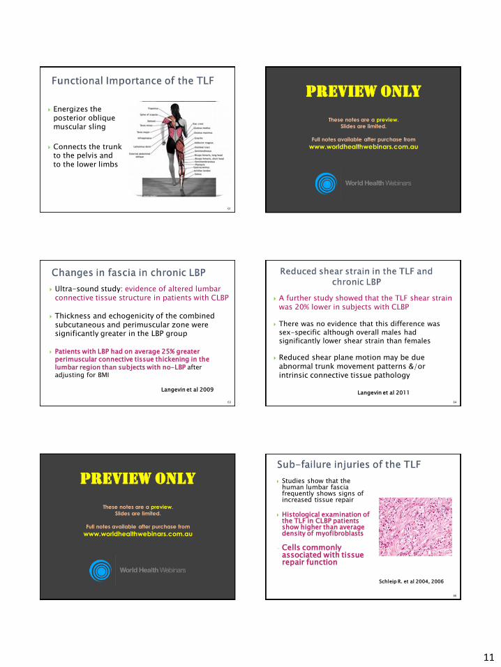
Studies show that the human lumbar fascia frequently shows signs of increased tissue repair
Histological examination of the TLF in CLBP patients show higher than average density of myofibroblasts
- Cells commonly associated with tissue repair function
Schleip R. et al 2004, 2006
66

12
Various soft-tissue treatments are directed to the muscle areas adjacent to the TLF
Exercise interventions for LBP rely on the functional link the TLF provides between trunk and limbs
Through its expansive fascial connections to deeper layers of fascia the TLF has an important role to play in providing stability:
1. to the lumbo-pelvic region but also
2. to integrate the pelvis & rib-cage
67
Myofascial Release provides a new paradigm challenging long-held beliefs about anatomy, biomechanics & human movement
Histological studies are beginning to provide a better understanding of the physiological function of fascia
Histological studies also show that fascia is adaptive – this has clinical implications
Myofascial Release is having fundamental impact on manual therapy as seen by its widespread use by a wide range of professions
68
PREVIEW ONLY
These notes are a preview.
Slides are limited.
Full notes available after purchase from
www.worldhealthwebinars.com.au
69
Thank you
World Health Webinars
http://worldhealthwebinars.com.au
Coming up next