Embed Size (px)
Citation preview
15th Symposium on the
Practical Applications of
Mass Spectrometry in the
Biotechnology Industry
(Mass Spec 2018)
Symposium Co-chairs:
Michael Boyne, COUR Pharmaceuticals
Development Company
Richard Rogers, Just Biotherapeutics
September 9-12, 2018
Hilton San Francisco Union Square
San Francisco, CA USA
Organized by
2
Table of Contents
Welcome Letter ................................................................................................. 3
Student Travel Grants ....................................................................................... 4
Program, Exhibitor and Media Partners ........................................................... 5
Scientific Program Summary ............................................................................ 8
Oral Abstracts ................................................................................................... 15
Workshop Abstracts .......................................................................................... 37
Roundtable Discussion Topics .......................................................................... 39
Technical Seminar Abstracts ............................................................................ 41
List of Posters .................................................................................................... 47
3
Welcome to the 15th Symposium on the Practical Applications of
Mass Spectrometry in the Biotechnology Industry We are pleased to welcome you to the 15th Symposium on the Practical Application of Mass
Spectrometry in the Biotechnology Industry. The focus of this Symposium is the application of mass
spectrometry (MS) for product characterization, process monitoring, formulation development and
release testing in the pharmaceutical industry. Since mass spectrometry is used routinely for a wide
array of applications, the meeting will provide scientists in the industry an opportunity to share their
data and learn from their colleagues. Although most of the applications may deal with
biopharmaceuticals (proteins, DNA, viral vectors), applications for conventional pharmaceuticals
will also be discussed. The Symposium will be held over a period of two and a half days devoted to
practical concerns in the use of MS within the biotechnology and pharmaceutical industries.
The success of this symposium will depend not only on our experienced and knowledgeable speakers
and leaders but also on the interactions and open discussion that take place among the attendees. We
encourage you to participate wholeheartedly in the discussion sections and roundtable sessions that
have been designed to stimulate the exchange of ideas and information.
We would like to thank the speakers who are giving generously of their time and resources, and you
for your attendance, which will make this a successful endeavor.
We gratefully acknowledge the generosity of our program partners and exhibitors. Thank you to
AbbVie, Inc., Agilent Technologies, Biogen, Bioinformatics Solutions, Inc., Bruker Daltonics, Inc.,
Charles River Laboratories, Eli Lilly and Company, Genedata Inc., Genentech, a Member of the
Roche Group, GenNext Technologies, Inc., Genovis AB, Merck & Co., Inc., Pfizer, Inc., Protein
Metrics Inc., Roche Diagnostics GmbH, Sanofi, SCIEX, Shimadzu Corporation, Thermo Fisher
Scientific, Waters Corporation and YMC America, Inc. We are also thankful for the expert assistance
and support of CASSS, as well as the audiovisual expertise of Michael Johnstone from MJ Audio-
Visual Productions.
We hope you enjoy the conference, build new contacts and return for new information in 2019!
THE ORGANIZING COMMITTEE:
Michael Boyne*, COUR Pharmaceuticals
Development Company
Steven Cohen, SAC Analytical Consultants
Terry Cyr, Health Canada
Ingo Lindner, Roche Diagnostics GmbH
Anders Lund, Synlogic
Yelena Lyubarskaya, Sanofi
Frances Namuswe, CDER, FDA
David Passmore, RubrYc Therapeutics
Richard Rogers*, Just Biotherapeutics
Sarah Rogstad, CDER, FDA
Jason Rouse, Pfizer, Inc.
Arjen Scholten, Janssen Vaccines and
Prevention
John Valliere-Douglass, Seattle Genetics, Inc.
Christopher Yu, Genentech, a Member of the
Roche Group
*Symposium Co-chair
4
CASSS Mass Spec Student Travel Grants
CASSS is pleased to provide a limited number of student travel grants for PhD students and post-
doctoral fellows who present applicable posters at the 15th Symposium on the Practical
Applications of Mass Spectrometry in the Biotechnology Industry (Mass Spec 2018). PhD
students or post-doctoral fellows conducting research at academic institutions throughout the
world are eligible.
This symposium offers insight to current topics and issues under discussion within the biotech
and biopharmaceutical industries, and as such, provides an opportunity to bridge between
industry, academia, and regulatory agencies. The presentations and workshops are focused on the
application of mass spectrometry to advance drug discovery and development in the
biotechnology industry. Topics will include the utility of MS as an alternative to conventional
assays such as peptide mapping, ion exchange, capillary electrophoresis, as well as for the
analysis of process-related impurities, such as host cell proteins; introduction of MS in the QC
laboratory, validation/transfer/compliance strategy, system suitability/assay acceptance criteria;
expectations from regulatory agencies for MS based assays for product characterization, release
and stability; technical challenges regarding quantitation, ionization, higher order structure
analysis, etc.; applications of MS for high throughput screening of cell-lines, raw materials, in-
process samples; and benchmarking of new technology.
Requirements are:
- Present a poster on a MS topic
- Proof of studentship/post-doc status
- Recommendation from the supervisor/advisor
CASSS has awarded student travel grants to the following individuals:
in-vivo Characterization of Antibodies Using a Nano-HPLC –MS/MS Approach
Annika Doell, University Duisburg-Essen, Germany
Reversible Self-association (RSA): A Stumbling Block for Developing High Concentration
IgG1 mAb Formulations
Yue (Martin) Hu, University of Kansas, USA
5
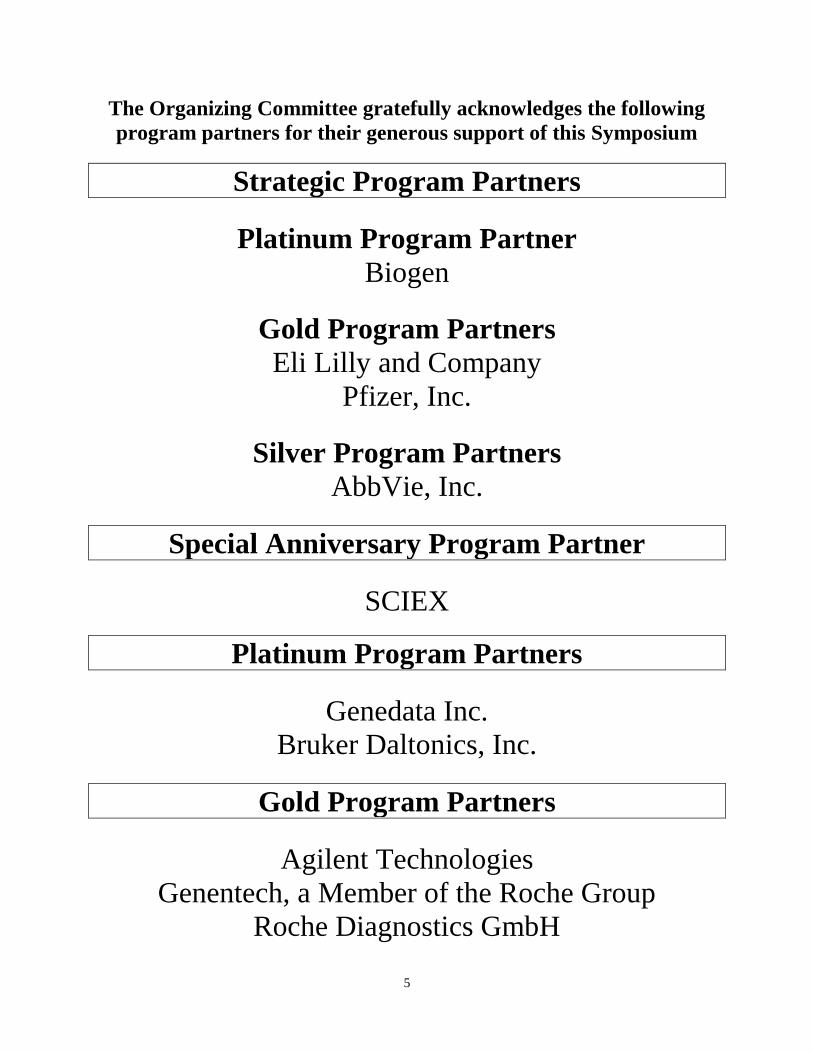
The Organizing Committee gratefully acknowledges the following
program partners for their generous support of this Symposium
Strategic Program Partners
Platinum Program Partner
Biogen
Gold Program Partners
Eli Lilly and Company
Pfizer, Inc.
Silver Program Partners
AbbVie, Inc.
Special Anniversary Program Partner
SCIEX
Platinum Program Partners
Genedata Inc.
Bruker Daltonics, Inc.
Gold Program Partners
Agilent Technologies
Genentech, a Member of the Roche Group
Roche Diagnostics GmbH
6
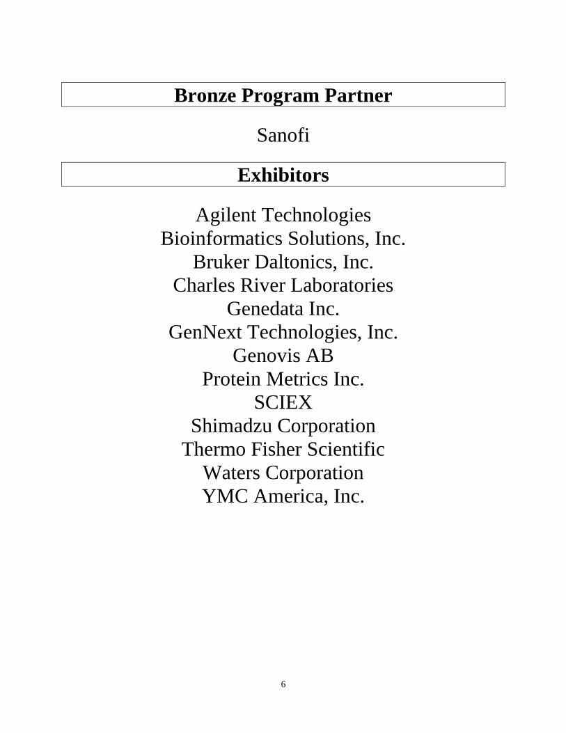
Bronze Program Partner
Sanofi
Exhibitors
Agilent Technologies
Bioinformatics Solutions, Inc.
Bruker Daltonics, Inc.
Charles River Laboratories
Genedata Inc.
GenNext Technologies, Inc.
Genovis AB
Protein Metrics Inc.
SCIEX
Shimadzu Corporation
Thermo Fisher Scientific
Waters Corporation
YMC America, Inc.
7
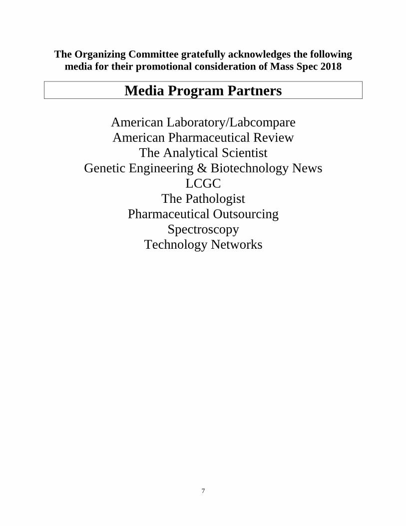
The Organizing Committee gratefully acknowledges the following
media for their promotional consideration of Mass Spec 2018
Media Program Partners
American Laboratory/Labcompare
American Pharmaceutical Review
The Analytical Scientist
Genetic Engineering & Biotechnology News
LCGC
The Pathologist
Pharmaceutical Outsourcing
Spectroscopy
Technology Networks
8
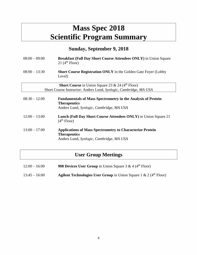
Mass Spec 2018
Scientific Program Summary
Sunday, September 9, 2018
08:00 – 09:00 Breakfast (Full Day Short Course Attendees ONLY) in Union Square
21 (4th Floor)
08:00 – 13:30 Short Course Registration ONLY in the Golden Gate Foyer (Lobby
Level)
Short Course in Union Square 23 & 24 (4th Floor)
Short Course Instructor: Anders Lund, Synlogic, Cambridge, MA USA
08:30 – 12:00 Fundamentals of Mass Spectrometry in the Analysis of Protein
Therapeutics
Anders Lund, Synlogic, Cambridge, MA USA
12:00 – 13:00 Lunch (Full Day Short Course Attendees ONLY) in Union Square 21
(4th Floor)
13:00 – 17:00 Applications of Mass Spectrometry to Characterize Protein
Therapeutics
Anders Lund, Synlogic, Cambridge, MA USA
User Group Meetings
12:00 – 16:00 908 Devices User Group in Union Square 3 & 4 (4th Floor)
13:45 – 16:00 Agilent Technologies User Group in Union Square 1 & 2 (4th Floor)
9
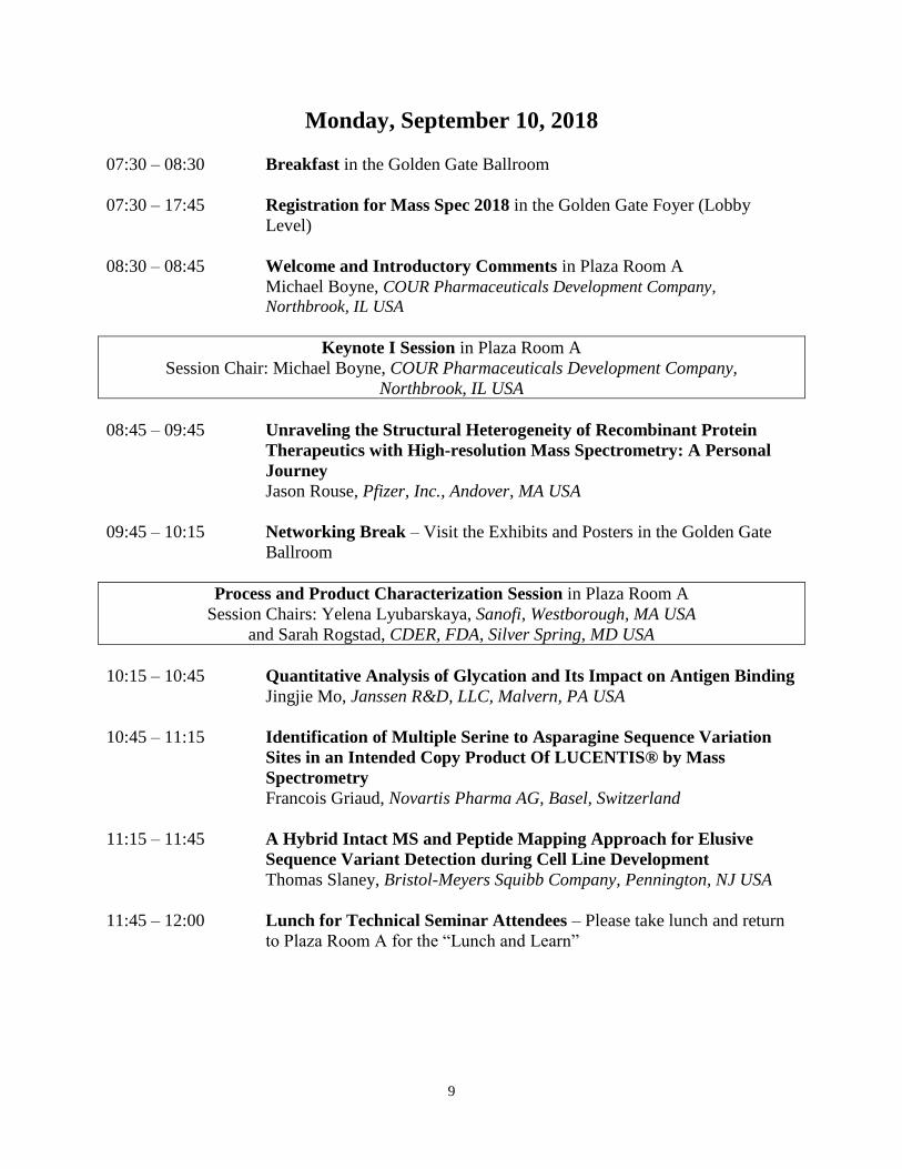
Monday, September 10, 2018
07:30 – 08:30 Breakfast in the Golden Gate Ballroom
07:30 – 17:45 Registration for Mass Spec 2018 in the Golden Gate Foyer (Lobby
Level)
08:30 – 08:45 Welcome and Introductory Comments in Plaza Room A
Michael Boyne, COUR Pharmaceuticals Development Company,
Northbrook, IL USA
Keynote I Session in Plaza Room A
Session Chair: Michael Boyne, COUR Pharmaceuticals Development Company,
Northbrook, IL USA
08:45 – 09:45 Unraveling the Structural Heterogeneity of Recombinant Protein
Therapeutics with High-resolution Mass Spectrometry: A Personal
Journey
Jason Rouse, Pfizer, Inc., Andover, MA USA
09:45 – 10:15 Networking Break – Visit the Exhibits and Posters in the Golden Gate
Ballroom
Process and Product Characterization Session in Plaza Room A
Session Chairs: Yelena Lyubarskaya, Sanofi, Westborough, MA USA
and Sarah Rogstad, CDER, FDA, Silver Spring, MD USA
10:15 – 10:45 Quantitative Analysis of Glycation and Its Impact on Antigen Binding
Jingjie Mo, Janssen R&D, LLC, Malvern, PA USA
10:45 – 11:15 Identification of Multiple Serine to Asparagine Sequence Variation
Sites in an Intended Copy Product Of LUCENTIS® by Mass
Spectrometry
Francois Griaud, Novartis Pharma AG, Basel, Switzerland
11:15 – 11:45 A Hybrid Intact MS and Peptide Mapping Approach for Elusive
Sequence Variant Detection during Cell Line Development
Thomas Slaney, Bristol-Meyers Squibb Company, Pennington, NJ USA
11:45 – 12:00 Lunch for Technical Seminar Attendees – Please take lunch and return
to Plaza Room A for the “Lunch and Learn”
10
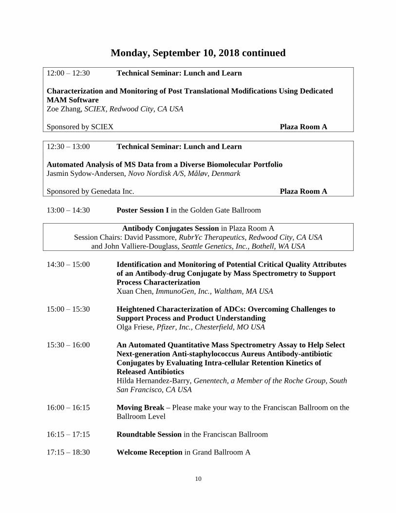
Monday, September 10, 2018 continued
12:00 – 12:30 Technical Seminar: Lunch and Learn
Characterization and Monitoring of Post Translational Modifications Using Dedicated
MAM Software
Zoe Zhang, SCIEX, Redwood City, CA USA
Sponsored by SCIEX Plaza Room A
12:30 – 13:00 Technical Seminar: Lunch and Learn
Automated Analysis of MS Data from a Diverse Biomolecular Portfolio
Jasmin Sydow-Andersen, Novo Nordisk A/S, Måløv, Denmark
Sponsored by Genedata Inc. Plaza Room A
13:00 – 14:30 Poster Session I in the Golden Gate Ballroom
Antibody Conjugates Session in Plaza Room A
Session Chairs: David Passmore, RubrYc Therapeutics, Redwood City, CA USA
and John Valliere-Douglass, Seattle Genetics, Inc., Bothell, WA USA
14:30 – 15:00 Identification and Monitoring of Potential Critical Quality Attributes
of an Antibody-drug Conjugate by Mass Spectrometry to Support
Process Characterization
Xuan Chen, ImmunoGen, Inc., Waltham, MA USA
15:00 – 15:30 Heightened Characterization of ADCs: Overcoming Challenges to
Support Process and Product Understanding
Olga Friese, Pfizer, Inc., Chesterfield, MO USA
15:30 – 16:00 An Automated Quantitative Mass Spectrometry Assay to Help Select
Next-generation Anti-staphylococcus Aureus Antibody-antibiotic
Conjugates by Evaluating Intra-cellular Retention Kinetics of
Released Antibiotics
Hilda Hernandez-Barry, Genentech, a Member of the Roche Group, South
San Francisco, CA USA
16:00 – 16:15 Moving Break – Please make your way to the Franciscan Ballroom on the
Ballroom Level
16:15 – 17:15 Roundtable Session in the Franciscan Ballroom
17:15 – 18:30 Welcome Reception in Grand Ballroom A
11
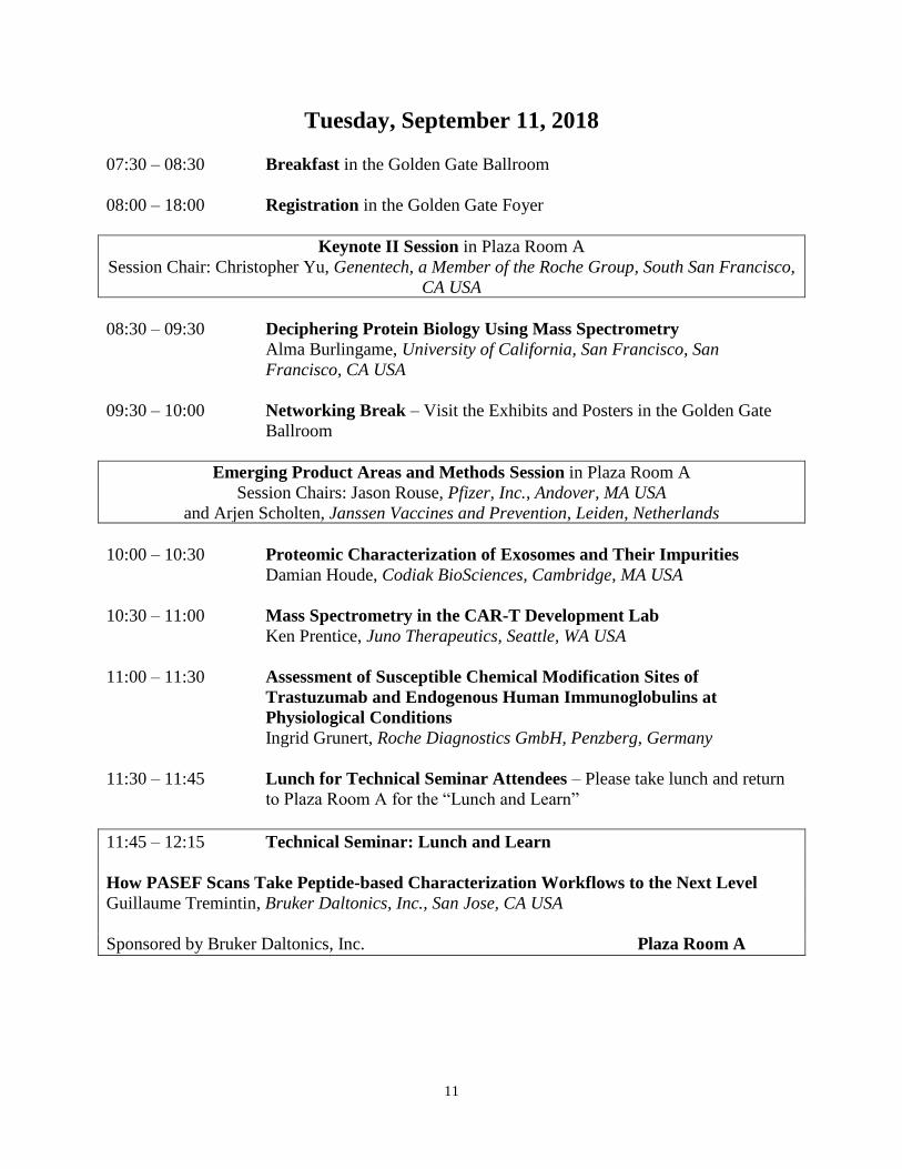
Tuesday, September 11, 2018
07:30 – 08:30 Breakfast in the Golden Gate Ballroom
08:00 – 18:00 Registration in the Golden Gate Foyer
Keynote II Session in Plaza Room A
Session Chair: Christopher Yu, Genentech, a Member of the Roche Group, South San Francisco,
CA USA
08:30 – 09:30 Deciphering Protein Biology Using Mass Spectrometry
Alma Burlingame, University of California, San Francisco, San
Francisco, CA USA
09:30 – 10:00 Networking Break – Visit the Exhibits and Posters in the Golden Gate
Ballroom
Emerging Product Areas and Methods Session in Plaza Room A
Session Chairs: Jason Rouse, Pfizer, Inc., Andover, MA USA
and Arjen Scholten, Janssen Vaccines and Prevention, Leiden, Netherlands
10:00 – 10:30 Proteomic Characterization of Exosomes and Their Impurities
Damian Houde, Codiak BioSciences, Cambridge, MA USA
10:30 – 11:00 Mass Spectrometry in the CAR-T Development Lab
Ken Prentice, Juno Therapeutics, Seattle, WA USA
11:00 – 11:30 Assessment of Susceptible Chemical Modification Sites of
Trastuzumab and Endogenous Human Immunoglobulins at
Physiological Conditions
Ingrid Grunert, Roche Diagnostics GmbH, Penzberg, Germany
11:30 – 11:45 Lunch for Technical Seminar Attendees – Please take lunch and return
to Plaza Room A for the “Lunch and Learn”
11:45 – 12:15 Technical Seminar: Lunch and Learn
How PASEF Scans Take Peptide-based Characterization Workflows to the Next Level
Guillaume Tremintin, Bruker Daltonics, Inc., San Jose, CA USA
Sponsored by Bruker Daltonics, Inc. Plaza Room A
12
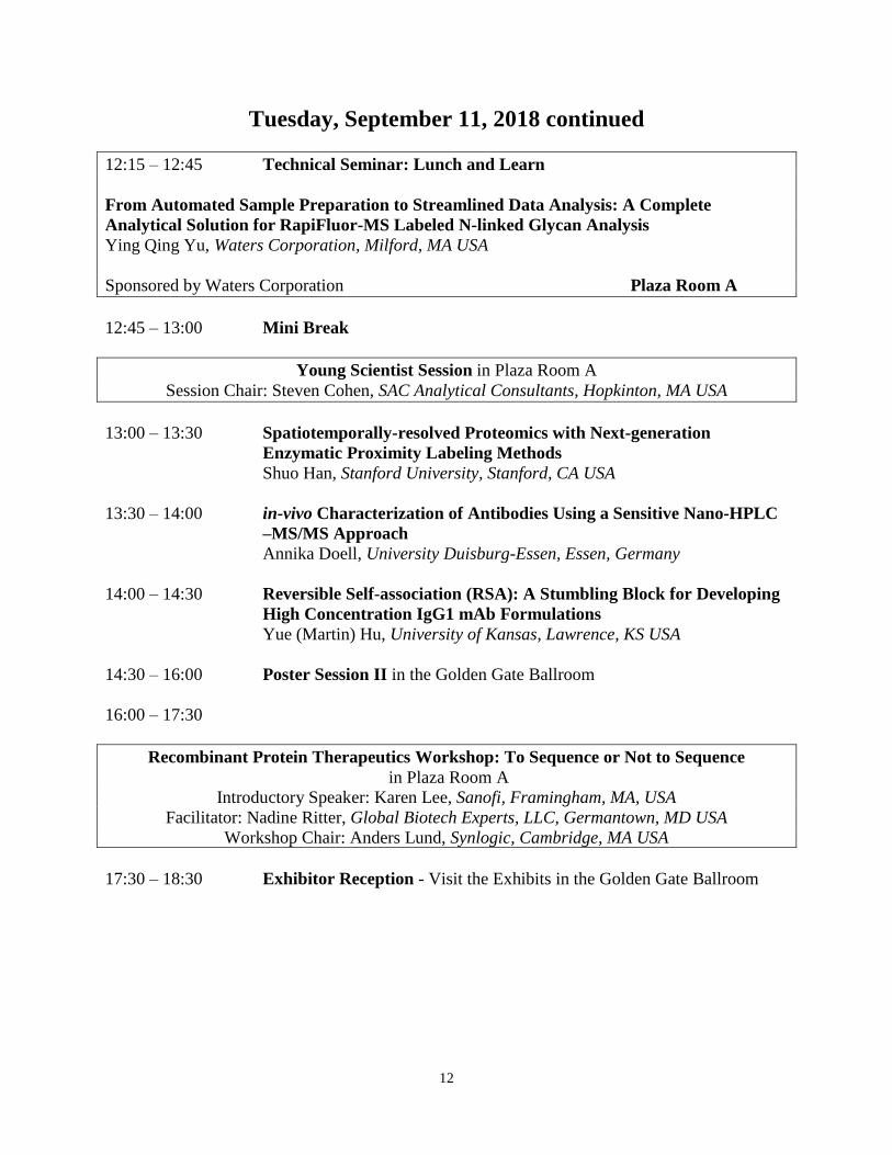
Tuesday, September 11, 2018 continued
12:15 – 12:45 Technical Seminar: Lunch and Learn
From Automated Sample Preparation to Streamlined Data Analysis: A Complete
Analytical Solution for RapiFluor-MS Labeled N-linked Glycan Analysis
Ying Qing Yu, Waters Corporation, Milford, MA USA
Sponsored by Waters Corporation Plaza Room A
12:45 – 13:00 Mini Break
Young Scientist Session in Plaza Room A
Session Chair: Steven Cohen, SAC Analytical Consultants, Hopkinton, MA USA
13:00 – 13:30 Spatiotemporally-resolved Proteomics with Next-generation
Enzymatic Proximity Labeling Methods
Shuo Han, Stanford University, Stanford, CA USA
13:30 – 14:00 in-vivo Characterization of Antibodies Using a Sensitive Nano-HPLC
–MS/MS Approach
Annika Doell, University Duisburg-Essen, Essen, Germany
14:00 – 14:30 Reversible Self-association (RSA): A Stumbling Block for Developing
High Concentration IgG1 mAb Formulations
Yue (Martin) Hu, University of Kansas, Lawrence, KS USA
14:30 – 16:00 Poster Session II in the Golden Gate Ballroom
16:00 – 17:30
Recombinant Protein Therapeutics Workshop: To Sequence or Not to Sequence
in Plaza Room A
Introductory Speaker: Karen Lee, Sanofi, Framingham, MA, USA
Facilitator: Nadine Ritter, Global Biotech Experts, LLC, Germantown, MD USA
Workshop Chair: Anders Lund, Synlogic, Cambridge, MA USA
17:30 – 18:30 Exhibitor Reception - Visit the Exhibits in the Golden Gate Ballroom
13
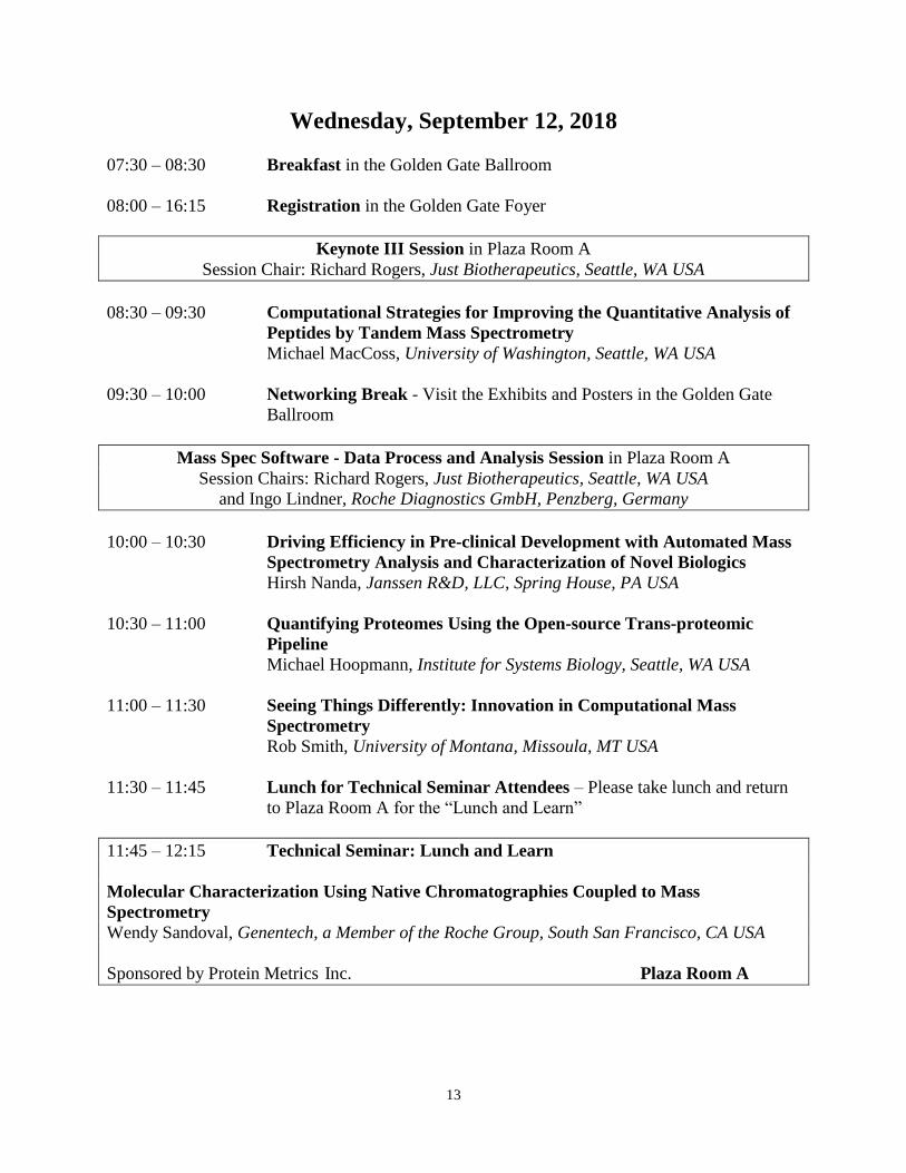
Wednesday, September 12, 2018
07:30 – 08:30 Breakfast in the Golden Gate Ballroom
08:00 – 16:15 Registration in the Golden Gate Foyer
Keynote III Session in Plaza Room A
Session Chair: Richard Rogers, Just Biotherapeutics, Seattle, WA USA
08:30 – 09:30 Computational Strategies for Improving the Quantitative Analysis of
Peptides by Tandem Mass Spectrometry
Michael MacCoss, University of Washington, Seattle, WA USA
09:30 – 10:00 Networking Break - Visit the Exhibits and Posters in the Golden Gate
Ballroom
Mass Spec Software - Data Process and Analysis Session in Plaza Room A
Session Chairs: Richard Rogers, Just Biotherapeutics, Seattle, WA USA
and Ingo Lindner, Roche Diagnostics GmbH, Penzberg, Germany
10:00 – 10:30 Driving Efficiency in Pre-clinical Development with Automated Mass
Spectrometry Analysis and Characterization of Novel Biologics
Hirsh Nanda, Janssen R&D, LLC, Spring House, PA USA
10:30 – 11:00 Quantifying Proteomes Using the Open-source Trans-proteomic
Pipeline
Michael Hoopmann, Institute for Systems Biology, Seattle, WA USA
11:00 – 11:30 Seeing Things Differently: Innovation in Computational Mass
Spectrometry
Rob Smith, University of Montana, Missoula, MT USA
11:30 – 11:45 Lunch for Technical Seminar Attendees – Please take lunch and return
to Plaza Room A for the “Lunch and Learn”
11:45 – 12:15 Technical Seminar: Lunch and Learn
Molecular Characterization Using Native Chromatographies Coupled to Mass
Spectrometry
Wendy Sandoval, Genentech, a Member of the Roche Group, South San Francisco, CA USA
Sponsored by Protein Metrics Inc. Plaza Room A
14
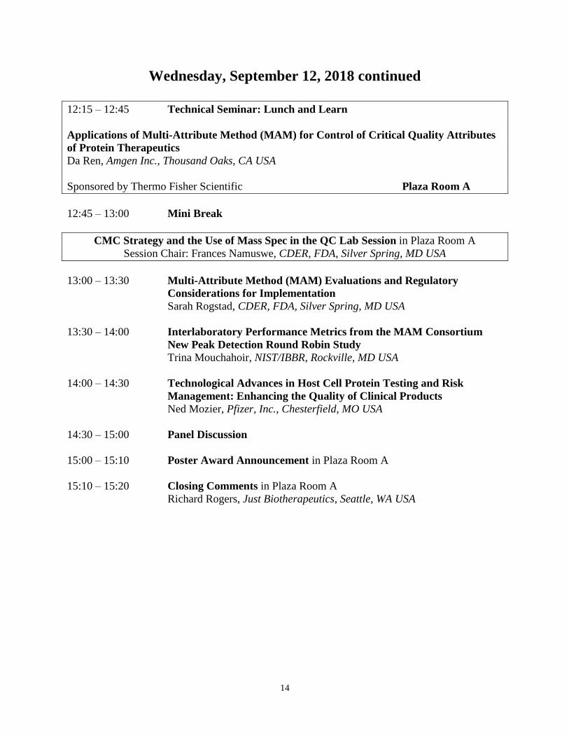
Wednesday, September 12, 2018 continued
12:15 – 12:45 Technical Seminar: Lunch and Learn
Applications of Multi-Attribute Method (MAM) for Control of Critical Quality Attributes
of Protein Therapeutics
Da Ren, Amgen Inc., Thousand Oaks, CA USA
Sponsored by Thermo Fisher Scientific Plaza Room A
12:45 – 13:00 Mini Break
CMC Strategy and the Use of Mass Spec in the QC Lab Session in Plaza Room A
Session Chair: Frances Namuswe, CDER, FDA, Silver Spring, MD USA
13:00 – 13:30 Multi-Attribute Method (MAM) Evaluations and Regulatory
Considerations for Implementation
Sarah Rogstad, CDER, FDA, Silver Spring, MD USA
13:30 – 14:00 Interlaboratory Performance Metrics from the MAM Consortium
New Peak Detection Round Robin Study
Trina Mouchahoir, NIST/IBBR, Rockville, MD USA
14:00 – 14:30 Technological Advances in Host Cell Protein Testing and Risk
Management: Enhancing the Quality of Clinical Products
Ned Mozier, Pfizer, Inc., Chesterfield, MO USA
14:30 – 15:00 Panel Discussion
15:00 – 15:10 Poster Award Announcement in Plaza Room A
15:10 – 15:20 Closing Comments in Plaza Room A
Richard Rogers, Just Biotherapeutics, Seattle, WA USA
15
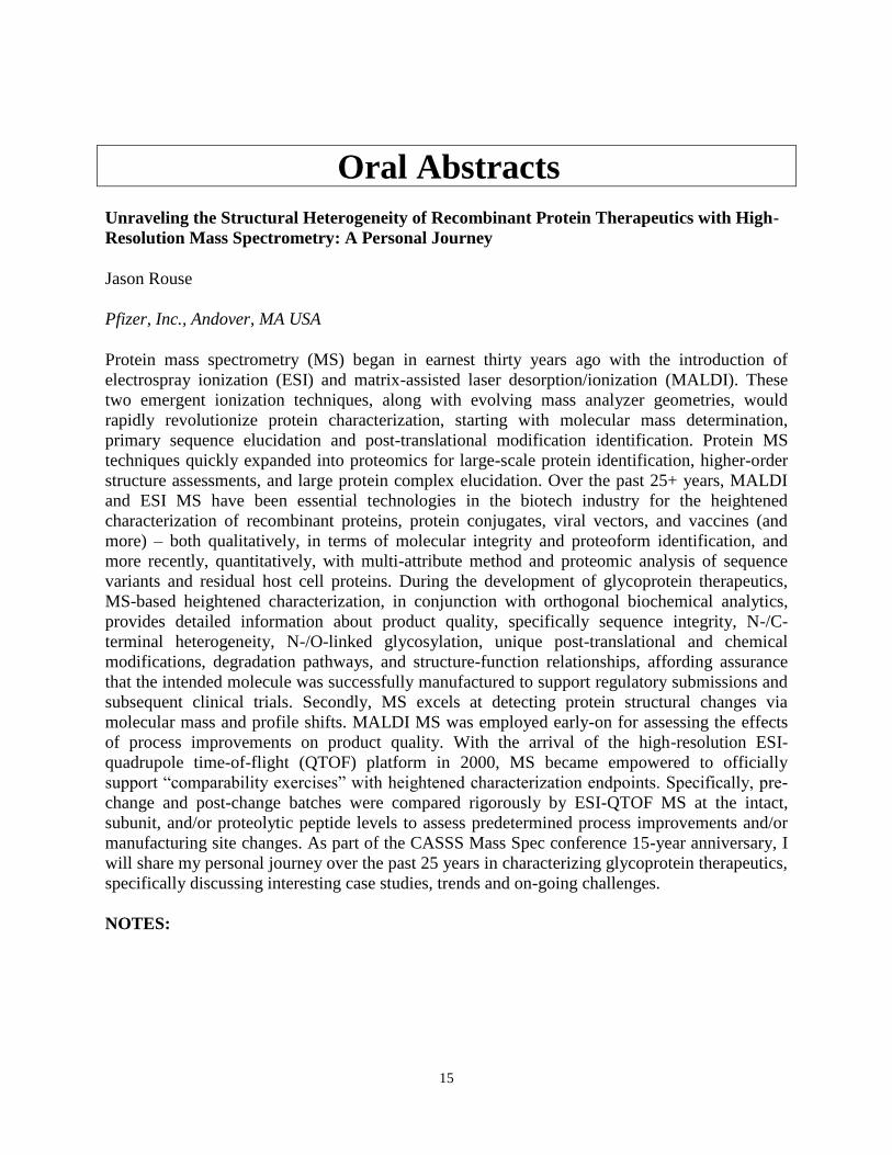
Oral Abstracts
Unraveling the Structural Heterogeneity of Recombinant Protein Therapeutics with High-
Resolution Mass Spectrometry: A Personal Journey
Jason Rouse
Pfizer, Inc., Andover, MA USA
Protein mass spectrometry (MS) began in earnest thirty years ago with the introduction of
electrospray ionization (ESI) and matrix-assisted laser desorption/ionization (MALDI). These
two emergent ionization techniques, along with evolving mass analyzer geometries, would
rapidly revolutionize protein characterization, starting with molecular mass determination,
primary sequence elucidation and post-translational modification identification. Protein MS
techniques quickly expanded into proteomics for large-scale protein identification, higher-order
structure assessments, and large protein complex elucidation. Over the past 25+ years, MALDI
and ESI MS have been essential technologies in the biotech industry for the heightened
characterization of recombinant proteins, protein conjugates, viral vectors, and vaccines (and
more) – both qualitatively, in terms of molecular integrity and proteoform identification, and
more recently, quantitatively, with multi-attribute method and proteomic analysis of sequence
variants and residual host cell proteins. During the development of glycoprotein therapeutics,
MS-based heightened characterization, in conjunction with orthogonal biochemical analytics,
provides detailed information about product quality, specifically sequence integrity, N-/C-
terminal heterogeneity, N-/O-linked glycosylation, unique post-translational and chemical
modifications, degradation pathways, and structure-function relationships, affording assurance
that the intended molecule was successfully manufactured to support regulatory submissions and
subsequent clinical trials. Secondly, MS excels at detecting protein structural changes via
molecular mass and profile shifts. MALDI MS was employed early-on for assessing the effects
of process improvements on product quality. With the arrival of the high-resolution ESI-
quadrupole time-of-flight (QTOF) platform in 2000, MS became empowered to officially
support “comparability exercises” with heightened characterization endpoints. Specifically, pre-
change and post-change batches were compared rigorously by ESI-QTOF MS at the intact,
subunit, and/or proteolytic peptide levels to assess predetermined process improvements and/or
manufacturing site changes. As part of the CASSS Mass Spec conference 15-year anniversary, I
will share my personal journey over the past 25 years in characterizing glycoprotein therapeutics,
specifically discussing interesting case studies, trends and on-going challenges.
NOTES:
16
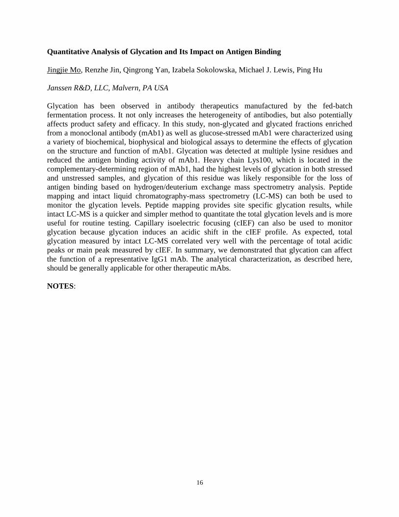
Quantitative Analysis of Glycation and Its Impact on Antigen Binding
Jingjie Mo, Renzhe Jin, Qingrong Yan, Izabela Sokolowska, Michael J. Lewis, Ping Hu
Janssen R&D, LLC, Malvern, PA USA
Glycation has been observed in antibody therapeutics manufactured by the fed-batch
fermentation process. It not only increases the heterogeneity of antibodies, but also potentially
affects product safety and efficacy. In this study, non-glycated and glycated fractions enriched
from a monoclonal antibody (mAb1) as well as glucose-stressed mAb1 were characterized using
a variety of biochemical, biophysical and biological assays to determine the effects of glycation
on the structure and function of mAb1. Glycation was detected at multiple lysine residues and
reduced the antigen binding activity of mAb1. Heavy chain Lys100, which is located in the
complementary-determining region of mAb1, had the highest levels of glycation in both stressed
and unstressed samples, and glycation of this residue was likely responsible for the loss of
antigen binding based on hydrogen/deuterium exchange mass spectrometry analysis. Peptide
mapping and intact liquid chromatography-mass spectrometry (LC-MS) can both be used to
monitor the glycation levels. Peptide mapping provides site specific glycation results, while
intact LC-MS is a quicker and simpler method to quantitate the total glycation levels and is more
useful for routine testing. Capillary isoelectric focusing (cIEF) can also be used to monitor
glycation because glycation induces an acidic shift in the cIEF profile. As expected, total
glycation measured by intact LC-MS correlated very well with the percentage of total acidic
peaks or main peak measured by cIEF. In summary, we demonstrated that glycation can affect
the function of a representative IgG1 mAb. The analytical characterization, as described here,
should be generally applicable for other therapeutic mAbs.
NOTES:
17
Identification of Multiple Serine to Asparagine Sequence Variation Sites in an Intended
Copy Product of LUCENTIS® by Mass Spectrometry
Francois Griaud
Novartis Pharma AG, Basel, Switzerland
Patent expiration of first-generation biologics and the high cost of innovative biologics are 2
drivers for the development of biosimilar products. There are, however, technical challenges to
the production of exact copies of such large molecules. In this presentation, a head-to-head
comparison between the originator anti-VEGF-A Fab product LUCENTIS® (ranibizumab) and
an intended copy product using an integrated analytical approach will be described. Published
and unpublished data will be presented. While no differences could be observed using size-
exclusion chromatography, capillary electrophoresis-sodium dodecyl sulfate and potency assays,
different acidic peaks were identified with cation ion exchange chromatography and capillary
zone electrophoresis. Further investigation of the intact Fab, subunits and primary sequence with
mass spectrometry demonstrated the presence of a modified light chain variant in the intended
copy product batches. This variant was characterized with a mass increase of 27.01 Da compared
to the originator sequence and its abundance was estimated in the range of 6–9% of the intended
copy product light chain. MS/MS spectra interrogation confirmed that this modification relates to
a serine to asparagine sequence variant found in the intended copy product light chain. We
demonstrated that the integration of high-resolution and sensitive orthogonal technologies was
beneficial to assess the similarity of an originator and an intended copy product.
NOTES:
18
A Hybrid Intact MS and Peptide Mapping Approach for Elusive Sequence Variant
Detection during Cell Line Development
Thomas Slaney, Hangtian Song, Neil Hershey, Erik Langsdorf, Wei Wu, Li Tao
Bristol-Myers Squibb Company, Pennington, NJ USA
Mass spectrometry has become an indispensable technology in process development for
biologics, mainly due to the comprehensive information it provides and the sensitivity it
achieves. Nevertheless, the detection and elucidation of sequence variants in therapeutic proteins
by mass spectrometry still presents a significant challenge, since variants can be generated by
multiple mechanisms and are usually at low levels. Although highest sensitivity is typically
obtained with peptide mapping assays, incomplete sequence coverage and atypical types of
sequence variants still render some sequence variants undetected. We have therefore adopted a
strategy combining intact and subunit mass analyses with peptide mapping to minimize chances
of missing sequence variants during cell line development. The combination approach allowed
us to detect uncommon sequence variants that would be hard to identify by regular peptide
mapping analysis. For example, a Gln-to-Lys variant with a 0.04 Da mass shift was observed in
peptide mapping but not intact MS, whereas a DNA frame shift mutation co-purified with our
protein of interest was missed by routine peptide mapping searches but was readily detected by
intact MS. Detailed description of the work flow and data analysis for these examples will be
presented.
NOTES:
19
Identification and Monitoring of Potential Critical Quality Attributes of an Antibody-drug
Conjugate by Mass Spectrometry to Support Process Characterization
Xuan Chen
ImmunoGen Inc., Waltham, MA USA
Antibody-drug conjugates (ADCs) are cancer-targeting biotherapeutic molecules that are
composed of monoclonal antibodies linked to small molecule cytotoxic payload.Mirvetuximab
soravtansine our lead compound, also known as IMGN853 , consists of the maytansinoid DM4
payload conjugated to M9346A antibody via sulfo-SPDB linker.
One of the important steps of drug development and preparation for regulatory filings is the
establishment of a robust manufacturing process for the ADCs and their components (e.g.,
antibody, linker, cytotoxic payload). Assessment of the criticality of quality attributes is essential
for planning process characterization activities. Structure-activity relationship (SAR) studies
provide information about the biological impacts of product variants. SAR studies were
performed by generating and/or isolating variants that were analyzed by physicochemical
methods and their bioactivities were monitored using in vitro binding and cell-based assays.
Mass spectrometry played a key role in identifying antibody and ADC product variants.
Process characterization studies evaluate the impact of process parameters on critical quality
attributes with the intent of identifying critical process parameters. These studies use design-of-
experiment approaches and generate a large number of samples from the multiple manufacturing
process steps. Several attributes of the process characterization samples (e.g., oxidation,
deamidation, trisulfides, and conjugation sites) were monitored by mass spectrometry. During
the talk, the mass spectrometric methods and results will be presented and their importance on
the development of consistent manufacturing processes will be discussed.
NOTES:
20
Heightened Characterization of ADCs: Overcoming Challenges to Support Process and
Product Understanding
Olga Friese1, Jacquelynn Smith1, Paul Brown1, Andrew Dawdy1, Thomas Powers1, Jason Rouse2
1Pfizer, Inc., Chesterfield, MO USA, 2Pfizer, Inc., Andover, MA USA
The development of analytical methods for the characterization of antibody drug conjugates
(ADCs) presents significant challenges given the complexity of ADCs resulting from the
addition of the drug payloads to already complex antibodies. Mass spectrometry is used for in-
depth characterization of ADCs to determine the level and sites of drug conjugation and
heterogeneities present due to the conjugation chemistry. The typical MS approach includes
mass analysis of both the intact and de-N-glycosylated 4-chain ADC, and two or three-part
subunit/domain analysis followed by proteolytic mapping. These MS-based analyses are used to
confirm the sequence fidelity of the ADC as well as extent and integrity of conjugation. Several
challenges that were encountered during heightened characterization of ADCs by LC/MS will be
presented. ESI MS analysis of many intact ADCs remains challenging due to extensive
conjugation with hydrophobic linker-payloads, non-covalent subunits, and/or chemically
unstable linkers. However, further method development to optimize the mobile phases during
LC separation, ESI source and ion transfer parameters allow establishment of reliable MS-based
assays for accurate drug-to-antibody ratio determination. Denaturing conditions of typical
LC/MS analyses impede the successful detection of intact, 4-chain ADCs generated via cysteine
site-directed chemistry approaches where hinge region disulfide bonds are partially reduced.
However, this class of ADCs is detected intact reliably under non-denaturing SEC/MS
conditions, also referred to as native MS. For ADCs with acid labile linkers such as one used for
conjugation of calicheamicin, a careful selection of mobile phase composition is critical to retain
intact linker-payload during LC/MS analysis. Increasing pH of the mobile phase prevented
cleavage of a labile bond in the linker moiety and resulted in retention of the intact linker-
payload. In-source fragmentation was also observed with typical ESI source parameters during
intact ADC mass analysis for a particular surface accessible linker-payload moiety conjugated to
the heavy chain C-terminal.
NOTES:

21
An Automated Quantitative Mass Spectrometry Assay to Help Select Next-generation Anti-
staphylococcus Aureus Antibody-antibiotic Conjugates by Evaluating Intra-cellular
Retention Kinetics of Released Antibiotics
Hilda Hernandez-Barry, Kimberly Kajihara, Daniel Tran, Martine Darwish, Richard Vandlen,
Leanna Staben, Thomas Pillow, Wouter Hazenbos, Kelly Loyet
Genentech, a Member of the Roche Group, South San Francisco, CA USA
Methicillin-resistant Staphylococcus aureus is the leading cause of hospital-acquired infections,
but it has become more difficult to treat due to evolved resistance to antimicrobial drugs and
intracellular bacterial reserves in phagolysosomes. For this purpose, a new kind of ADC
therapeutic has recently emerged, namely antibody-antibiotic conjugates (AACs). An AAC
combines the binding specificity of an antibody with an antibiotic and can deliver the
bactericidal activity needed for complete killing. However, this AAC has no direct antibacterial
activity on S. aureus until the AAC opsonized bacteria is taken up by the host cells and the
intracellular proteases act on the cathepsin cleavable linker and release the antibiotic in its active
form.
Kinetics and concentration of free drug released intracellularly are a couple of key parameters
that contribute to the efficacy of an AAC. To this end, we have developed an in vitro cell-based
LC/MS drug release automated assay in a 96-well format in order to measure the intracellular
concentration (ICC) of released antibiotic at specific time-points. This quantitative LC/MS assay
employs a QTRAP® 6500 Sciex instrument in MRM mode for detection of the antibiotics of
interest. The ICC has been determined for numerous AAC candidates in order to select an AAC
that is both retained in the cell and efficacious at killing bacteria. When the ICC is greater than
the minimum efficacious concentration to eliminate S. aureus at pH 5 (MEC5), it is a reasonable
predictor of AAC potency. Thus, using ICC and MEC5 data together with catalytic cleavage and
potency assays, we can more fully characterize the fate and action of the AAC payload release
and ultimately guide the design of more effective and targeted AAC therapeutics.
NOTES:
22
Deciphering Protein Biology Using Mass Spectrometry
Alma Burlingame
University of California, San Francisco, San Francisco, CA USA
The number of human genes is relatively small and known, and the sequencing at both the DNA
and RNA levels is now routine. However, protein studies at the detailed molecular level provide
experimental challenges of daunting complexity by comparison.
Topics of current interest in protein biology include:
(1) direct measurement of nascent protein synthesis;
(2) modulation of protein function by posttranslational processes; and
(3) studies of the interactions of proteins and the architecture of protein complexes.
This presentation will discuss examples from these areas of our research where mass
spectrometry has played a key role.
Acknowledgement:
Financial support has been provided by UCSF Program for Breakthrough Biomedical Research,
the Adelson Medical Research Foundation and HHMI.
NOTES:
23
Proteomic Characterization of Exosomes and Their Impurities
Damian Houde, Rane Harrison
Codiak BioSciences, Cambridge, MA USA
Extracellular vesicles (EVs) are a heterogeneous population of nano-sized cell-derived
membrane vesicles that are actively released into the extracellular space. Once released, EVs
perform critical roles in intercellular communication by transferring their biological content (i.e.,
proteins, lipids, nucleic acids, and other compounds) between cells. Consequently, and because
EVs are nontoxic and nonimmunogenic, they have become an attractive option for delivering
pharmaceutical and biopharmaceutical payloads for a variety of medical conditions. However,
given their inherent heterogeneity, EVs are often difficult to purify and characterize. Here, EVs
from a human cell-line were purified by two different methods. The EV samples were then
extensively characterized by proteomic analysis, which enabled the identification of proteins,
their content, and supported the comparison of different isolated populations. These data were
critical for the identification of EV associated proteins and proteinaceous particulate impurities
from the host cell line, which helped drive improvements to the EV purification process.
NOTES:
24
Mass Spectrometry in the CAR-T Development Lab
Ken Prentice
Juno Therapeutics, Seattle, WA USA
Cell based therapeutics have been hailed as the third pillar of medicine and there has been rapid
acceptance of this class of drugs in both academic and industry settings in the last few years.
Specifically chimeric antigen receptor expressing t-cells (CAR T) have garnered significant
attention with the recent FDA approval of the first cell-based immunotherapies for oncology.
Cells and cell based therapeutics have been traditionally analyzed using immunochemistry (i.e.
flow cytometry) or genomic (i.e. PCR/NGS) based assays. While mass spectrometry has had
limited use in this class of therapies, most applications have focused on target discovery. This
talk will focus on mass spectrometry as a powerful analytical tool for structural elucidation and
process development support of CAR T products. Applications encompassing raw materials, cell
culture, and CAR T protein detection and characterization will be discussed.
NOTES:
25
Assessment of Susceptible Chemical Modification Sites of Trastuzumab and Endogenous
Human Immunoglobulins at Physiological Conditions
Ingrid Grunert
Roche Diagnostics GmbH, Penzberg, Germany
The quality control testing of chemical degradations in the bio-pharmaceutical industry is
currently under controversial debate. Here we have systematically applied in vitro and in vivo
stress conditions to investigate the influence of protein degradation on structure-function.
Extensive purification and characterization enabled identification and functional assessment of
the physiological degradation of chemical modification sites in the variable complementarity-
determining regions (CDRs) and conserved region of trastuzumab. We demonstrate that the
degradation of the solvent accessible residues located in the CDR and the conserved fragment
crystallizable region (Fc) occurs faster in vivo (within days) compared to the levels observed for
bio-process and real-time storage conditions. These results hence question the rationality of
extreme monitoring of low level alterations in such chemical modifications as critical patient
safety parameters in product quality control testing, given that these modifications merely mirror
the natural/physiological aging process of endogenous antibodies.
NOTES:
26
Spatiotemporally-resolved Proteomics with Next-generation Enzymatic Proximity Labeling
Methods
Shuo Han
Stanford University, Stanford, CA USA
The ability to characterize endogenous proteins – their structures, localization, trafficking, and
function – within the native context of living cells is necessary to advance our understanding of
cellular processes and pathologies. Traditional methods to study the protein constituents of
subcellular compartments or their respective interactions are limited, and often require the use of
perturbing conditions such recombinant protein tags, cellular lysis and fractionation. We will
describe two recent technologies based on genetically-targetable enzymes that catalyze
proximity-dependent biotinylation of endogenous proteins in living cells, which can be
subsequently enriched and identified using mass spectrometry. Engineered ascorbate peroxidase
(APEX) has been used to gain insight into the proteomic compositions of various cellular
structures and macromolecular complexes that are traditionally inaccessible, including the
synaptic clefts and several mitochondrial sub-compartments. In addition, APEX can also
biotinylate RNAs for spatial transcriptomic analysis. BioID is a complementary method to
APEX that utilizes a promiscuous biotin ligase to perform proximity-dependent labeling.
However, while APEX labeling requires 1 minute, BioID requires long labeling periods of 18-24
hours. Using directed evolution, our lab has developed TurboID: a smaller, faster promiscuous
biotin ligase that catalyzes proximity labeling upon 10-minute addition of biotin. Together, these
new methods will enable scientists to map proteomes of organelles and signaling pathways, with
high spatiotemporal resolution, in a wide range of cell types and model organisms.
NOTES:
27
in-vivo Characterization of Antibodies using a Sensitive Nano-HPLC–MS/MS Approach
Annika Doell1, Markus Hollmann2, Oliver Schmitz1
1University Duisburg-Essen, Essen, Germany, 2AbbVie Deutschland GmbH & Co KG,
Ludwigshafen, Germany
Biotherapeutics are products of genetically engineered cells, and include hormones, regulatory
peptides and proteins. In particular, therapeutic proteins have become increasingly important in
the treatment of numerous severe diseases. Antibodies are an example of therapeutic proteins,
which are used in medicine for treatment of e.g. autoimmune diseases as well as different types
of cancer.
The analysis of protein modifications on peptide level using liquid chromatography coupled to
mass spectrometry (LC-MS) is a common approach to characterize protein therapeutics. While
protein characterization of in-vitro samples is performed routinely, the analysis of protein
therapeutics after in-vivo administration has recently gained significant attention. There is a
profound interest in getting an understanding of what happens to the biotherapeutic molecules in-
vivo and correlating critical quality attributes with immunogenicity and bioavailability. With the
Open Flow Microperfusion (OFM) approach, subcutaneously applied antibodies are extracted
via probes from the interstitial fluid. Those samples are then analyzed using the sensitive Nano-
HPLC-MS/MS approach.
The combination of the above-mentioned methods can potentially provide new insights into the
field of antibody modifications that may occur in the subcutaneous layer. A deeper
understanding of metabolization mechanisms after subcutaneous administration would bring a
remarkable benefit for the patient’s safety and also the development of biopharmaceutics.
NOTES:
28
Reversible Self-association (RSA): A Stumbling Block for Developing High Concentration
IgG1 mAb Formulations
Yue (Martin) Hu
University of Kansas, Lawrence, KS USA
Monoclonal antibodies (mAbs) have become a class of drugs of high importance for treating
numerous human diseases. Subcutaneous (SC) administration is an increasingly common and
convenient route for patients to be able to do home-based treatments. Since mAbs often require
high mg/kg dosing and SC injections are limited to a small injection volume (~1.5 mL), this
necessitates the development of high-concentration mAb formulations. High concentration mAb
solutions pose many pharmaceutical challenges including physical instability during
manufacturing, storage, and delivery. In addition, reversible self-association (RSA) has emerged
as an important formulation challenge in terms of significantly increasing solution viscosity,
turbidity and even phase separation. In this work, four different human immunoglobulin G1
molecules were obtained from MedImmune with varying solution properties at high
concentration. By evaluating mAbs with both “good” and “bad” solution properties at different
protein concentrations in a single formulation buffer, the goal is to use hydrogen-deuterium
exchange mass spectrometry (HX-MS) to identify peptide segments involved in the protein-
protein interactions (PPIs) of the mAbs. These results will help to identify patterns that may lead
to the identification of RSA “hot-spots” for high concentration mAb formulation issues. First,
intact mass analysis and peptide mapping were performed to confirm sequences and post-
translational modifications of different mAbs, trying to identify important chemical modification,
truncation, glycosylation, and get high peptide coverage on the sequence. Second, HX-MS
experiments were performed on mAbs solutions (in D2O) using lyophilization-reconstitution
approach to obtain site-specific information on the regions of “bad” mAbs (peptide segments)
that are primarily responsible for RSA at high concentrations. In addition, excipient effects on
mAbs RSA were also examined by HX-MS on their ability of promoting or disrupting protein
interactions. In terms of future work, HX-MS and biophysical data will be correlated to obtain a
more comprehensive understanding of RSA behaviours of the four mAbs, and to determine if
common motifs are present that may correspond to molecular “hot spots” of PPIs, and see if a
particular RSA effect on solution correlates with a particular molecular hot spot sequence.
NOTES:
29
Computational Strategies for Improving the Quantitative Analysis of Peptides by Tandem
Mass Spectrometry
Michael MacCoss
University of Washington, Seattle, WA USA
Proteomics technology has improved dramatically over the last decade. The technology
developments have largely been directed around instrument hardware, where instruments have
been developed that scan faster, are more sensitive, and have greater mass measurement
accuracy. However, the basic workflow has remained largely unchanged -- mass spectrometers
are directed toward the acquisition of tandem mass spectra on the most abundant molecular
species eluting from a chromatography column. More recently, efforts have been focused on the
acquisition of mass spectrometry data on target peptides of interest. With improvements in
instrument hardware and instrument control software, the practical experimental difference
between a targeted and discovery proteomics is beginning to become blurred. These analyses are
a significant change from the traditional proteomics workflow and have required the
development of novel computational strategies to analyze, visualize, and interpret these data. We
will present work illustrating our efforts in the development of targeted proteomics and provide a
vision for challenges that still need to be overcome before these analyses become routine and
replace more traditional discovery proteomics methodology.
NOTES:
30
Driving Efficiency in Pre-clinical Development with Automated Mass Spectrometry
Analysis and Characterization of Novel Biologics
Hirsh Nanda1, Bo Zhai1, Andy Mahan1, Eric Carlson2, Yong Kil2, Li Jing2, Andrew Nichols2
1Janssen R&D, LLC, Spring House, PA USA, 2Protein Metrics Inc., San Carlos, CA USA
Large molecule therapeutics are susceptible to numerous posttranslational modifications and
other variants which may have detrimental effects on efficacy, stability or immunogenicity. Mass
spectrometry-based peptide mapping provides information on many important molecule
attributes such as posttranslational modification, clipping, glycosylation and sequence variants.
As biotherapeutic portfolios progress from mAbs to more complicated architectures (e.g. multi-
specifics, scaffold proteins, ADCs and antigens) both the challenges and the demands for MS
based characterization has only grown. Given the complexity of analysis, data processing often
limits the throughput of the assays as it requires multiple software packages and extensive
manual interpretation. To manage characterization and PTM analysis of multiple candidate
molecules with fast turn-around times, an automated end-to-end data analysis pipeline has been
developed to accelerate clone selection and developability characterization within pre-clinical
development. The pipeline is a vendor agnostic end-to-end solution, which provides a UI for
efficient data validation and report generation. Based on the Byonic MS/MS search engine and
Byologic software (Protein Metrics) for label-free quantitation, search parameters were designed
to detect in a single run for PTMs (oxidation, deamidation, isomerization), glycans, N/C-terminal
heterogeneity such as unprocessed signal peptide or clipping as well as sequence variants.
Examples from non-platform therapeutics will be presented and a comparison to other software
solutions will be discussed.
NOTES:
31
Quantifying Proteomes Using the Open-source Trans-proteomic Pipeline
Michael Hoopmann1, Jason Winget2, Luis Mendoza1, Robert Moritz1
1Institute for Systems Biology, Seattle, WA USA, 2Proctor & Gamble, Mason, OH USA
The Trans-Proteomic Pipeline (TPP) is a suite of open-source, freely available software tools for
the analysis of proteomes using mass spectrometry. Highly modular and customizable, the TPP
is both a complete toolset and also capable of interfacing with many popular data analysis tools.
While several quantitative proteomics workflows have been supported since its inception, recent
developments have resulted in new tools for label-free quantitation of proteins from shotgun
mass spectrometry. Here we present StPeter, a tool capable of implementing both Normalized
Spectral Abundance Factor and Normalized Spectral Index quantification metrics. The tool has
been seamlessly integrated into the TPP for reproducibility and ease of use. We demonstrate the
use of the TPP to obtain quantitative protein results from shotgun mass spectra, and compare the
quantitative methods of StPeter to many state-of-the-art stand-alone tools and packages. We also
demonstrate that the software is computationally efficient and supports data from a variety of
instrument platforms and experimental designs. An obvious advantage of using the TPP is it
negates the need to manage inputs and outputs between the many software utilities required to
perform quantitative analyses, dramatically streamlining quantitative workflows and simplifying
user interactions. Results can be viewed within the TPP graphical user interfaces and exported in
standard formats for downstream statistical analysis. The TPP is freely available
at http://www.tppms.org and additional information about StPeter can be found in
DOI:10.1021/acs.jproteome.7b00786
NOTES:
32
Seeing Things Differently: Innovation in Computational Mass Spectrometry
Rob Smith
University of Montana, Missoula, MT USA
By many accounts, innovation in mass spectrometry data processing has lagged far behind
innovation in instrumentation. In this talk, we suggest strategies for accelerating innovation in
computational mass spectrometry through questioning both what current limitations are and why
they exist. The impact of greater problem understanding on innovation is explored, from explicit
identification of problem assumptions to creating non-ambiguous vocabularies for describing
problems in greater detail and accuracy. In aggregate, these approaches provide a practical
pathway to greater innovation in mass spectrometry data processing.
NOTES:
33
Multi-attribute Method (MAM) Evaluation and Regulatory Considerations for
Implementation
Sarah Rogstad
CDER, FDA, Silver Spring, MD USA
Currently, mass spectrometry (MS) is primarily used for drug substance characterization in the
protein therapeutic field. However, there has been a recent push toward the use of MS-based
methods for quality control (QC) purposes, collectively known as multi-attribute methods
(MAM). As these methods have not been used previously in this context, new regulatory
questions must be addressed prior to their full implementation. Such questions include whether
these methods are fit for purpose, the extent of the capabilities of these methods, the ability of
technicians to make quick pass/fail decisions, and how to implement and assess appropriate
system suitability, method validation and comparisons with traditional methods. FDA has
identified four major points to consider for MAM implementation: method validation,
performance comparisons to traditional methods, capabilities and specificities of new peak
detection, and risk assessment. A research program was developed to address these
considerations to better assess the applicability of method. An in-house MAM platform was
developed for the relative quantitation of specific product quality attributes (PQAs) of rituximab,
which was chosen as a model protein. Samples included a range of expiration dates for both US
approved and unapproved products. Analysis included method development, forced degradation,
system suitability assessment, and comparisons to traditional methods. The study found high
reproducibility between users with varying experience levels. However, higher coefficient of
variation (CV) values (>15%) were found for lower abundance PQAs (<5%). These data were
found to be generally consistent with data from released glycan HILIC profiling analyses. During
a forced degradation study with US approved rituximab (40 °C/75% RH for 28 days), oxidation
and deamidation were found to increase linearly over the time course while C-terminal lysine
clipping, N-terminal pyroglutamination and glycosylation did not show significant changes over
time. Using the new peak detection feature of the method, one peak, that was not specifically
targeted in the analysis, was found to increase over the time course (> 10-fold change in area,
peak intensity threshold of 0.1% of the TIC). This peak was determined to be an isoaspartic acid
modification. Additional studies may be needed to further examine the efficacy of the new peak
detection feature. Forced degradation results were also compared to results from orthogonal
methods. Charge variant analysis showed an increase in acidic peaks over time. MAM
deamidation data was found to have a linear correlation (R2 = 0.94) with the acidic peak data.
Further assessment and statistical analyses will be conducted on this relationship as well as on
the clipped variant data. System suitability testing approaches were also assessed.
NOTES:
34
Interlaboratory Performance Metrics from the MAM Consortium New Peak Detection
Round Robin Study
Trina Mouchahoir1, John Schiel1, Richard Rogers2
1NIST/IBBR, Rockville, MD USA, 2Just Biotherapeutics, Seattle, WA USA
The Multi-attribute Method (MAM) is an emerging application of ultrahigh-performance liquid
chromatography coupled to mass spectrometry (UHPLC-MS) useful for simultaneous
monitoring of multiple biopharmaceutical product quality attributes. The MAM Consortium was
initially formed as a venue to share regulatory experiences, harmonize best practices, and
generate innovative methodology to facilitate widespread integration. Its members recently
contributed to a new peak detection (NPD) inter-laboratory study to evaluate performance
metrics and reproducibility of the MAM utilizing pre-digested samples of the NISTmAb RM
8671. Evaluation of the data focused on attribute analytics as well as the NPD component of
MAM. The initial results provide a global overview of the current state of the industry as well as
valuable insight regarding the robustness and reproducibility of the MAM NPD platform.
NOTES:
35
Technological Advances in Host Cell Protein Testing and Risk Management: Enhancing
the Quality of Clinical Products
Ned Mozier
Pfizer, Inc., Chesterfield, MO USA
Improvements in databases and mass spectrometry (MS) technology have radically changed the
landscape for HCP testing and impurity management. The oft debated question as to whether
MS has a function in the quality control testing laboratory is no longer hindered by its
capabilities, although this remains a strategic decision for corporations. The power of MS for
identification of HCPs is now common in development, especially in regard to process
development and for risk management. The immunoassay remains the bedrock by which most
quality decisions are made, but increasingly MS is being used as an orthogonal method. This has
enabled more useful risk analysis and intelligent decision-making. Together, immunoassay and
MS are the most important tools for managing the risk of impurities such as HCPs and have led
to higher quality products. How various technologies are applied during the development
paradigm has evolved and been accelerated with the advent of biosimilars. This presentation
will review the known and unknown risks of HCPs and provide cases where individual proteins
have been shown to affect clinical trials, portfolio decisions and how the technological
innovations have influenced this direction.
NOTES:

36
NOTES:
37
Recombinant Protein Therapeutics Workshop:
To Sequence or Not to Sequence
Tuesday, September 11
16:00 – 17:30
Plaza Room A
Workshop Chair:
Anders Lund, Synlogic, Cambridge, MA USA
Facilitator:
Nadine Ritter, Global Biotech Experts, LLC, Germantown, MD USA
Speaker:
Karen Lee, Sanofi, Framingham, MA USA
Scribes:
Steve Cohen, SAC Analytical Consultants, Hopkinton, MA USA
Yelena Lyubarskaya, Sanofi, Westborough, MA USA
Our industry is constantly changing and evolving. This workshop is focused on the use of
LCMS and peptide mapping in the primary characterization of protein and peptide biologics.
One of our goals for this meeting is to write a “peptide mapping best practices” document. This
document will be used to help discern the guidance around LCMS analysis of biopharmaceutical
products, specifically protein based biotherapeutics. To help, we invited all Mass Spec 2018
attendees to contribute by filling out a survey. We are trying to recognize if there are industry
best practices around protein characterization and protein lot release.
As a preface to this workshop, ICH guidelines Q5B and Q6B for novel molecular entities
describe the analysis of the expression constructs, specifically "Segments of the expression
construct should be analyzed using nucleic acid techniques in conjunction with other tests
performed on the purified recombinant protein for assuring the quality and consistency of the
final product." Given this statement, how do we as an industry apply “other tests”? Please
participate in the discussion – ask QUESTIONS, add your observations! Our goal is to use the
data from the survey (and the discussion from the workshop) to compile a best practices
document to be published late 2018, or early 2019 by the CASSS Mass Spec Organizing
Committee.
NOTES:

38
NOTES:
39
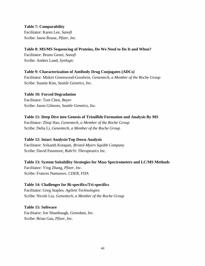
Roundtable Discussion
Monday, September 10
16:15 – 17:15
There are 15 roundtable topics. The plan is for these to be active discussions, not presentations
or lectures. To create useful discussion, we are going to try and limit each topic to 10 attendees.
Seating will be on a first come, first serve basis. These discussions will include a facilitator,
whose role is to help assist the discussion and ensure a lively exchange, and a scribe, whose role
is to make general, anonymous notes about the discussion that will be posted on the Mass Spec
2018 website.
Listed below is a quick view of the Roundtable Topics, Facilitators and Scribes:
Table 1: Challenges During Regulatory Submissions
Facilitator: Ramsey Saleem, Amgen Inc.
Scribe: John Valliere-Douglass, Seattle Genetics, Inc.
Table 2: Process Analytics Technology and Mass Spectrometry
Facilitator: Ananya Dubey, Waters Corporation
Scribe: Nalini Sadagopan, Agilent Technologies
Table 3: MS in the Biomanufacturing Environment
Facilitator: Hirsh Nanda, Janssen R&D LLC
Scribe: Sean McCarthy, SCIEX
Table 4: Host Cell Protein Analysis
Facilitator: Annemiek Verwilligen, Janssen Vaccines and Prevention
Scribe: Sarah Rogstad, CDER, FDA
Table 5: Automation
Facilitator: Sreekanth Suravajjala, Amgen Inc.
Scribe: Kristin Boggio, Pfizer, Inc.
Table 6: in vivo Biotransformation Analysis Strategies and Real Lab Experiences
Facilitator: Olga Friese, Pfizer, Inc.
Scribe: Yelena Lyubarskaya, Sanofi
40
Table 7: Comparability
Facilitator: Karen Lee, Sanofi
Scribe: Jason Rouse, Pfizer, Inc.
Table 8: MS/MS Sequencing of Proteins, Do We Need to Do It and When?
Facilitator: Bruno Genet, Sanofi
Scribe: Anders Lund, Synlogic
Table 9: Characterization of Antibody Drug Conjugates (ADCs)
Facilitator: Midori Greenwood-Goodwin, Genentech, a Member of the Roche Group
Scribe: Sunnie Kim, Seattle Genetics, Inc.
Table 10: Forced Degradation
Facilitator: Tom Chen, Bayer
Scribe: Jason Gilmore, Seattle Genetics, Inc.
Table 11: Deep Dive into Genesis of Trisulfide Formation and Analysis By MS
Facilitator: Zhiqi Hao, Genentech, a Member of the Roche Group
Scribe: Delia Li, Genentech, a Member of the Roche Group
Table 12: Intact Analysis/Top Down Analysis
Facilitator: Srikanth Kotapati, Bristol-Myers Squibb Company
Scribe: David Passmore, RubrYc Therapeutics Inc.
Table 13: System Suitability Strategies for Mass Spectrometers and LC/MS Methods
Facilitator: Ying Zhang, Pfizer, Inc.
Scribe: Frances Namuswe, CDER, FDA
Table 14: Challenges for Bi-specifics/Tri-specifics
Facilitator: Greg Staples, Agilent Technologies
Scribe: Nicole Liu, Genentech, a Member of the Roche Group
Table 15: Software
Facilitator: Joe Shambaugh, Genedata, Inc.
Scribe: Brian Gau, Pfizer, Inc.
41
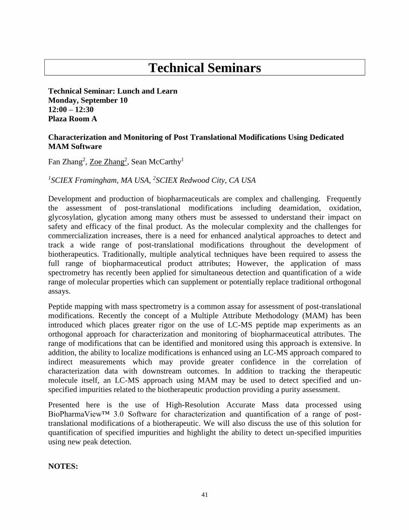
Technical Seminars Technical Seminar: Lunch and Learn
Monday, September 10
12:00 – 12:30
Plaza Room A
Characterization and Monitoring of Post Translational Modifications Using Dedicated
MAM Software
Fan Zhang2, Zoe Zhang2, Sean McCarthy1
1SCIEX Framingham, MA USA, 2SCIEX Redwood City, CA USA
Development and production of biopharmaceuticals are complex and challenging. Frequently
the assessment of post-translational modifications including deamidation, oxidation,
glycosylation, glycation among many others must be assessed to understand their impact on
safety and efficacy of the final product. As the molecular complexity and the challenges for
commercialization increases, there is a need for enhanced analytical approaches to detect and
track a wide range of post-translational modifications throughout the development of
biotherapeutics. Traditionally, multiple analytical techniques have been required to assess the
full range of biopharmaceutical product attributes; However, the application of mass
spectrometry has recently been applied for simultaneous detection and quantification of a wide
range of molecular properties which can supplement or potentially replace traditional orthogonal
assays.
Peptide mapping with mass spectrometry is a common assay for assessment of post-translational
modifications. Recently the concept of a Multiple Attribute Methodology (MAM) has been
introduced which places greater rigor on the use of LC-MS peptide map experiments as an
orthogonal approach for characterization and monitoring of biopharmaceutical attributes. The
range of modifications that can be identified and monitored using this approach is extensive. In
addition, the ability to localize modifications is enhanced using an LC-MS approach compared to
indirect measurements which may provide greater confidence in the correlation of
characterization data with downstream outcomes. In addition to tracking the therapeutic
molecule itself, an LC-MS approach using MAM may be used to detect specified and un-
specified impurities related to the biotherapeutic production providing a purity assessment.
Presented here is the use of High-Resolution Accurate Mass data processed using
BioPharmaView™ 3.0 Software for characterization and quantification of a range of post-
translational modifications of a biotherapeutic. We will also discuss the use of this solution for
quantification of specified impurities and highlight the ability to detect un-specified impurities
using new peak detection.
NOTES:
42
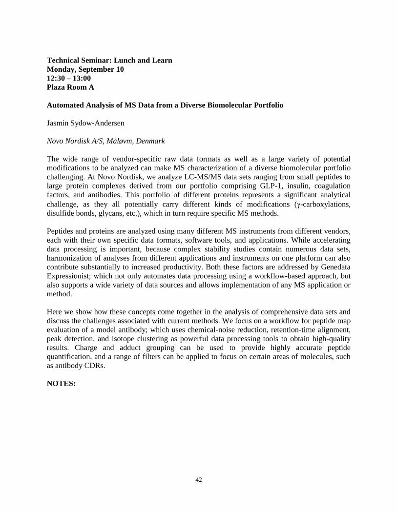
Technical Seminar: Lunch and Learn
Monday, September 10
12:30 – 13:00
Plaza Room A
Automated Analysis of MS Data from a Diverse Biomolecular Portfolio
Jasmin Sydow-Andersen
Novo Nordisk A/S, Måløvm, Denmark
The wide range of vendor-specific raw data formats as well as a large variety of potential
modifications to be analyzed can make MS characterization of a diverse biomolecular portfolio
challenging. At Novo Nordisk, we analyze LC-MS/MS data sets ranging from small peptides to
large protein complexes derived from our portfolio comprising GLP-1, insulin, coagulation
factors, and antibodies. This portfolio of different proteins represents a significant analytical
challenge, as they all potentially carry different kinds of modifications (-carboxylations,
disulfide bonds, glycans, etc.), which in turn require specific MS methods.
Peptides and proteins are analyzed using many different MS instruments from different vendors,
each with their own specific data formats, software tools, and applications. While accelerating
data processing is important, because complex stability studies contain numerous data sets,
harmonization of analyses from different applications and instruments on one platform can also
contribute substantially to increased productivity. Both these factors are addressed by Genedata
Expressionist; which not only automates data processing using a workflow-based approach, but
also supports a wide variety of data sources and allows implementation of any MS application or
method.
Here we show how these concepts come together in the analysis of comprehensive data sets and
discuss the challenges associated with current methods. We focus on a workflow for peptide map
evaluation of a model antibody; which uses chemical-noise reduction, retention-time alignment,
peak detection, and isotope clustering as powerful data processing tools to obtain high-quality
results. Charge and adduct grouping can be used to provide highly accurate peptide
quantification, and a range of filters can be applied to focus on certain areas of molecules, such
as antibody CDRs.
NOTES:
43
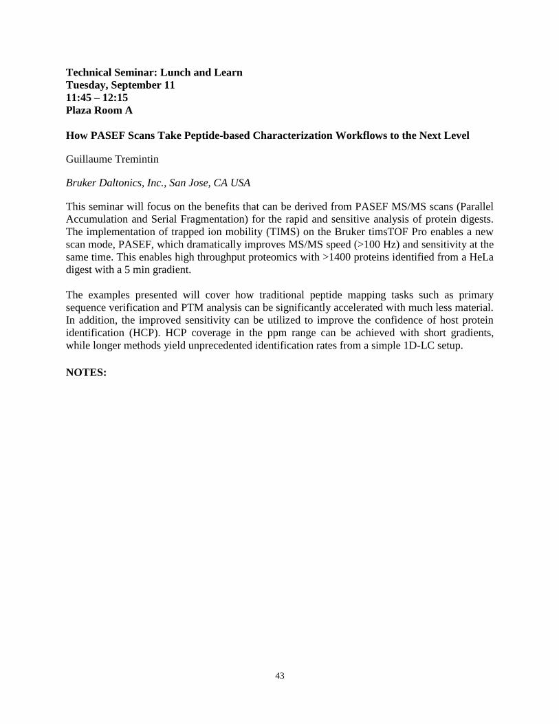
Technical Seminar: Lunch and Learn
Tuesday, September 11
11:45 – 12:15
Plaza Room A
How PASEF Scans Take Peptide-based Characterization Workflows to the Next Level
Guillaume Tremintin
Bruker Daltonics, Inc., San Jose, CA USA
This seminar will focus on the benefits that can be derived from PASEF MS/MS scans (Parallel
Accumulation and Serial Fragmentation) for the rapid and sensitive analysis of protein digests.
The implementation of trapped ion mobility (TIMS) on the Bruker timsTOF Pro enables a new
scan mode, PASEF, which dramatically improves MS/MS speed (>100 Hz) and sensitivity at the
same time. This enables high throughput proteomics with >1400 proteins identified from a HeLa
digest with a 5 min gradient.
The examples presented will cover how traditional peptide mapping tasks such as primary
sequence verification and PTM analysis can be significantly accelerated with much less material.
In addition, the improved sensitivity can be utilized to improve the confidence of host protein
identification (HCP). HCP coverage in the ppm range can be achieved with short gradients,
while longer methods yield unprecedented identification rates from a simple 1D-LC setup.
NOTES:
44
Technical Seminar: Lunch and Learn
Tuesday, September 11
12:15 – 12:45
Plaza Room A
From Automated Sample Preparation to Streamlined Data Analysis: A Complete
Analytical Solution for RapiFluor-MS Labeled N-linked Glycan Analysis
Ying Qing Yu
Waters Corporation, Milford, MA USA
Glycosylation is an important attribute of biopharmaceutical products to monitor from
development through manufacturing. However, glycosylation analysis has traditionally been a
time-consuming process with long sample preparation protocols and manual interpretation of the
data. To address the challenges associated with glycan analysis, we have developed a
streamlined analytical solution that covers the entire analysis from automated sample preparation
to data analysis. In this presentation, we will discuss the recent implementation of automation
platforms including Hamilton, Tecan, and Andrew Alliance for the GlycoWorks N-linked
Glycans sample preparation kit with RapiFluor-MS (RFMS) labeling chemistry. The
combination of automated sample preparation with Waters rapid RFMS labeling technology
provides a scalable yet flexible solution that offers significant time savings and increased data
consistency in the analysis of glycans.
To support the streamlined sample preparation, Waters has developed an efficient analytical data
processing workflow for structural assignment of RFMS labeled N-glycans using the compliant-
ready UNIFI Scientific Information System. Following HILIC-UPLC/FLR/MS analyses,
complementary FLR and MS data is processed simultaneously in an automated fashion to
expedite the task of structural assignment and relative glycoform quantification. Using the
integrated glycan structural library, a search based on calibrated retention time in glucose units
(GU) and accurate mass are used in the identification and structural elucidation of released
glycans. Data generated from mAbs and fusion proteins are discussed to show the full capability
of this enabling analytical workflow.
NOTES:
45
Technical Seminar: Lunch and Learn
Wednesday, September 12
11:45 – 12:15
Plaza Room A
Molecular Characterization Using Native Chromatographies Coupled to Mass
Spectrometry
Wendy Sandoval
Genentech, a Member of the Roche Group, South San Francisco, CA USA
We employ native chromatographies coupled to high resolution mass spectrometry to probe the
extent and impact of minor antibody variants, post-translational modifications and monitor
correct assembly. Intact mass analysis of separated moieties allows for a facile and complete
description of the molecule in question. Charge variant native mass spectrometry (CVMS), an
integrated native ion exchange mass spectrometry-based charge variant analytical approach that
delivers detailed molecular information in a single, semi-automated analysis will be
presented. Characterization of variants such as deamidation, which are traditionally unattainable
by intact mass due to their minimal molecular weight differences, were measured unambiguously
by mass and retention time to allow confident MS1 identification. An example of ligand
screening using native online size separation (SEC-MS) will be provided in which non-covalent
protein-small molecule interactions are interrogated and affinity ranked. Although
chromatographic separation offers the time resolution of species, a key component of the
analysis is the ability of intact mass deconvolution algorithms to accurately report and quantify
the species present.
NOTES:

46
Technical Seminar: Lunch and Learn
Wednesday, September 12
12:15 – 12:45
Plaza Room A
Applications of Multi-Attribute Method (MAM) for Control of Critical Quality Attributes
of Protein Therapeutics
Da Ren
Amgen Inc., Thousand Oaks, CA USA
Abstract not available at time of print.
NOTES:
47
Poster Abstracts
ADC and Bispecifics
P-200-M
Overcoming Unusual Challenges in the Characterization of Monoclonal Antibodies by
Mass Spectrometry
Bruno Genet, Séverine Clavier, Armelle Martelet, Nelly Lechat
Sanofi, Vitry sur seine, France
Different modalities of monoclonal antibodies are pushing the limits of characterization by mass
spectrometry. With this new class of biomolecules, new critical quality attributes need to be
carefully monitored to ensure product quality as efficacy and safety, in particular absence of
immunogenicity. From developability in early phase, during process development and routine
monitoring in first clinical phases, Mass Spectrometry is more widely used to monitor post
translational modifications or to identify and monitor unwanted HCPs.
Several case studies will illustrate the first stages of development of a monoclonal antibody and
the characterization of unusual modifications (such as sulfation, additional glycosylation).
The enzymatic desulfation of the antibody allowed to generate different levels of sulfation to
monitor the impact on biological activities.The oxidation need to be carefully monitored as all
the components of a formulation buffer can impact its level. Finally, a presence of an additional
glycosylation on one chain complexify the global pattern and need a monitoring of separate
glycoforms on each chains.
In addition to traditional methods (intact mass in denaturing conditions and peptide mapping),
the use of non-denaturing native SEC-MS method for the investigation of structural
heterogeneity complete the toolbox for the in-depth characterization of atypical behavior.
NOTES:
48
P-201-T
A New LC-MS Approach for Enhancing Subunit-Level Profiling of mAbs and ADCs
Xiaoxiao Liu1, Jennifer Nguyen1, Jacquelynn Smith2, Olga Friese2, Jason Rouse3, Daniel Walsh2,
Ximo Zhang1, Nilini Ranbaduge1, Matthew Lauber1
1Waters Corporation, Milford, MA USA, 2Pfizer, Inc., Chesterfield, MO USA, 5Pfizer, Inc.,
Andover, MA USA
Protein reversed phase chromatography is heavily dependent on the conditions under which it is
performed. Methods employing polymeric columns and trifluoroacetic acid (TFA) have been
preferred by chromatographers but are inherently restricted to low pressure, low throughput
analyses and compromised MS detection. Accordingly, a novel LC-MS platform has been
developed for the subunit profiling of mAb-based therapeutics, and it includes three critical
breakthroughs: a new particle technology to afford increased throughput, a unique high coverage
phenyl surface to lessen ion pairing dependence, and a more MS-friendly mobile phase system
based on highly purified difluoroacetic acid (DFA).
Our investigations have shown that it is possible to achieve higher resolution separations of mAb
subunits when DFA is used in place of TFA. Along with a newly developed column technology
based on an optimized superficially porous particle and novel phenyl surface chemistry, it has
thus been possible to achieve unprecedented resolution and to accelerate analyses via the use of
high flow rates. In addition, a 4-fold increase in MS signal has been observed when 0.1% DFA is
used in place of 0.1% TFA. The newly developed phenyl-based stationary phase, used in
combination with DFA mobile phases, has also dramatically improved the recovery of
challenging samples, such as ADC subunit digests. Ultimately, it is now possible to envision a
new platform method where unforeseen levels of detail can be observed with high fidelity using
15 minute or shorter LC-MS runs.
NOTES:
49
P-202-M
Identifying and Characterizing Bispecific-related Impurities Using Intact Mass Analysis
Agatha Wieczorek
Amgen Inc., Los Angeles, CA USA
Therapeutic bispecific antibodies (B-Abs) can target two pathways implicated in disease. In
general, a B-Ab has two different light chains (LC1 and LC2), and two different heavy chains
(HC1 and HC2) that ideally correctly pair via engineered charge pair mutations. Because of the
challenges associated with multi-chain expression and assembly, product related variants exist
for bispecifics that are not encountered in a monovalent antibody. These include half molecules,
mis-paired light chains, and homodimers. Due to the complexity and co-elution of the product
related impurities, the bispecific antibody herein was not able to leverage platform methods;
primarily by size exclusion chromatography (SEC) or reduced and non-reduced SDS based
capillary electrophoresis (CE-SDS). However, cation exchange chromatography (CEX) was
heavily utilized and found to be an ideal means of separating impurities by exploiting localized
surface charges. Additionally, intact mass methods were used to identify and characterize the
product related attributes. Extensive characterization using mass spectrometry demonstrated the
removal of these impurities throughout the process and in the final product. Data obtained by the
CEX and MS methods was able to verify that 1) varying cell culture temperature conditions
resulted in the formation of different mis-pairs (HC1:LC1 homodimer and LC2:HC2 half mAb)
in the aggregate species, and 2) mRNA expression levels for each culture condition confirmed
increased expression of LC1 in the varying culture conditions. In summary, we present a mass
spectrometry based screening strategy to ensure proper molecule assembly, and subsequently
characterize and control product related impurities during manufacturing, purification and
release testing. Additionally, varying cell culture temperature conditions resulted in the
formation of different mis-pairs. RNA expression levels for each culture confirmed increased
expression of LC1 in the culture conditions where increased HC1:LC1 homodimer was
observed, suggesting different cell culture conditions can affect chain expression and subsequent
molecule assembly.
NOTES:

50
NOTES:
51
CMC Strategy and the
Use of Mass Spec in the QC Lab
P-203-T
Streamlined MAM Analysis for New Peak Detection, Comprehensive Data Inspection, and
Confident Reporting
Eric Carlson, Rose Lawler, Ilker Sen, Michelle English, Kossi Lekpor
Protein Metrics Inc., San Carlos, CA USA
Mass spectrometry plays an increasingly important role in the characterization and comparison
of innovator protein products as well as biosimilar samples. Recently, there is increasing interest
in relying on mass spectrometry to apply Multi-Attribute Methods (MAM) to characterize
biotherapeutics in a consistent manner with the aim of replacing (or at a minimum
supplementing) other traditional analyses. In this poster, we show an example of Protein Metrics
Byos™ which is able to perform fully and semi-automated quality control (QC) ready data
analysis for MAM applications. Byos is used to generate a clear list of product quality attributes
(PQAs) which is presented to an analyst for inspection and curation. Byos then uses the curated
PQA list to annotate a reference chromatogram. The annotated chromatogram is used as a basis
to compare to additional files for new peak detection, quantitative comparison and
comprehensive reports. There are also options for full automation of the process. Results show
MAM analysis of a stressed sample and highlight the results of new peak detection and
differential quantification of PQAs.
NOTES:
52
P-204-M
A Quantitative MS-based Multiple-attribute Method (MAM) Approach for New Biological
Entities and Biosimilar Candidates Presented by a Full Method Qualification for an IgG1
Market Product
Anke Schnabel, Gerhard Körting, Julia Brückner, Yvonne Jasper, Heiner Falkenberg, Roland
Moussa
Protagen Protein Services GmbH, Dortmund, Germany
Detailed characterization including assessment of critical quality attributes of biopharmaceuticals
is essential across process development, manufacturing and release. The multiple-attribute
method (MAM) Peptide Mapping with quantitative LC-ESI-MS provides detailed and site-
specific information on product quality attributes (PQAs), like amino acid sequence, terminal
processing, truncation, deamidation, oxidation or glycosylation. The MAM approach has been
proven to be a powerful tool during development and production of new biological entities and
biosimilar candidates and has the potential to replace conventional protein analytical methods for
characterization and quantification of PQAs. Thereby, the MAM approach has to meet various
demands in the different development phases regarding sample throughput, standardization, and
quality level with the ultimate discipline GMP-release testing. In originator monitoring and
subsequent biosimilarity exercise the MAM needs to be qualified according to current guidelines
with special regards to the comparability assessment and robustness.
We present the application of MAM in IgG1 biosimilar development using Thermo Scientific Q
Exactive instrumentation and the software solution (Protein Metrics Inc.) for fast processing of
post translational modification (PTM) levels, terminal variants and N-glycosylation profile.
Based on a case study the principle of a molecule-specific method qualification of a MAM
peptide mapping MS approach will be introduced including the evaluation of the quality
parameters: analyte autosampler stability, repeatability, intermediated precision, linearity and
specificity of the method using appropriate stress samples.
The MAM approach offers the technical potential for validation and usage as GMP release
testing for biopharmaceuticals. However, multiple challenges need to be solved to use MS in
fully compliant routine analyses, such as the variability for low-level impurities, and GMP-
compliant software for data acquisition and automated data processing.
NOTES:
53
P-205-T
A Method for the Rapid Identification of Monoclonal Antibodies Using a Waters Acquity®
QDa®
Nicole Stephens
Genentech, a Member of the Roche Group, South San Francisco, CA USA
In this study we utilized the Waters Acquity® QDa® mass detector to develop a rapid, QC
friendly peptide mapping ID method with mass detection. By using instruments employing
Empower software, we were able to develop detection and reporting solutions to augment the
already established UV peptide mapping method. As an initial step, we developed a Lys-C
peptide mapping method employing reversed phase UHPLC followed by MS1 mass detection.
For product identification, mAb specific Empower processing methods were developed utilizing
3-4 unique identifying peptides along with 5-6 system suitability peptides. The Empower
processing methods use retention time, peak area and m/z (multiple charge states when possible)
for the identification of a peptide. Building product specific processing methods allows for
automated processing of data without analyst manipulation. Finally, an Empower report method
was created for easy visualization of peak identifications and confirmation of product
identification.
NOTES:
54
P-206-M
Application of an MS Method for Critical Quality Attributes Characterization of
Therapeutic Antibody and Biosimilar Product
Terry Zhang, Haichuan lIU, Stephane Houel, Michael Blank, Aaron Bailey, Jonathan Josephs,
Jennifer Sutton
Thermo Fisher Scientific, San Jose, CA USA
Recombinant monoclonal antibodies (mAb) and derivatives have become the fastest growing
class of human therapeutics since 1980s. As an element of regulated manufacturing process, the
determination and monitoring of critical quality attributes (CQAs) for a biotherapeutic
compound is essential for lot release and acceptance with regulatory agencies. Traditionally, a
combination of multiple separation techniques including HILIC, SEC, CEX, and RP-HPLC in
conjunction with UV spectroscopy and NMR is used for attribute analysis and purity
assessment. A multi-attribute method (MAM), developed recently by Rich Rogers, has gained
increasing interest in biopharmaceutical industry. The MAM depends upon high resolution and
accurate mass (HRAM) mass spectrometry to provide quantitative information for CQAs
monitoring and new peak detection. The patent expiration of first generation mAb therapeutics
provides opportunity for biosimilar mAbs to enter biotherapeutics market. FDA expects that
side-by-side comparison analysis of suitable attributes of the biosimilar and innovator reference
products should be performed. In this study, the MAM was used to compare differences in
selected CQAs of a biosimilar and the innovator product of Trastuzumab.
Therapeutic antibodies, Trastuzumab and its biosimilar, were reduced, alkylated and digested
with trypsin. The peptides were separated and eluted using a Vanquish Horizon uHPLC system
(Thermo Scientific) with an Accucore C18 column. The MS and MS/MS data were acquired
using a Chromeleon-controlled Q Exactive Plus (Thermo Scientific) with Biopharma option The
BioPharma Finder 3.0 was used for antibody peptide mapping and CQAs selection. The selected
CQAs were imported into Chromeleon for CQAs quantiation and comparison between the
innovator and biosimilar products. The preliminary results showed that the percentages of some
of the CQAs were very similar between the biosimilar and innovator products, whereas other
CQAs (e.g. glycosylation) showed differences between the two products.
NOTES:
55
Data Process and Analysis
P-207-T
Supporting Bioprocess Development and Method Transfer using Multi Attribute
Monitoring Methods
Robert Birdsall, Weibin Chen, Ximo Zhang, Ying Qing Yu
Waters Corporation, Milford, MA USA
As part of a sound pharmaceutical quality system (ICH Q10), an increasing trend of MS-based
techniques in lab settings traditionally associated with optical-based assays has been observed in
an effort to improve and expand the body of knowledge surrounding a drug product. This has
facilitated the introduction of LCMS-based peptide mapping methods for semi-targeted
monitoring of biotherapeutic protein attributes or “Multi-Attribute Monitoring” (MAM)
workflows in an effort to reduce redundant assays and increase productivity. Challenges
associated with these approaches include managing extensive data sets for identification of
PQAs, determining assay criteria for CQA’s as methods migrate from development to
manufacturing environments, and assessing platform suitability for deployment into regulated
development and QC/lot release roles.
In this study, we have generated a data set representative of a process development setting to
demonstrate how these challenges can be addressed with a single Chromatography Data
Software (CDS) platform. A peptide map (N=48) of a therapeutic mAb was acquired with 19
CQA candidates monitored using an LCMS platform incorporating GXP practices. Thresholding
of data using CV and % Area (% modification) as screening parameters was used for efficient
data visualization for rapid identification of process deviations in existing PQA’s across 1800
data result sets. Successful method migration to a manufacturing environment using the same
LCMS-based CDS platform is demonstrated with the incorporation of system suitability to assess
system readiness in a regulated environment. Using targeted monitoring, CQA abundance of a
biological replicate (N=3) representing a release assay was quickly assessed using pass/fail
criteria integrated into reporting for efficient review of data and lot release. Results from this
study have been compiled to enable data-based discussions of fit-for-purpose MS for
implementing peptide map based attribute monitoring in process development and regulated
environments as part of life cycle management.
NOTES:
56
P-208-M
Towards the Comprehensive Mass Spectrometry-based Biopharmaceutical Analysis using
a Novel Software Suite
Lin He, Yi Liu, Weiping Sun, Di Wang, Tom Yang, Lei Xin, Wen Zhang, Qingyang Li, Baozhen
Shan
Bioinformatics Solutions Inc., Waterloo, ON Canada
High-resolution mass spectrometry (MS) is widely used for the characterization of therapeutic
proteins, e.g., monoclonal antibodies (mAb), in both discovery and development stages. More
specifically, MS is an effective method to measure the molecular weight of intact proteins,
retrieve amino acid sequence information, peptide mapping, and identification of post-
translational modifications (PTMs) and sequence variants. Deep studies also involve disulfide
bond linkage analysis and glycan profiling. However, the higher performance of emerging
instruments outstrips the capacity of the data analysis. A fully equipped analytical pipeline
handling the aforementioned tasks is highly demanded by both industrial and academic
communities. Here we propose a software suite, PEAKS Pharma, for readily transit the
experimental data to comprehensive knowledge.
The proposed software suite includes three main functions: 1) intact mass spectrum
deconvolution for molecular weight analysis, 2) complete protein de novo sequencing for
accurate protein sequence retrieval, and 3) protein sequence validation, including peptide
mapping and protein characterization.
The evaluation of the software suite was performed on NIST mAb data generated from different
labs for different purposes. The result showed that 1) our intact mass analysis module could
efficiently deconvolute the intact mass spectra and retrieve accurate molecular masses, 2) with
the multiple enzyme digestion approaches, our complete protein de novo sequencing module
could give the accurate amino acid sequences of both heavy and light chains, and 3) using the
tryptic digest data, our peptide mapping could get >97% sequence coverage, as well as the
expected disulfide bond linked peptides and N-linked glycan profiling result. Furthermore, the
peptide features without acquisition of tandem mass spectra could also be automatically and
accurately matched to the given proteins in the peptide mapping analysis, according to the
accurate precursor mass and retention time.
NOTES:
57
P-209-T
An Automated Approach for Comprehensive Characterization and Quantification of Low-
abundance Sequence Variants in a Standard Monoclonal Antibody
Peter Haberl1, Albert van Wyk2, Maurizio Bronzetti3, Aude Tartiere4, Joe Shambaugh3, Arnd
Brandenburg6
1Genedata GmbH, Munich, Germany, 2Genedata Limited, Cambridge, United
Kingdom, 3Genedata, Inc., Lexington, MA USA, 4Genedata, Inc., San Francisco, CA USA,
6Genedata AG, Basel, Basel, Switzerland
In the production of biopharmaceuticals, sequence variants (SVs) are protein species that contain
unintended changes to the target amino acid sequence. The presence of SVs can adversely affect
the safety and efficacy of biopharmaceutical preparations, and therefore an analytical method
that enables sensitive detection of SVs would represent a significant step toward ensuring
product quality. Mass spectrometric methods enable identification of SVs, but the minimization
of false positives remains a challenge during data analysis. We present an automated processing
workflow for LC-MS/MS data that provides comprehensive characterization and sensitive
quantification of sequence variants and describe an effective strategy to minimize the number of
false positive and false negative identifications.
Traditional approaches to SV identification rely solely on the efficacy of search engines. Using
the dedicated software platform Genedata Expressionist® (Genedata AG, Basel, Switzerland),
we developed a data analysis strategy that incorporated a systematic reduction of the space
searched for SV candidates. Briefly, all peptides (both unmodified and with expected post-
translational modifications) belonging to the target mAb sequence were identified with high
confidence and excluded from the search space before SV analysis. This reduction of the search
space dramatically reduced the number of false positive identifications. Subsequent steps in the
automated workflow were created to search for specific groups of misincorporations; such as
Phe→Tyr and those occurring within under-alkylated peptides. These targeted searches were key
to identifying variants that might otherwise have been overseen (false negatives). Applied to an
NIST antibody sample, our workflow generated only a few dozen SV candidates. Investigation
of these potential SVs — through manual validation of MS/MS spectra — required significantly
less time than would have been required to investigate the large number of candidates generated
using traditional approaches.
NOTES:
58
P-210-M
A New N-glycan Workflow in Byos
Elaine Sun1, Marshall Bern2, Mario DiPaola1, Andrew Hanneman1
1Charles River Laboratories, Woburn, MA USA, 2Protein Metrics Inc., San Jose, CA USA
The analysis of released N-linked glycans from next-generation biotherapeutic glycoproteins is
challenging due to numerous factors including multiple glycosylation sites, highly-branched
structures, isomers, and a potential for motifs not commonly encountered with monoclonal
antibodies like phosphorylation, NeuGc, and polylactosamine repeats. Here, we describe
efficiency-enhancing new software tools designed to support data processing for analyzing
released N-glycans, achieving dual goals of quantitation, and of mass spectrometry-based
characterization. The processing workflow includes automatic integration of HILIC
fluorescence chromatograms (FC), association of FC peak quantification with mass spectrometry
data, and quick query of an N-glycan database to provide peak-by-peak compositional analysis
supporting structural interpretation. To further enhance the confident identification of N-glycan
structures including highly-branched and uncommon motifs, we are building MS/MS spectrum
matching tools to support both high-energy and low-energy CID. We match observed MS/MS
peaks to theoretical fragment ions, and store well-identified m/z-corrected MS/MS spectra in
spectral libraries for future matching of known N-glycans. The workflow will also detect new
and high-fold-change elution peaks in comparison projects, and flag incompletely identified
elution peaks for further elucidation using multiple levels of MS, exoglycosidase sequencing, or
other methods.
NOTES:
59
P-211-T
High Throughput LC-MS Analysis of Intact Light and Heavy Chain Supporting mAb
Screening and Cell Line Characterization
Elaine Sun1, Ahmet Cansizoglu1, Andrew Hanneman1, Mario DiPaola1, Marshall Bern2
1Charles River Laboratories, Woburn, MA USA, 2Protein Metrics Inc., San Jose, CA USA
LC-MS of reduced mAbs to provide intact molecular mass determination is a widely used
characterization tool supporting monoclonal antibody drug development, where it is used to
assess for the correct amino acid sequences of light chain and heavy chains, as well as any post-
translational modifications including glycosylation. Molecular weight confirmation of LC/HC is
also useful in support of cell line development; however, higher sample throughput is required.
Here we present high-throughput LC-MS mass analysis of mAb light/heavy chain by employing
the Intact Mass software program from Protein Metrics Incorporated, where the time-consuming
data analysis bottleneck has effectively been removed. Using batch processing to support high-
throughput, Q-TOF LC-MS data from more than 100 mAb samples was quickly analyzed with
results consistently showing comparable data quality to a more traditional low-throughput
characterization work flow, including high mass accuracy, identification of major and minor
species, and a simple data reporting structure.
NOTES:
60
P-212-M
Four Levels of Approach for Comprehensive Glycosylation Profiling of Monoclonal
Antibodies
David Wong
Agilent Technologies, Santa Clara, CA USA
Protein glycosylation is one of the major post-translational modifications (PTMs) of mAbs that
play an important role in many biological processes. The distribution and composition of the
glycans bound to the mAb molecules can influence therapeutic efficacy and immunogenicity.
Consistent glycosylation-associated quality control of therapeutic mAbs has become a high
priority in pharmaceutical bioprocessing. In this study, we demonstrated all four levels of
LC/MS workflows (Intact mAb, mAb subunits, glycopeptides and released glycans) that utilize
an automated liquid-handling robot for sample preparation, a UHPLC system, an accurate mass
LC/Q-TOF system, and automated data processing using BioConfirm software for
glycan/glycoform quantitative analysis.
The intact mAb workflow provided rapid assessment of the major glycoforms of the mAb. We
can perform a relative quantitation comparison on the selected glycoforms among different
samples. The mAb subunits analysis workflow after complete protein reduction offered detailed
quantitative information about individual glycans such as G0F, G1F, and G2F. Our results
showed that the relative quantitation (%) results of the glycoforms from intact mAb were
comparable to the results from the NISTmAb subunit workflow. The glycopeptide analysis
through peptide mapping allowed relative quantitation of glycans and also offered N-
glycosylation site(s) information. Finally, the released glycan workflow with both fluorescence
and mass spectrometric detection provided high analytical sensitivity and the best quantitation. A
Personal Compound Database (PCD) containing accurate mass and structural information of
glycans was used for identification using our proprietary Find-by-Formula algorithm. The
glycosylation pattern of the major abundant glycans, such as the G0F, G1F isoforms, and G2F,
was comparable between the fluorescent and MS data. Overall, our complete automated
workflow solution dramatically improves not only productivity by allowing convenient sample
preparation and streamlined data acquisition, but also accuracy in mAb glycan quantitation data
analysis.
NOTES:
61
Emerging Product Areas
P-213-T
Assessing Higher Order Structure Similarity with HDX-MS
Asish Chakraborty, Ying Qing Yu, Weibin Chen
Waters Corporation, Milford, MA USA
Hydrogen deuterium exchange mass spectrometry (HDX-MS) is a key technology that provides
critical information on both protein conformation and dynamics. The automation of sample
handling, data collection, and data analysis has catalyzed the expansion of HDX-MS from
academia into the world of biopharmaceuticals. Ease of use, sensitivity, speed, and resolution in
the study of the higher order structure of proteins and protein complexes make this technique a
uniquely valuable tool in the biophysical toolbox.
In recent years, HDX-MS has become an indispensable tool in the biopharmaceutical industry to
answer important questions concerning biotherapeutic structure, stability, and interactions. In
this study, we used HDX-MS to compare the higher order structure similarity between innovator
and biosimilar versions of a mAb. Our results show that the structures of the innovator and
biosimilar mAbs are comparable. However, minute differences were observed in the Fc-CH2
domain and a few overlapping peptides attracted our attention in this region. Details will be
discussed in the presentation.
NOTES:
62
P-214-M
Advanced MS-based Evaluation of Biologic Compatibility with Intravenous-administered
Small Molecule Medications
Dongdong Wang1, Annie He1, Wanlu Qu1, Nazila Salamat-Miller2, Paul A. Salinas2, Jennifer S.
Chadwick1, Shiaw-Lin Wu1
1BioAnalytix Inc., Cambridge, MA USA, 2Shire, Lexington, MA USA
A recent trend in biologic therapeutics has been the development of increasingly potent
molecules, including growth factors, enzymes, antibody-drug conjugates and bispecifics, which
are dosed in clinical settings at very low concentrations (<10 mg/mL). However, knowledge of
chemical compatibility of intravenous (IV) biologic drugs co-administered with small molecule
drugs at such low concentration is limited. As such, there is a significant need for approaches
that enable accurate detection and effective characterization of drug-drug compatibility at low
concentrations. In the present study, a robust sample preparation method with hybrid mass
spectrometry was developed to assess quantitation levels and characterize integrity at very low
levels (<10 mg/mL) of rhIGFBP-3/rhIGF-1, a heterodimeric biologic complex intended for
preventing complications of prematurity in preterm infants. The method was further developed
to determine relative amounts of low-abundance variants, specifically oxidized species and
fragments, when prepared as an admixture with several small-molecule drugs, including
furosemide, caffeine citrate, ampicillin, penicillin and vancomycin, which vary widely in pH and
formulation and have a diverse set of chemical properties. No increase in oxidation or
fragmentation levels for rhIGF-1/rhIGFBP-3 was observed post mixing and acceptable recovery
of biologic was observed with most co-mixed drugs. Lower biologic recovery and drug-
modified rhIGFBP-3 was observed with ampicillin and penicillin. The results of this study
demonstrate accurate quantitation can be reproducibly achieved for low concentration
biotherapeutics (LOQ at 3 mg/mL) and associated variants (present at ~1% of the biotherapeutic
level) when admixed with massive excess of small molecule drugs, further providing valuable
information about the compatibility and stability of the active biologic at clinically relevant
concentrations.
NOTES:

63
P-215-T
Flash Oxidation (FOX): A New and Improved Platform for Biopharmaceutical Hydroxyl
Radical Protein Foot-printing Higher Order Structural Analysis
Scot Weinberger1, Joshua Sharp2, Ronald Orlando2
1GenNext Technologies, Inc., Montara, CA USA, 2University of Mississippi, Oxford, MS USA
Hydroxyl Radical Protein Foot-printing (HRPF) is an emerging and promising higher order
structural (HOS) analysis technique that involves the irreversible labeling of a protein’s exterior
by reaction with hydroxyl radicals followed with MS analysis to identify the outer portions of the
protein by its increased mass. HRPF has been used to detect: defects in protein HOS and
function; monoclonal antibody production (Mab) defects; the interplay of Mab HOS and drug
function; as well as biosimilar failure and storage induced defects for innovator products.
Academic laboratories have demonstrated the utility of HRPF for HOS analysis, however
adoption in pharma is slow, and only in laboratories that have hired researchers from or closely
collaborate with HRPF academic labs. We have identified barriers that have limited the adoption
of HRPF in biopharma. These include: 1) the use of dangerous and expensive lasers that demand
substantial safety precautions; 2) the irreproducibility of HRPF caused by background
scavenging of OH radicals that limit comparative studies; and 3) the absence of data processing
tools to facilitate HRPF data analysis. As such, there are no commercial HRPF sample
preparation devices, despite the demonstrated need for its HOS analytical power.
The FOX System replaces expensive and hazardous UV lasers with proprietary plasma lamp
technology, creating a facile and safe bench-top means to perform biopharmaceutical HRPF
HOS analysis. Real-time measurement of effective hydroxyl radical yield provides reproducible
labeling irrespective of background scavenging. Moreover, our FoxWare data processing
software processes results in minutes, in lieu of days as presently experienced. This poster
describes the FOX system platform and demonstrates utility in protein HOS analysis when
compared to the current-day, laser-based approach.
NOTES:
64
P-216-M
Universal S-Trap Sample Processing: Standardized Reproducible Sample Workup from
Sub-ug to Multi mg Scales for all Sample Types
John Wilson
Cold Spring Harbor Laboratory, New York, NY USA
Variability in sample preparation has long hampered bottom-up proteomics. Proteins’ vastly
different solubilities and myriad extraction protocols yield very different data depending on what
proteins are [not] extracted. S-Trap sample processing solves this problem by integrating: 1)
complete protein solubilization with 5% SDS; 2) total protein denaturation (pH < 1 and > 70%
organic); 3) simultaneous sample concentration/cleanup; and 4) rapid reactor-type proteolytic
digestion.
S-Traps were obtained from ProtiFi (www.protifi.com, Huntington NY). S-Trap micros were
used from sub-ug to 100 ug max, S-Trap minis from 100 – 300 ug, S-Trap midis > 300 ug, and
96 well plates for high throughput applications. Briefly, all samples were extracted with 5%
SDS, fully denatured, captured on the S-Trap where they were cleaned of all contaminants
including Laemmli sample buffer, PEG, detergents, urea, reduction and alkylation reagents, etc.
and digested.
S-Trap sample processing allowed unbiased protein analysis including poorly soluble proteins
(membrane proteins, chromatin and muscle, etc.) and was successfully applied without alteration
to all samples including tissues, FFPE blocks, cell lines, biological fluids, etc. Using only
standard lab equipment, S-Traps enabled reproducible sample processing with < 10% CVs from
protein to peptides in around 1 – 2 hrs from sub-microgram to multi-milligram scales.
S-Trap processing is highly reproducible (CVs typically < 10%) and independent of the
properties of the proteins under study. Without change, its harmonized protocol is applicable to
all kinds of samples from liquid biopsies (serum and dried blood spots [DBS]) to FFPE blocks to
tissues or cell lines. S-Trap processing increases protein recovery, affording analysis of very
small sample quantities, and can enable simultaneous proteomics and metabolomics.
NOTES:
65
Intact/Subunit/Top-down Analysis
P-217-T
Assessing Biosimilarity of an Intact Monoclonal Antibody Drug by Simultaneously
Monitoring Charge Heterogeneity and Glycoform Profile using Orbitrap Native LC-MS
Aaron Bailey1, Roberto Gamez1, Guanghui Han2, Jennifer Sutton1, Terry Zhang1, Stephane
Houel1, Shanhua Lin4, Kate Southwick1, Wendy Sandoval2, Jonathan Josephs1
1Thermo Fisher Scientific, San Jose, CA USA, 2Genentech, a Member of the Roche Group, South
San Francisco, CA USA, 4Thermo Fisher Scientific, Sunnyvale, CA USA
Manufacturing of innovator biologics can be accurately mimicked to produce generic
“biosimilar” drug products. In order to satisfy safety and efficacy requirements, a biosimilar drug
must be reasonably comparable to an innovator. Comparability is directly assessed by measuring
a panel of critical quality attributes (CQAs). Multi-attribute method (MAM) has been
demonstrated to measure several CQAs simultaneously using LC-MS peptide mapping data.
Additional CQAs, such as charge variants and intact glycoform profile are conventionally
monitored at the intact level using ion exchange chromatography (IEC) or capillary
electrophoresis (CE). Our IEC-MS platform combines charge variant separation with native
protein accurate mass measurement to allow isoform-specific monitoring of multiple CQAs of
intact therapeutic proteins.
Intact trastuzumab was separated by IEC (ProPac WCX-10) directly coupled to MS. Mobile
phases consisted of aqueous 50 mM ammonium acetate using a pH gradient from 6.8 to 10.1.
LC-MS was accomplished using a Vanquish H-Class UHPLC system with a variable wavelength
detector directly connected to a Thermo Scientific™ Q Exactive™ HF-X Orbitrap™ mass
spectrometer with BioPharma Option. Native LC-MS raw data were analyzed using a time-
resolved deconvolution approach utilizing Sliding Window and ReSpect algorithms in Thermo
Scientific™ BioPharma Finder™ software. Thermo Scientific Chromeleon software was used to
integrate XIC peak areas and relatively compare CQAs of innovator and biosimilar drug
samples.
Time-resolved deconvolution analysis of the innovator drug resulted in identification of several
specific isoforms comprised of differential N-glycosylation in combination with a variety of low
level charge-imparting PTMs, such as deamidation, C-terminal lysine removal, or sialylation of
N-glycans. We observed marked differences in glycoform profile and charge variant isoforms
when comparing innovator and biosimilar. These differences observed were consistent with a
comparison of these same two samples via MAM Orbitrap peptide mapping analysis.
NOTES:
66
P-218-M
Structural Analysis of the Multimers of Bovine Serum Albumin by Ion Mobility-mass
Spectrometry
Rebecca Glaskin1, Dawn Stickle2
1Agilent Technologies, Lexington, MA USA, 2Agilent Technologies, Odessa, FL USA
Proteins have dynamic structures that often adopt multiple conformational states dependent on
their function. Mass Spectrometry (MS) is a powerful tool for the analytical characterization of
biomolecules that provides sensitivity, speed, small sample consumption, and ability to analyze
complex mixtures. When coupled to ion mobility spectrometry (IMS), characterization of both
mass and molecular structure of these biomolecules can be obtained to probe the conformation
states these proteins can adopt. This can reveal information in regards to subunit composition as
well as the overall topology of protein complexed, even examine the range of conformation
states protein complexes can adopt and allows the detection of protein assemblies in a single
spectrum.
An Agilent G1992A nano-electrospray ionization source was coupled to an Agilent 6560 IM-
QTOF instrument where 20 mg•mL-1 of Bovine Serum Albumin (BSA) was infused at a flow
rate of 16 L•hr-1 from one of the following solution conditions: 150 mM ammonium acetate,
0.1% formic acid (by volume), and 50:50:0.1 water/acetonitrile/formic acid (by volume).
With the additional dimension of separation provided by the drift tube, we utilize the fact that
different charge states fall along trendlines based on the increased force larger charge states
experience as they travel through the drift tube. These different trendlines allow for increased
selectivity in the detection of the various multimers of BSA. The resulting multidimensional
distributions obtained from these three different solutions show the formation of different
multimers. The 150 mM ammonium acetate solution displayed the largest multimeric complexes
compared to the other two solutions. As the solution conditions changes, the observed multimers
decrease to predominantly monomer in the 50:50:0.1 water/acetonitrile/formic acid solution.
Comparisons of the resulting maximum entropy deconvolution and two-dimensional plots of
drift time vs m/z show insight into how BSA monomers combine to form these multimeric
species.
NOTES:
67
P-219-T
Middle-down Analyses of Unmodified and Stressed Monoclonal Antibodies using an
Orbitrap Fusion Lumos Tribrid Mass Spectrometer
Stephane Houel1, Romain Huguet2, Jennifer Sutton2, Aaron Bailey2, Vlad Zabrouskov2, Jonathan
Josephs2
1Thermo Fisher Scientific, Cambridge, MA USA, 2Thermo Fisher Scientific, San Jose, CA USA
Peptide mapping is the gold standard for in-depth characterization of biotherapeutics, but the
sample preparation, LC runtime and data analysis can be time consuming. Considering that
subunit mass analysis is a common assay in the biopharmaceutical industry, getting sequence
information using the same sample preparation and LC settings would be advantageous.
Automated data processing has historically been one of the bottlenecks for middle-down
experiments; however, dedicated software developed for biotherapeutics characterization is
available today. Finally, high sequence coverage for middle-down experiments on
biotherapeutics is imperative. In this study, we show how to achieve high sequence coverage on
mAbs and pinpoint sites of oxidation resulting from oxidative stress.
Mabs were digested with IdeS followed by denaturation and reduction. Data were acquired using
a Thermo Scientific™ Orbitrap Fusion™ Lumos™ Tribrid™ mass spectrometer coupled to a
Thermo Scientific™ Vanquish™ Horizon UHPLC system. Targeted MS/MS spectra were
collected by isolating and combining multiple charge states. Higher-energy Collision
Dissociation (HCD), Electron Transfer Dissociation (ETD) and Ultra-Violet Photo-Dissociation
(UVPD) fragmentation techniques were used and the resulting LC-MS/MS data were searched
with Thermo Scientific™ Biopharma Finder™ 3.0 software.
High sequence coverage was obtained by combining multiple modes of fragmentation even when
subunit chromatographic peak widths were only around 30 seconds. These results suggest that
middle-down experiments could be a valuable alternative assay to peptide mapping for fast
amino acid sequence confirmation.
The subunit mass analysis of mAbs stressed with hydrogen peroxide, showed that up to two sites
could be oxidized at the same time. Multiplexing different charge states representing different
level of oxidation of a stressed mAb can be a strategy to quickly identify sites of oxidation and
can provide unique information.
NOTES:
68
P-220-M
Application of a Domain-specific Free Thiol Method to Monoclonal Antibody
Characterization
Yi Pu, Rachel Chen, Tai Nguyen, Chongfeng Xu, Zoran Sosic, Tyler Carlage, Li Zang
Biogen, Cambridge, MA USA
Characterization of free thiol variants in antibody therapeutics is important for biopharmaceutical
development, as the presence of free thiols may have an impact on aggregate formation,
structural and thermal stability, as well as antigen-binding potency. Most current methods for
free thiol quantification involves labeling of free thiol groups by different tagging molecules
followed by UV, fluorescence or mass spectrometry (MS) detection. Here, we optimized a label-
free liquid chromatography (LC)-UV/MS method for free thiol quantification at a subunit level
and studied free thiol variants in several monoclonal antibodies (mAbs) with different processes,
formulations and stability, with a focus on understanding of possible correlations between free
thiols and aggregates.
The subunit free thiol separation and quantification was based on a combined proteolysis
method, which generated three antibody fragments, Fab, single chain Fc and Hinge. The free
thiol forms could be separated from their corresponding subunits using reserved-phase LC and
subsequently detected by UV and MS. This method was successfully applied to various in-house
IgG1 mAbs for characterization of domain-specific free thiol variants. The quantification results
were compared with two orthogonal approaches, Ellman’s assay and bottom-up peptide
mapping. Ellman's assay measured total free thiol content in intact antibodies, while bottom-up
approach, with site-specific free thiol information, was achieved by differential alkylation of
cysteine residues using isotope labeling followed by peptide mapping.
Due to the potential roles free thiols play in the formation of disulfide-linked aggregates, we
applied these methods to understand correlations between free thiol content and the aggregate
formation tendency in antibodies via analysis of a variety of samples with elevated levels of
aggregates, different formulations and from process changes. The insights provided by this study
as well as a discussion of the various free thiol methods will be presented.
NOTES:
69
P-221-T
Rapid On-column Monoclonal Antibody Subunit Generation for Automated Glycan
Analysis
Kevin Cook1, Stephan Björk2, Maria Nordgren2, Helén Nyhlén2, Jonathan Sjögren2, Fredrik
Olsson2
1Genovis, Inc., Cambridge, MA USA, 2Genovis AB, Lund, Sweden
Antibody subunit analysis using IdeS enzymatic digestion and LC-MS has become a widely
accepted analytical strategy for rapid characterization of therapeutic antibodies and related
products. The IdeS enzyme specifically digests IgG just below the hinge, generating F(ab’)2 and
Fc/2 fragments. The IdeS based middle-level LC-MS workflow enables analysis of multiple
antibody quality attributes such as glycosylation, oxidation, and c-terminal lysine clipping.
Detailed knowledge of post-translational modifications is required for development and
manufacturing of biopharmaceuticals.
Here we present a solution for rapid and consistent subunit generation facilitated by on-column
digestion of a monoclonal antibody. We developed and characterized a HPLC column with
immobilized IdeS for automated middle-level workflows and tested the robustness by studying
the Fc-glycan profile of a therapeutic antibody over time. The IdeS column showed consistent
digestion over prolonged time periods at 37°C and allowed for more than 200 injections of
samples with relevant antibody concentrations without noticing significant reduction of digestion
performance. The subunit enzyme column could be used with standard HPLC-MS setup and
potential be connected directly to a bioreactor. Initially the column was tested for rapid Fc-
glycan determination as the first of many possible critical quality attributes that could be
monitored automatically by this online digestion method. Additionally, automated antibody multi
attribute method analysis could be developed using the HPLC IdeS column reducing sample
handling errors and increasing throughput.
NOTES:
70
P-222-M
Quantitation of Intact Therapeutic Protein in Plasma Matrix by LC/MS
Yihan Li, Ian Moore, Fan Zhang, Sahana Mollah, Lei Xiong
SCIEX, Redwood City, CA USA
LC/MS based quantitation of intact therapeutic proteins using HRMS has become a significant
new workflow in biopharmaceutical development. Here we present a workflow to quantify intact
therapeutic proteins in plasma matrix, including target protein enrichment, LC/MS data
acquisition and data analysis.
Immunocapture workflow using magnetic beads was utilized to capture trastuzumab from 50 mL
of plasma standards. After target capture, plasma was removed and the beads were washed twice.
0.5% formic acid was added to elute trastuzumab from beads. The eluents were analyzed with a
10 min gradient in SCIEX ExionLCTM AD system coupled to SCIEX X500B QTOF system.
Data was acquired in TOF-MS mode and processed in PeakView®.
To reduce interference from matrix proteins and enable quantitation of low-concentration
samples, all the data files were reconstructed with Bio Tools micro-application in a none
commercial version of PeakView® prior to quantitative analysis: raw data was converted into
.wiff format; then every two TOF-MS spectra in the converted file were averaged and
deconvoluted (input m/z range 2200-3600, output mass range 130-170 kDa, resolution 5000); the
original spectra in the converted file were replaced by the corresponding deconvoluted data to
form a reconstructed .wiff file. The deconvoluted data of trastuzumab revealed three major
components: 148056.08 (with two units of G0F), 148218.22 (with G0F and G1F) and 148380.36
Da (with G0F and G2F). Extracted ion chromatograms were generated with these three masses
with a tolerance window of ±2 Da. A gaussian smooth was performed with a smoothing width of
1 point. Peaks were automatically selected in PeakView® and the calculated peak areas were used
for quantitation. The linear dynamic range of quantitation is 100 ng/mL to 50000 ng/mL. This
workflow could serve as a generic quantitation method for intact therapeutic proteins in complex
biological matrix.
NOTES:
71
LC and LC/MS
P-223-T
A Fit-for-purpose Accurate Mass MS for Routine Biotherapeutic Analysis
Henry Shion1, Jonathan Pugh2, Jing Fang1, William Alley1, Robert Lewis2, Ying Qing Yu1, John
Gebler1, Scott Berger1, Weibin Chen1
1Waters Corporation, Milford, MA USA, 2Waters Corporation, Wilmslow, United Kingdom
The adoption of accurate mass MS for routine analysis has been hindered by instrument size,
operation complexity, hardware robustness, and reproducibility. Yet, there is a need for this
approach; for example, efficient data collection and analysis of quality-relevant information
within the pharmaceutical industry. Here, we describe the performance evaluation of a prototype
small-footprint time-of-flight MS that could overcome many of the above challenges.
For intact mass analysis, a dilution series for both trastuzumab and NIST mAb established a limit
of detection (LOD) and intra-system LOD reproducibility. Results were comparable to other
high-resolution MS systems. More structurally complex proteins (infliximab, trastuzumab
emtansine, and EPOs), were analyzed and data demonstrated the greater heterogeneity of these
molecules. The average drug-to-antibody ratio (DAR) for a cysteine-conjugated ADC agreed
with values from commercial MS systems and hydrophobic interaction chromatography (HIC).
NIST mAb subunits showed good mass accuracy. Low oxidation levels were observed for both
subunits after forced degradation experiments.
Peptide mapping of the NIST mAb was used to evaluate system robustness. A high sequence
coverage was determined through precursor accurate masses and identification of fragment ions.
Glycan analysis was performed on the NIST mAb. All major glycans were correctly identified as
well as many lower abudance glycans, including some potentially immunogenic glycans.
Several key biopharmaceutical experiments were performed on a small-footprint prototype TOF
MS instrument. The data generated for all experiments (intact mAb mass, peptide mapping,
glycan analysis) were very comparable to currently available commercial high resolution MS
instrumentation. Thus, these data show great promise that this instrument could be in appropriate
choice for routine MS-based biopharmaceutical analyses.
A prototype small-footprint oa-TOF MS provides fit-for-purpose data for multiple experiment
types relevant to routine biotherapeutic analysis.
NOTES:
72
P-224-M
Mass Spectrometric Investigations to Dissect N-terminal Heterogeneity of Protein
Therapeutics
Harsha Gunawardena, Jeff Brelsford, Andy Mahan, Hirsh Nanda
Janssen R&D, LLC, Spring House, PA USA
Therapeutics are susceptible to enzymatic and non-enzymatic fragmentation specific to structural
motifs within the protein. Any significant clipped species and the analytical methods used to
monitor the purity and integrity become part of the critical quality attributes of the product or
process. Herein we describe several mass spectrometry methods to identify and quantify the
levels of the clipped species. Our first approach involves an unbiased high throughput screening
approach to understand potential N-terminal clipping by combing TMPP or dimethyl labeling
with intact mass analysis and short gradient tryptic peptide mapping. The ability to apply
multiple dissociation methods in tandem with the ability to trigger these MS events based on
reporters (TRT)1 is essential to selectively sequence peptides specific to TMPP labeled peptides.
We show the utility of this approach in a bench mark study where a clipped species was spiked
in at predefined ratios TMPP-labeled and analyzed by intact mass and tryptic peptide mapping.
The methodology is then applied to monitor potential degradation products over the course of
bioreactor expression study conducted over a period of 14 days. Clipping is further validated by
examining the host cell proteome in the context of identifying and finding associations between
the putative enzyme and clipped site. Our second approach is a label-free quantitative (LFQ)
approach to detect non-enzymatic clipped species during full developability undergoing stress
conditions of bispecific mAbs. A novel search strategy is used for detecting non-enzymatic
clipping while LFQ is used to obtain occupancy of the clipped species by measuring the peak
areas of the unclipped peptide counterpart between release material where clipping is absent, and
thermally or high pH stressed samples where clipping is present. We report on the accuracy of
the occupancy over the typical %ratio estimates for clipping and compare with intact-MS, UV-
based, and stable isotope dilution estimates.
NOTES:
73
P-225-T
Bottom Up Approach with Low pH Digestion for Differentiation of Monoclonal Antibody
Therapeutics using High Resolution Accurate Mass LC-MS
Lianji Jin
ORA, FDA, Cincinnati, OH USA
The Forensic Chemistry Center is an FDA laboratory that conducts forensic analyses of multiple
categories of drug products. As monoclonal antibody (mAb) therapeutics become more common
in the treatment of a variety of medical conditions, rapid and in-depth screening methodologies
for analyzing these products are needed to protect consumers from suspected counterfeits,
unapproved or misbranded products. Bottom up protein analysis is commonly used since it can
yield a lot of information about the protein from the enzymatic digestion. The sample preparation
artifacts typically associated with this approach were addressed by others using a low pH
digestion procedure that minimizes oxidation and deamidation reactions. In this work, bottom up
approach with the low pH digestion was evaluated for differentiating mAb therapeutics at the
peptide level using two different high-resolution accurate mass LC-MS systems: Orbitrap Elite
and Q-Exactive. Sequence coverage was examined for five IgG subtype mAb therapeutics: two
reference materials and three therapeutics products (denosumab, tocilizumab and infliximab).
Extensive sequence coverage for both heavy and light chains was obtained for each mAb using
either LC-MS system. A few abundant glycopeptides were also detected, consistent with
previous findings during comprehensive Fc N-glycosylation profiling, which allow the peptide
with the N-glycosylation site counted toward the overall sequence coverage. Tocilizumab
sequence discrepancy present in public domain was pinpointed at the peptide level combined
with our previous findings at the intact, subunit and subdomain levels. Similarly, infliximab full
sequence is deduced using the partially available public sequence information. Additionally,
minor peptide charge variants were readily resolved which helped with determining the
heterogeneity of the N- and C-terminal ends. Bottom up approach with a low pH digestion was
well suited for differentiating five mAb therapeutics at the peptide level with increased
resolution.
NOTES:
74
P-226-M
A 2D LC-MS/MS Strategy for Reliable Detection of 10-ppm Level Residual Host Cell
Proteins in Therapeutic Antibodies
Delia Li, Feng Yang, Donald Walker, Jeannine Schoenfelder, Joseph Carver, Reed Harris,
Christopher Yu, David Michels, Alice Zhang
Genentech, a Member of the Roche Group, South San Francisco, CA USA
One of the challenges for producing biotherapeutics is clearing host cell proteins (HCPs) during
the purification process. HCPs in the final product can pose an immunogenicity risk in patients
or potentially impact the efficacy and toxicity of the biologic, and therefore residual HCPs must
be monitored. While ELISA is currently most commonly used, methodologies employing LC-
MS/MS have been increasingly used to identify and quantify residual HCPs in biopharmaceutical
products.
We developed an approach to improve the sensitivity and reliability for low-level HCP detection
that uses a high pH-low pH two-dimensional reversed phase LC-MS/MS in conjunction with
offline fraction concatenation. Proof-of-concept was established using a model of seven proteins,
spanning a size range of 29-78 kDa, spiked into a purified antibody product to simulate the
presence of low-level HCPs. Our approach enabled consistent identification of all seven proteins
at 10 ppm with 100% success rate following LC-MS/MS analysis of six concatenated fractions
across multiple analysts, column lots and injection loads.
For the first time, we demonstrate an effective LC-MS/MS strategy that has not only high
sensitivity, but also high reliability for HCP detection. The method performance has high impact
on pharmaceutical company practices in using advanced LC-MS/MS technology to ensure
product quality and patient safety.
NOTES:
75
P-227-T
A Plate-based Immunoaffinity LC-MS/MS Assay for Universal Quantification of
Monoclonal Antibodies in Animal Serum
Yue Lu, Kevin Ray, Pegah Jalili, Jeffrey Turner, Nicolas Caffarelli, Tom Juehne
MilliporeSigma, St. Louis, MO USA
Monoclonal antibodies are the largest class of biotherapeutics approved for a variety of clinical
indications, particularly in oncology and autoimmune diseases. During preclinical drug
development, assays are required to understand the absorption, distribution, metabolism and
excretion of monoclonal antibodies, which is crucial for their design and selection. Hereby, we
demonstrate a robust, high-throughput workflow for quantification of human IgG1 antibodies in
animal sera by LC-MS/MS utilizing a stable isotope labeled universal monoclonal antibody
internal standard which is introduced prior to immunoaffinity enrichment and tryptic digestion
on an easy-to-use 96-well plate format. The novel aspects of this workflow are the use of a
microtiter plate format without magnetic beads and the low sample volume requirements.
Biotinylated anti-human Fc capture antibody was added to the wells of a streptavidin-coated
plate and incubated for 30 min at room temperature. To each well, 10 ng of SIL internal standard
and 5 mL of sample/standard were added with subsequent incubation at room temperature for 2
hours. After washing, the contents of each well were digested by trypsin at 60 °C for 2 hours.
Three tryptic peptides from the conserved IgG1 antibody heavy chain region, along with the
corresponding tryptic stable isotope labeled internal standard peptides, were monitored in each
sample. Assay parameters were developed using a generic monoclonal human antibody. The
linear assay range was established in the range of 0.1 to 12.5 μg/ml using a 5 μl serum sample
volume. The calibration curves of triplicate injections of standards prepared in cynomolgus
monkey serum show a linear curve with regression coefficient of >0.99, %CV values of <20%,
and accuracies ranging from 85-115%. The upper assay range could be extended to 200 ug/mL
using a non-linear 5PL regression analysis. Universal assay performance was verified with
several biotherapeutic antibodies, including adalimumab, infliximab, rituximab and cetuximab.
NOTES:
76
P-228-T
Biotherapeutic Protein Analysis by MS-compatible Size Exclusion Chromatography
Veronica Qin
Agilent Technologies, Wilmington, DE USA
High molecular weight (HMW) aggregates and low molecular weight (LMW) fragments of
biotherapeutic proteins may be induced by light, temperature fluctuations, pH changes and other
stress conditions such as mechanical forces. Close monitoring of these size variants is critical
because immunogenic responses and differences in pharmacokinetics or potency could arise due
to the existence of these species in the drug product.
In this presentation, size exclusion chromatography (SEC)-UV and SEC coupled to mass
spectrometry were applied to analyze the size variants derived from therapeutic proteins, and MS
compatible mobile phases under non-denaturing conditions were explored. LC/MS conditions
and mobile phase composition were optimized to achieve best resolution and MS sensitivity.
This SEC-MS method provided high resolution and MS sensitivity of the size variants including
HWM aggregates, monomer and LMW species, and with shorter analysis time compared to
conventional SEC-HPLC method. With deconvoluted ESI mass spectrum molecular weight of
these species was determined.
NOTES:
77
P-229-M
High-throughput Screening of Antibodies using RapidFire Mass Spectrometry for Clone
Selection of Bispecific Antibodies
William Sawyer
Genentech, a Member of the Roche Group, South San Francisco, CA USA
With high sensitivity and throughput, ELISA and FRET assays are commonly used to detect and
quantitate biotherapeutics. However, challenges associated with assay development time, sample
specificity, detection interference have led to a major drive in developing high throughput mass
spectrometry. With recent new advances, high-throughput label-free screening is now realized.
Indeed, the Agilent RapidFire, a trap-n-elute liquid chromatography system coupled to mass
spectrometry, has been routinely utilized for screening small molecule and peptide libraries.
However, there are relatively few reported applications using this system to screen protein drugs
such as the emerging bispecific antibody therapeutics. In our application, bispecific IgGs were
generated through coexpressing two different light and heavy chains in a single host cell. While
this strategy is potentially more efficient, unwanted mispaired IgG species can be produced in
addition to the desired bispecific IgG. The goal is to rapidly screen through hundreds to
thousands of clones through characterizing and quantitating bispecific antibodies to select for the
best pair match. In order to achieve the desired throughput, we have developed a RapidFire-MS
method towards this purpose. A variety of chromatography resins were evaluated and loading
and elution parameters were optimized. Here, we report a high-throughput RapidFire-TOF
platform for detecting and characterizing antibodies at low nanomolar range at a rate of 25
seconds per sample. This platform enables direct analysis of bi-specific antibodies and potential
impurities in cell culture media for clone selection.
NOTES:
78
P-230-T
Hydroxyl Radical Protein Footprinting for Measurement of Protein-Ligand Interactions in
gp120
Joshua Sharp1, Sandeep K. Misra1, Amika Sood2, Paulo A. Soares3, Vitor H. Pomin1, Robert J.
Woods2
1University of Mississippi, Oxford, MS USA, 2Complex Carbohydrate Research Center, Athens,
GA USA, 3Institute of Medical Biochemistry Leopoldo de Meis, Rio de Janeiro, Rio de Janeiro,
Brazil
Experimentally measuring sites of ligand binding in proteins can be challenging, especially when
the protein and/or the ligand is not a homogeneous entity but exists as a mixture or in multiple
proteoforms/glycoforms. Here, we present recent results using a technology known as hydroxyl
radical protein footprinting (HRPF) by Fast Photochemical Oxidation of Proteins (FPOP). This
method uses in situ hydroxyl radical-protein chemistry to covalently label amino acid side
chains, with the chemistry completed on the order of microseconds. Once the protein surface is
labeled, the protein is processed with a standard bottom-up proteomics workflow, and the
amount of oxidation at each amino acid side chain is measured by bottom-up LC-MS/MS.
This analysis measures changes in the protein surface due to structural events based on changes
in the apparent rate of oxidation by the radical when comparing two or more structural states
(e.g. ligand-bound versus ligand-free).
We use HRPF by FPOP to determine the sites of binding for a model heparin pentasaccharide
(fondaparinux) on HIV gp120 from the JR-FL strain. Heparan sulfate is known to play an
essential role in viral adhesion and entry, so understanding how gp120 interacts with heparan
sulfate at a molecular level is important. Additionally, we demonstrate how HRPF data can be
coupled with computational methods such as blind and targeted ligand docking approaches to
both cross-validate the results, as well as to provide atomic-resolution models that are supported
by empirical HRPF data. Using these methods, we identify two sites of heparan sulfate binding
for JR-FL gp120: the V3 loop and the N-/C-terminal tail domain. Molecular docking identifies
basic residues that modulate the interaction and provide a molecular model to test for disruption
of the interaction. These data demonstrate the usefulness of HRPF by FPOP to analyze complex
protein-ligand interactions.
NOTES:
79
P-231-M
Host Cell Protein Analysis of Biopharmaceuticals Using Automated Sample Preparation
and LC-MS/MS
Linfeng Wu, Shuai Wu
Agilent Technologies, Santa Clara, CA USA
Biopharmaceuticals such as monoclonal antibody (mAb) have been growing fast in the past few
years. Since biopharmaceuticals are generated from biological sources, some of the low-level
host cell proteins (HCPs) can remain in the final products even after multiple purification steps.
Due to their potential to affect product safety and efficacy, identification and quantification of
HCPs in drug product are required by regulatory.
In this study, a workflow coupling automated sample preparation and LC-MS/MS analysis was
demonstrated. A CHO cell-cultured purified IgG1 mAb spiked with UPS2 proteomic standards
were used to evaluate the whole workflow. Digested samples were fractionated on cartridges
using a sample preparation robot followed by LC-MS/MS analysis using a LC-QTOF for protein
discovery and semi-quantification analysis. These results were also compared to samples without
fractionation. The digest sample was also subjected to quantification analysis using LC-MRM
assay coupled with stable isotope dilution method on a triple quadrupole (TQ) to evaluate the
platform.
Our results show that all the spiking proteins above 2 ppm were identified with high confidence
using the sample automation robot and LC-QTOF system. Peptides from HCPs could be
quantified at sub-ppm level with excellent precision and accuracy using the LC-TQ system.
NOTES:
80
P-232-T
Ultra-sensitive Quantification of Monoclonal Antibodies and ADCs in Mouse Plasma using
Trap-Elute MicroLC-MS/MS Method
Lei Xiong, Ji Jiang, Remco van Soest
SCIEX, Redwood City, CA USA
Quantification of monocolonal antibodies (mAbs) and antibody-drug conjugates (ADCs) in
biological fluids plays critical roles cross multiple stages of the biotherapeutic development.
Nowadays, LC-MS has been routinely adopted for biologics quantitation serving as the
orthogonal technology to the traditional ligand binding assays (LBAs). As the amount of
biological sample that can be collected from a small animal is limited, sensitivity improvement
of the biologics quantitation assay has called specific importance in LC-MS method
development. The implementation of micro flow chromatographic technique and immunoaffinity
based sample preparation method provides significant improvement on assay sensitivity.
MicroLC provides multiple fold boost on signal intensity, while immunoaffinity based sample
preparation dramatically improves the sample cleanness, thereby reducing baseline interference.
Herein we introduce a hybrid LBA/microLC-MS/MS workflow for ultra-sensitive quantification
of Trastuzumab Emtansine and SILuLite SigmaMab in mouse plasma.
Immunocapture workflow using magnetic beads was utilized to capture mAb and ADC from 25
mL of plasma. The analytes were eluted from the beads and digested by trypsin/lysineC. The
digested samples were then subjected to LC-MS/MS analysis with MRM mode. In order to
achieve the desired assay sensitivity with limited sample, a 5 µL/min HPLC flow rate was
applied for improved ionization efficiency; a trap-elute LC profile was used to increase sample
injection volume and shorten sample loading time. The MRM parameters for signature peptides
were extensively optimized.
With the optimized method condition, the presented micro-flow assay achieved a LLOQ of 1
ng/mL for trastuzumab emtansine quantification and 2 ng/mL for SILuLite SigmaMab in mouse
plasma. The assay accuracies are 85-115% and CV%s are below 15% for all tested samples. The
calibration curves cover 4-4.5 orders of magnitude and display regression coefficients (r) > 0.995
using a weighting of 1/x2.
NOTES:
81
P-233-M
Assessment of the Impact of IgG Antibody Post-translational Modifications (PTMs) on the
Neonatal Fc Receptor (FcRn) Binding Using a Rapid Affinity Chromatography- Mass
Spectrometry Method
Xueqing Zhao
Regeneron Pharmaceuticals, Inc., Tarrytown, NY USA
The neonatal Fc Receptor (FcRn) is critical to protect IgG from lysosomal degradation and to
regulate the IgG half-life in circulation. The presence of PTMs in the Fc region of an IgG may
affect FcRn-IgG interaction and thereby influence the pharmacokinetic properties of an IgG in
vivo. We have developed an online two-dimension liquid chromatography (2D-LC) –mass
spectrometry (MS) method to quickly assess the effect of PTMs on the binding between the
antibody and FcRn. The first-dimension affinity separation is conducted on a stationary phase
containing immobilized FcRn (Roche Diagnostics, Germany). Antibodies and antibody PTM
variants, bound to the FcRn affinity column are eluted from the column using a pH gradient,
which mimics the physiological FcRn-IgG binding and dissociation process. The eluted antibody
species of interest can be sequentially fractionated, desalted and further separated online with the
second-dimension reverse phase column and then analyzed directly by MS. With the 2D-LC
setup, sample handling is minimized, and potential contamination and sample loss are also
reduced.
This novel approach has been successfully applied to characterize monoclonal antibodies and
their oxidation variants. Compared to the native antibodies, decreased binding was observed for
antibodies exhibiting high levels of oxidation; this result has been confirmed by Biacore assay.
This FcRn 2D-LC-MS method can simultaneously evaluate FcRn-IgG binding, while identifying
critical PTMs on the separated variants that can influence FcRn binding.
NOTES:

82
NOTES:
83
Process and Product Characterization
P-234-T
Epitope Mapping of Diphtheria Toxin by Hydrogen Deuterium Exchange Mass
Spectrometry (HDXMS)
Shaolong Zhu1, Peter Liuni2, Derek Wilson2, Andrew James1
1Sanofi Pasteur, Toronto, ON Canada, 2York University, Toronto, ON Canada
Hydrogen Deuterium Exchange Mass Spectrometry (HDXMS) has emerged as a critical tool for
characterizing protein conformation, protein-protein and protein-ligand interactions. This
technology has been adopted in biopharmaceutical settings for evaluating protein stability,
structure, and for analysis of antigen/antibody epitopes. In this study, HDXMS was implemented
to conduct epitope mapping of diphtheria toxin (DTx) against two monoclonal antibodies
(mAbs) in an effort to better understand mAb/antigen interactions during development of next-
generation antigenicity/potency assays. DTx is composed of three domains: 1) Receptor (R)
domain for entry into the cell through receptor-mediated endocytosis. 2) catalytic (C) domain for
inhibiting protein synthesis through ADP ribosylation of elongation factor 2. 3) translocation (T)
domain for insertion and creation of pore into the endosome facilitating the transfer of C domain
into the cytosol.
Waters Synapt G2-S coupled to a nanoAcquity UPLC system with HDX module was used to
perform all the HDX time course experiments. 10µM of free DTx and 10µM:20µM (DTx:mAb)
of the complex was allowed to mix with deuterated buffers (90%) at pH/pD 7.5 for times ranging
from 2 min to an hour. The reaction was quenched at pH 2.5 with subsequent digestion of
deuterated protein in pepsin/protease XIII (1:1) column.
HDXMS profiles comparing free DTx, and two DTx-mAbs complexes, yielded approximately
90% sequence coverage. HDX data for one mAb showed reduction in deuterium uptake spanning
the T and R domains whereas the other mAb showed reduction of deuterium uptake in the C
domain partly encompassing separate regions of the catalytic loop. In both instances, the
epitopes were conformational. Taken together, our HDX results are consistent with western blot
experiments conducted to corroborate DTx domain recognition by the selected mAbs.
NOTES:
84
P-235-M
Identification and Quantification of Glycans from Proteins with Multiple Glycosylation
Sites by Automated LC-MS Data Processing
Sven Bahrke
Glycotope GmbH, Berlin, Germany
Glycosylation is a critical quality attribute of therapeutic proteins for development of innovator
biotherapeutics as well as biosimilars. LC-MS based methods are powerful tools for
glycoprofiling. As manual evaluation of the LC-MS data is time consuming and prone to errors
automation of the data processing appears to be an advantageous improvement.
The present study demonstrates the versatility of an automated solution for glycan
characterization of commercial therapeutic proteins comprising different numbers of
glycosylation sites (2 - 8). Data processing time, regulatory considerations and quality (total
assigned peak area and the number of false annotations) are evaluated. Products resulting from
different rodent and human cell lines were investigated.
N-glycans were released enzymatically from the proteins by N-glycanase F and subsequently
labeled with fluorescent dyes. Data were recorded by using an HILIC-UPLC-FLD-ESI-QTOF
MS/MS system. Recorded data were automatically processed (GlycoFiler) including integration
of fluorescence data, deconvolution of MS/MS spectra and matching to a glycan library,
calculation of glycan parameters of biologically relevant quality attributes like core fucosylated
glycans, high mannose type structures, mono- to tetrasialylated glycans etc. With each data
acquisition a set of standards is run to prove validity of the analyses, automation features
comparison of the samples to the standards.
The absolute number of N-glycan structures automatically detected and quantified ranged from
46 (Trastuzumab) to 368 (EGFR) depending on the number of N-glycosylation sites and the
expression cell line.
Automated structure identification is based on an MS/MS spectra library comprising
characteristic fragments for isobaric structures like core vs. antennary fucose.
Increasing complexity of the N-glycosylation neither compromises speed of the automated data
processing, ranging from 2 – 4 minutes, nor quality of the analyses in comparison to manual data
evaluation: The non-assigned peak area ranged from 1-2%, false positives were below 2%.
Replicate analyses resulted in standard deviations of < 2%.
NOTES:
85
P-236-T
Biotherapeutic Thermal Stability and Aggregation Tracked Using Ion Mobility
Spectrometry
Henry Benner, Mike Bogan
IonDx, Monterey, CA USA
We introduce a new type of ion mobility spectrometry to investigate variations in the higher-
order structure (HOS), aggregation and thermal stability of biotherapeutic drugs. A first technical
benefit of our ion mobility measurements is the capability to study M(1+) ions that have not been
previously distorted during the electrospray process. We show the variation in CCS of 6
antibodies that were electrosprayed but prevented from passing through high charge states and
therefore assumed to be in a near native conformation. In contrast, CCS data for electrosprayed
biotherapeutics measured using ion mobility-mass spectrometry show CCS increasing with the
number of charges. The large number of charges on an electrosprayed protein stretches its
conformation, thus leading to CCS values that are distorted. Our technique eliminates the
coulombic distortion problem. A second technical benefit of our technology is a capability for
measuring the thermal stability of biotherapeutics. Our method connects forced thermal
degradation with our ion mobility spectrometer and monitors the concentration of M(1+) ions as
a function of sample temperature. We present thermograms for 6 antibodies including NISTmAb
and use these plots to calculate melting point temperatures. The thermograms lead to melting
point temperatures that are similar to Tm values derived from differential scanning calorimetry
(DSC). The biggest benefit of this method for determining Tm, is the sample requirement – only
0.5 ug of sample is needed compared to mg for DSC, thus allowing thermal stability tests to be
run at all stages of product development, including early stages when bacterial expression
vectors are used before manufacturers switch to mammalian expression systems. The simplicity
of this methodology opens the possibility for real-time process monitoring.
NOTES:
86
P-237-M
Refinement of the Multi-attribute Method to Monitor Product Quality Attributes in
Biotherapeutics
Kristin Boggio1, Carly Daniels2, Andrew Dawdy2, Anastasiya Manuilov1, Wenqin Ni1, Himakshi
Patel1, Thomas Powers2, David Ripley1, Joshua Woods2, Ying Zhang1, Olga Friese2, Matt
Thompson1, Keith Johnson1, Justin Sperry2, Jason Rouse1
1Pfizer, Inc., Andover, MA USA, 2Pfizer, Inc., Chesterfield, MO USA
Biotherapeutics, with modalities ranging from monoclonal antibodies to gene therapies, comprise
a large portion of pharmaceutical development pipelines. Development of biotherapeutics
requires a thorough understanding of product quality attributes (PQAs) to ensure that the product
meets desired quality, safety, and efficacy profiles. Conventionally, an array of
chromatographic, electrophoretic, and ELISA-based methods are used to monitor a wide range of
PQAs including product-related structural heterogeneity such as glycosylation, charge isoforms,
fragmentation, aggregation, and oxidation. Execution of multiple methods, however, is time
consuming, costly, and often provides an indirect measure of biologically relevant PQAs. We
have incorporated the mass spectrometry (MS)-based multi-attribute method (MAM) at various
stages of the non-GMP product development lifecycle to monitor multiple PQAs simultaneously
within a single experiment, to simplify analyses.
MAM involves low-artifact LC/MS-peptide mapping with a high-resolution mass spectrometer
and bioinformatics with new peak detection (Biopharma Finder and Chromeleon (Thermo
Scientific)). Data analysis using Biopharma Finder allows for the identification of PQAs, while
Chromeleon allows for semi-automated monitoring and quantitation of known PQAs and the
detection of new peaks. In this presentation, a Design of Experiment (DoE) based refinement of
the MAM digestion method was performed across a panel of IgG monoclonal antibodies (IgG1,
IgG2, and IgG4) by optimizing reduction/alkylation conditions, digestion time, buffer exchange,
protein concentration, and choice of enzyme, to produce a robust method. ANOVA analysis of
identified PQAs revealed that reduction/alkylation conditions were robust and didn’t greatly
impact results, while protein concentration and choice of digestion enzyme had a greater impact
on observed digestion efficiency.
NOTES:
87
P-238-T
Mass Spec Approaches to Detect Clipped Forms of Target-related CHO Homolog in a High
Concentration rhumAb X
Scott Chamberlain1, Viswanatham Katta2
1Genentech, a Member of the Roche Group, South San Francisco, CA USA, 2MS4CMC, Inc.,
Foster City, CA USA
When a therapeutic antibody (rhumAb X) targeted to a human 5k peptide (hu-pep5k) was
expressed in CHO cells, it is possible that a CHO homolog of peptide (CHO-pep5k), if present,
gets copurified. It is challenging to detect and measure such low-level impurities in the antibody
formulated at ~150 mg/mL. The technique must be ‘blind’ to the antibody and/or the impurity
has to be enriched in some fashion for the impurity peptide to be detected, characterized and
amounts measured.
At the exploratory stage, MALDI-TOFMS turned out to be uniquely capable of detecting
different low-level clipped forms of the CHO-pep5k. It provided high sensitivity for low MW
peptides even in the presence of large excess of the antibody and formulation components. The
spectra showed intense signals from low mass peptides (<3kDa) that could be identified by TOF-
TOF method to be clipped forms 11-33, 11-34 of the homolog CHO-pep5k. A simplified sample
prep with 4HCCA matrix allowed generation of linear maldi-tof spectra of the peptide impurities
and intact antibody from the same spot. LC/MS/MS (Q-Exactive) allowed injection of larger
volumes of supernatant after precipitating the antibody. This data verified the presence of the 11-
33 and 11-34 forms and also showed other minor clipped forms. Spiking experiments with N15-
labeled CHO-pep5k indicated the intact peptide to be at lower levels than some of the clipped
forms. All the observed clipped forms contain the binding epitope of the target for this antibody.
The observed cleavage sites are attributable to a less specific enzyme known to play a role in
CHO-pep5k formation.
NOTES:
88
P-239-M
Development of a Procedure to Minimize Oxidation and Deamidation During Protease
Digestion
Ken Chanthamontri, Amber Henry, Pegah Jalili, Kevin Ray
MilliporeSigma, St. Louis, MO USA
Non-enzymatic deamidation and oxidation is a major source of recombinant monoclonal
antibody instability and microheterogeneity. In order to obtain an accurate assessment of site-
specific Asparagine (Asn) deamidation and Methionine (Met) oxidation, it is critical to reduce
artifactual modifications that occur during sample preparation steps. However, typical protein
digestion workflows often introduce a significant amount of artifacts such as deamidation and
oxidation. In this study we have developed an optimized protocol utilizing an improved digestion
buffer which allows efficient protein digestion in less than 6 hours with minimized artificial
modifications such as deamidation and oxidation.
The optimized digestion protocol was used to investigate two case studies. During the first case
study, a model monoclonal antibody known to be particularly susceptible to oxidation during
downstream processing was analyzed using the optimized workflow. The levels of oxidation and
deamidation were compared in stressed, protein A-purified samples using the standard
purification procedure and control samples. The results show that the average level of oxidation
was 4% for control antibody sample and increased to 12% and 11.3% for purified sample and
stressed sample, respectively. A second case study was performed to compare our optimized
protocol with a protocol recently published by NIST. The levels of deamidation, oxidation
under-alkylation, and missed cleavages were calculated for both protocols using peak areas from
extracted ion chromatograms. Our results indicate that the levels of deamidation and oxidation,
missed cleavages, and under-alkylation were similar for both protocols e.g. the average amount
of oxidation of six Met containing peptides for triplicate digestions was observed to be 1.8% and
1.9% for the NIST protocol and our digestion protocol, respectively. The average amount of
deamidation of 13 Asn containing peptides was 0.3% and 0.2% for the NIST protocol and our
protocol, respectively.
NOTES:
89
P-240-T
Metabolomic Profiling of Fed-batch CHO Cell Culture Samples Using LC-MS and GC-MS
Methods
Rachel Chen, Amr Ali, Zoran Sosic, Li Zang
Biogen, Cambridge, MA USA
Most of recombinant therapeutic proteins are currently produced by mammalian cell culture
processes, such as Chinese Hamster Ovary (CHO) cell line. Process engineering efforts,
including optimization of culture media, feeding strategies and culture conditions, were
extensively applied to increase the productivity and improve product quality of therapeutic
proteins. To better understand the underlying cellular metabolism associated with a cell culture
process change, metabolomics analysis using three methodologies, including non-targeted LC-
MS and GC-MS methods as well as a targeted LC-MS quantitation method, was performed on
fed-batch CHO cell culture samples and the results were compared.
Non-targeted LC-MS and GC-MS methods were employed for metabolite detection from the
spent media taken from bioreactors at different time points. The non-targeted LC-MS method,
which involved hydrophilic interaction chromatography (HILIC) separation coupled with MS
analysis on an LTQ-Orbitap, identified ~140 compounds, through accurate mass and MS2
fragmentation matching. GC-MS analysis on a TOF MS instrument identified similar number of
compounds through library mass spectrum matching. In addition, a targeted LC-MS method
using MRM-based quantitation strategy (developed by Shimadzu) was employed for the
monitoring of 95 metabolites that are commonly observed in cell culture media.
The non-targeted LC-MS and GC-MS methods provided complementary compound coverage
from the cell culture analysis. LC-MS method identified most amino acids, their derivatives and
degradants, dipeptides, vitamins, nucleotides, etc., while GC-MS analysis detected other
compound classes, including carbohydrates, sugar phosphates, hydroxyl acids, and fatty acids.
The targeted LC-MS quantitation method captured the relative abundance change of major
culture medium components and cellular metabolites in a high-throughput fashion. A principle
component analysis (PCA) of the data revealed a significantly different metabolomic profile
under the selected culture condition. The differentially expressed compounds were mapped to
KEGG pathways, which allowed for a better understanding of cell culture metabolism affected
by the process change.
NOTES:
90
P-241-M
Robust Automated Platform for Routine Analysis and Investigations Regarding
Metabolites in Cell Culture using the Waters Quadrupole Dalton (QDa®)
Scott Corley, Eike Zimmermann
Boehringer Ingelheim Pharmaceuticals, Inc., Fremont, CA USA
We present a novel robust method for the routine analysis of metabolites in cell culture using the
Waters Quadrupole Dalton (QDa®) single quadrupole mass spectrometer. The method is
comprehensive with single analysis detection of amino acids, vitamins, organic acids, glucose,
and other small molecules. This method represents an improvement upon UPLC methods
typically used to monitor metabolites, such as the Waters AccQTag Ultra method to monitor
amino acids and UV methods to monitor organic acids. These improvements include
quantification of a broader range of metabolites in a single assay, a linear range of quantification
over 2 orders of magnitude through the use of internally labeled standards (ILS), streamlined
sample preparation including a consistent single dilution, elimination of any derivatization, and
automation using a TECAN liquid handling robot. The method is robust enough to allow
automated data processing, saving additional analyst efforts. We have demonstrated excellent
comparability to typical techniques used to monitor cell culture media. The use of a
comprehensive metabolite method in-house delivers our upstream colleagues faster process
understanding and expedites process improvements. Finally, the use of the QDa represents an
alternative to numerous reports of higher resolution mass spectrometry methods to monitor
metabolites, where the QDa is an easy to use, lower cost addition amenable to most analytical
labs in biopharma where operation does not require expertise in mass spectrometry. Additionally,
we will present a case study, where the QDa was used to confirm addition of lipid metabolites
into media designed to increase specific productivity in our cell line.
NOTES:
91
P-242-T
Identification, Quantification and Monitoring of Low-Abundance Host Cell Proteins
During Monoclonal Antibody Purification
Catalin Doneanu
Waters Corporation, Milford, MA USA
In recent years, LC/MS-based assays have been adopted as orthogonal techniques to ELISA for
HCP analysis due to their flexibility and sensitivity. Here we explored the capabilities of a single
dimension chromatographic assay, coupled with mass spectrometry using two different data
independent acquisition modes for detection of peptide precursors and their fragments. While in
MSE mode all peptide ions produced by the electrospray source are transmitted by the
quadrupole analyzer, in SONAR mode the quadrupole slides over the mass range of interest
during the time required for recording a single MS-spectrum by the TOF-analyzer. Co-eluting
precursor ions with different m/z are separated during the rapid quadrupole scan and their
corresponding fragmentation spectra are acquired using an identical quadrupole separation. Mass
spectra are recorded with high-resolution (~ 25,000) for precursors/fragments. The first step of
the HCP identification and quantification workflow is the HCP Discovery Assay performed in
SONAR mode using extensive (90 min) peptide separations. Following data processing with
Progenesis QI for proteomics 4.0, the HCPs are identified by a proteome-wide database search.
In addition, SONAR MS/MS fragmentation spectra can be assembled into spectral libraries,
containing peptide precursors, charge states and retention times. In the second step of the HCP
workflow, additional HCP samples resulted from the purification of the same biopharmaceutical,
are analyzed by higher–throughput HCP Monitoring assays employing data-independent MSE
acquisition with 30 min peptide separations. The LC/MSE dataset is then searched against the
spectral library for HCP identification and quantification at every step during biopharmaceutical
purification. An antibody product initially purified by Protein A affinity chromatography was
further purified by SCX (strong-cation exchange) chromatography using different elution
conditions. Two HCPs and 4 spiked protein standards were identified across 5 mAb preparations
(one Protein A and 4 SCX fractions). The sensitivity of both assays (Discovery and Monitoring)
was 10 ppm.
NOTES:
92
P-243-M
Leveraging Proteomics to Improve CHO Bioreactor Productivity
Brian Gau1, Revati Wani1, James Guthrie1, Peter Slade1, Olga Friese1, Jason Rouse2
1Pfizer, Inc., Chesterfield, MO USA, 2Pfizer, Inc., Andover, MA USA
Increasing cell culture productivity is an imperative for economical biotherapeutic drug
manufacture. With late stage projects, the bioprocess levers for productivity are limited to
exogenous controls, such as media constituent levels, feed frequency, and pH. Historically these
have been optimized by design-of-experiment studies on smaller-scale bioreactors. While such
an empirical approach is powerful, it provides little molecular insight as to how the most
important changes mediate improved productivity. Proteomics can illuminate this underlying
biochemistry by highlighting the multitude of host cell proteins—in addition to the expression
target—that are down- or up-regulated.
We report the highlights of a proteomics study of two treatments which gave substantial
improvement in mAb production in CHO cells. Nocodazole is a drug used to arrest mitosis.
Treatment of bioreactor cultures with Nocodazole resulted in doubling of the Qp , relative to that
of the control, 72 h after inoculation at Day 5. This trend was accelerated at higher pH. 27 cell
pellet and spent media samples were trypsin-digested and analyzed by reversed-phase LC-
MS/MS using a long-gradient. The Orbitrap Elite ultrahigh-resolution mass spectrometer ensured
accurate HCP ID, with MS/MS spectra matched to peptides by a Mascot search of the NCBInr
and Pfizer databases. Relative quantitation of HCPs was based on component peptide peak areas.
Protein annotation and interaction data were collated using String-db.org and pathway databases.
We found that Nocodazole treatment promotes increases in expression of proteins associated
with oxidative phosphorylation, fuel metabolism, and protein maturation in the ER, and
decreases in expression of proteins associated with the cell cycle.
NOTES:
93
P-244-T
Automated MS Analysis for High-volume Quality Control of Recombinant Protein
Variants
Albrecht Gruhler1, Arnd Brandenburg2
1Novo Nordisk A/S, Måløv, Denmark, 2Genedata AG, Basel, Switzerland
Samples from a variant screen containing hundreds of ~40 kDa proteins were prepared in 96-
well plates, analyzed by Size Exclusion (SE)-UHPLC, and characterized by LC-MS intact mass
analysis. SE-UHPLC was used to determine the protein concentration and the amount of high-
and low-molecular-weight protein. Subsequently, each sample was subjected to LC-MS analysis
using a Bruker maXis II instrument. Collected MS data were analyzed using an automated batch
processing workflow running on the Genedata Expressionist MS data processing platform.
Briefly, data processing comprised loading raw data files, generating average spectra for the RT
ranges in which proteins eluted, deconvolution over the relevant mass ranges, and identification
of different species through mapping to the protein’s sequence. Deconvolution preserved the
isotopic structure of the peaks and allowed highly accurate calculation of the monoisotopic
masses of the isotope clusters. Automatic protein mapping, which took several known post-
translational modifications into account, was performed for each sample. Finally, a report listing
all identified protein peaks and the highest-intensity unidentified protein peaks was generated.
In this way, we could efficiently and reproducibly process the ~50 GB of MS data generated
during the analysis of each 96-well plate.
NOTES:
94
P-245-M
Amino Acid Profiling to Improve Production and Quality Targets of Therapeutic
Monoclonal Antibodies using Rapid, High Throughput Capillary Electrophoresis-
Electrospray Ionization-Mass Spectrometry
Diksha Gupta, Brian Lee, Daniel Tengco, SJ Diong, Vangipuram Rangan, Gavin Dollinger
Bristol-Myers Squibb Company, Redwood City, CA USA
Amino acids are the basic supplements of cell culture feeds with a known effect on productivity,
therefore, cell culture feeds optimization is a critical step. Using a microfluidic capillary
electrophoresis (CE)-MS platform (ZipChip, 908 Devices) we can rapidly (<2 min) analyze
amino acids directly from cell culture with virtually no sample preparation. This strategy aims to
identify the most effective amino acids and related concentrations for target product
enhancement. Here we demonstrate rapid, high throughput screening of amino acids for the
optimization of feed strategy in both hybridoma and CHO cells.
NOTES:
95
P-246-T
O-Glycosylation in a CDR of an IgG1 Antibody -Structural Characterization of a Rare
Variant Using High Resolution LC-MS Analysis
Zhiqi Hao1, Diana Liu1, Tomasz Baginski11, Will Haskins2, Yan Chen1
1Genentech, a Member of the Roche Group, South San Francisco, CA USA, 2Catalent Pharma
Solutions, Emeryville, CA USA
During clinical product development, it is critical to characterize unknown product variants, gain
knowledge regarding potential impact on safety and potency, and develop an appropriate control
strategy to ensure adequate product quality. Recently, an IgG1 antibody in early stage
development was found to contain an elevated level of acidic charge variants. In this study,
comprehensive analysis using mass spectrometry and other analytical tools was performed to
identify and characterize the variants.
Liquid chromatography-mass spectrometry (LC-MS) analysis of the intact protein revealed that
the product contains a variant 947 Da larger than the expected mass. With peptide map analysis,
it was determined that the unknown modification is located in one of the light chain
complementarity determining regions (CDRs). Further detailed characterization was performed
with a unique data-dependent LC-MS/MS method using two different fragmentation
mechanisms, higher-energy collision dissociation (HCD) and electron transfer dissociation
(ETD). Data generated with this method provided complete structural information, including the
sequence of the modified peptide, the modification attachment site, and the structure of the
modification. The results indicated that one of the serine residues of the CDR-containing peptide
was modified with negatively charged, sialic acid-containing O-glycans. More than 80% of the
determined O-glycan species is Hex1HexNAc1NeuAc2, which contains two sialic acid residues.
This identification was confirmed by sequential treatment of the variant with sialidase and O-
glycanase enzymes, which removed specific components of the glycan, followed by LC-MS
analysis. The impact of the variant on bioactivity was assessed in an antigen-binding assay using
a sample enriched with the variant and the results were used to establish the control strategy for
the product.
NOTES:
96
P-247-M
Development of a Targeted nanoLC-MS/MS Method for Quantitation of Residual Toxins
from Bordetella Pertussis
Andrew James1, Lisa Szymkowicz1,2, Derek Wilson2
1Sanofi Pasteur, Toronto, ON Canada, 2York University, Toronto, ON Canada
Pertussis, also known as whooping cough, is a highly contagious respiratory disease caused by
infection by Bordetella pertussis (B. pertussis). Pertussis pathogenesis is driven by cell-surface
adhesion proteins and secreted toxins; some of which have been harnessed for their
immunogenic properties as purified components in acellular vaccines. Three of these virulence
factors, tracheal cytotoxin (TCT peptide), adenylate cyclase toxin (ACT) and dermonecrotic
toxin (DNT), are toxins with potential for co-purification and thus must be monitored as process-
related impurities. Residual toxins are traditionally detected using animal test or cell-based
approaches. Like all biologically-driven methods, these assays are subject to variability from
matrix effects and are unable to match the specificity provided by targeted mass spectrometry
(MS)-based workflows.
This work describes the development of a targeted MS-based analysis for sensitive and selective
detection of residual TCT, ACT and DNT from B. pertussis. In silico tryptic digests of
ACT/DNT protein sequences were used to generate synthetic peptide libraries comprised of
candidate peptides for detecting low concentrations of ACT/DNT in biological samples purified
from B. pertussis. Candidate peptides were screened by nano LC-MS/MS on a high resolution,
high mass (Q-Exactive HF) instrument with peptide performance assessed at both the MS1 and
MS2 level. Parallel reaction monitoring (PRM) and data dependent acquisitions (DDA) were
used to establish specificity, linearity and reproducibility of precursor and fragment ion
responses for targeted, analyte-specific fragmentation patterns. Collectively, the results
demonstrate that a synthetic peptide screening approach can be used to develop a targeted
method for detecting residual toxins from B. pertussis. The targeted method will be evaluated as
an in vitro strategy for quantitation of residual protein toxins in biological matrixes.
NOTES:
97
P-248-T
Employing MS-based Multi-attribute Methods (MAMs) for Automated Quality Monitoring
of Biotherapeutics
Joe Shambaugh1, Peter Haberl2, Albert van Wyk3, Aude Tartiere4, Maurizio Bronzetti1, Arnd
Brandenburg5
1Genedata, Inc., Lexington, MA USA, 2Genedata GmbH, Munich, Germany, 3Genedata Limited,
Cambridge, United Kingdom, 4Genedata, Inc., San Francisco, CA USA, 5Genedata AG, Basel,
Switzerland
Production of biopharmaceuticals requires costly process monitoring strategies and quality
systems to ensure final product quality. Critical quality attributes (CQAs) — typically measured
using a variety of analytical techniques — are used to analyze biopharmaceuticals before release.
However, CQAs are generally related to physical properties of biopharmaceuticals and do not
characterize the product at the molecular level. Many biopharmaceutical producers are exploring
MS-based multi-attribute methods (MAMs), which enable measurement of multiple quality
attributes in a single test using a single technology and provide detailed insights at the molecular
level. Such methods are especially attractive because they offer the potential of reducing
development and manufacturing costs while at the same time, increasing product quality and
safety. We present an automated MAM processing workflow using the Genedata Expressionist®
software platform (Genedata AG, Basel, Switzerland) for automated quality monitoring of
biotherapeutics. In an initial step, a system suitability test provides confirmation that the
performance of the analytical instrumentation — and the quality of the data that it delivers — is
adequate for the intended analyses. Dedicated data processing workflows are tailored to measure
the CQAs for a given biomolecule. Optimized data processing applied to large data sets and
execution times scaled linearly with the number of samples. Multiple quality attributes — such
as the presence, location, and relative ratios of amino acid modifications in the target molecule
— could be determined from a single data set. Comparison of test and reference samples enabled
the identification of potential contaminants. A data analysis workflow used to monitor a
bioreactor in real time demonstrated that an implementation of the MAM approach created using
the software platform was amenable to automation. The availability of a compliance module
offering GxP functionalities — such as audit trails, electronic signatures, and data- and user-
management — facilitates the deployment of such MAM implementations in regulated
environments.
NOTES:
98
P-249-M
Unexpected Asp-isomerization Behavior in Therapeutic Proteins: Connecting Primary
Structure with Higher Order Structure and Dynamics
Andy Mahan, Hirsh Nanda, Dorina Saro, Sandeep Somani, Jeff Brelsford, Weiping Shen, Harsha
Gunawardena, Eric Beil, Bo Zhai
Janssen R&D, LLC, Spring House, PA USA
The complexity of protein-based therapeutics contributes to their overall liability to chemical
modification and degradation that can negatively impact the shelf-life of drug products and
significantly increases burden during manufacturing process development. In discovery and early
development stages of research and development, multiple candidates are evaluated for potency
and stability; however, timeframes are often very short for making decisions on optimal
molecules. One of the more challenging modifications, Asp isomerization, results in only a slight
local re-arrangement of the poly-peptide chain and is often only apparent after prolonged
stability testing. Yet iso-Asp modifications can have a deleterious effect on biological activity,
especially when located in the active CDR region. In an effort to determine accelerated stress
conditions that predict long-term stability behavior, three therapeutic mAbs containing ‘DS’,
‘DG’ & ‘DD’ isomerization motifs were exposed to varying pH and temperature conditions.
Mass-spectrometry which has established itself as an exquisite tool for identifying protein
modifications was used to monitor and quantify the chromatographically separated iso-species.
Asp isomerization is known to be catalyzed under low pH conditions, however surprisingly for
one mAb, a significantly faster rate of modification was detected under high pH conditions.
Biophysical characterization of the molecule at the different pH suggested a correlation between
higher order protein structure and primary structure modification. Molecular dynamics
simulation of the fab with residue protonation states corresponding to high and low pH showed
that the CDR loop containing the Asp site was substantially more flexible at high pH. The
simulations suggest that the atypical pH dependence of Asp isomerization is due to pH-
dependent flexibility of the CDR loop. The results of this study suggest that accelerated stress
studies complimented with molecular simulation can provide predictive and mechanistic insight
into long-term stability of protein therapeutics.
NOTES:
99
P-250-T
Characterization and Monitoring of Post Translational Modifications Using Dedicated
MAM Software
Sean McCarthy1, Fan Zhang2
1SCIEX, Framingham, MA USA, 2SCIEX, Redwood City, CA USA
Development and production of biopharmaceuticals are complex and challenging. Frequently
the assessment of post-translational modifications including deamidation, oxidation,
glycosylation, glycation among many others must be assessed to understand their impact on
safety and efficacy of the final product. As the molecular complexity and the challenges for
commercialization increases, there is a need for enhanced analytical approaches to detect and
track a wide range of post-translational modifications throughout the development of
biotherapeutics. Traditionally, multiple analytical techniques have been required to assess the
full range of biopharmaceutical product attributes; However, the application of mass
spectrometry has recently been applied for simultaneous detection and quantification of a wide
range of molecular properties which can supplement or potentially replace traditional orthogonal
assays.
Peptide mapping with mass spectrometry is a common assay for assessment of post-translational
modifications. Recently the concept of a Multiple Attribute Methodology (MAM) has been
introduced which places greater rigor on the use of LC-MS peptide map experiments as an
orthogonal approach for characterization and monitoring of biopharmaceutical attributes. The
range of modifications that can be identified and monitored using this approach is extensive. In
addition, the ability to localize modifications is enhanced using an LC-MS approach compared to
indirect measurements which may provide greater confidence in the correlation of
characterization data with downstream outcomes. In addition to tracking the therapeutic
molecule itself, an LC-MS approach using MAM may be used to detect specified and un-
specified impurities related to the biotherapeutic production providing a purity assessment.
Presented here is the use of High-Resolution Accurate Mass data processed using
BioPharmaView™ 3.0 Software for characterization and quantification of a range of post-
translational modifications of a biotherapeutic. We will also discuss the use of this solution for
quantification of specified impurities and highlight the ability to detect un-specified impurities
using new peak detection.
NOTES:
100
P-251-M
Pharma Industry Case Studies with a Focus on Therapeutic Protein Development and
Characterization
Michael Molhoj
Roche Diagnostics GmbH, Penzberg, Germany
In the pharmaceutical industry, mass spectrometry goes hand-in-hand with therapeutic protein
development and characterization. The talk will include real-world applications of mass
spectrometry and among other topics include case studies on the detection and verification (and
challenges) of rarely reported PTMs like pSer, 4-Hyp, sulfation, and succinimide cleavage
during product characterization; differences or no differences between early-stage HEK and late-
stage CHO cells derived materials; novel verification of hinge region O-glycosylation site by
MS; how we approach preventing PTMs; development of our domain crossover bispecific
antibody formats (CrossMab CH1-CL, VH-VL, and Fab) to ensure correct light chain assembly
and prevent side products; subunit generation and analysis for functional analysis; and next level
online therapeutic protein characterization using a flexible custom-made multidimensional liquid
chromatography (MS/MS) system.
NOTES:
101
P-252-T
LC-MS Based Characterization of an eNIST-Fab Fragment Produced in Escherichia coli
Michael Pettit1, Robert Brinson2, Prasad Reddy1, Zvi Kelman1, John Schiel2
1NIST, Rockville, MD USA, 2NIST/IBBR, Rockville, MD USA
The NIST monoclonal antibody (NISTmAb) reference material, RM 8671, provides a
representative test molecule for performing system suitability tests, assisting in method
qualification, and evaluating method/instrument performance and variability. For example, the
Fab fragment of the NISTmAb RM 8671 (NIST-Fab) has been prepared and distributed for
interlaboratory determination of higher order structure using techniques such as hydrogen-
deuterium exchange mass spectrometry (HDX-MS) and nuclear magnetic resonance
spectroscopy (NMR). Stable isotopically labelled versions of the NIST-Fab are required to
facilitate NMR resonance assignment, provide contrast for small angle neutron scattering, and
serve as internal standards for quantitative mass spectrometry. Mammalian cell expression,
however, is not traditionally used as a practical or cost effective means of obtaining isotopically
labeled mAbs; isotopically labeled proteins are commonly expressed in Escherichia coli (E. coli)
gown in minimal media.
A recently developed E. coli strain was engineered with the intention of enhancing the fidelity of
correct disulfide bond formation; this E. coli strain was subsequently used to produce a prototype
Fab fragment from the NISTmAb, denoted as eNIST-Fab. Herein we utilize LC-MS based
strategies for characterization of the eNIST-Fab prototype and subsequent comparison to the Fab
fragment of the NISTmAb 8671. Although mAbs expressed in E. coli are not glycosylated, many
features in the critical Fab fragment are shared with their mammalian counterparts. Product
quality of the eNIST-Fab and evaluation of molecular sameness to the NIST-Fab are critical for
assuring suitability of eNIST-Fab for intended use in the development of improved structural and
biophysical techniques.
NOTES:

102
P-253-M
Using Mass Spectrometry to Identify Host Cell Hydrolases in Biopharmaceuticals
Bernd Reisinger
Boehringer Ingelheim Gmbh & Co. KG, Biberach, Germany
The importance of biopharmaceuticals in the treatment of severe diseases is steadily increasing.
The bioprocess leading to high quality pharmaceuticals is complex and consists of a wide range
of different steps and techniques. In the first part of biopharmaceutical production, the product of
interest is expressed in a suitable host cell. The obtained cell culture fluid still contains– beside
the desired therapeutic product – plenty of host cell impurities. The vast majority of these protein
impurities are removed during the chromatographic purification steps applied in the downstream
process. However, some host cell proteins are still present at the final process stage, e.g. due to
the ability to interact with the antibody under the conditions applied. The remaining host cell
proteins can adversely affect product quality due to different reasons. For example, hydrolases
can either degrade the therapeutic product (proteases) or polysorbates (lipases) which are widely
used to stabilize biopharmaceutical products like therapeutic antibodies.
In order to clarify the identity of host cell proteins impurities, mass spectrometry is the method
of choice. However, the sensitivity of this analysis is hampered by the tremendous excess of the
desired antibody over the host cell impurities. Hence, we focused on two different strategies to
enhance the level of MS-based host cell protein identifications: MS analysis of antibody-depleted
process samples and enrichment of low abundant protein impurities. Towards the latter approach,
unknown host cell proteins were chromatographically enriched via a particular resin.
Furthermore, current enrichment strategies focus on host cell proteins interacting with the
therapeutic antibody as well as on fishing enzymes containing an active serine as found in the
majority of hydrolases.
NOTES:
103
P-254-T
High Throughput CE-based N-Glycan Analysis for Clone Screening in Biotherapeutic
Protein Production
Amita Goel1, Ruchika Srivastava1, Thiago Millen1, Nikhil Goel1, Nicy Varghese1, Divya Goel1,
John Yan2, Tom Rice2, Jim Torrence2, Aled Jones2, Ted Haxo2
1Celltheon Corporation, Union City, CA USA, 2ProZyme, a part of Agilent, Hayward, CA USA
The structure of N-linked glycans can play a critical role in the pharmacology of therapeutic
proteins, potentially affecting immunogenicity, pharmacokinetics and pharmacodynamics,
making glycan characterization an essential part of the development process. Glycan profiling is
becoming more common and increasingly necessary in the early stages of biotherapeutic cell line
development. Cell culture process optimization conditions calls for continuous assessment of the
glycan profile. This requires significantly increased throughput for sample preparation, analytical
instrumentation, data processing and expertise in glycan characterization. Unfortunately, these
factors can cause a bottleneck to results when using traditional technologies.
Here we present a streamlined glycan screening solution that combines rapid sample preparation
and analysis with a simplified data processing approach. The sample preparation includes a 5-
minute deglycosylation step to release N-glycans, followed by glycan labeling and cleanup, and
may be completed in under 1 hour. Labeled N-glycans are separated using a small and user-
friendly capillary electrophoresis (CE) instrument, with a run time of 2 minutes per sample. This
complete and focused approach allows for the preliminary screening of hundreds of single clones
and cell culture conditions, enabling the selection of top candidates based on the desired N-
glycan profile. Top clone candidates and optimized cell culture conditions can be efficiently
selected for higher resolution profiling such as LC-FLR and LC-MS.
NOTES:
104
P-255-M
Routine Analysis of Host Cell Proteins in Antibodies using PASEF
Stuart Pengelley1, Guillaume Tremintin2, Waltraud Evers1, Mike Greig3, Detlev Suckau1
1Bruker Daltonik GmbH, Bremen, Germany, 2Bruker Daltonics, Inc., San Jose, CA USA, 3Bruker
Daltonics, Inc., Billerica, MA USA
Therapeutic antibodies are manufactured in cell culture and downstream purification steps are
applied to remove host cell proteins (HCPs). Since residual HCPs in monoclonal antibodies
(mAbs) can cause immunogenic reactions or cause product degradation, there is a requirement to
monitor these impurities during purification and in the final product. Bottom-up mass
spectrometry approaches have already been established for analyzing HCPs but the low
abundance of these proteins, in the region of 1 – 100 ppm in relation to the mAb, still present a
significant challenge. Here, PASEF (parallel accumulation and serial fragmentation) scans,
implemented on the timsTOF PRO QTOF, have been used to separate ions by m/z and mobility
to detect residual HCPs in a single experiment using a standard LC-MS configuration.
The NISTmAb Reference Material 8671 and the Universal Proteomics Standard (UPS1, Sigma)
were reduced using DTT in TFE solution and alkylated with iodacetamide prior to overnight
digestion with trypsin (Promega). Peptides were separated with an analytical HPLC platform
(Elute UHPLC, Bruker) and a nano hPLC platform (nanoElute, Bruker) coupled to a timsTOF
Pro mass spectrometer (Bruker) using the PASEF scan mode. The detection limit for spiked
protein as well as actual NISTmAb HCPs was evaluated.
The increased sensitivity obtained with the PASEF scan mode allowed improving on prior
results both in terms of number of proteins identified and number of peptides identified for the
detected HCPs. Over 200 HCPs could be identified.
NOTES:
105
P-256-T
Rapid Identity Assays for mAb Development, Production Control and Release
Anja Resemann1, Waltraud Evers1, Yue Ju2, Guillaume Tremintin3, Detlev Suckau1
1Bruker Daltonics GmbH, Bremen, Germany, 2Bruker Daltonics Inc., Billerica, MA
USA, 3Bruker Daltonics Inc., San Jose, CA USA
During biopharmaceutical development and production there is a need for fast analysis return
times to accelerate decision making and reduce costs. We utilized rapid protein digest methods
and automated MALDI-TOF sample analysis with a software workflow to compare
measurements against a target attribute profile. This comparison was used in clone selection
workflows to screen glycan profiles in intact Fc-domains and to provide antibody identities
rapidly, based on differentiating abundant peptides in peptide mass fingerprints.
Several antibodies were used in this study either as standard materials (NIST mAb) or as
commercial formulations (Adalimumab, Trastuzumab. Panitumumab, Cetuximab and
Natalizumab). For clone selection they were digested using IdeS (Genovis), diluted into DHAP
or sinapinic acid MALDI matrix and the 2+ charge state was used for Fc-glycan profiling in
linear mode MALDI-MS analysis. For rapid release identity testing they were incubated in 25%
trifluoroethanol/ 5mM DTT (5min, 60°C) digested using trypsin/Lys-C (Promega), 5min after
dilution with digestion buffer. Samples were MALDI analyzed in reflector mode using CCA
matrix. Automatically acquired spectra were analyzed in BioPharma Compass 3.0 (Bruker).
Antibody identity was confirmed based on the peptide profile similarity while rapid glycan
profiling was based on Fc-linked glycan analysis.
These protocols allowed achieving 15min return times from intact antibody sample to automatic
identity confirmation based on trypsin/Lys-C digests and of 30 min for Fc-glycoprofiling
samples. This shows the potential of MALDI as versatile platform to accelerate characterization
and identification tasks to support mAb development.
NOTES:
106
P-257-M
Identification of Adeno-associated Virus Capsid Proteins Using ZipChip CE/MS
Yun Zhang, Yan Wang, Zoran Sosic, Li Zang, Svetlana Bergelson, Wei Zhang
Biogen, Cambridge, MA USA
A simple and rapid identity test of adeno-associated virus (AAV) serotypes is important for
supporting the AAV gene therapy development, as it relates to efficacy and safety of an AAV
and is also recommended for AAV vector product release. However, the commonly used mass
spectrometry-based identity tests by either MS peptide mapping or RP-LC/MS intact mass
method have the limitations of extensive sample preparation, and relatively large amount of
material needed due to low protein sample concentration. Herein, we describe a simple and novel
microfluidic ZipChip CE/MS method used to characterize AAV capsid proteins and identify the
AAV serotype. The analysis consumed 5 nL of sample, which is 10,000 times less than the
amount for RP-LC/MS intact mass analysis. The three capsid proteins of either wild-type AAV2
or an AAV2 mutant with three amino acid mutations were separated and identified within 4
minutes directly from a polysorbate-containing formulation buffer. This rapid method can be
suitable to confirm AAV serotype identity or monitor potential capsid protein heterogeneity to
support the recombinant AAV gene therapy development.
NOTES:
107
P-258-T
Direct Characterization of Charge Isoforms by Online Cation-exchange HPLC/Mass
Spectrometry
Ying Zhang, Himakshi Patel, David Ripley, Michael Jankowski, Matt Thompson, Qing-tian
Shang, Jason Rouse
Pfizer, Inc., Andover, MA USA
Monoclonal antibody (mAb) products derived from mammalian cells are heterogeneous in their
biochemical properties due to both intracellular posttranslational modifications (PTMs) as well
as chemical changes induced by extracellular processes. Regulatory guidelines require
characterization of primary sequence and PTMs present on mAbs to ensure acceptable quality,
safety and efficacy profiles. MAb charge isoforms are characterized and monitored during
product development lifecycle for consistency as well as evaluated for potential impact to
biological activity. Common chromatographic and electrophoretic methods used to analyze
charge isoforms include ion exchange chromatography (CEX-HPLC and AEX-HPLC),
isoelectric focusing (IEF), capillary zone electrophoresis (CZE), and imaged capillary
electrophoresis (iCE). In these methods, charge isoforms are generally classified as either acidic
or basic species relative to the major species.
For mAbs, characterization of acidic and basic species involves CEX-HPLC and fractionation of
the peak of interest, and then analysis by intact, subunit, and peptide mapping via liquid
chromatography/mass spectrometry (LC/MS). The procedure is tedious, time consuming, and
due to extensive sample manipulation, has the potential to introduce artifactual modifications
that confound the interpretation of each species. Direct characterization of the charge
heterogeneity profile by LC/MS through early process and product development activities,
stability studies, and forced degradation studies would provide better understanding of charge
isoforms present on the molecule in terms of pathways and kinetics. A CEX-HPLC/MS method
using pH gradient elution with volatile, low ionic strength mobile phases coupled to a high-
resolution mass spectrometer was evaluated. The method facilitated separation, identification,
and quantitation of multiple common mAb charge-altering modifications (sialylated N-glycans,
glycation, C-terminal lysine, C-terminal amidation, and N-terminal extensions) in a single
experiment without the time-consuming fractionation of individual species. CEX-HPLC/MS
provided rapid, comprehensive charge isoform monitoring and enhanced product and process
understanding at an early stage of the development lifecycle.
NOTES:

108
NOTES:
109
Young Scientist
P-259-M
Elucidating Disulfide Bonds in Biologics Through Semi-specific Proteolysis with
Thermolysin and LC-MS/MS Analysis
Evan Bunnage, Michael Cammarata, Christopher Baxter Rath
AbSci, Vancouver, WA USA
Identification and characterization of disulfide bonds is necessary to assess protein quality,
structure, and function. Established methods for identifying disulfide bonds in proteins typically
use highly specific proteolytic enzymes, such as trypsin, followed by LC-MS/MS and
identification algorithms. Although effective, there are significant dead-times in these
workflows owing to lengthy digestion. Additionally, antibody therapeutics are often resistant to
proteolysis and may require additional denaturation steps prior to digestion. We explored
alternative semi-specific proteases to a) reduce sample handling steps, b) reduce digestion time,
and c) increase confidence in assignments through assignment of overlapping digest products.
Thermolysin (Geobacillus stearothermophilus), a semi-specific thermostable protease, functions
optimally between 70-90 °C which is above the melting temperature of many antibodies. Thus
our high temperature digestion alleviates the need for denaturants and helps facilitate rapid
proteolysis.
While our thermolysin digestion protocols performed well experimentally, they presented
bioinformatics challenges. Although semi-specific, thermolysin is thought to be more specific
than other enzymes utilized in mass spectrometry, such as pepsin. A challenge in assigning
disulfide bonds in non-specific and semi-specific digestions is the explosion in permutations of
possible sequences to be cross-linked. This large search space presents a practical challenge due
to an exponential increase in algorithm search time and decreasing confidence in assignments.
To address this challenge, we utilized Protein Metrics Software for facile iterative generation of
defined enzymatic models for thermolysin digestion. Once digestion models were defined for
each protein substrate, disulfide bonds can be assigned in an automated fashion. Methodologies
and data presented are for trastuzumab (trade name Herceptin), a well-characterized monoclonal
antibody (mAb) standard whose structural integrity relies on the correct arrangement of 16
disulfide linkages.
NOTES:

110
NOTES:
111
Late Breaking
LB-20-M
Microchip Capillary Electrophoresis-ESI-MS for Rapid, Multi-level Analysis of Complex
Proteins
Ashley Bell1, Erin Redman2, Aditya Kulkarni1, J. Scott Mellors2
1908 Devices Inc., Boston, MA USA, 2908 Devices Inc., Carrboro, NC USA
Mass spec characterization of proteins often requires multiple “levels” of analysis to generate a
complete picture of the molecule. These levels are commonly referred to as top-down, middle-
down, and bottom-up. While LC-MS methods have been developed for each of these levels,
those methods typically require completely different columns and mobile phases for the different
levels; and the methods typically yield relatively poor results for middle and top down analysis.
Microchip CE-ESI-MS can achieve excellent performance for all of these levels, using the same
exact experimental conditions. This enables rapid, back-to-back multilevel characterization of a
protein, with no down-time between samples. This presentation will demonstrate that capability
for a variety of proteins, including complex glycoproteins and monoclonal antibodies.
All work was performed using a commercially available microfluidic CE-ESI system (ZipChip,
908 Devices Inc.), attached to an orbitrap mass spectrometer (Thermo). The microfluidic devices
utilized a covalently attached, neutral polymer surface coating to prevent analyte interactions and
suppress electroosmotic flow. Proteins were analyzed at the top-down level by simply diluting to
an appropriate concentration before loading into the microchip. Middle-down analysis was
performed on monoclonal antibodies by reducing them with DTT and subjecting them to limited
proteolysis using the IDeS enzyme. Bottom-up analysis utilized a standard enzymatic digestion
method. All samples were analyzed using the same microfluidic device (ZipChip HR) and the
same background electrolyte (methanol/water/formic acid, pH 2.2).
To achieve successful analysis at all three structural levels, a BGE with a relatively low pH was
chosen. This BGE is not necessarily optimal for all levels of analysis of all proteins, but we
found that it provided good results in all cases. Of particular note is the fact that this BGE fully
denatures the structure of intact mAbs; so while we have previously demonstrated baseline
resolution of intact mAb charge variants by microchip CE-MS, the work presented here did not
yield resolution of intact charge variants. In this denaturing BGE we observe just a single mAb
peak, but the electrophoretic separation still functions to electrokinetically “desalt” the sample.
This yields a clean mass spectrum with a simple dilute and shoot method. Middle-down analysis
of mAbs yields separation of three main segments of the molecule: the free light chain (LC), and
the Fc and Fd domains of the heavy chain. We also observe separation of variant forms of each
of these segments. Top-down analysis of smaller proteins using this method yields similar
separation of variants. The separation of sialic acid variants of alpha-1-acid glycoprotein will be
presented as an example. For middle-down analysis of mAbs and top-down analysis of smaller

112
proteins, we will present results for fragmentation via ETD, CID, and HCD. Of particular interest
is the ability to achieve sufficient fragmentation in short enough times to be compatible with
narrow microchip CE-MS peaks. Results that address this topic will be presented. Bottom-up
analysis for a variety of proteins will also be presented. Results will focus on the ability to
rapidly characterize protein structure and determine biologically relevant information.
NOTES:
113
LB-21-T
A Mass Spectrometry based “ELISA” Assay for Multi-attribute. Vaccine Potency Testing.
A Mass Spectrometry based “ELISA” Assay for Vaccine Potency Testing Abrogates
Antibody Dependency
Annemiek Verwilligen1, Jonathan Knibbe1, Marc Verhemeldonck2, Lieve Dillen2, Arjen
Scholten1
1 Janssen Vaccines and Prevention, Leiden, Netherlands, 2 Janssen Pharmaceutica NV, Beerse,
Belgium
At Janssen we are developing a candidate tetravalent HIV vaccine based on the AdVac®
platform. The HIV vaccine contains four different are live, harmless, adenoviruses which are
each genetically modified to encode for a different mosaic protein. These so-called transgenes
consist of a variety of HIV antigens to protect against the multitude of HIV subtypes and clades
that have been identified worldwide. Upon vaccination, the adenoviruses infect the human cells,
which then express the desired mosaic antigens to elicit a broad, protective immune response
against HIV. Potency testing of such vaccines is therefore expected to include a quantitative
determination of transgene protein expression after infection of an appropriate cell substrate. The
in vitro potency test is traditionally a cell-based assay, of which the result would be based on an
ELISA read-out. In the case of our candidate tetravalent HIV vaccine, four selective antibodies
would be needed for quantification of the expression of each of the four mosaic proteins. As
these proteins have sequences with over 90% homology, generation of such antibodies is very
challenging, and time consuming.
To overcome this problem, we developed a mass spectrometry potency method which allows for
quantification of the expression of the four mosaic antigens in one single analysis. The advantage
of mass spectrometry is that detection is based on monitoring the mass, retention time and
fragmentation behavior of unique transgene specific peptides (proteotypic peptides). Selective
detection by antibody binding – such as in ELISA – may not even be possible in the case of
highly homologous proteins, whereas for MS detection a difference of one amino acid is already
selective enough. Quantification of the proteotypic peptides, which each represent one of the
four mosaic proteins, allowed us to determine their expression levels simultaneously. This MS-
based “ELISA” assay can be performed directly in crude harvest cell lysate, without prior
enrichment or use of immune reagents. In adherence with the USP guidance for industry the
relative potency for the four different mosai proteins is determined by parallel line analysis
versus a reference standard. The “MS-ELISA” is a promising assay platform, which can
potentially be widely applied in the field of biologics and vaccines manufacturing and testing;
even in a GMP environment.
NOTES:
114
LB-22-M
High-Resolution Hydroxyl Radical Protein Footprinting Introduction and Workflow
John Schenkel1, Janna Kiselar2, Mark Chance1,2
1NeoProteomics, Inc., Cleveland, OH USA, 2Case Western Reserve University School of
Medicine, Cleveland, OH USA
Hydroxyl radical protein footprinting (HRF) with mass spectrometry (MS) is a recognized
approach for assessing structure of proteins. Hydroxyl radicals react with a wide variety of side-
chains resulting in their oxidation. Advances in liquid chromatography and MS approaches
permit an examination of the labeling of individual residues, transforming the approach to high-
resolution. Overall, the technology can provide an accurate measures of side-chain solvent
accessibility in a wide range of interesting and useful contexts for the study of protein structure
and dynamics in both academia and industry. Here we demonstrate the extension of the HRF
workflow to include single residue examination, showcasing the importance of individual labels
as significant telltales of local conformational change to a complex.
The typical workflow for high-resolution HRF is illustrated here by examination of a free antigen
and an antigen-FAb complex. All samples are exposed to hydroxyl radicals for 0-800
microseconds of the x-ray beam. The amount of exposure for both samples is assessed using
fluorophore Alexa-488. After exposure, the samples are subjected to deglycosylation using
PNGase F. Deglycosylated samples are precipitated with 10% trichloroacetic acid
(TCA)/acetone, reduced and alkylated with 10 mM iodoacetamide and 25 mM of DTT
respectively, and digested with trypsin. The MS data are acquired using high-resolution LC-MS
and analyzed, resulting in dose response plots for each peptide. Peptides from the antigen that
show changes in modification against the complex are further analyzed for labeling at each
residue.
HRF introduces side-chain oxidative modifications resulting in specific mass shifts at the
specific site, which are identified from the tandem MS. To quantify the extent of modification,
the selected ion chromatograms (SIC) are extracted from MS1 and integrated for the unmodified
and all modified forms of peptide ion. These peak area values are used to characterize reaction
kinetics in the form of dose response (DR) plots for each peptide or residue that measure the loss
of unmodified as a function of the X-ray exposure. All modified peptide products for a given
peptide in which modification occurs at the different residues are separated by LC due to their
different hydrophobicity and quantified by MS enhancing resolution.
The solvent exposed regions of antigen are modified, and their rates of modification are
compared for free antigen against the complex. Reduction in the rate of modification in the
antigen-FAb complex (called rate constant, RC) indicate protection of the region to hydroxyl
radical attack and potential locations of the binding interface. These protected regions are further
analyzed for the extent of modification at each labeled residue. The residues within these regions
that showed protection upon complex formation are candidates for the mutagenesis experiments
and potential targets for mAb design.
115
The protection ratio (PR) is calculated as ratio of RCAntigen/RCComplex. The PR value for
given peptide < 1 suggests that corresponding region experienced gain in solvent accessibility,
PR 1 indicates that solvent accessibility of region remains unchanged, while PR>1 suggests that
corresponding region exhibits protection from the solvent as a function of complex formation.
Peptides with the highest PR value are examined individually as these residues are proven to be
the binding interface between antibody and antigen. Importantly, the HRF technique identifies
conformational epitopes. We will report several examples of epitope or paratope identification in
the presentation.
High-resolution HRF provide specific residues that are candidates for mutagenesis experiments
and potential targets for mAb design.
NOTES:
116
LB-23-T
Profiling the N-Glycosylation of Biotheraputic IgGs with HILIC-MRM Analysis of Trypsin
Digested Culture Media
Ron Orlando1,2, Marla Popov2, Ben Libert3, Barry Boyes2,3
1Complex Carbohydrate Research Center, Athens, GA USA, 2GlycoScientific, LLC, Athens, GA
USA, 3Advanced Materials Technology Inc, Wilmington, DE USA
The ability to accurately quantitate the glycan chains attached to glycoproteins has wide-ranging
implications. Numerous studies over the past 40 years have demonstrated that abnormal
glycosylation occurs in virtually all types of human cancers and demonstrate the potential of
using glycan markers in either a diagnostic or a prognostic manner. The glycosylation on
recombinant protein therapeutics is also known to have profound effects, with one of the better-
known examples being the increased serum half-life of erythropoietin (EPO) resulting from
glycoengineering. Hence, the quantification of glycoprotein glycans play important roles from
the discovery of new diagnostic/prognostic markers to the development of therapeutic agents.
The focus of this presentation is the evaluation of an IgG with isotopically labeled glycans as an
internal standard for the relative and absolute quantitation of N-linked glycans attached to human
IgGs. We have developed a HILIC-MRM protocol that permits us to monitor 36 glycoforms
attached to the conserved N-linked glycosylation site in the FC region of human IgGs. This
procedure can be applied to the analysis of IgGs in cell culture media without the need for
extensive sample cleanup and involves minimal sample processing. Essentially, the sample is
spiked with an internal standard consisting of an isotopically labeled IgG. The material is then
reduced, alkylated, digested with trypsin, and analyzed by HILIC-MRM. The internal standard
allows for both absolute and relative quantitation across the multiple samples and reduces the
experimental accuracy to <10%. To demonstrate utility our HILIC-MRM approach, we have
performed a time course experiment to evaluate how glycosylation changes over the course of an
expression and compared glycosylation profiles obtained from IgGs expressed under different
conditions.
We have also compared the results obtained from the HILIC-MRM method to the standard
purify/release/analyze approach. We have demonstrated that HILIC-MRM provides similar
glycan profiles to those achieved with the standard approach. However, there are several notable
exceptions, including the following. 1) The HILIC-MRM approach has a lower limit of detection
compared to LC-MS analysis of released glycans, which provides for a larger linear dynamic
range. 2) Several of the low-level species detected by LC-MS analysis of the released glycans
appear to result from a contaminant glycoprotein, not the IgG under investigation. These
contaminating glycans do not affect the HILIC-MRM approach because the glycans are left
attached to the peptide, which serves as an identifier for the glycan’s source glycoprotein.
Information on the glycoprotein of origin is lost when the glycans are released from the peptide
backbone. 3) The reproducibility of the HILIC-MRM approach was found to be superior,
presumably resulting from reducing the number of sample handling steps.

117
NOTES:
118
LB-25-T
The Impact of Covalent Modifications on Antibody Aggregation
Francis Kinderman, Carl G. Kolvenbach, Marisa Joubert, Quanzhou Luo, Xichdao Nguyen, Jie
Wen, Cynthia Li, Andy Goetze, Riki Stevenson, Yijia Jiang, Linda Narhi, Greg Flynn, Nic
Angell
Amgen Inc., Thousand Oaks, CA USA
Protein aggregation is a complex phenomenon that impacts biopharmaceutical development in
virtually all stages. There are multiple pathways that lead to aggregation and all mechanisms of
aggregation are not well defined yet control of this event is critical. In this study we attempt to
decipher whether covalent modifications play a role in the stability or aggregation propensity of
representative antibodies. We selected a panel of four antibodies, two IgG2’s and two IgG1’s for
this study. They were selected based on two main criteria: the presence of conserved framework
sequences and minimal non-germline mutations, and the presence of a minimal number of
hotspots for glycation, methionine oxidation, and deamidation in the variable domains. This
would allow us to focus on conserved Fc hotspots. The four molecules were then subjected to
forced degradation to maximize glycation, oxidation or deamidation with the caveat being that
the degree of modification may be beyond normal physiological ranges. Biophysical analyses
and peptide mapping were performed on the degraded material to confirm conformation and
quantify the degree of modification after treatment. The degraded molecules were then placed on
stability to monitor levels of aggregation.
NOTES: