Embed Size (px)
Citation preview

Supporting Information
© Wiley-VCH 2007
69451 Weinheim, Germany

Supporting Information for Höbartner & Silverman, Angewandte Chemie page S1
Engineering a Selective Small-Molecule Substrate Binding Site into a Deoxyribozyme
Claudia Höbartner and Scott K. Silverman*
Department of Chemistry, University of Illinois at Urbana-Champaign, 600 South Mathews Avenue, Urbana, IL 61801 (USA)
Table of Contents
Materials and oligonucleotides....................................................................................................... page S2 General description of kinetic assays ............................................................................................. page S3 Formation of GTP binding site in 10DM24 requires RΔ ................................................................ page S3 Dependence of ligation rate kobs on Mg2+ concentration ................................................................ page S3 Determination of Kd,app for GTP ..................................................................................................... page S4 Characterization of single-nucleotide branched RNA product....................................................... page S4 Examining requirements for 5′-activated RNA in 10DM24-catalyzed RNA ligation ................... page S5 Assessing the selectivity of the 10DM24-catalyzed NTP ligation reaction ................................... page S5 Assessing the Watson-Crick base pair requirement at the second position of P4 .......................... page S7 Synthesis of C2–C3-cleaved GTP (GclvTP).................................................................................... page S8 Synthesis of acyclovir triphosphate (GacvTP) ................................................................................. page S9 Demonstrating the multiple-turnover capability of 10DM24......................................................... page S11 Investigating the cofactor binding site next to the substrate binding site in P4 ............................. page S11 Demonstrating the unreactivity of the single-nucleotide branched product with GTP .................. page S13 Synthesis of pppGpG...................................................................................................................... page S13 10DM24-catalyzed ligation of pppGpG......................................................................................... page S14 References for Supporting Information.......................................................................................... page S15
Supporting Information for Höbartner & Silverman, Angewandte Chemie page S2
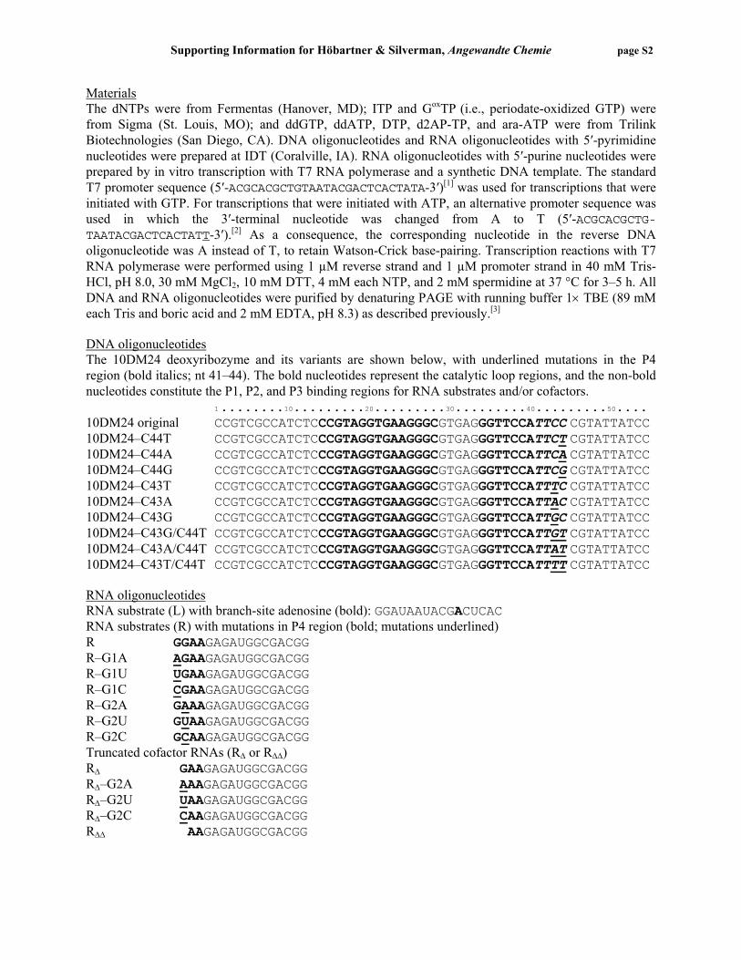
Materials The dNTPs were from Fermentas (Hanover, MD); ITP and GoxTP (i.e., periodate-oxidized GTP) were from Sigma (St. Louis, MO); and ddGTP, ddATP, DTP, d2AP-TP, and ara-ATP were from Trilink Biotechnologies (San Diego, CA). DNA oligonucleotides and RNA oligonucleotides with 5′-pyrimidine nucleotides were prepared at IDT (Coralville, IA). RNA oligonucleotides with 5′-purine nucleotides were prepared by in vitro transcription with T7 RNA polymerase and a synthetic DNA template. The standard T7 promoter sequence (5′-ACGCACGCTGTAATACGACTCACTATA-3′)[1] was used for transcriptions that were initiated with GTP. For transcriptions that were initiated with ATP, an alternative promoter sequence was used in which the 3′-terminal nucleotide was changed from A to T (5′-ACGCACGCTG-TAATACGACTCACTATT-3′).[2] As a consequence, the corresponding nucleotide in the reverse DNA oligonucleotide was A instead of T, to retain Watson-Crick base-pairing. Transcription reactions with T7 RNA polymerase were performed using 1 µM reverse strand and 1 µM promoter strand in 40 mM Tris-HCl, pH 8.0, 30 mM MgCl2, 10 mM DTT, 4 mM each NTP, and 2 mM spermidine at 37 °C for 3–5 h. All DNA and RNA oligonucleotides were purified by denaturing PAGE with running buffer 1× TBE (89 mM each Tris and boric acid and 2 mM EDTA, pH 8.3) as described previously.[3] DNA oligonucleotides The 10DM24 deoxyribozyme and its variants are shown below, with underlined mutations in the P4 region (bold italics; nt 41–44). The bold nucleotides represent the catalytic loop regions, and the non-bold nucleotides constitute the P1, P2, and P3 binding regions for RNA substrates and/or cofactors. 1 ........10.........20.........30.........40.........50.... 10DM24 original CCGTCGCCATCTCCCGTAGGTGAAGGGCGTGAGGGTTCCATTCC CGTATTATCC 10DM24–C44T CCGTCGCCATCTCCCGTAGGTGAAGGGCGTGAGGGTTCCATTCT CGTATTATCC 10DM24–C44A CCGTCGCCATCTCCCGTAGGTGAAGGGCGTGAGGGTTCCATTCA CGTATTATCC 10DM24–C44G CCGTCGCCATCTCCCGTAGGTGAAGGGCGTGAGGGTTCCATTCG CGTATTATCC 10DM24–C43T CCGTCGCCATCTCCCGTAGGTGAAGGGCGTGAGGGTTCCATTTC CGTATTATCC 10DM24–C43A CCGTCGCCATCTCCCGTAGGTGAAGGGCGTGAGGGTTCCATTAC CGTATTATCC 10DM24–C43G CCGTCGCCATCTCCCGTAGGTGAAGGGCGTGAGGGTTCCATTGC CGTATTATCC 10DM24–C43G/C44T CCGTCGCCATCTCCCGTAGGTGAAGGGCGTGAGGGTTCCATTGT CGTATTATCC 10DM24–C43A/C44T CCGTCGCCATCTCCCGTAGGTGAAGGGCGTGAGGGTTCCATTAT CGTATTATCC 10DM24–C43T/C44T CCGTCGCCATCTCCCGTAGGTGAAGGGCGTGAGGGTTCCATTTT CGTATTATCC RNA oligonucleotides RNA substrate (L) with branch-site adenosine (bold): GGAUAAUACGACUCAC RNA substrates (R) with mutations in P4 region (bold; mutations underlined) R GGAAGAGAUGGCGACGG R–G1A AGAAGAGAUGGCGACGG R–G1U UGAAGAGAUGGCGACGG R–G1C CGAAGAGAUGGCGACGG R–G2A GAAAGAGAUGGCGACGG R–G2U GUAAGAGAUGGCGACGG R–G2C GCAAGAGAUGGCGACGG Truncated cofactor RNAs (RΔ or RΔΔ) RΔ GAAGAGAUGGCGACGG RΔ–G2A AAAGAGAUGGCGACGG RΔ–G2U UAAGAGAUGGCGACGG RΔ–G2C CAAGAGAUGGCGACGG RΔΔ AAGAGAUGGCGACGG

Supporting Information for Höbartner & Silverman, Angewandte Chemie page S3
General description of kinetic assays All kinetic assays with the 10DM24 deoxyribozyme and its variants were performed similarly to previously described experiments.[3] The 5′-32P-labeled RNA substrate (L for “left-hand substrate”) that provides the branch-site adenosine was the limiting reagent relative to 10DM24 (E) and the truncated cofactor RNA (RΔ; R originally derived from “right-hand substrate”). The ratio L:E:RΔ was 1:10:30, with 0.25 µM E. The cofactor RΔ was 5′-phosphorylated unless otherwise stated. The RNA substrate L, deoxyribozyme E and cofactor RΔ were annealed in 5 mM HEPES, pH 7.5, 15 mM NaCl, 0.1 mM EDTA by heating at 95 °C for 3 min and cooling on ice for 5 min. The ligation reactions were performed with the appropriate concentration of NTP substrate (0.05–50 mM) at a final buffer concentration of 100 mM CHES, pH 9.0, 150 mM NaCl, 2 mM KCl, and 40–400 mM MgCl2 at 37 °C for up to 5 h. The combination of 1 mM NTP and 40 mM MgCl2 was defined as “standard incubation conditions”; 10 mM NTP and 150 mM MgCl2 was defined as “enhanced incubation conditions”. At appropriate timepoints, aliquots were removed from the sample, quenched into stop solution (80% formamide, 1× TB [89 mM each Tris and boric acid, pH 8.3], and 50 mM EDTA containing 0.025% bromophenol blue and xylene cyanol) and stored at –20 °C prior to analysis. Samples were separated by 20% denaturing PAGE at 30 W for 115 min and imaged with a PhosphorImager. The resulting data were fit to the equation yield = Y•(1–e–kt), where k = kobs and Y = final yield. Formation of GTP binding site in 10DM24 requires presence of RΔ The ligation reaction of GTP with the 2′-hydroxyl group of the branch-site adenosine in the substrate RNA (L) requires the presence of the cofactor RNA RΔ (Figure S1). Different phosphorylation states of RΔ (non-phosphorylated, 5′-monophosphorylated, and 5′-triphosphorylated) are tolerated. The highest ligation efficiency was observed with 5′-p-RΔ.
Figure S1. 10DM24-catalyzed ligation reaction of GTP with the branch-site adenosine of the 16-mer RNA substrate requires the RNA cofactor RΔ. The ligation reactions were performed under standard incubation conditions using 1 mM GTP and 40 mM MgCl2. The 20% denaturing PAGE images show the 0, 20, 40, 90, and 180 min timepoints for each reaction.
Supporting Information for Höbartner & Silverman, Angewandte Chemie page S4
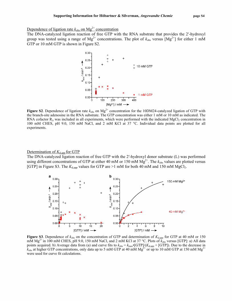
Dependence of ligation rate kobs on Mg2+ concentration The DNA-catalyzed ligation reaction of free GTP with the RNA substrate that provides the 2′-hydroxyl group was tested using a range of Mg2+ concentrations. The plot of kobs versus [Mg2+] for either 1 mM GTP or 10 mM GTP is shown in Figure S2.
Figure S2. Dependence of ligation rate kobs on Mg2+ concentration for the 10DM24-catalyzed ligation of GTP with the branch-site adenosine in the RNA substrate. The GTP concentration was either 1 mM or 10 mM as indicated. The RNA cofactor RΔ was included in all experiments, which were performed with the indicated MgCl2 concentration in 100 mM CHES, pH 9.0, 150 mM NaCl, and 2 mM KCl at 37 °C. Individual data points are plotted for all experiments. Determination of Kd,app for GTP The DNA-catalyzed ligation reaction of free GTP with the 2′-hydroxyl donor substrate (L) was performed using different concentrations of GTP at either 40 mM or 150 mM Mg2+. The kobs values are plotted versus [GTP] in Figure S3. The Kd,app values for GTP are >1 mM for both 40 mM and 150 mM MgCl2.
Figure S3. Dependence of kobs on the concentration of GTP and determination of Kd,app for GTP at 40 mM or 150 mM Mg2+ in 100 mM CHES, pH 9.0, 150 mM NaCl, and 2 mM KCl at 37 °C. Plots of kobs versus [GTP]: a) All data points acquired. b) Average data from (a) and curve fits to kobs = kmax•[GTP]/(Kd,app + [GTP]). Due to the decrease in kobs at higher GTP concentrations, only data up to 5 mM GTP at 40 mM Mg2+ or up to 10 mM GTP at 150 mM Mg2+ were used for curve fit calculations.
Supporting Information for Höbartner & Silverman, Angewandte Chemie page S5
Characterization of single-nucleotide branched RNA product For isolation of the 2′,5′-branched ligation product by PAGE, the ligation reaction was performed under standard incubation conditions (1 mM GTP and 40 mM MgCl2 in 100 mM CHES, pH 9.0, 150 mM NaCl, and 2 mM KCl at 37 °C) with 150 pmol unlabeled L RNA, 180 pmol 10DM24 and 225 pmol RΔ in 40 µL reaction volume for 3.5 h. The RNA product with the single added guanosine at the branch-site adenosine was characterized by MALDI-MS (m/z calcd. 5433, found 5437 ± 5).
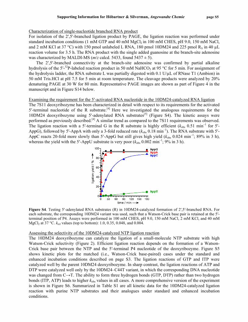
The 2′,5′-branched connectivity at the branch-site adenosine was confirmed by partial alkaline hydrolysis of the 5′-32P-labeled reaction product in 50 mM NaHCO3 at 95 °C for 5 min. For assignment of the hydrolysis ladder, the RNA substrate L was partially digested with 0.1 U/µL of RNase T1 (Ambion) in 50 mM Tris.HCl at pH 7.5 for 5 min at room temperature. The cleavage products were analyzed by 20% denaturing PAGE at 30 W for 80 min. Representative PAGE images are shown as part of Figure 4 in the manuscript and in Figure S14 below. Examining the requirement for the 5′-activated RNA nucleotide in the 10DM24-catalyzed RNA ligation The 7S11 deoxyribozyme has been characterized in detail with respect to its requirements for the activated 5′-terminal nucleotide of the R substrate.[4] Here we investigated the analogous requirements for the 10DM24 deoxyribozyme using 5′-adenylated RNA substrates[5] (Figure S4). The kinetic assays were performed as previously described.[4] A similar trend as compared to the 7S11 requirements was observed. The ligation reaction with a 5′-terminal G in the R substrate is highly efficient (kobs 0.51 min–1 for 5′-AppG), followed by 5′-AppA with only a 3-fold reduced rate (kobs 0.18 min–1). The RNA substrate with 5′-AppC reacts 20-fold more slowly than 5′-AppG but still gives high yield (kobs 0.024 min–1; 89% in 3 h), whereas the yield with the 5′-AppU substrate is very poor (kobs 0.002 min–1; 9% in 3 h).
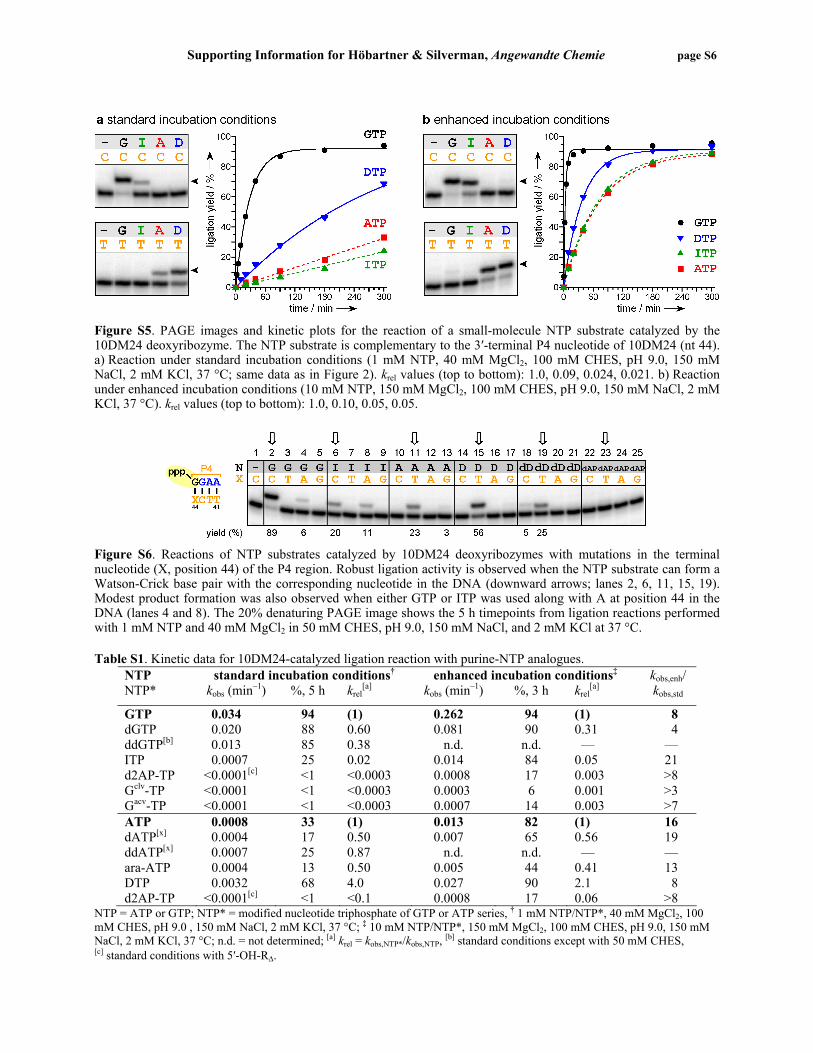
Figure S4. Testing 5′-adenylated RNA substrates (R) in 10DM24-catalyzed formation of 2′,5′-branched RNA. For each substrate, the corresponding 10DM24 variant was used, such that a Watson-Crick base pair is retained at the 5′-terminal position of P4. Assays were performed in 100 mM CHES, pH 9.0, 150 mM NaCl, 2 mM KCl, and 40 mM MgCl2 at 37 °C. krel values (top to bottom): 1.0, 0.35, 0.048, and 0.004. Assessing the selectivity of the 10DM24-catalyzed NTP ligation reaction The 10DM24 deoxyribozyme can catalyze the ligation of a small-molecule NTP substrate with high Watson-Crick selectivity (Figure 2). Efficient ligation reaction depends on the formation of a Watson-Crick base pair between the NTP and the 5′-terminal P4 nucleotide of the deoxyribozyme. Figure S5 shows kinetic plots for the matched (i.e., Watson-Crick base-paired) cases under the standard and enhanced incubation conditions described on page S3. The ligation reactions of GTP and ITP were catalyzed well by the parent 10DM24 deoxyribozyme. In sharp contrast, the ligation reactions of ATP and DTP were catalyzed well only by the 10DM24–C44T variant, in which the corresponding DNA nucleotide was changed from C→T. The ability to form three hydrogen bonds (GTP, DTP) rather than two hydrogen bonds (ITP, ATP) leads to higher kobs values in all cases. A more comprehensive version of the experiment is shown in Figure S6. Summarized in Table S1 are all kinetic data for the 10DM24-catalyzed ligation reaction with purine NTP substrates and their analogues under standard and enhanced incubation conditions.
Supporting Information for Höbartner & Silverman, Angewandte Chemie page S6
Figure S5. PAGE images and kinetic plots for the reaction of a small-molecule NTP substrate catalyzed by the 10DM24 deoxyribozyme. The NTP substrate is complementary to the 3′-terminal P4 nucleotide of 10DM24 (nt 44). a) Reaction under standard incubation conditions (1 mM NTP, 40 mM MgCl2, 100 mM CHES, pH 9.0, 150 mM NaCl, 2 mM KCl, 37 °C; same data as in Figure 2). krel values (top to bottom): 1.0, 0.09, 0.024, 0.021. b) Reaction under enhanced incubation conditions (10 mM NTP, 150 mM MgCl2, 100 mM CHES, pH 9.0, 150 mM NaCl, 2 mM KCl, 37 °C). krel values (top to bottom): 1.0, 0.10, 0.05, 0.05.
Figure S6. Reactions of NTP substrates catalyzed by 10DM24 deoxyribozymes with mutations in the terminal nucleotide (X, position 44) of the P4 region. Robust ligation activity is observed when the NTP substrate can form a Watson-Crick base pair with the corresponding nucleotide in the DNA (downward arrows; lanes 2, 6, 11, 15, 19). Modest product formation was also observed when either GTP or ITP was used along with A at position 44 in the DNA (lanes 4 and 8). The 20% denaturing PAGE image shows the 5 h timepoints from ligation reactions performed with 1 mM NTP and 40 mM MgCl2 in 50 mM CHES, pH 9.0, 150 mM NaCl, and 2 mM KCl at 37 °C. Table S1. Kinetic data for 10DM24-catalyzed ligation reaction with purine-NTP analogues.
NTP standard incubation conditions† enhanced incubation conditions‡ NTP* kobs (min–1) %, 5 h krel
[a] kobs (min–1) %, 3 h krel[a]
kobs,enh/ kobs,std
GTP 0.034 94 (1) 0.262 94 (1) 8 dGTP 0.020 88 0.60 0.081 90 0.31 4 ddGTP[b] 0.013 85 0.38 n.d. n.d. — — ITP 0.0007 25 0.02 0.014 84 0.05 21 d2AP-TP <0.0001[c] <1 <0.0003 0.0008 17 0.003 >8 Gclv-TP <0.0001 <1 <0.0003 0.0003 6 0.001 >3 Gacv-TP <0.0001 <1 <0.0003 0.0007 14 0.003 >7 ATP 0.0008 33 (1) 0.013 82 (1) 16 dATP[x] 0.0004 17 0.50 0.007 65 0.56 19 ddATP[x] 0.0007 25 0.87 n.d. n.d. — — ara-ATP 0.0004 13 0.50 0.005 44 0.41 13 DTP 0.0032 68 4.0 0.027 90 2.1 8 d2AP-TP <0.0001[c] <1 <0.1 0.0008 17 0.06 >8
NTP = ATP or GTP; NTP* = modified nucleotide triphosphate of GTP or ATP series, † 1 mM NTP/NTP*, 40 mM MgCl2, 100 mM CHES, pH 9.0 , 150 mM NaCl, 2 mM KCl, 37 °C; ‡ 10 mM NTP/NTP*, 150 mM MgCl2, 100 mM CHES, pH 9.0, 150 mM NaCl, 2 mM KCl, 37 °C; n.d. = not determined; [a] krel = kobs,NTP*/kobs,NTP, [b] standard conditions except with 50 mM CHES, [c] standard conditions with 5′-OH-RΔ.
Supporting Information for Höbartner & Silverman, Angewandte Chemie page S7
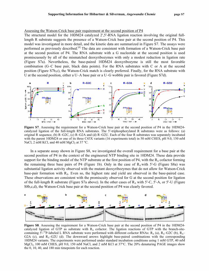
Assessing the Watson-Crick base pair requirement at the second position of P4 The structural model for the 10DM24 catalyzed 2′,5′-RNA ligation reaction involving the original full-length R substrate suggests the presence of a Watson-Crick base pair at the second position of P4. This model was investigated in more detail, and the kinetic data are summarized in Figure S7. The assays were performed as previously described.[4] The data are consistent with formation of a Watson-Crick base pair at the second position of P4. The RNA substrate with a G nucleotide at the second position is used promiscuously by all of the mismatched deoxyribozymes with only a modest reduction in ligation rate (Figure S7a). Nevertheless, the base-paired 10DM24 deoxyribozyme is still the most favorable combination (G–C base pair, black data points). For the RNA substrates with C or A at the second position (Figure S7b,c), the Watson-Crick match is clearly preferred. Finally, for the RNA substrate with U at the second position, either a U–A base pair or a U–G wobble pair is favored (Figure S7d).
Figure S7. Assessing the requirement for a Watson-Crick base pair at the second position of P4 in the 10DM24-catalyzed ligation of the full-length RNA substrates. The 5′-triphosphorylated R substrates were as follows: (a) original R sequence, (b) R–G2C, (c) R–G2A and (d) R–G2U. Each of the four R substrates was separately incubated with the parent 10DM24 or one of its three C43X variants (16 experiments total) in 50 mM CHES, pH 9.0, 150 mM NaCl, 2 mM KCl, and 40 mM MgCl2 at 37 °C.
In a separate assay shown in Figure S8, we investigated the overall requirement for a base pair at the second position of P4 in the context of the engineered NTP binding site in 10DM24. These data provide support for the binding model of the NTP substrate at the first position of P4, with the RΔ cofactor forming the remaining three base pairs of P4 (Figure 1b). Only in the case of RΔ with 5′-G (Figure S8a) was substantial ligation activity observed with the mutant deoxyribozymes that do not allow for Watson-Crick base-pair formation with RΔ. Even so, the highest rate and yield are observed in the base-paired case. These observations are consistent with the promiscuity observed for G at the second position for ligation of the full-length R substrate (Figure S7a above). In the other cases of RΔ with 5′-C, 5′-A, or 5′-U (Figure S8b,c,d), the Watson-Crick base pair at the second position of P4 was clearly favored.
Figure S8. Assessing the requirement for a Watson-Crick base pair at the second position of P4 in the 10DM24-catalyzed ligation of GTP as substrate with RΔ cofactor. The ligation reactions of GTP with the branch-site-containing 5′-32P-labeled L RNA substrate were performed with different cofactor RNAs: RΔ (a), RΔ–G2C (b), RΔ–G2A (c), and RΔ–G2U (d). The downward arrows highlight base-paired combinations with the corresponding 10DM24 variants. The experiments were performed under standard incubation conditions using 1 mM GTP, 40 mM MgCl2, 100 mM CHES, pH 9.0, 150 mM NaCl, and 2 mM KCl at 37°C. The 20% denaturing PAGE images show the 0, 10, 40, and 180 min timepoints for each reaction.
Supporting Information for Höbartner & Silverman, Angewandte Chemie page S8
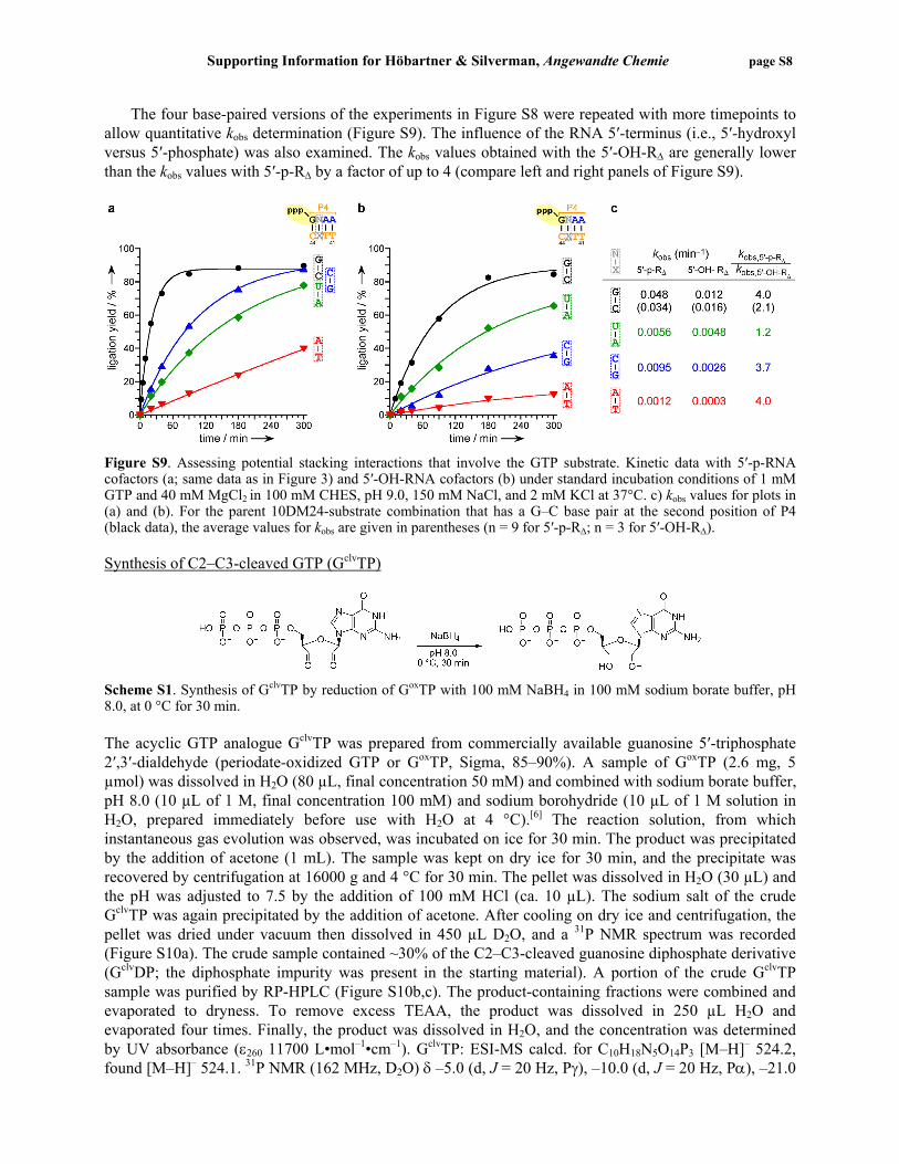
The four base-paired versions of the experiments in Figure S8 were repeated with more timepoints to allow quantitative kobs determination (Figure S9). The influence of the RNA 5′-terminus (i.e., 5′-hydroxyl versus 5′-phosphate) was also examined. The kobs values obtained with the 5′-OH-RΔ are generally lower than the kobs values with 5′-p-RΔ by a factor of up to 4 (compare left and right panels of Figure S9).
Figure S9. Assessing potential stacking interactions that involve the GTP substrate. Kinetic data with 5′-p-RNA cofactors (a; same data as in Figure 3) and 5′-OH-RNA cofactors (b) under standard incubation conditions of 1 mM GTP and 40 mM MgCl2 in 100 mM CHES, pH 9.0, 150 mM NaCl, and 2 mM KCl at 37°C. c) kobs values for plots in (a) and (b). For the parent 10DM24-substrate combination that has a G–C base pair at the second position of P4 (black data), the average values for kobs are given in parentheses (n = 9 for 5′-p-RΔ; n = 3 for 5′-OH-RΔ). Synthesis of C2–C3-cleaved GTP (GclvTP)
Scheme S1. Synthesis of GclvTP by reduction of GoxTP with 100 mM NaBH4 in 100 mM sodium borate buffer, pH 8.0, at 0 °C for 30 min. The acyclic GTP analogue GclvTP was prepared from commercially available guanosine 5′-triphosphate 2′,3′-dialdehyde (periodate-oxidized GTP or GoxTP, Sigma, 85–90%). A sample of GoxTP (2.6 mg, 5 µmol) was dissolved in H2O (80 µL, final concentration 50 mM) and combined with sodium borate buffer, pH 8.0 (10 µL of 1 M, final concentration 100 mM) and sodium borohydride (10 µL of 1 M solution in H2O, prepared immediately before use with H2O at 4 °C).[6] The reaction solution, from which instantaneous gas evolution was observed, was incubated on ice for 30 min. The product was precipitated by the addition of acetone (1 mL). The sample was kept on dry ice for 30 min, and the precipitate was recovered by centrifugation at 16000 g and 4 °C for 30 min. The pellet was dissolved in H2O (30 µL) and the pH was adjusted to 7.5 by the addition of 100 mM HCl (ca. 10 µL). The sodium salt of the crude GclvTP was again precipitated by the addition of acetone. After cooling on dry ice and centrifugation, the pellet was dried under vacuum then dissolved in 450 µL D2O, and a 31P NMR spectrum was recorded (Figure S10a). The crude sample contained ~30% of the C2–C3-cleaved guanosine diphosphate derivative (GclvDP; the diphosphate impurity was present in the starting material). A portion of the crude GclvTP sample was purified by RP-HPLC (Figure S10b,c). The product-containing fractions were combined and evaporated to dryness. To remove excess TEAA, the product was dissolved in 250 µL H2O and evaporated four times. Finally, the product was dissolved in H2O, and the concentration was determined by UV absorbance (ε260 11700 L•mol–1•cm–1). GclvTP: ESI-MS calcd. for C10H18N5O14P3 [M–H]– 524.2, found [M–H]– 524.1. 31P NMR (162 MHz, D2O) δ –5.0 (d, J = 20 Hz, Pγ), –10.0 (d, J = 20 Hz, Pα), –21.0
Supporting Information for Höbartner & Silverman, Angewandte Chemie page S9
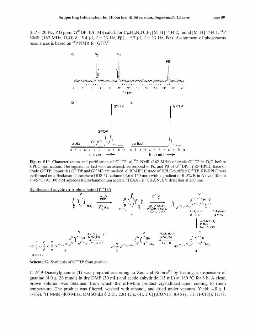
(t, J = 20 Hz, Pβ) ppm. GclvDP: ESI-MS calcd. for C10H16N5O11P2 [M–H]– 444.2, found [M–H]– 444.1. 31P NMR (162 MHz, D2O) δ –5.4 (d, J = 23 Hz, Pβ), –9.7 (d, J = 23 Hz, Pα). Assignment of phosphorus resonances is based on 31P NMR for GTP.[7]
Figure S10. Characterization and purification of GclvTP. a) 31P NMR (162 MHz) of crude GclvTP in D2O before HPLC purification. The signals marked with an asterisk correspond to Pα and Pβ of GclvDP. b) RP-HPLC trace of crude GclvTP. Impurities GclvDP and GclvMP are marked. c) RP-HPLC trace of HPLC-purified GclvTP. RP-HPLC was performed on a Beckman Ultrasphere ODS 5U column (4.6 × 150 mm) with a gradient of 0–5% B in A over 10 min at 45 °C (A: 100 mM aqueous triethylammonium acetate (TEAA), B: CH3CN; UV detection at 260 nm). Synthesis of acyclovir triphosphate (GacvTP)
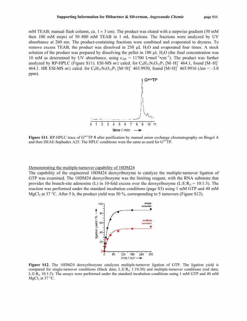
Scheme S2. Synthesis of GacvTP from guanine. 1. N2,9-Diacetylguanine (1) was prepared according to Zou and Robins[8] by heating a suspension of guanine (4.0 g, 26 mmol) in dry DMF (30 mL) and acetic anhydride (15 mL) at 180 °C for 8 h. A clear, brown solution was obtained, from which the off-white product crystallized upon cooling to room temperature. The product was filtered, washed with ethanol, and dried under vacuum. Yield: 4.8 g 1 (78%). 1H NMR (400 MHz, DMSO-d6) δ 2.21, 2.81 (2 s, 6H, 2 CH3CONH), 8.46 (s, 1H, H-C(8)), 11.78,

Supporting Information for Höbartner & Silverman, Angewandte Chemie page S10
12.24 (2 s, 2H, 2 NH) ppm. 13C NMR (100 MHz, DMSO-d6) δ 23.9, 24.7 (2 CH3CONH), 121.5, 137.5, 148.4, 154.6, 168.0, 173.8 (2 CONH) ppm. 2. N2-Acetyl-9-(2-acetoxyethoxymethyl)guanine (2) was prepared according to Gao and Mitra.[9] A mixture of acetic acid (0.26 mL, 4.5 mmol, 1.3 equiv.) and acetic anhydride (1.5 mL, 16.2 mmol, 4.8 equiv.) was cooled to 0 °C in an ice-water bath, and pTsOH (112 mg, 0.6 mmol, 0.2 equiv.) was added. 1,3-Dioxolane (1.2 mL, 17.4 mmol, 5.1 equiv.) was added dropwise to the stirred solution under continued cooling in the ice-water bath. A suspension of N2,9-diacetylguanine 1 (800 mg, 3.4 mmol) in toluene (5 mL) was added. The solution was allowed to warm to room temperature and was then heated at reflux for 2.5 h (120 °C oil bath temperature). TLC (9:1 dichloromethane/methanol) showed complete consumption of 1 and the formation of two products in ~1:1 ratio. The biphasic mixture (oily brown phase underneath colorless phase) was cooled to room temperature and the solvent was evaporated. The oily residue was separated by column chromatography on SiO2 with 2–10% methanol in dichloromethane (1% steps, 100 mL each). Both products were isolated and characterized as the 9-alkylation (2) and 7-alkylation (3) products by the characteristic chemical shift differences of H-C(8) and NCH2O in the 1H NMR spectrum.[9] The undesired 7-alkyation product 3 is less polar and was recovered from early fractions (#1-16), and the desired N2-acetyl-9-(2-acetoxyethoxymethyl)guanine 2 was recovered from late fractions (#19-55). Yield: 300 mg 2 (29%). 1H NMR (400 MHz, DMSO-d6) δ 1.93, 2.16 (2 s, 6H, 2 CH3CONH), 3.66, 4.05 (2 m, 4H, OCH2CH2OCO), 5.46 (s, 2H, NCH2O), 8.13 (s, 1H, H-C(8)), 11.79, 12.06 (2 s, 2H, 2 NH) ppm. Yield: 300 mg 3 (29%). 1H NMR (400 MHz, DMSO-d6) δ 1.93, 2.15 (2 s, 6H, 2 CH3CONH), 3.69, 4.04 (2 m, 4H, OCH2CH2OCO), 5.66 (s, 2H, NCH2O), 8.36 (s, 1H, H-C(8)), 11.63, 12.16 (2 s, 2H, 2 NH) ppm. 3. 9-(2-Hydroxyethoxymethyl)guanine (acyclovir; 4) was prepared by treatment of 3 (100 mg, 0.32 mmol) with 40% aqueous methylamine solution (1 mL) at room temperature for 1 h in a tightly stoppered round-bottom flask.[9] The solvent was evaporated and the residue was triturated with ethanol. The product was recovered by centrifugation (16000 g, at room temperature for 10 min) and dried under vacuum. Yield: 67 mg 4 (92%). 1H NMR (400 MHz, DMSO-d6) δ 3.44 (s, 4H, OCH2CH2OCO), 4.67 (s, 1H, OH), 5.32 (s, 2H, NCH2O), 6.49 (br. s, 2H, NH2), 7.80 (s, 1H, H-C(8)), 10.60 (br. s, 1H, NH) ppm. ESI-MS: m/z calcd. for C8H10N5O3 [M–H]– 224.2, found [M–H]– 224.1. 4. Acyclovir triphosphate has previously been synthesized via enzymatic and chemical routes.[10] Many chemical approaches are known for the synthesis of nucleoside analog triphosphates.[11] Here we followed a common procedure that involves formation of a dichlorophosphate intermediate. Acyclovir 4 (50 mg, 0.22 mmol) was coevaporated twice with pyridine (2 × 5 mL), dried under vacuum, placed under argon atmosphere, and dissolved in trimethyl phosphate (0.5 mL). Tributylamine (58 µL, 0.24 mmol, 1.1 equiv.) and POCl3 (23 µL, 0.24 mmol, 1.1 equiv.) were added under argon and the solution was stirred at room temperature for 1 h. Then, a solution of tributylammonium pyrophosphate (108 mg, 0.24 mmol, 1 equiv. based on POCl3) in trimethyl phosphate (1 mL) was added, and the clear solution was stirred at room temperature for 30 min. For TLC monitoring (6:3:1 iPrOH/NH4OH/H2O), a sample was quenched into triethylamine. The precipitate was recovered by centrifugation and dissolved in 50 mM triethylammonium bicarbonate, pH 8.0 (TEAB) for spotting onto a silica gel TLC plate. Multiple products and unreacted starting material were observed. After 50 min, triethylamine (1.8 mL, 60 equiv.) was added to the reaction mixture. The resulting precipitate was recovered by centrifugation and dissolved in 2 mL 50 mM TEAB. The aqueous solution was allowed to stand at room temperature for 2 h to ensure hydrolysis of the cyclic triphosphate intermediate. The crude product was loaded onto an anion-exchange column (Biogel A, Bio-Rad, 1 × 12 cm) which was previously equilibrated with 50 mM TEAB. The products were eluted with a stepwise gradient (50 mM then 100 mM steps) of 50–400 mM TEAB, and the fractions were monitored by TLC. The separation was poor, and a mixture of products was obtained. The presence of the desired GacvTP was confirmed by ESI-MS (not shown). A fraction of the sample was further purified by anion-exchange chromatography on DEAE-Sephadex A25 (0.5 g of DEAE-Sephadex A25 was swelled in 50
Supporting Information for Höbartner & Silverman, Angewandte Chemie page S11
mM TEAB; manual flash column, ca. 1 × 3 cm). The product was eluted with a stepwise gradient (50 mM then 100 mM steps) of 50–800 mM TEAB in 1 mL fractions. The fractions were analyzed by UV absorbance at 260 nm. The product-containing fractions were combined and evaporated to dryness. To remove excess TEAB, the product was dissolved in 250 µL H2O and evaporated four times. A stock solution of the product was prepared by dissolving the pellet in 180 µL H2O (the final concentration was 10 mM as determined by UV absorbance, using ε260 = 11700 L•mol–1•cm–1). The product was further analyzed by RP-HPLC (Figure S11). ESI-MS m/z calcd. for C8H13N5O12P3 [M–H]– 464.1, found [M–H]– 464.1. HR ESI-MS m/z calcd. for C8H15N5O12P3 [M+H]+ 465.9930, found [M+H]+ 465.9916 (Δm = –3.0 ppm).
Figure S11. RP-HPLC trace of GacvTP 5 after purification by manual anion exchange chromatography on Biogel A and then DEAE-Sephadex A25. The HPLC conditions were the same as used for GclvTP. Demonstrating the multiple-turnover capability of 10DM24 The capability of the engineered 10DM24 deoxyribozyme to catalyze the multiple-turnover ligation of GTP was examined. The 10DM24 deoxyribozyme was the limiting reagent, with the RNA substrate that provides the branch-site adenosine (L) in 10-fold excess over the deoxyribozyme (L:E:RΔ = 10:1:3). The reaction was performed under the standard incubation conditions (page S3) using 1 mM GTP and 40 mM MgCl2 at 37 °C. After 5 h, the product yield was 50 %, corresponding to 5 turnovers (Figure S12).
Figure S12. The 10DM24 deoxyribozyme catalyzes multiple-turnover ligation of GTP. The ligation yield is compared for single-turnover conditions (black data; L:E:RΔ 1:10:30) and multiple-turnover conditions (red data; L:E:RΔ 10:1:3). The assays were performed under the standard incubation conditions using 1 mM GTP and 40 mM MgCl2 at 37 °C.

Supporting Information for Höbartner & Silverman, Angewandte Chemie page S12
Investigating the cofactor binding site next to the substrate binding site in P4 The 10DM24-catalyzed ligation reaction of GTP in the presence of the two-nucleotide short cofactor RΔΔ was performed with the 5′-32P-labeled RNA substrate (L) that provides the branch-site adenosine as the limiting reagent. For kinetic assays, the ratio of L:E:RΔΔ was 1:10:30 with 0.25 µM E. For isolation of the reaction products, the ratio L:E:RΔΔ was 1:2:3 with 1.5 µM E. The reaction was performed under the enhanced incubation conditions with 20 mM GTP and 150 mM MgCl2 in 100 mM CHES, pH 9.0, 150 mM NaCl, and 2 mM KCl at 37 °C for up to 7 h. The ligation reaction with GTP resulted in the formation of two reaction products (Figure S13a). Both products were isolated and shown by partial alkaline hydrolysis (Figure S14) to be branched with the connectivities A–G and A–GG (branch-site adenosine underlined). Incubation with dGTP in place of GTP produced only the single-nucleotide addition product (Figure S13b). When the first two nucleotides of P4 in the deoxyribozyme (nt 43 and 44) were changed from CC→TT, ATP was accepted as both substrate and cofactor, because incubation with 20 mM ATP and 150 mM MgCl2 in the presence of RΔΔ and the 10DM24 variant resulted in a small amount of the ATP ligation product (Figure S13c).
Incubation at high pH for prolonged times results in partial random degradation of the RNA. Under the reaction conditions for ligation of GTP in the presence of RΔΔ, this random RNA degradation was <25% after 7 h at pH 9.0 and 37 °C (data not shown).
Figure S13. Investigating the cofactor binding site next to the substrate binding site in P4. a) Incubation of 5′-32P-labeled L RNA with GTP in the presence of 10DM24 and RΔΔ results in formation of two products, corresponding to addition of one and two guanosine nucleotides at the branch-site adenosine. b) Incubation with dGTP leads to formation of only the single-nucleotide branched product. c) The 10DM24 double mutant with CC→TT catalyzes ligation of ATP to the branch-site adenosine. All reactions in (a–c) were performed with 20 mM NTP and 150 mM MgCl2 in 100 mM CHES, pH 9.0, 150 mM NaCl, and 2 mM KCl at 37 °C for up to 7 h. The illustrated timepoints were at 3, 5, and 7 h for GTP and dGTP, and at 1, 3, and 6 h for ATP. d) Kinetic plots for the 10DM24-catalyzed NTP ligation in the two-nucleotide binding site architecture. The kobs values obtained from linear curve fits are given under the gel images in (a–c).

Supporting Information for Höbartner & Silverman, Angewandte Chemie page S13
Figure S14. Partial alkaline hydrolysis of isolated products from 10DM24-catalyzed ligation of GTP in the two-nucleotide binding site architecture. For both isolated products, comparison to the analogous assay for the linear precursor RNA confirms the 2′,5′-connectivity at the branch-site adenosine (underlined). The lane marked T1 shows partial digestion of L with RNase T1 (G-specific) for ladder calibration. Partial alkaline hydrolysis and T1 digestion were performed under the conditions described on page S5 for characterization of the single-nucleotide branched RNA product. Demonstrating the unreactivity of the single-nucleotide branched product with GTP The purified A–G product was tested as a substrate for 10DM24-catalyzed ligation of GTP in the presence of the RΔΔ cofactor [(A–G):E:RΔΔ ~1:10:30]. The reaction was performed under the enhanced incubation conditions of 20 mM GTP and 150 mM MgCl2. After incubation at 37 °C for 3 h, no new product formation was observed (data not shown). This is consistent with the hypothesis that two GTP nucleotides cannot be attached successively to the branch-site adenosine. Synthesis of pppGpG The dinucleotide substrate pppGpG was synthesized by abortive in vitro transcription using T7 RNA polymerase.[12,13] The transcription template was the reverse oligonucleotide 5′-TATAGTGAGTCGTAT-TATCCTATAGTGAGTCGTATTACAGCGTGCGT (initiation site for pppGpG synthesis underlined), together with the standard T7 promoter oligonucleotide (see page S2). Using 5′-CCCTATAGTGAGTCGTATTA-CAGCGTGCGT as template led to a smaller amount of desired dinucleotide product due to ‘slippage’ and therefore synthesis of a ladder of oligoguanosine nucleotides.[13] The in vitro transcription reaction was performed in the presence of GTP as the only nucleotide triphosphate. In a typical experiment, 200 pmol of the reverse oligonucleotide was annealed with 200 pmol of promoter oligonucleotide in 5 mM Tris, pH 7.5, followed by adjustment to final concentrations of 40 mM Tris, pH 8.0, 20 mM MgCl2, 10 mM GTP, 10 mM DTT, and 2 mM spermidine in a total reaction volume of 200 µL. The transcription was initiated by the addition of T7 RNA polymerase and the sample was incubated at 37 °C for 4 h. A solution of EDTA (25 µL of 0.5 M EDTA, pH 8.0) was added to the turbid reaction mixture, and the sample was vortexed until the magnesium pyrophosphate precipitate was completely dissolved. The clear solution was then extracted with 200 µL of 25:24:1 phenol/chloroform/isoamyl alcohol, and the products were precipitated by the addition of 1.2 mL of acetone. Turbidity appeared immediately; the mixture was frozen at –80 °C for 30 min, and the precipitate was recovered by centrifugation at 16000 g and 4 °C for 50 min. The supernatant was removed. The white pellet was dried under vacuum and redissolved in 15 µL H2O. The desired dinucleotide product was isolated from the crude mixture by RP-HPLC on a Beckman Ultrasphere ODS 5U column (4.6 × 150 mm) with a gradient of 0–10% B in A over 15 min at 45 °C (A: 100 mM aqueous triethylammonium acetate (TEAA), B: CH3CN; UV detection at 260 nm). The product-

Supporting Information for Höbartner & Silverman, Angewandte Chemie page S14
containing fractions were combined and evaporated to dryness. To remove excess TEAA, the product was dissolved in 250 µL H2O and evaporated four times. Finally, the product was dissolved in 30 µL H2O, and the concentration was determined by UV absorbance (ε260 23400 L•mol–1•cm–1). Yield: 120 nmol pppGpG. ESI-MS calcd. for C20H28N10O21P4 [M–H]– 867.4, found [M–H+Et3N]– 968.6. 10DM24-catalzyed ligation of pppGpG For the DNA-catalyzed ligation of pppGpG, the 5′-32P-labeled RNA with the branch-site adenosine (L) was incubated with the 10DM24 deoxyribozyme (E) and the cofactor RΔΔ in the ratio of L:E:RΔΔ = 1:10:30 in the presence of 1 mM pppGpG and 40 mM MgCl2 in 100 mM CHES pH 9.0, 150 mM NaCl, and 2 mM KCl at 37 °C. The analysis of the ligation reaction was performed as described on page S3. The ligation with pppGpG was efficient in the presence of RΔΔ (Figure S15, black circles); in contrast, in the absence of RΔΔ no product formation was observed (black crosses). This is consistent with the model that pppGpG and the first two nucleotides of RΔΔ together reconstitute the P4 helix. When the ligation reaction with pppGpG was tested in the presence of RΔ instead of RΔΔ, the ligation product A–GG was formed, but with a 10-fold decrease in kobs (red squares in Figure S15). Because RΔ provides an extraneous G nucleotide when combined with pppGpG (see right side of Figure S15), some disruption in activity is perhaps anticipated. Mutation of the second deoxyribozyme nucleotide in P4 from C→T resulted in a 130-fold decrease in kobs (blue triangles in Figure S15) No product formation was observed when the first DNA nucleotide in P4 (nt 44) was changed from C→T (green triangles in Figure S15). This is consistent with the observation that GTP was also not a substrate for the 10DM24–C44T mutant deoxyribozyme (compare Figure S6, lane 3).
Figure S15. 10DM24-catalyzed ligation of pppGpG. Reactions were performed at 1 mM pppGpG and 40 mM MgCl2 in 100 mM CHES, pH 9.0, 150 mM NaCl, and 2 mM KCl at 37 °C. kobs values (top to bottom): 0.19 min–1
(black), 0.01 min–1 (red), and 0.0014 min–1 (blue).

Supporting Information for Höbartner & Silverman, Angewandte Chemie page S15
The ligation reaction of pppGpG was performed using a variety of pppGpG concentrations with the parent 10DM24 deoxyribozyme in the presence of RΔΔ and 40 mM MgCl2. In Figure S16, the kobs values are plotted versus [pppGpG] and fit to kobs = kmax•[pppGpG]/(Kd,app + [pppGpG]). The Kd,app for pppGpG is >1 mM.
Figure S16. Dependence of kobs on the concentration of pppGpG and determination of Kd,app for pppGpG at 40 mM Mg2+ in 100 mM CHES, pH 9.0, 150 mM NaCl, and 2 mM KCl at 37 °C. References for Supporting Information [1] J. F. Milligan, D. R. Groebe, G. W. Witherell, O. C. Uhlenbeck, Nucleic Acids Res. 1987, 15, 8783-
8798. [2] a) T. M. Coleman, G. Wang, F. Huang, Nucleic Acids Res. 2004, 32, e14; b) F. Huang, G. Wang, T.
Coleman, N. Li, RNA 2003, 9, 1562-1570. [3] a) A. Flynn-Charlebois, Y. Wang, T. K. Prior, I. Rashid, K. A. Hoadley, R. L. Coppins, A. C. Wolf,
S. K. Silverman, J. Am. Chem. Soc. 2003, 125, 2444-2454; b) Y. Wang, S. K. Silverman, Biochemistry 2003, 42, 15252-15263.
[4] R. L. Coppins, S. K. Silverman, J. Am. Chem. Soc. 2005, 127, 2900-2907. [5] Y. Wang, S. K. Silverman, RNA 2006, 12, 1142-1146. [6] D. M. Hawley, J. J. Sninsky, G. N. Bennett, P. T. Gilham, Biochemistry 1978, 17, 2082-2086. [7] D. Solomon, M. Fridman, J. Zhang, T. Baasov, Org. Lett. 2001, 3, 4311-4314. [8] R. Zou, M. J. Robins, Can. J. Chem. 1987, 65, 1436-1437. [9] H. W. Gao, A. K. Mitra, Synth. Commun. 2001, 31, 1399-1419. [10] a) J. E. Reardon, T. Spector, J. Biol. Chem. 1989, 264, 7405-7411; b) P. A. Furman, M. H. St Clair,
J. A. Fyfe, J. L. Rideout, P. M. Keller, G. B. Elion, J. Virol. 1979, 32, 72-77. [11] K. Burgess, D. Cook, Chem. Rev. 2000, 100, 2047-2059. [12] F. Huang, Z. Yang, M. Yarus, Chem. Biol. 1998, 5, 669-678. [13] I. Kuzmine, C. T. Martin, J. Mol. Biol. 2001, 305, 559-566.


![69451 Weinheim, Germany - Wiley-VCH 20 % DMSO aqueous solution containing 3-(2-pyridyldithio) propionic acid N-hydroxysuccinimide ester (76.8 mM) or (2-[Biotinamido]ethylamido)-3,3](https://img.dokumen.tips/doc/110x75/5aafa8eb7f8b9a190d8d95e2/69451-weinheim-germany-wiley-20-dmso-aqueous-solution-containing-3-2-pyridyldithio.jpg)