Embed Size (px)
Citation preview

Radiation-Induced Vascular Damage in Tumors:Implications of Vascular Damage in AblativeHypofractionated Radiotherapy (SBRT and SRS)
Authors: Park, Heon Joo, Griffin, Robert J., Hui, Susanta, Levitt,Seymour H., and Song, Chang W.
Source: Radiation Research, 177(3) : 311-327
Published By: Radiation Research Society
URL: https://doi.org/10.1667/RR2773.1
BioOne Complete (complete.BioOne.org) is a full-text database of 200 subscribed and open-access titlesin the biological, ecological, and environmental sciences published by nonprofit societies, associations,museums, institutions, and presses.
Your use of this PDF, the BioOne Complete website, and all posted and associated content indicates youracceptance of BioOne’s Terms of Use, available at www.bioone.org/terms-of-use.
Usage of BioOne Complete content is strictly limited to personal, educational, and non - commercial use.Commercial inquiries or rights and permissions requests should be directed to the individual publisher ascopyright holder.
BioOne sees sustainable scholarly publishing as an inherently collaborative enterprise connecting authors, nonprofitpublishers, academic institutions, research libraries, and research funders in the common goal of maximizing access tocritical research.
Downloaded From: https://bioone.org/journals/Radiation-Research on 03 Apr 2020Terms of Use: https://bioone.org/terms-of-use

RADIATION RESEARCH 177, 311–327 (2012)0033-7587/12 $15.00�2012 by Radiation Research Society.All rights of reproduction in any form reserved.DOI: 10.1667/RR2773.1
REVIEW
Radiation-Induced Vascular Damage in Tumors: Implications of VascularDamage in Ablative Hypofractionated Radiotherapy (SBRT and SRS)
Heon Joo Park,a,b Robert J. Griffin,c Susanta Hui,a Seymour H. Levitta,d and Chang W. Songa,1
a Department of Therapeutic Radiology-Radiation Oncology, University of Minnesota Medical School, Minneapolis, Minnesota; b Department ofMicrobiology, Center for Advanced Medical Education by BK21 Project, College of Medicine, Inha University, Inchon, Korea; c Department ofRadiation Oncology, University of Arkansas for Medical Sciences, Little Rock, Arkansas; and d Department of Oncology-Pathology, Karolinska
Institute, Stockholm, Sweden
Park, H. J., Griffin, R. J., Hui, S., Levitt, S. H. and Song,C. W. Radiation-Induced Vascular Damage in Tumors:Implications of Vascular Damage in Ablative Hypofractio-nated Radiotherapy (SBRT and SRS). Radiat. Res. 177, 311–327 (2012).
We have reviewed the studies on radiation-inducedvascular changes in human and experimental tumorsreported in the last several decades. Although the reportedresults are inconsistent, they can be generalized as follows. Inthe human tumors treated with conventional fractionatedradiotherapy, the morphological and functional status of thevasculature is preserved, if not improved, during the earlypart of a treatment course and then decreases toward the endof treatment. Irradiation of human tumor xenografts orrodent tumors with 5–10 Gy in a single dose causes relativelymild vascular damages, but increasing the radiation dose tohigher than 10 Gy/fraction induces severe vascular damageresulting in reduced blood perfusion. Little is known aboutthe vascular changes in human tumors treated with high-dosehypofractionated radiation such as stereotactic body radio-therapy (SBRT) or stereotactic radiosurgery (SRS). However,the results for experimental tumors strongly indicate thatSBRT or SRS of human tumors with doses higher than about10 Gy/fraction is likely to induce considerable vasculardamages and thereby damages the intratumor microenviron-ment, leading to indirect tumor cell death. Vascular damagemay play an important role in the response of human tumorsto high-dose hypofractionated SBRT or SRS. � 2012 by Radiation
Research Society
INTRODUCTION
It is well accepted that the intratumor microenvironment,such as oxygenation status, greatly influences the radiosen-sitivity of tumor cells and that the intratumor microenvi-ronment is closely related to the functional status of tumormicrovasculature. Therefore, detailed insight into theradiation-induced changes in tumor microvasculature isimportant for maximizing the efficacy of radiotherapyagainst cancer. The radiation-induced changes in tumorblood vessels have been demonstrated to be markedlyvariable, depending on the total radiation dose, dose rate,fraction size and the number of fractions as well as onbiological factors such as the tumor type, tumor site and thestage of tumor growth. In the early years of radiotherapy,tumors were irradiated with single or a few fractionatedlarge doses (1, 2). However, after the landmark observationsby Regaud and Ferroux (3) and Coutard (4) about 80 yearsago that the therapeutic ratio in treating cancer withradiation could be increased by delivering the radiation inmultiple fractions of small doses, fractionated radiotherapyhas been an almost universally accepted clinical practice. Asearly as 1936, Mottram (5) reported that the cancer cells inthe peripheral tumor volume with good blood supply weremore sensitive to radiation than the cells in the central tumorvolume. Subsequent studies demonstrated that the responseof tumor cells to radiation is closely related to oxygensupply through blood perfusion and that fractionatedradiotherapy minimizes radiation-induced vascular damage,thereby allowing reoxygenation of hypoxic tumor cells. Inthe 1950s, radiosurgery using gamma knives was intro-duced by Leksell (6) to deliver high-dose hypofractionatedradiation to confined vascular lesions and malignancies inthe brain. As a result of the recent remarkable advances inimaging technology, computer-aided field shaping, treat-ment planning, dosimetry and radiation delivery systems, ithas now become possible to conformally deliver 20–60 Gyto tumors in a single fraction or 2–5 fractions (7–11). This is
1Address for correspondence: Radiobiology Laboratory, Depart-ment of Therapeutic Radiology-Radiation Oncology, University ofMinnesota Medical School, MMC494, 424 Harvard St. S.E.Minneapolis, MN 55455; e-mail: [email protected].
311
Downloaded From: https://bioone.org/journals/Radiation-Research on 03 Apr 2020Terms of Use: https://bioone.org/terms-of-use
referred to as stereotactic radiosurgery (SRS) for thetreatment of intracranial lesions and stereotactic bodyradiation therapy (SBRT) for the treatment of extracranialtumors. A relevant and important question is the fate oftumor blood vessels when tumors are exposed to such high-dose hypofractionated radiation. Another pertinent questionis whether the well-established radiobiological principles ofconventional fractionated radiotherapy such as reoxygena-tion of hypoxic cells play any role in the response of tumorsto SRS or SBRT. We have briefly discussed these topics inour recent publication (12). Numerous reports havedescribed the radiation-induced vascular changes in tumorsand their implications for radiotherapy but have oftenreached conflicting conclusions. The purpose of the presentreview is to examine the previous studies on radiation-induced vascular changes in both human and experimentaltumors to establish an up-to-date view of the subject withgreater relevance to the rapidly growing interest in treatingtumors with high-dose per fraction radiotherapy, such asSBRT and SRS.
TUMOR BLOOD VESSELS AND BLOOD FLOW
Solid tumors acquire blood vessels by coopting neigh-boring vessels in normal tissues and angiogenesis, that is,sprouting or intussusceptive microvascular growth fromexisting arteries or veins (13, 14). Tumor blood vessels arealso formed by vasculogenesis by adding endothelialprogenitor and other stem-like cells that derived from thebloodborne and bone marrow-delivered stem cells to thegrowing tumor vessels (15). As tumors grow and invadenormal tissues, the arteries of normal tissues are incorpo-rated into the tumors. Therefore, varying fractions of tumorvascular beds originate from normal tissues. The hastilyformed capillary-like tumor microvasculature is comprisedof a single layer of endothelial cells often separated by gapsand is devoid of underlying basement membrane or smoothmuscle cells (pericytes) and innervations. However, some ofthe microvasculature of slowly growing human tumorsexhibit a thin layer of smooth muscle similar to thecapillaries of normal tissues (13). In general, tumor bloodvessels are irregular in diameter with rather narrow tubesconnected next to dilated and often sinusoid-like structures.Compared to the well-organized web-like network ofhomogeneous capillaries in normal tissues, the tumorcapillaries are sharply bent, tortuous and branched withmultiple dead ends (16). Blood perfusion through suchdisorganized vascular networks tends to be sluggish andoften intermittently stationary. However, it should bestressed that the architecture and morphology of tumormicrovasculature and the blood perfusion are markedlyheterogeneous and variable, depending on various factorssuch as tumor type, tumor size and site of tumor growth.Even within a tumor, the vascular distribution and bloodperfusion are rather heterogeneous. For example, in some
tumors, the central volume is well vascularized and wellperfused compared to the periphery, while in other types oftumors the opposite is true. The tumor blood vessels areusually more permeable and leaky compared with the bloodvessels in the surrounding normal tissues probably becausethe tumor vasculatures are morphologically immature (17).The tumor vascular permeability is also significantlyvariable depending on tumor type. For example, theblood-brain barrier in the normal brain tissue is retainedin many brain tumors, and thus the blood vessels in braintumors tend to be less permeable than the blood vessels ofother types of tumors. The interstitial fluid pressure (IFP) ofcertain types of tumors is known to be elevated owing to thehigh vascular permeability in tumors (14).
RADIATION-INDUCED VASCULAR CHANGES INTUMORS
We have summarized the 43 representative studies on theradiation-induced changes in tumor vasculature reported inthe last 60 years in Table 1. Of the 43 reports (18–60), thefirst seven (18–24) describe vascular changes in humantumors and the remainder are related to radiation-inducedvascular changes in either human tumor xenografts inrodents or transplanted mouse or rat tumors. Variousmethods, including colpophotography (18), 133Xe clearance(19), Doppler sonography (20), MR imaging (22) and CTimaging (23, 24), have been used to study the radiation-induced vascular changes in human tumors. All the studieswith human tumors are concerned with the vascular changescaused by conventional fractionated radiotherapy. Bergsjo(18) studied the vascular changes in cervical tumors causedby radiotherapy with 17 Gy delivered in 10–12 fractions.Colphophotographic examination of the tumor surfaceindicated that the vascularity on the surface of tumorsimproved slightly at the end of the therapy. However, itshould be realized that the changes in the vasculature on thesurface of tumors as determined with colphophotographymay not represent the overall vascular changes in tumors.The conclusions of other studies on human tumors (19–24)follow a general trend that blood flow remains unchanged orincreases slightly during the early period of fractionatedradiotherapy but decreases toward the end of treatments. Forexample, in the study by Mayr et al. (22), human cervicalsquamous carcinoma and adenocarcinoma were treated with40–50 Gy over 4–5 weeks in 5 fractions/week (2 Gy/fraction). Blood flow, as determined by MRI, increasedduring the first 2 weeks of therapy and decreased thereafter.The authors reported that high cervical tumor perfusionbefore the treatment and increasing or persistent highperfusion during the early part of the therapy was afavorable sign of the treatment outcome. Pirhonen et al. (20)reported that a decrease of tumor vasculature during thefractionated radiotherapy of advanced cervical carcinomawas associated with a better treatment outcome and that the
312 REVIEW
Downloaded From: https://bioone.org/journals/Radiation-Research on 03 Apr 2020Terms of Use: https://bioone.org/terms-of-use
TABLE 1Summary of Studies on the Radiation-Induced Vascular Changes in Human Tumors, Human Tumor Xenografts Grown in
Animals, and Animal Tumors
Tumors and sites Methods Vascular changesAuthor(s)
(year) (ref.)
Human cervicalcarcinoma(143 patients)
Colpophotography Irradiation with 1700 R in 10–12 fraction in 15 days, in general,slightly increased the surface vascularity at the end of fractionatedradiation therapy.
Bergsjo (1968)(18)
Human superficialmetastatic tumors(43 patients with 48tumors)
133Xe clearance Tumors were irradiated with 1100–3000 rad/week in 5 fraction/week. Blood flow increased during the 1st week of the treatmentand decreased from the 2nd week of the treatment. In a longerfollow up, the tumor blood flow decreased continuously.
Mantyla et al.(1982) (19)
Human advancedcervical carcinoma(14 patients)
Color Doppler, ultrasound Irradiated with 30–65 Gy at 1.9 Gy/fraction and 5 fraction/week. In11 out of 14 tumors, vascularity and blood flow decreasedsignificantly. The decrease in tumor vasculature during thetreatment was associated with better outcome. Eight of 10patients with increased tumor vascularity at the end of radiationneeded further treatment or died of disease.
Pirhonen et al.(1995) (20)
Human uterine cervicalcancer
Immunostaining of biopsysections for factor VIII-related antigen
Radiotherapy with 50 Gy in 2 Gy/day for 5 times/week. Endocavitarybrachytherapy was delivered in 7–8 fraction to a total dose of 29–34Gy. No change in vascular density was observed, but vasculardamage occurred as indicated by endothelial swelling and hypertrophyduring early phase of radiotherapy. Tumor oxygen tension varieddepending on various factors beside vascular functions.
Lyng et al.(2000) (21)
Human squamouscarcinoma (14) andadenocarcinomas (3)of cervix
MR imaging Irradiated with 40–45 Gy/4–5 week, 5 fraction/week. Some tumorsshowed increased blood perfusion during the first 2 weeks, butthe perfusion decreased thereafter. High tumor perfusion beforetherapy and increasing or persistent high perfusion early duringthe course of therapy appeared to be favorable signs.
Mayr et al.(1996) (22)
Human non-small-celllung cancer(16 patients)
Volumetric perfusioncomputed tomography
Vascular blood volume and permeability were greater in tumor rimthan tumor center. After fractionated irradiation with 9 Gy in 2fraction, 18 Gy in 4 fraction and 27 Gy in 6 fraction, vascularvolume increased significantly in tumor rim and slightly in tumorcenter. Vascular permeability also increased in tumor rim, but notin tumor center.
NG et al.(2007) (23)
Human rectal cancer(23 patients)
Perfusion CT imaging Irradiated with 25 Gy in 5 fraction (5 Gy ’ 5) in 1 week. From 3days after the hypofractionated treatment, trans-endothelialvolume constant (K trans) (permeability) slightly increased. Theincreased vascular permeability may improve the bioavailabilityof cytotoxic agents in rectal tumors.
Janssen et al.(2009) (24)
Human melanomaxenograft in athymicnude mice in theflank
Angiography In 1 week after irradiation with 10.0–15.0 Gy in a single dose, 35–45% of 5–15-lm-diameter vessels were nonfunctional. The dosesrequired for loss of 50% of the functional vessels with diametersof 5–15, 15–25, and 25–35 lm were 16, 21 and 20 Gy,respectively. In spite of early loss of functional vessels, tumorsbecame supervascularized as tumors regressed after 20 or 25 Gyirradiation. Regrowth of irradiated tumors appeared to bepreceded by efficient neovascularization.
Solesvik (1984)(25)
Human colon tumorxenografts in theflank (s.c.) ofathymic mice
99mTcO4-RBC forfunctional vascularvolume and 125I-plasmaprotein for vascularpermeability
Irradiation with 4–16 Gy in a single dose increased the vascularpermeability in 24–72 h and decreased the functional vascularvolume in 24 h. The increase in vascular permeability byirradiation is potentially valuable to increase monoclonal antibodyuptake by tumors.
Kalofonos et al.(1990) (26)
Human laryngealsquamous cellcarcinoma xenograftsin nude mice
Histological imaging ofendothelial marker forvessels and Hoechst33342 injection forvascular perfusion
After irradiation with 10 Gy, the number of perfused vessels slightlyincreased within 1 day, and then significantly decreased at 26 hfollowed by recovery to control level in 7–11 days. The hypoxiccell fraction decreased at 7 h after irradiation but significantlyincreased to pre-irradiation levels at 11 days after irradiation.
Bussink et al.(2000) (27)
Human MA148 ovariancarcinoma xenograftsin nude mice, s.c.
Immunohistochemistry forPECAM (CD31-redfluorescence
After irradiation with 5 Gy/week for 4 weeks, the total vesseldensity decreased by 50%. Irradiation and anginex synergisticallyreduced the functional vascularity in tumors.
Dings et al.(2005) (28)
Human A-07 melanomaxenografts in nudemice
Dynamic contrast-enhanced magneticresonance imagingusing Gd-DTPA
At 72 h after 10 Gy irradiation in a single dose, tumor bloodperfusion decreased by 40%. However, intratumor mean pO2 andpO2 fluctuation were not altered by irradiation with 5 or 10 Gy,indicating that intratumor microenvironment was not changed by5–10 Gy irradiation.
Brurberg (2006)(29)
Continued on next page
REVIEW 313
Downloaded From: https://bioone.org/journals/Radiation-Research on 03 Apr 2020Terms of Use: https://bioone.org/terms-of-use

TABLE 1Continued.
Tumors and sites Methods Vascular changesAuthor(s)
(year) (ref.)
Human A549 lungadenocarcinomaxenografts in the hindlegs of nude mice,s.c.
Hoechst 33342 for bloodperfusion and DynamicMagnetic Resonanceimaging of GD-DTPAfor functionalvascularization
Analysis with Hoechst 333342 indicated a rich blood vesselperfusion in the peripheral part of the tumors. Irradiation with 20Gy in a single dose caused no changes in vascular densitywhereas apoptosis of tumor cells was significant at 10.5 hpostirradiation. Blood perfusion, as determined with GD-DTPAimaging increased at 1 h postirradiation. Hypoxic area in thetumors decreased for 30.5 h after irradiation.
Fokas et al.(2010) (30)
Human U251glioblastomaxenograft grown s.c.in the back orintracranially (i.c.) innude mice
Fluorescence imaging oflectin for i.c. tumorsand ultrasound analysisof contrast agent fors.c. tumors
Irradiation with 15 Gy in a single dose decreased blood perfusion to10% of control in i.c. tumors and to 30% of control in s.c.tumors in 2 weeks. In i.c. tumors, CD31-stained cells (endothelialcells) were reduced to 25% of control accompanied by markedincrease in hypoxic area (pimonidazole staining). Thereafter, thedamaged vasculatures were restored by virtue of vasculogenesisthrough recruitment of bone marrow-derived cells in both s.c. andi.c. tumors. AMD3100, an inhibitor of vasculogenesis, preventedthe recovery of tumor vasculature. Vasculogenesis needs to beblocked for complete control of tumor by radiotherapy.
Kioi et al.(2010) (31)
Mouse adenocarcinomaof C3H mice intransparent chambers
Transparent chamber.Microsocpicobservation
Irradiated with 2,000 or 3,000 R in a single fraction caused pronouncednarrowing of microvessels for approximately 1 week. By 2–4 daysafter irradiation, the circulation was slowed. The retardation ofcirculation during 2–5 days postirradiation was responsible for tumorcell death. Irradiated vessels were unable to regrow.
Merwin et al.(1950) (32)
Hamster neurilemmomain the cheek pouchchambers
Cheek pouch transparentchamber. Microscopicobservation
Irradiation with 3,000 R caused variable degrees of edema andextensive reduction in blood flow in 24–30 h, with subsequentrestoration toward normalcy accompanied by small focalhemorrhaging. Subsequent tumor growth with neovascularizationbegan in the perimeter of the tumor.
Eddy (1980)(33)
Rat R3240 Acmammaryadenocarcinoma inwindow chambers
Dorsal flap transparentwindow chamber.Microscopicobservation
Irradiation with 5 Gy caused conjoint increase in both vasculardensity and perfusion during 24–72 h post-irradiation, althoughthe degree of change was variable from one individual to thenext. The degree of change in vascular density was inverselyrelated to median pretreatment diameter.
Dewhirst et al.(1990) (34)
Mouse adenocarcinomain the thigh
Histological examination Irradiated with 2400–2600 R in 1 fraction. Slight dilation of bloodvessels occurred immediately after irradiation. From 24 hpostirradiation, blood vessels dilated markedly and ruptured, andblood extravasated.
Lasnitzki (1947)(35)
Mouse malignanttumors in the flank,s.c., 5 differenttumors
Angiography Irradiated with 680–3,000 R in 1 fraction. From 3rd day after 680 Rirradiation, the vascularity started to decrease. Abrupt taperingand narrowing of vessels peaked 9–13 days after 680-2000 Rirradiation.
McAlister et al.(1963) (36)
Rat Walker carcinomaand Murphy-Sturnlymphoma in theflank, s.c
Angiography andHistology
After irradiation with 500 R/day for 3 days (1500 R),supervascularization developed. When the tumors were treatedwith fractionated irradiation with relatively small fraction sizes,progressive destruction of tumor parenchymal cells preceded theregression of the microcirculation leading to development ofsupervascularization. However, after exposure to 1,500–6,000 Rin 1 fraction, tumor vasculatures fragmented.
Rubin et al.(1966) (37)
Mouserhabdomyosarcoma inthe flank, s.c.
133Xe clearance Irradiation with 2000 R markedly reduced the blood flow ratebetween 1 and 9 days post-irradiation.
Robert (1967)(38)
Rat Walker carcinomain the flank, s.c
51Cr-RBC for functionalvascular volume and125I-plasma protein forvascular permeability.
Soon after irradiation with 2 Gy, the extravasation of plasma protein(permeability) increased while the vascular volume remainedunchanged. In 2–12 days after irradiation with 10–60 Gy, thevascular volume significantly decreased. Revascularizationoccurred as the tumor began to regrow about 15 days after 30 Gyirradiation.
Song et al.(1971) (39)
Rat Walker carcinomain the flank, s.c.
133Xe clearance Irradiation with 20 Gy in 1 fraction increased the 133Xe clearancerate in 1–6 days whereas it decreased the vascular volume.
Song et al.(1972) (40)
Rat Walker carcinomain the flank, s.c
51Cr-RBC for functionalvascular volume and125I-plasma protein forvascular permeability
While irradiation with 2.5 Gy increased the vascular volume, 5–20Gy decreased the vascular volume at 24 h post-irradiation. Theextravasation of plasma protein increased at 24 h after irradiationwith 2.5–20 Gy.
Wong et al.(1973) (41)
Continued on next page
314 REVIEW
Downloaded From: https://bioone.org/journals/Radiation-Research on 03 Apr 2020Terms of Use: https://bioone.org/terms-of-use

TABLE 1Continued.
Tumors and sites Methods Vascular changesAuthor(s)
(year) (ref.)
Mouse KHT sarcoma inthe flank, i.m.
133Xe clearance Blood flow (133Xe clearance) tended to decrease 3 h after irradiationwith .1,000 rad and increased 3–4 days and 7 days afterirradiation with 1000 and 2,000 or 4,000 rad. Reoxygenation ofhypoxic cells occurred as a result of increased blood perfusionafter irradiation. Blood flow decreased after irradiation with8,000–16,000 rad.
Kallman et al.(1972) (42)
Mouse neuroblastoma inthe flank, s.c
51Cr-RBC for functionalvascular volume and125I-plasma protein forvascular permeability.Histopathology
Irradiation with 2.5 and 5 Gy slightly increased the intravascularvolume at 24 h, but it decreased thereafter. Irradiation with 10and 20 Gy caused progressive decrease in the vascular volume toabout 30% of control at days 12. Extravasation of plasma proteinincreased during 3 days after 2.5–20 Gy irradiation. As thetumors regressed after 20 Gy irradiation, the vascular networkbecame disorganized, aggregated and condensed.
Song et al.(1974) (43)
Mouse mammarycarcinoma in the leg,s.c.
Morphometry (colloidalcarbon filling)
After 500 R irradiation, the vascular volume slightly decreased in 1day and recovered in 4 days. Vascular volume decreased andfailed to recover after irradiation with 1,500 R. Nevertheless, thetransient vascular changes conceivably rendered the hypoxic cellsreoxygenated. Irradiation with 4500 R caused extensive damageto microvasculature.
Hilmas et al.(1975) (44)
Mouse mammarycarcinoma in the leg,s.c
51Cr-RBC for functionalvascular volume
After irradiation with 10 and 20 Gy, vascular volume increasedwithin 24 h and subsequently decreased. The numbers of viabletumor cells significantly decreased in 3 days after irradiation. Thetransitional increase in blood supply and the decline in theoxygen consumption due to death of tumor cells may lead totransitional increase in oxygenation status of surviving tumorcells after tumors are exposed to high dose radiation.
Clement et al.(1976) (45)
Rat BA-1112rhabodomyosarcomain posterior portionof scalp
Photon activation ofoxygen and 15Opositron decay
After irradiation with 16.5, 38.5 or 60.5 Gy, blood flow decreasedby 35–50%. However, the blood flow in the tumor irradiated with16.5 Gy recovered in 24–48 h while that of tumor irradiated with60.5 Gy remained decreased at 72 h after irradiation. Irradiationwith small doses used in fractionated radiotherapy did not causevascular changes.
Emami et al.(1981) (46)
Mouse RIF-1 tumorgrown in the leg, s.c.
14C-iodo-antipyrineuptake
Blood perfusion increased 1–2 days after irradiation with 20 Gy butnot after irradiation with 2 Gy. Tumor necrosis increased about 3times at 1 day after 20 Gy irradiation and then declined to abouttwice its control value at 2 days.
Tozer et al.(1989) (47)
Rat RIHrhabdomyosarcomasgrown in the flank.
Electron micrography ofvascular wall of tumorcapillaries. Tumor pO2was measured withpolarographicelectrodes
Tumors were irradiated with 60 Gy in 3 Gy 3 20 fraction for 4weeks. After 30 Gy irradiation, inner surface of the lumenbecame rough, and the endothelial cells and pericytes wereswollen. After 60 Gy irradiation, endothelial cells becameelongated, sometimes detached from each other and from basallamina. In the early phase of the fractionated treatment (up to 30Gy) tumor oxygenation slightly improved, but it distinctlydecreased in the later phase of treatment.
Zywietz et al.(1996) (48)
Rat BT4C gliomagrown intracerebrally
Immunohistochemicalstaining for bloodvessels
Whole brains treated with 20 Gy in 4 Gy 3 5 fraction tumorvolume decreased to 77% of original volume and themicrovascular density decreased to 72% of original value at 5thafter the treatment.
Johansson et al.(1999) (49)
Mouse KHTfibrosarcoma in thehind limb, i.m.
Immunohistochemicalstaining for bloodvessels and fluorescentDiOC7 injection forperfused vessels
Many tumor vessels were not perfused before irradiation. Thedensity of perfused vessels decreased at 24 h after 10 Gyirradiation and recovered to control level by 72 h. Despite thedecrease in the perfusion, O2 availability appeared to be increaseddue to reduction in O2 consumption and yet the radiobiologicallyhypoxic cell fraction did not change.
Fenton et al.(2001) (50)
Mouse glioblastoma inthe thighs, s.c.
Power Doppler ultrasound for vascularity(vascular index) andImmunofluorescencestaining of tissuesections
The majority of the functional vasculatures were in the periphery oftumor. Irradiation with 10 Gy in 2 fractions reduced the tumorvascularity to 37% of control in 3 days. The immunofluorescencestaining for endothelial cell demonstrated a similar pattern to thatof the quantified power Doppler US study.
Donnelly et al.(2001) (51)
Continued on next page
REVIEW 315
Downloaded From: https://bioone.org/journals/Radiation-Research on 03 Apr 2020Terms of Use: https://bioone.org/terms-of-use
TABLE 1Continued.
Tumors and sites Methods Vascular changesAuthor(s)
(year) (ref.)
Rat autochthonousmammary tumorsinduced by s.cinjection of N-nitrosoN-methyl urea
Power Dopplersonography forDoppler index(PDI)(vascularity)
Irradiation with 18 Gy in a single fraction caused variable reduction(12–63%) in PDI (vascularity). The degree of reduction in PDI at7 days after irradiation was correlated with the subsequent tumorregression. The early changes in tumor perfusion after irradiationappeared to precede the long-term tumor regression.
Denis et al.(2003) (52)
Mouse SCCVII tumorsgrown in the hindlegs of C3H mice,s.c.
Selective irradiation oftumor vesselsendothelial cells withBNCT using BSH-liposome
Selective irradiation of tumor vessel endothelial cells with 30–33 Gyirradiation with 10B(na)7Li reaction completely suppressed thetumor growth without direct irradiation of tumor cells.Destruction of tumor vasculatures alone can cause tumorregression.
Ono et al.(2003) (53)
MCF/129 fibrosarcomawere induced inapoptosis sensitiveAmaseþ/þ orapoptosis resistantAmase–/– mice, andB1F1 melanomaswere induced inBaxþ/þ or Bax–/– mice
Endothelial cell apoptosisin tumors wasdetermined by TUNEL.Radiation-inducedtumor growth delaywas related toendothelial cellapoptosis.
Irradiation of tumors with 15 Gy in a single dose caused far moretumor endothelial cell apoptosis in Amaseþ/þ mice than inAmase–/– mice. Tumors grown in apoptosis-resistant Amase–/–
mice and Bax–/– mice were much more radioresistant than thetumors grown in Amaseþ/þ mice or Baxþ/þ mice. Conclusion:Microvascular damage regulates tumor response to radiation atthe clinically relevant dose range.
Garcia-Barroset al. (2003)(54)
Mouse squamous cellcarcinoma (SCCVII)in the hind legs ofC3H mice, s.c.
Dynamic magneticresonance imaging ofG8-Gd-D (13 nm) forvascular permeability
After irradiation with 15 Gy, vascular permeability increased toabout 1.4 times of control occurring between 7 and 24 h afterirradiation. Irradiation with 5 Gy 3 5 decreased the vascularpermeability whereas 2 Gy 3 5 and 3 Gy 3 5 did not.
Kobayashi et al.(2004) (55)
Mouse K1735melanoma grown inC3H/HeN mice
Doppler ultrasound forblood flow.Immunostaining forvascular density, HIF-1and VEGF. TUNELstaining for endotheliacell (EC)apoptosis
Tumors were exposed to 12 Gy. Blood flow decreased significantlyand the histological preparations of tumors grown 10-fold involume after irradiation showed the following: prominent regionsof hypoxia (EF5 positive), HIF-1a and VEGF, and decreasedmicrovascular density. Significant EC death occurred next dayafter irradiation.
Tsai et al.(2005) (56)
Lewis lung carcinomaof C57BL/mice.Grown s.c. in thehind limb and dorsalskin window chamber
Doppler ultrasound forblood flow,microvascular density,endothelial cellsapoptosis, proliferationof tumor cells andtumor growth
Irradiation of tumors in window chambers with 20 Gy completelyobliterated tumor vessels. The blood flow and vascular density inthe s.c. tumors markedly decreased at 2 days after 20 Gyirradiation but blood flow recovered substantially at day 4. Second20-Gy irradiation 2 days after the 1st 20 Gy was far more effectivethan that 4 days after the 1st 20 Gy for sustained reduction ofblood flow and also for suppression of tumor growth.
Kim et al.(2006) (57)
Rat colorectal tumorgrown s.c. in the hindlegs of WAG/Rij rats
DCE-MRI withmacromolecularcontrast agent (P792)
Irradiation with 5 3 5 Gy decreased the neovascular leakage(endothelial transfer constant), fractional interstitial space, andmicrovessel density in the tumor rim at day 5 and increased thetumor pO2. No correlation was found between the DCE-MRI andthe histological parameters. Tumor PO2 is related to vascularleakage and fractional interstitial space.
Ceelen et al.(2006) (58)
Radiosensitive FSC1-3(DNA-PK–/–) andradioresistantT53(DNA-PKþ/þ)tumors in nude andSCID mice, s.c.
Labeling of endothelialcells in situ by i.v.injection ofbiotinylated lectinfollowed byhistological staining
Irradiation with 15 Gy in a single dose markedly reduced thefunctional vascularity in the tumors grown in SCID mice but onlymoderately in the tumors grown in nude mice. Tumor cellradiosensitivity was the major determinant of radiation-inducedgrowth delay in nude mice while both tumor cell death and vasculardamage contributed to the tumor growth delay in SCID mice.
Ogawa et al.(2007) (59)
Mouse TRAMP-C1prostate tumors in thethigh of C57BL/6Jmice, i.m.
Immunohistochemistryand molecular assays
Irradiation with 25 Gy in a single dose or 60 Gy in 15 fractiondecreased the tumor microvascular density over a 3-week periodto nadirs of 25% and 40% of control, respectively. Consequently,chronic and persistent hypoxia regions infiltrated with CD68þ
tumor-associated macrophages developed in the irradiated tumors.Central dilated vessels developed surrounded by avascularizedhypoxic regions.
Chen et al.(2009) (60)
Note. The tumor types studied, methods for the determination of vascular changes, radiation-induced vascular changes, and the authors of thestudies are shown.
316 REVIEW
Downloaded From: https://bioone.org/journals/Radiation-Research on 03 Apr 2020Terms of Use: https://bioone.org/terms-of-use
patients with increased tumor vascularity at the end oftreatment needed further treatment. One may assume that,during the course of fractionated radiotherapy, tumormicrovasculature beds gradually become nonfunctional asthe demands for nutrients, including oxygen, decline due toradiation-induced death of tumor cells.
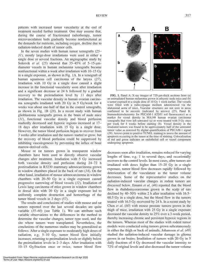
In the seven studies with human tumor xenografts (25–31), mostly large-dose irradiations were used in either asingle dose or several fractions. An angiographic study bySolesvik et al. (25) showed that 35–45% of 5–15-lm-diameter vessels in human melanoma xenografts becamenonfunctional within a week after irradiation with 10–15 Gyin a single exposure, as shown in Fig. 1A. In a xenograft ofhuman squamous cell carcinoma of the larynx (27),irradiation with 10 Gy in a single dose caused a slightincrease in the functional vascularity soon after irradiationand a significant decrease at 24 h followed by a gradualrecovery to the preirradiation level by 11 days afterirradiation. The vascular density in human ovarian carcino-ma xenografts irradiated with 20 Gy in 5 Gy/week for 4weeks was about one-half of that in the control xenografts,as shown in Fig. 1B (28). In a recent study with humanglioblastoma xenografts grown in the brain of nude mice(31), functional vascular density and blood perfusionmarkedly decreased and hypoxic regions increased at 17–18 days after irradiation with 15 Gy in a single dose.However, the tumor blood perfusion began to recover from3 weeks after irradiation and the tumors started to grow, butthe recovery of blood perfusion could be suppressed byinhibiting vasculogenesis by preventing the influx of bonemarrow-derived cells.
Mouse or rat tumors grown in transparent windowchambers have been used to directly observe vascularchanges after treatment. Irradiation with 5 Gy increasedboth vascular density and perfusion during 24–72 hpostirradiation in R3230 mammary adenocarcinoma grownin window chambers placed in the back of rats (34). On theother hand, irradiation of mouse adenocarcinoma in windowchambers with 20–50 Gy in a single exposure causedprogressive narrowing of blood vessels (32). Irradiation ofLewis lung carcinoma of mice grown in window chambersin dorsal skin with 20 Gy in a single exposure led touniformly complete destruction and hemorrhage of thetumor blood vessels in 2 days (57).
The results and conclusions of studies with mouse and rattumors reported over the last several decades are quitevariable (35–60) (see Table 1). We can attribute suchvariable observations to the differences in the method todetermine the vascular changes, tumor type used, and thesite where tumors were transplanted. Nevertheless, theconclusions of the numerous studies may be generalized asfollows. After a single exposure to moderately high doses ofradiation, e.g. 5–10 Gy, tumor blood flow initiallyincreases, returning to preirradiation levels or slightly belowthe preirradiation levels in 2–3 days. After irradiation with10–15 Gy/fraction once or twice, tumor blood flow
decreases soon after irradiation, remains reduced for varying
lengths of time, e.g. 1 to several days, and occasionallyrecovers to the control levels. In most cases, after tumors are
irradiated with doses higher than 15–20 Gy in a singleexposure, tumor blood flow decreases rapidly followed by
deterioration of the vasculature as the tumor volumedecreases. Some of the representative studies on the
radiation-induced vascular changes in rodent tumors are
discussed below. Emami et al. (46) reported that the bloodflow in rhabdomyosarcomas grown in the scalp of rats
declined by 40–50% within 2 h after irradiation with 16.5–60.5 Gy in a single dose, but the blood flow in the tumors
treated with 16.5 Gy recovered by 24 h. In a recent study byChen et al. (60) with mouse prostate tumors grown in the
thigh of mice, irradiation with 25 Gy in a single exposure
decreased the vascular density to 25% over a 3-week period,thereby increasing chronic and persistent hypoxic regions in
the tumors. Whereas most of the studies with rodent tumormodels were conducted using tumors grown subcutaneously
in either the thigh or back of animals, Johansson et al. (49)studied the radiation-induced vascular damage in glioma
grown in rat brains. Irradiation of tumors with 20 Gy in 5
daily fractions of 4 Gy decreased the vascular intensity to72% of original levels and also decreased the tumor volume
FIG. 1. Panel A: X-ray images of 720-lm-thick sections from (a)an unirradiated human melanoma grown in athymic nude mice and (b)a tumor exposed to a single dose of 10 Gy 1 week earlier. The vesselswere filled with a radio-opaque medium administered via theabdominal aorta of mice. Vascular structures are not seen in areasconfirmed to be necrotic (indicated by arrows) (25). Panel B:Immunohistochemistry for PECAM-1 (CD31 red fluorescence) as amarker for vessel density in MA148 human ovarian carcinomaxenografts that were left untreated (a) or were treated with 5 Gy onceper week for 4 weeks before staining (b). Vessel density in theirradiated tumors was found to be approximately half of the untreatedtumor value as assessed by digital quantification of PECAM-1 signal(28). Arrows point to positive TUNEL staining to assess the amount ofapoptosis occurring in the tumor at the time of staining. Colocalizationof red and green indicates an endothelial cell or vessel componentundergoing apoptosis.
REVIEW 317
Downloaded From: https://bioone.org/journals/Radiation-Research on 03 Apr 2020Terms of Use: https://bioone.org/terms-of-use

to 77% of original size by 5 days after completion of the
treatment. Using Doppler sonography, Kim et al. (57)
noninvasively determined the blood flow in Lewis lung
tumors of mice grown s.c. in the hind limbs after irradiation
with 20 Gy once or twice separated by 2 or 4 days. The
tumor blood flow decreased significantly by 2 days after 20
Gy irradiation, but it recovered substantially by 4 days after
irradiation. Such recovery of blood flow after the initial 20-
Gy irradiation could be successfully suppressed by
irradiating the tumors again with 20 Gy at 2 days after the
initial irradiation. Reirradiation at 4 days after the first
irradiation was less effective than that at 2 days after the
first irradiation for sustained reduction of tumor blood flow
and for suppressing tumor growth. Tsai et al. (56) also used
Doppler sonography and immunohistochemistry to deter-
mine the blood flow and intratumor microenvironment in
murine melanoma tumors irradiated with 12 Gy in a single
exposure. The authors reported that there were marked
defects in vascular perfusion and decline in tumor
vascularity accompanied by prominent regions of hypoxia,necrosis and hemorrhage when the tumor volume increased
10-fold after irradiation.
We have extensively investigated the radiation-induced
vascular change in the Walker 256 tumors grown
subcutaneously in the hind legs of rats (39–41). In general,irradiation with doses smaller than 2.5 Gy caused a slight
decrease in the functional vascular volume for 6–12 h,
followed by a return to preirradiation levels. Irradiation with
5–20 Gy in a single exposure decreased the vascular volume
in dose- and time-dependent manner. As shown in Fig. 2,the functional intravascular volume in Walker 256 tumors
decreased for 2–6 days after irradiation with 5–10 Gy in
FIG. 2. Effects of radiation on the functional intravascular volume in Walker 256 tumors (s.c.) grown in thelegs of Sprague-Dawley rats (39). Panel A: Vascular volume in the control tumors. Panels B–F: Vascular volumein the tumors irradiated with various doses of X rays in a single exposure. The dotted line in panel A representsthe vascular volume of 68 control tumors of different weights, and it is shown in panels B–F for comparison withthe vascular volume of the irradiated tumors. The distributions of the marks representing functional vascularityin individual tumors shifted to below the dotted line after irradiation with .5 Gy, indicating that radiation causedvascular damage. The statistical significances between the control group and 10-Gy group as well as the 30-Gygroup were analyzed using linear regression. The vascular volumes at 2 days and 6 days after 10 Gy irradiationwere combined, and the vascular volumes at 2 days and 12 days after 30 Gy irradiation were combined. Thevascular volumes in the tumors irradiated with 10 Gy as well as 30 Gy were significantly smaller than that in thecontrol tumors with P , 0.001.
318 REVIEW
Downloaded From: https://bioone.org/journals/Radiation-Research on 03 Apr 2020Terms of Use: https://bioone.org/terms-of-use
many but not all tumors (39). However, irradiation with 30
or 60 Gy in a single dose caused marked and lasting
decreases in functional vascular volume or vascularity in the
tumors. The decrease in the vascular volume by irradiation
with doses higher than 10 Gy was statistically significant (P, 0.001). The vascular permeability in the Walker 256
tumors of rats, as assessed by the extravasation of 125I-
labeled albumin, was 20–30 times greater than that in the
muscle (17). This report was probably the first demonstra-
tion that tumor blood vessels are highly permeable
compared with the blood vessels of normal tissue. The
vascular permeability in Walker 256 tumors increased
immediately after irradiation throughout the delivered dose
range of 2–20 Gy and then returned to preirradiation levels
in 2–3 days (39–41). Others have observed similar increases
in vascular permeability in tumors or normal tissues after
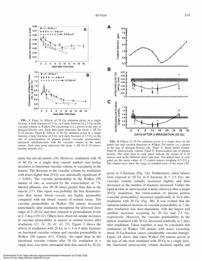
irradiation (23, 24, 26, 41, 43, 55). Figure 3 shows the
effects of irradiation with 20 Gy in 1, 4 or 8 daily fractions
on functional vascular volume and vascular permeability in
Walker 256 tumors (61). Clearly, the rapid drop in the
functional vascular volume after 20 Gy irradiation in a
single dose was more substantial than that caused by 20 Gy
given in 4 fractions (Fig. 3A). Furthermore, when tumorswere exposed to 20 Gy in 8 fractions (8 3 2.5 Gy), thevascular volume initially increased slightly and thendecreased as the number of fractions increased. Unlike therapid decline in intravascular volume observed after a single20-Gy irradiation, the extravasation of plasma protein(vascular permeability) increased significantly at 24 h afterirradiation with 20 Gy (Fig. 3B). It was evident that theradiation-induced increase in vascular permeability at 1 dayafter irradiation was dose-dependent, with the largest andsmallest increases occurring by 20 Gy and 2.5 Gy,respectively. However, the vascular permeability in thetumors irradiated with 20 Gy decreased markedly at 2 daysafter irradiation. Taken together, it may be concluded thatirradiation of Walker 256 tumors with doses exceedingabout 10 Gy/fraction causes considerable vascular damage.Figure 4A shows that when Walker 256 tumors grown inthe legs of rats were irradiated with 30 Gy in a single dose,the functional intravascular volume declined rapidly and
FIG. 3. Panel A: Effects of 20 Gy radiation given in a singlefraction, 4 daily fractions of 5 Gy, or 8 daily fractions of 2.5 Gy on thevascular volume in Walker 256 carcinomas (s.c.) grown in the legs ofSprague-Dawley rats. Each data point represents the mean 6 SE for8–10 tumors. Panel B: Effects of 20 Gy radiation given in a singlefraction, 4 daily fractions of 5 Gy, or 8 daily fractions of 2.5 Gy on therate of extravasations of plasma protein (vascular permeability)measured simultaneously with the vascular volume in the sametumors. Each data point represents the mean 6 SE for 8–10 tumor-bearing animals (61).
FIG. 4. Effects of 30 Gy radiation given in a single dose on thetumor size and vascular functions in Walker 256 tumors (s.c.) grownin the legs of Sprague-Dawley rats. Panel A: Dried tumor weight.Panel B: Intravascular volume. Panel C: Extravasation rate of plasmaprotein. The solid lines in each panel indicate the means of 6–10tumors used at the different times indicated. The dotted lines in eachpanel are the mean values of 15 control tumors weighing 0.3–2.0 g.The shaded areas show the range of standard error of the mean (39).
REVIEW 319
Downloaded From: https://bioone.org/journals/Radiation-Research on 03 Apr 2020Terms of Use: https://bioone.org/terms-of-use
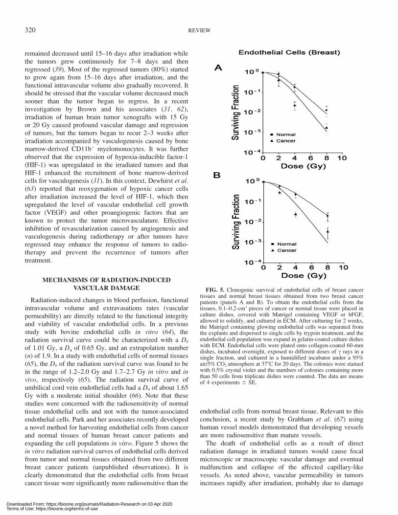
remained decreased until 15–16 days after irradiation whilethe tumors grew continuously for 7–8 days and thenregressed (39). Most of the regressed tumors (80%) startedto grow again from 15–16 days after irradiation, and thefunctional intravascular volume also gradually recovered. Itshould be stressed that the vascular volume decreased muchsooner than the tumor began to regress. In a recentinvestigation by Brown and his associates (31, 62),irradiation of human brain tumor xenografts with 15 Gyor 20 Gy caused profound vascular damage and regressionof tumors, but the tumors began to recur 2–3 weeks afterirradiation accompanied by vasculogenesis caused by bonemarrow-derived CD11bþ myelomonocytes. It was furtherobserved that the expression of hypoxia-inducible factor-1(HIF-1) was upregulated in the irradiated tumors and thatHIF-1 enhanced the recruitment of bone marrow-derivedcells for vasculogenesis (31). In this context, Dewhirst et al.(63) reported that reoxygenation of hypoxic cancer cellsafter irradiation increased the level of HIF-1, which thenupregulated the level of vascular endothelial cell growthfactor (VEGF) and other proangiogenic factors that areknown to protect the tumor microvasculature. Effectiveinhibition of revascularization caused by angiogenesis andvasculogenesis during radiotherapy or after tumors haveregressed may enhance the response of tumors to radio-therapy and prevent the recurrence of tumors aftertreatment.
MECHANISMS OF RADIATION-INDUCEDVASCULAR DAMAGE
Radiation-induced changes in blood perfusion, functionalintravascular volume and extravasations rates (vascularpermeability) are directly related to the functional integrityand viability of vascular endothelial cells. In a previousstudy with bovine endothelial cells in vitro (64), theradiation survival curve could be characterized with a D0
of 1.01 Gy, a Dq of 0.65 Gy, and an extrapolation number(n) of 1.9. In a study with endothelial cells of normal tissues(65), the D0 of the radiation survival curve was found to bein the range of 1.2–2.0 Gy and 1.7–2.7 Gy in vitro and invivo, respectively (65). The radiation survival curve ofumbilical cord vein endothelial cells had a D0 of about 1.65Gy with a moderate initial shoulder (66). Note that thesestudies were concerned with the radiosensitivity of normaltissue endothelial cells and not with the tumor-associatedendothelial cells. Park and her associates recently developeda novel method for harvesting endothelial cells from cancerand normal tissues of human breast cancer patients andexpanding the cell populations in vitro. Figure 5 shows thein vitro radiation survival curves of endothelial cells derivedfrom tumor and normal tissues obtained from two differentbreast cancer patients (unpublished observations). It isclearly demonstrated that the endothelial cells from breastcancer tissue were significantly more radiosensitive than the
endothelial cells from normal breast tissue. Relevant to thisconclusion, a recent study by Grabham et al. (67) usinghuman vessel models demonstrated that developing vesselsare more radiosensitive than mature vessels.
The death of endothelial cells as a result of directradiation damage in irradiated tumors would cause focalmicroscopic or macroscopic vascular damage and eventualmalfunction and collapse of the affected capillary-likevessels. As noted above, vascular permeability in tumorsincreases rapidly after irradiation, probably due to damage
FIG. 5. Clonogenic survival of endothelial cells of breast cancertissues and normal breast tissues obtained from two breast cancerpatients (panels A and B). To obtain the endothelial cells from thetissues, 0.1–0.2-cm3 pieces of cancer or normal tissue were placed inculture dishes, covered with Matrigel containing VEGF or bFGF,allowed to solidify, and cultured in ECM. After culturing for 2 weeks,the Matrigel containing glowing endothelial cells was separated fromthe explants and dispersed to single cells by trypsin treatment, and theendothelial cell population was expand in gelatin-coated culture disheswith ECM. Endothelial cells were plated onto collagen-coated 60-mmdishes, incubated overnight, exposed to different doses of c rays in asingle fraction, and cultured in a humidified incubator under a 95%air/5% CO2 atmosphere at 378C for 20 days. The colonies were stainedwith 0.5% crystal violet and the numbers of colonies containing morethan 50 cells from triplicate dishes were counted. The data are meansof 4 experiments 6 SE.
320 REVIEW
Downloaded From: https://bioone.org/journals/Radiation-Research on 03 Apr 2020Terms of Use: https://bioone.org/terms-of-use

in the endothelial cells followed by widening of the gapsbetween endothelial cells (Fig. 3B, Fig. 4C) (23, 24, 39, 41,43, 48, 55, 68). The increase in extravasation of plasma dueto the increase in vascular permeability may increase theerythrocyte concentration within the narrow capillaries,thereby leading to retardation or stasis of blood perfusion. Inaddition, the increased permeability of capillaries mayincrease the extravascular or interstitial plasma proteinconcentrations, thereby elevating interstitial fluid pressure.The elevation of interstitial fluid pressure above theintravascular blood pressure will cause vascular collapse.Therefore, it is probable that the early decline in functionalvascularity after irradiation in tumors may be caused at leastin part by collapse of blood vessels as a result of elevationof interstitial fluid pressure. When tumor volume shrinksdue to death of parenchymal cells after irradiation, thetumor vascular beds may become further disorganized,aggregated, condensed and fragmented (43). Figure 3 showsthat the extent of vascular damage in tumors treated withfractionated radiation was less than that caused by high-dose single fractions, in accordance with the reports byothers (55, 60). It is likely that sublethal radiation damage inendothelial cells is repaired during fractionated irradiationand thus the functional integrity of tumor vessels is lessimpaired.
It is noteworthy that in most of the previous studies on theeffects of radiation on tumor vascular functions using rodenttumors or human tumor xenografts, the tumors and varyingvolumes of the surrounding normal tissues were irradiatedsimultaneously. Since tumor vascular beds are connected tothe vascular networks of normal tissues, it is likely that thevascular damage in the adjacent normal tissues significantlyinfluenced the tumor blood perfusion in the previousstudies. Therefore, the inconsistent results on the radia-tion-induced vascular changes observed in the previousstudies with experimental tumors may be attributed in partto the differences in the type and site of tumors studied andalso the differences in the volume of normal tissuesirradiated. Kioi et al. (31) reported that irradiation ofhuman U251 glioblastoma xenografts growing in the brainand in the back of nude mice reduced blood flow to 10%and 30% of the original value, respectively. In this respect,it is known that tumors growth is significantly retardedwhen tumors are transplanted into previously irradiatedtissues rather than unirradiated tissues, which is commonlyknown as the tumor bed effect (69). It is believed thatangiogenesis in tumors, which originates from existingnormal tissue blood vessels, is retarded due to radiation-induced vascular damages in the surrounding normaltissues, and thus the supply of oxygen and other nutrientsessential for the growth of tumors is limited. It remains to beinvestigated whether the vascular changes in tumors treatedwith conformal irradiation such as SBRT or SRS signifi-cantly differ from those in the tumors treated with adjacentnormal tissues.
VASCULAR CHANGES AND OXYGEN TENSION INIRRADIATED TUMORS
The intratumor oxygen tension is controlled by theoxygen supply through blood perfusion and the oxygenconsumption rate mainly by the tumor cells. Therefore, theradiation-induced vascular changes may affect the tumoroxygenation. Surprisingly, however, there have been only afew studies that simultaneously measured the radiation-induced changes in tumor microvasculature and the intra-tumor oxygen tension. In the 1960–1970, Carter and Silver(70), Evans and Naylor (71), Kolstad (72), Bergsjo andEvans (73), and Badib and Webster (74) pioneeredinvestigations of the effects of radiotherapy on the oxygentension in various human tumors. Unfortunately, the resultsof these early studies are rather inconsistent and difficult tointerpret because the studies were conducted with equip-ment and methods with limited accuracy and reliability (75).For example, Badib and Webster (74) reported thatradiotherapy increased tumor oxygenation, but in this study,the tumor pO2 was measured at only a single point in eachtumor. In recent years, using more advanced and reliablemethods, investigators determined the changes in pO2 invarious human tumors caused by conventional fractionatedradiotherapy. Dunst et al. (76) determined the pO2 in humancervical cancer treated with fractionated radiotherapy andreported that the median tumor pO2 increased significantlywhen the total dose reached 20 Gy, particularly in tumorsthat had low baseline pO2 values. However, the tumor pO2
declined at the end of treatment, and this appeared to be dueto vascular damage. Cooper et al. (77) also reported thatfractionated radiotherapy increased median pO2 in humancervical cancer. On the other hand, Lyng et al. (21, 78) andFyles et al. (79) observed no significant changes in pO2 incervical cancer, and Brizel et al. (80) also reported nochanges in pO2 in head-neck tumors during the course ofconventional fractionated radiotherapy. Interestingly, in thestudy by Lyng et al. (21), little changes occurred in pO2,while there was a clear evidence of vascular damage in thecervical cancer treated with fractionated radiation. In a well-designed study by Stadler et al. (81), the pO2 in head-necktumors decreased significantly by the end of a first course ofsplit-course radiotherapy with 30 Gy, recovered during a 2-week break, and then decreased again by the end of thesecond course of treatment with 40 Gy. These studiesshowed that fractionated radiotherapy of human tumors mayincrease, cause no significant change, or decrease in tumoroxygen tension. As pointed out by Molls et al. (75), the onlytrend observed in studies was that the pO2 in human tumorsdecreased by the end of the course of fractionatedradiotherapy. It is entirely unclear why the direction andmagnitude of changes in tumor pO2 are so inconsistentamong different studies and even in the same tumor types,e.g. cervical cancer (76–79) and head-neck cancer (80, 81).Tumor size, oxygen measurement technique, pO2 levelbefore treatment, radiation dose and different time-dose
REVIEW 321
Downloaded From: https://bioone.org/journals/Radiation-Research on 03 Apr 2020Terms of Use: https://bioone.org/terms-of-use

schedules are some of the many factors that may control thedirection of changes in tumor pO2. Importantly, unlike thechanges in tumor pO2 during treatment, the tumor pO2 priorto fractionated radiotherapy has been shown to be related tothe outcome of the treatment. The human cervical tumorswith high pO2 before receiving fractionated radiotherapyresponded better than those with low pO2 to the treatments(82).
Radiation-induced changes in pO2 in human tumorxenografts or animal tumors have also been investigated.Brurberg (29) studied possible relationships betweenvascular changes and pO2 in human melanoma xenograftsin nude mice. Irradiation with 10 Gy in a single dose causedno changes in pO2 in the xenografts in 72 h, while theirradiation reduced the blood perfusion by as much as 40%.In the study by Ceelen et al. (58), irradiation of ratcolorectal tumors grown in the hind legs of rats with 5 3 5Gy significantly reduced the microvascular density butslightly increased the intratumor pO2. Zywietz et al. (48)treated rhabdomyosarcoma in rats with a total dose of 60 Gyin 20 fractions over 4 weeks and observed that tumor pO2
increased slightly in the early phase of the treatment butdeclined as the treatment progressed. The investigatorsattributed the decrease in tumor pO2 at the end of treatmentto damage in the tumor capillary endothelial cells. The pO2
in mouse adenocarcinoma decreased significantly in 6 hafter 20 Gy irradiation in a single exposure, recovered tocontrol levels by 48 h, and then gradually declined (83).Vaupel et al. (84) reported that irradiation of mousemammary adenocarcinoma with a single dose of 60 Gymarkedly increased tumor pO2 at 72–74 h after exposure.However, Endrich and Vaupel (85) later suggested that asingle large dose of radiation would destroy the tumormicrovasculature and lead to parenchymal cell death. In astudy by Koutcher et al. (86), irradiation of mousemammary carcinoma with single doses of 32 or 65 Gysignificantly increased the mean tumor pO2 and reduced thefrequency of pO2 values lower than 2.5 mmHg at 3–4 daysafter radiation exposure. Unfortunately, in those two studies(84, 86), the tumor pO2 was measured within 3–4 days afterirradiation, where as tumor pO2 may decline later as theradiation-induced vascular damage becomes significant. Inthis regard, Goda et al. (83) reported that tumor pO2
underwent dynamic changes after irradiation with 10, 20and 40 Gy in a mouse tumor model, and they concluded thatrepeated monitoring is necessary to know the precisechanges in tumor oxygenation in irradiated tumors.Nevertheless, it is rather curious that the tumor pO2
increased after irradiation with 60 Gy or 65 Gy in view ofthe possibility that irradiation with such large doses wouldcause severe damage in the tumor microvasculature, asdiscussed in the previous section. One may speculate that, inthe tumors irradiated with 50–60 Gy, the oxygen demand intumors is drastically diminished due to rapid death of tumorcells or severe damage to tumor cells that would reduceoxygen consumption before vascular damage is fully
expressed (12). Another conceivable explanation is that asingle large dose of radiation causes a transient vascularnormalization by preferentially destroying the most imma-ture and abnormal portions of the vascular bed, allowing fora reorganization of perfusion through the remainingfunctional, more mature vasculature. However, it is highlylikely that an increase in tumor pO2 after irradiation withdoses as high as 60 Gy is a transitional phenomenonbecause marked increases in the hypoxic areas could beobserved in the immunohistochemical preparations ofhuman tumor xenografts 2–3 weeks after irradiation with15 Gy or 20 Gy (31, 62) or in mouse prostate tumors afterirradiation with 25 Gy in a single dose (60). Likewise,necrotic and hypoxic areas increased significantly in humansquamous cell carcinoma xenografts after irradiation with10 Gy (27) and in mouse melanoma irradiated with 12 Gy(56). Fractions of hypoxic cells in rodent tumors have beendemonstrated to be reoxygenated after an exposure to dosesas high as 10–20 Gy. It should be noted that thereoxygenation of hypoxic cells refers to an improvementof oxygenation status of hypoxic cells that survived theinitial high-dose irradiation, and it does not necessarilyindicate that the overall oxygenation status in the irradiatedtumors is increased. Furthermore, it does not indicate theextent of cell death including hypoxic cells after the initialhigh-dose irradiation (45). To our knowledge, the oxygentension in human tumors treated with high-dose hypofrac-tionated SBRT or SRS has not been investigated.
ROLE OF VASCULAR DAMAGE IN THE RESPONSEOF TUMORS TO RADIOTHERAPY
There have been considerable discussions in the radio-therapy community as to whether the primary effect ofionizing radiation in destroying tumors is directly killingcancer cells or indirectly killing cancer cells via vasculardamage. Cramer (87) reported as early as 1932 thatinterference with tumor blood flow caused by radiationdamage to tumor stroma played an important role in theoverall response of tumors to radiation. Denis et al. (52)reported that the radiosensitivity of rat mammary tumorscorrelated with early vessel changes. In support of thenotion that the major target of radiotherapy is tumorendothelial cells or vasculature and not tumor parenchymalcells, investigators reported that irradiation caused rapidapoptosis in tumor endothelial cells by promoting acidicsphingomyelinase (ASMase)-mediated generation of cer-amide, a proapoptotic second messenger (54, 88, 89).Garcia-Barros et al. (54) concluded that ceramide-mediatedapoptosis in tumor endothelial cells leads to secondarydeath in tumor cells and that radiation-induced endothelialcell death is thus the major player in the response of tumorsto radiation at the clinically relevant dose range. Fuks andKolesnick (90) reported that irradiation of tumors withdoses higher than 8–10 Gy in a single exposure causes
322 REVIEW
Downloaded From: https://bioone.org/journals/Radiation-Research on 03 Apr 2020Terms of Use: https://bioone.org/terms-of-use
ceramide-mediated apoptosis in endothelial cells, therebycausing indirect death of parenchymal cells, whereasfractionated irradiation with 1.3–3.0 Gy per fraction inducesapoptosis in endothelial cells through other signalingpathways. However, above contention that radiosensitivityof endothelial cells dictates the response of tumor toradiotherapy has been strongly rebuffed by other investiga-tors (91, 92). Budach and his coinvestigators in Suit’slaboratory (93) previously reported that the TCD50 (radia-tion dose that cures 50% of tumors treated) for human ormurine tumors transplanted into two different strains ofmice with different radiosensitivities was dependent on theradiosensitivity of the tumor cells and not the radiosensi-tivity of the host stromal cells. To further assess therelationship between the intrinsic tumor cell radiosensitivityand tumor response, Gerweck et al. (94) transplantedradiosensitive DNA-PKcs–/– and radioresistant DNA-PKcsþ/þ
tumor cells into the same strain of nude mice and studied theradiation-induced tumor growth delay. The growth delay ofthe tumors derived from DNA-PKcs–/– cells were signifi-cantly longer than that of the tumors derived fromradioresistant DNA-PKcsþ/þ cells, indicating that theradiosensitivity of tumor cells, not that of stromal cells,dictates the response of tumors to radiotherapy. In asubsequent study by the same group of investigators (59),DNA-PKcs–/– and DNA-PKcsþ/þ tumor cells were trans-planted into nude mice and radiosensitive SCID mice, andthe resultant tumors were irradiated with 15 Gy in a singleexposure. Whereas the irradiation reduced the functionalvascularity only modestly in the tumors induced in the nudemice, the irradiation caused considerable reductions in thenumber of functional vessels in the tumors grown in SCIDmice regardless of the intrinsic radiosensitivity of thetransplanted tumor cells. An analysis of the radiation-induced growth delay of the tumors indicated that whereasdirect killing of tumor cells was the major determinant oftumor response in nude mice, both direct killing and indirect
killing of tumor cells as a result of vascular damagecontributed to tumor response in SCID mice. It may beconcluded that the contribution of radiation-inducedvascular damage to the response of tumors to radiation willbe significant only when tumors are irradiated with doseshigh enough to cause substantial vascular damages in thetumors. Therefore, it is likely that the radiosensitivity ofendothelial cells is relatively insignificant in the conven-tional fractionated radiotherapy using 1.5–2.0 Gy/fraction.
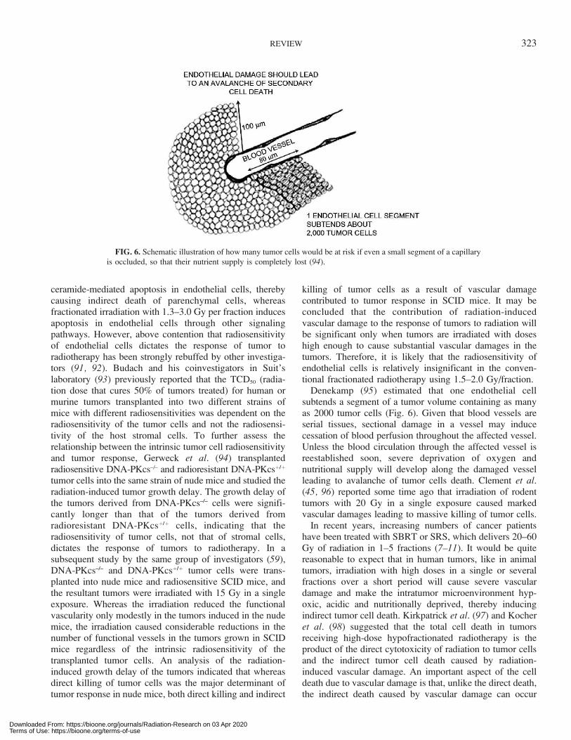
Denekamp (95) estimated that one endothelial cellsubtends a segment of a tumor volume containing as manyas 2000 tumor cells (Fig. 6). Given that blood vessels areserial tissues, sectional damage in a vessel may inducecessation of blood perfusion throughout the affected vessel.Unless the blood circulation through the affected vessel isreestablished soon, severe deprivation of oxygen andnutritional supply will develop along the damaged vesselleading to avalanche of tumor cells death. Clement et al.(45, 96) reported some time ago that irradiation of rodenttumors with 20 Gy in a single exposure caused markedvascular damages leading to massive killing of tumor cells.
In recent years, increasing numbers of cancer patientshave been treated with SBRT or SRS, which delivers 20–60Gy of radiation in 1–5 fractions (7–11). It would be quitereasonable to expect that in human tumors, like in animaltumors, irradiation with high doses in a single or severalfractions over a short period will cause severe vasculardamage and make the intratumor microenvironment hyp-oxic, acidic and nutritionally deprived, thereby inducingindirect tumor cell death. Kirkpatrick et al. (97) and Kocheret al. (98) suggested that the total cell death in tumorsreceiving high-dose hypofractionated radiotherapy is theproduct of the direct cytotoxicity of radiation to tumor cellsand the indirect tumor cell death caused by radiation-induced vascular damage. An important aspect of the celldeath due to vascular damage is that, unlike the direct death,the indirect death caused by vascular damage can occur
FIG. 6. Schematic illustration of how many tumor cells would be at risk if even a small segment of a capillary
is occluded, so that their nutrient supply is completely lost (94).
REVIEW 323
Downloaded From: https://bioone.org/journals/Radiation-Research on 03 Apr 2020Terms of Use: https://bioone.org/terms-of-use
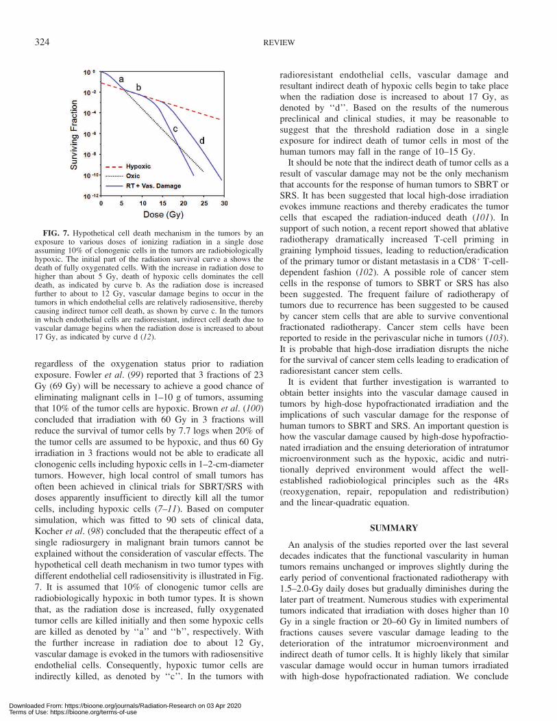
regardless of the oxygenation status prior to radiationexposure. Fowler et al. (99) reported that 3 fractions of 23Gy (69 Gy) will be necessary to achieve a good chance ofeliminating malignant cells in 1–10 g of tumors, assumingthat 10% of the tumor cells are hypoxic. Brown et al. (100)concluded that irradiation with 60 Gy in 3 fractions willreduce the survival of tumor cells by 7.7 logs when 20% ofthe tumor cells are assumed to be hypoxic, and thus 60 Gyirradiation in 3 fractions would not be able to eradicate allclonogenic cells including hypoxic cells in 1–2-cm-diametertumors. However, high local control of small tumors hasoften been achieved in clinical trials for SBRT/SRS withdoses apparently insufficient to directly kill all the tumorcells, including hypoxic cells (7–11). Based on computersimulation, which was fitted to 90 sets of clinical data,Kocher et al. (98) concluded that the therapeutic effect of asingle radiosurgery in malignant brain tumors cannot beexplained without the consideration of vascular effects. Thehypothetical cell death mechanism in two tumor types withdifferent endothelial cell radiosensitivity is illustrated in Fig.7. It is assumed that 10% of clonogenic tumor cells areradiobiologically hypoxic in both tumor types. It is shownthat, as the radiation dose is increased, fully oxygenatedtumor cells are killed initially and then some hypoxic cellsare killed as denoted by ‘‘a’’ and ‘‘b’’, respectively. Withthe further increase in radiation doe to about 12 Gy,vascular damage is evoked in the tumors with radiosensitiveendothelial cells. Consequently, hypoxic tumor cells areindirectly killed, as denoted by ‘‘c’’. In the tumors with
radioresistant endothelial cells, vascular damage andresultant indirect death of hypoxic cells begin to take placewhen the radiation dose is increased to about 17 Gy, asdenoted by ‘‘d’’. Based on the results of the numerouspreclinical and clinical studies, it may be reasonable tosuggest that the threshold radiation dose in a singleexposure for indirect death of tumor cells in most of thehuman tumors may fall in the range of 10–15 Gy.
It should be note that the indirect death of tumor cells as aresult of vascular damage may not be the only mechanismthat accounts for the response of human tumors to SBRT orSRS. It has been suggested that local high-dose irradiationevokes immune reactions and thereby eradicates the tumorcells that escaped the radiation-induced death (101). Insupport of such notion, a recent report showed that ablativeradiotherapy dramatically increased T-cell priming ingraining lymphoid tissues, leading to reduction/eradicationof the primary tumor or distant metastasis in a CD8þ T-cell-dependent fashion (102). A possible role of cancer stemcells in the response of tumors to SBRT or SRS has alsobeen suggested. The frequent failure of radiotherapy oftumors due to recurrence has been suggested to be causedby cancer stem cells that are able to survive conventionalfractionated radiotherapy. Cancer stem cells have beenreported to reside in the perivascular niche in tumors (103).It is probable that high-dose irradiation disrupts the nichefor the survival of cancer stem cells leading to eradication ofradioresistant cancer stem cells.
It is evident that further investigation is warranted toobtain better insights into the vascular damage caused intumors by high-dose hypofractionated irradiation and theimplications of such vascular damage for the response ofhuman tumors to SBRT and SRS. An important question ishow the vascular damage caused by high-dose hypofractio-nated irradiation and the ensuing deterioration of intratumormicroenvironment such as the hypoxic, acidic and nutri-tionally deprived environment would affect the well-established radiobiological principles such as the 4Rs(reoxygenation, repair, repopulation and redistribution)and the linear-quadratic equation.
SUMMARY
An analysis of the studies reported over the last severaldecades indicates that the functional vascularity in humantumors remains unchanged or improves slightly during theearly period of conventional fractionated radiotherapy with1.5–2.0-Gy daily doses but gradually diminishes during thelater part of treatment. Numerous studies with experimentaltumors indicated that irradiation with doses higher than 10Gy in a single fraction or 20–60 Gy in limited numbers offractions causes severe vascular damage leading to thedeterioration of the intratumor microenvironment andindirect death of tumor cells. It is highly likely that similarvascular damage would occur in human tumors irradiatedwith high-dose hypofractionated radiation. We conclude
FIG. 7. Hypothetical cell death mechanism in the tumors by anexposure to various doses of ionizing radiation in a single doseassuming 10% of clonogenic cells in the tumors are radiobiologicallyhypoxic. The initial part of the radiation survival curve a shows thedeath of fully oxygenated cells. With the increase in radiation dose tohigher than about 5 Gy, death of hypoxic cells dominates the celldeath, as indicated by curve b. As the radiation dose is increasedfurther to about to 12 Gy, vascular damage begins to occur in thetumors in which endothelial cells are relatively radiosensitive, therebycausing indirect tumor cell death, as shown by curve c. In the tumorsin which endothelial cells are radioresistant, indirect cell death due tovascular damage begins when the radiation dose is increased to about17 Gy, as indicated by curve d (12).
324 REVIEW
Downloaded From: https://bioone.org/journals/Radiation-Research on 03 Apr 2020Terms of Use: https://bioone.org/terms-of-use
that the radiation-induced vascular damage and the resultingindirect death of tumor cells play important roles in theresponse of tumors to high-dose hypofractionated SBRTand SRS. In addition, enhanced immune reactions andincreased eradiation of cancer stem cells might be involvedin the response of tumors to SBRT or SRS. Further studiesto gain better insights into the effects of high-dosehypofractionated irradiation on tumor vasculature arewarranted. In addition, whether the 4Rs and the linear-quadratic equation are applicable for SBRT or SRS remainsto be investigated.
ACKNOWLEDGMENTS
We wish to thank Drs. Jack Fowler and Martin Brown for their valuable
discussion and advice in preparation of this article. We are also grateful to
Dr. Kaethrlyn Dusenbery for her continuous support and encouragement.
This work was supported by National Cancer Institute (USA) grant R01-
CA116725, Joseph Wargo Fund from the Minnesota Medical Foundation
and Nuclear R&D Program of KOSEF (2009-0093747) (Korea).
Received: August 15, 2011; accepted: December 1, 2011; published
online: January 9, 2012
REFERENCES
1. Dessauer F. My studies on the special foundations of deeptherapy treatment. Am J Roentgenol 1921; 8:578–88.
2. Hall E, Giaccia AJ. Time, dose, and fractionation in radiotherapy.In: Radiobiology for the radiologist. 6th edition. Philadelphia:Lippincott Williams & Wilkins; 2006. Chapter 22.
3. Regaud C, Ferroux R. Discordance des effects de rayons X, d’unepart dans le testicule, par le fractionnement de la dose. C R SocBiol 1927; 97:431–4.
4. Coutard J. Roentgen therapy of epitheliomas of the tensile region,hypopharynx, and larynx from 1920 to 1926. Am J Roentgenol1932; 28:313–31.
5. Mottram JC. A factor of importance in the radiosensitivity oftumors. Br J Radiol 1936; 9:606–14.
6. Leskell L. The stereotactic method and radiosurgery of the brain.Acta Chirurg Scand 1951; 102:316–9.
7. Hiraoka M, Matsuo Y, Nagata Y. Stereotactic body radiationtherapy (SBRT) for early stage lung cancer. Cancer/Radiother2007; 11:32–5.
8. Timmerman RD, Kavanagh BD, Cho LC, Papiez L, Xing L.Stereotactic body radiation therapy in multiple organ sites. J ClinOncol 2007; 25:947–52.
9. Levitt SH, Perez CA, Hui S, Purdy JA. Evolution ofcomputerized radiotherapy in radiation oncology. Int J RadiatBiol Oncol Phys 2008; 70:978–86.
10. Ritter M. Rationale conduct and outcome using hypofractionatedradiotherapy in prostate cancer. Semin Radiat Oncol 2008; 18:249–56.
11. Kim Y, Cho KH, Kim J, Lim YK, Min HS, Lee SH, et al. Single-dose versus fractionated stereotactic radiotherapy for brainmetastases. Int J Radiat Oncol Biol Phys 2011; 81:483–9.
12. Song CW, Park H, Griffin RJ, Levitt SH. Radiobiology ofstereotactic radiosurgery and stereotactic body radiation therapy.In: Technical basis of radiation therapy. Levitt SH, Purdy JA,Perez CA, Vijayakumar S, editors. Berlin, Heidelberg: Springer-Verlag; 2012.
13. Carmeliet P, Jain RK. Angiogenesis in cancer and other diseases.Nature 2000; 407:249–57.
14. Jain RK. Molecular regulation of vessel maturation. Nat Med2003; 9:685–93.
15. Reyes M, Dudek A, Jahagirdar B, Koodie L, Marker PH,Verfaillie CM. Origin of endothelial progenitors in humanpostnatal bone marrow. J Clin Invest 2002; 109:337–46.
16. Konerding MA, Miodonski AJ, Lametschwandtner A. Microvas-cular corrosion casting in the study of tumor vascularity: areview. Scanning Microsc 1995; 9:1233–44.
17. Song CW, Levitt SH. Quantitative study of vascularity in Walkercarcinoma 256. Cancer Res 1971; 31:587–9.
18. Bergsjom P. Radiation-induced early changes in size andvascularity of cervical carcinoma. Acta Radiol 1967; Suppl274:7.
19. Mantyla MJ, Toivanen JT, Pitkanen MA, Rekonen AH.Radiation-induced changes in regional blood flow in humantumors. Int J Radiat Oncol Biol Phys 1982; 8:1711–7.
20. Pirhonen JP, Grenman SA, Breadbacka AB, Bahado-Singh RO,Salmi TA. Effects of external radiotherapy on uterine blood flowin patients with advanced cervical carcinoma assessed by colorDoppler ultrasonography. Cancer 1995; 76:67–71.
21. Lyng H, SundfØr K, Rofstad EK. Changes in tumor oxygentension during radiotherapy of uterine cervical cancer: Relation-ship to changes in vascular density, cell density, and frequency ofmitosis and apoptosis. Int J Radiat Biol Oncol Phys 2000;46:935–46.
22. Mayr NA, Yuh WT, Magnotta VA, Ehrhardt JC, Wheeler JA,Sorosky JI, et al. Tumor perfusion studies using fast magneticresonance imaging technique in advanced cervical cancer: a newnoninvasive predictive assay. Int J Radiat Oncol Biol Phys 1996;36:623–33.
23. Ng QS, Goh V, Milner J, Padhani AR, Saunders MI, Hoskin PJ.Acute tumor vascular effects following fractionated radiotherapyin human lung cancer: in vivo whole tumor assessment usingvolumetric perfusion computed tomography. Int J Radiat OncolBiol Phys 2007; 67:417–24.
24. Janssen MA, Aerts HJ, Kierkels RG, Backes WH, Ollers MC,Buijsen J, et al. Tumor perfusion increases during hypofractio-nated short-course radiotherapy in rectal cancer: sequentialperfusion-CT findings. Radiother Oncol 2010; 94:156–60.
25. Solesvik OV, Rofstad EK, Brustad T. Vascular changes in ahuman malignant melanoma xenograft following single-doseirradiation. Radiat Res 1984; 98:115–28.
26. Kalofonos H, Rowlinon G, Epenetos AA. Enhancement ofmonoclonal antibody uptake in human colon tumor xenograftsfollowing irradiation. Cancer Res 1990; 50:159–63.
27. Bussink J, Kaanders JA, Rijken PF, Raleigh JA, Van der KogelAJ. Changes in blood perfusion and hypoxia after irradiation of ahuman squamous cell carcinoma xenografts tumor line. RadiatRes 2000; 153:398–404.
28. Dings RP, Williams BW, Song CW, Griffioen AW, Mayo KH,Griffin RJ. Antiangiogenic peptide anginex synergizes withradiation therapy to cause tumor growth inhibition and regressionvia endothelial cell radiosensitization. Int J Cancer 2005;115:312–9.
29. Brurberg KG, Thuen M, Ruud EB, Rofstad EK. Fluctuations inpO2 in irradiated human melanoma xenografts. Radiat Res 2006;165:16–25.
30. Fokas E, Hanze J, Kamlah F, Eul BG, Lang N, Keil B, et al.Irradiation-dependent effects on tumor perfusion and endogenousand exogenous hypoxia markers in an A549 xenograft model. IntJ Radiat Oncol Biol Phys 2010; 77:1500–8.
31. Kioi M, Vogel H, Schultz G, Hoffman RM, Harsh GR, BrownJM. Inhibition of vasculogenesis, but not angiogenesis, preventsthe recurrence of glioblastoma after irradiation in mice. J ClinInvest 2010; 120:694–705.
32. Merwin R, Algire GH. Transparent-chamber observations of the
REVIEW 325
Downloaded From: https://bioone.org/journals/Radiation-Research on 03 Apr 2020Terms of Use: https://bioone.org/terms-of-use
response of a transplantable mouse mammary tumor to localroentgen irradiation. J Natl Cancer Inst 1950; 2:593–623.
33. Eddy HA. Tumor vascular response following irradiation.Microvasc Res 1980; 20:195–211.
34. Dewhirst MW, Oliver R, Tso CY, Gustafson C, Secomb T, GrossJF. Heterogeneity in tumor microvascular response to radiation.Int J Radiat Oncol Biol Phys 1990; 18:559–68.
35. Lasnitzki I. A quantitative analysis of the direct and indirectaction of x radiation on malignant cells. Br J Radiol 1947;20:234–47.
36. McAlister WH, Margulis AR. Angiography of malignant tumors.Radiology 1963; 81:664–75.
37. Rubin P, Casarett G. Microcirculation of tumors Part II: Thesupervascularized state of irradiated regressing tumors. ClinRadiol 1966; 17:346–55.
38. Robert J, Martin J, Burg C. Action des rayons X sur lavasculatization specifique de tumeurs isologues de la sourid et durat. Compt Rend Soc Biol 1967; 161:867–71.
39. Song CW, Levitt SH. Vascular changes in Walker 256 carcinomaof rats following x irradiation. Radiology 1971; 100:397–407.
40. Song CW, Payne T, Levitt SH. Vascularity and blood flow in x-irradiated Walker carcinoma 256 of rats. Radiology 1972;104:693–7.
41. Wong HH, Song CW, Levitt SH. Early changes in the functionalvasculature of Walker carcinoma 256 following irradiation.Radiology 1973; 108:429–34.
42. Kallman RF, DeNardo GL, Stasch MJ. Blood flow in irradiatedmouse sarcoma as determined by the clearance of xenon-133.Cancer Res 1972; 32:483–90.
43. Song CW, Sung JH, Clement JJ, Levitt SH. Vascular changes inneuroblastoma of mice following x-irradiation. Cancer Res 1974;34:2344–50.
44. Hilmas DE, Gillette EL. Microvasculature of C3H/Bi mousemammary tumors after x-irradiation. Radiat Res 1975; 61:128–43.
45. Clement JJ, Song CW, Levitt SH. Changes in functionalvascularity and cell number following x-irradiation of a murinecarcinoma. Int J Radiat Oncol Biol Phys 1976; 1:671–8.
46. Emami B, Ten Haken RK, Nussbaum GH, Hughes WL. Effectsof single-dose irradiation on tumor blood flow studied by 15Odecay after photon activation in situ. Radiology 1981; 141:207–9.
47. Tozer GM, Bhujwalla AM, Griffiths JR, Maxwell RJ. Phospho-rus-31 magnetic resonance spectroscopy and blood perfusion ofthe RIF-1 tumor following x-irradiation. Int J Radiat Oncol BiolPhys 1989; 16:155–64.
48. Zywietz F, Reeker W, Kochs E. Studies on tumor oxygenation ina rat rhabdomyo-sarcoma during fractionated irradiation. In:Oxygen transport to tissue XVII. Ince C, Kesecioglu J, Telci L,Akpir K, editors. New York: Plenum Press; 1996. p. 445–55.
49. Johansson M, Bergenheim AT, Widmark A, Henriksson R.Effects of radiotherapy and estramustine on the microvasculaturein malignant glioma. Br J Cancer 1999; 80:142–8.
50. Fenton B, Lord EM, Paoni SF. Effects of radiation on tumorintravascular oxygenation, vascular configuration, developmentof hypoxia, and clonogenic survival. Radiat Res 2001; 155:360–8.
51. Donnelly EF, Geng L, Wojcicki WE, Fleischer AC, Hallahan DE.Quantified power Doppler US of tumor blood flow correlateswith microscopic quantification of tumor blood vessels. Radiol-ogy 2001; 219:166–70.
52. Denis F, Bougnoux P, Paon L, le Floch O, Tranquart F.Radiosensitivity of rat mammary tumors correlates with earlyvessel changes assessed by power Doppler sonography. JUltrasound Med 2003; 22:921–9.
53. Ono K, Kinashi Y, Suzuki M. Selective irradiation of the bloodvessels by using boron neutron capture reaction-development and
its utilization. In: 12th International Congress of RadiationResearch. Brisbane, Australia, August 17–22, 2003. Abstractp08/1659.
54. Garcia-Barros M, Paris F, Cordon-Cardo C, Lyden D, Rafii S,Haimovitz-Friedman A, et al. Tumor response to radiotherapyregulated by endothelial cell apoptosis. Science 2003; 300:1155–9.
55. Kobayashi H, Reijnders K, English S, Yordanov AT, MilenicDE, Sowers AL, et al. Application of a macromolecular contrastagent for detection of alterations of tumor vessel permeabilityinduced by radiation. Clin Cancer Res 2004; 10:7712–20.
56. Tsai JH, Makonnen S, Hyman T, Feldman M, Sehgal CM, MaityA, et al. Ionizing radiation inhibits tumor neovascularization byinducing ineffective angiogenesis. Proc Am Assoc Cancer Res2005; 46:Abstract #3032.
57. Kim DWN, Huamani J, Niemann KJ, Lee H, Geng L, Leavitt LL,et al. Noninvasive assessment of tumor vasculature response toradiation-mediated, vasculature-targeted therapy using quantifiedpower Doppler sonograpy. J Ultrasound Med 1006; 25:1507–17.
58. Ceelen W, Smeet P, Backes W, Damme NV, Boterberg T,Demetter P, et al. Noninvasive monitoring of radiotherapy-induced microvascular changes using dynamic contrast enhancedmagnetic resonance imaging (DCE-MRI) in a colorectal tumormodel. Int J Radiat Oncol Biol Phys 2006; 64:1188–96.
59. Ogawa K, Boucher Y, Kashiwagi, Fukumura D, Chen D,Gerweck LE. Influence of tumor cell and stroma sensitivity oftumor response to radiation. Cancer Res 2007; 67:4016–21.
60. Chen FH, Chiang CS, Wang CC, Tsai CS, Jung SM, Lee CC, etal. Radiotherapy decreases vascular density and causes hypoxiawith macrophage aggregation in TRAMP-C1 prostate tumors.Clin Cancer Res 2009; 15:1721–9.
61. Song CW, Park HJ, Tanaka N, Kwak HJ, Park MJ, Levitt SH.Implication of vascular damage in the response of tumors toablative high-dose radiotherapy. In: Proceedings of the 51stAnnual ASTRO Meeting, Chicago, Nov. 1–5, 2009. Abstract#2859.
62. Ahn GO, Brown JM. Matrix metalloproteinase-9 is required fortumor vasculogenesis but not for angiogenesis: role of bonemarrow-derived myelomonocytic cells. Cancer Cell 2008;13:193–205.
63. Dewhirst MW, Cao Y, Li CY, Moeller B. Exploring the role ofHIF-1 in early angiogenesis and response to radiotherapy.Radiother Oncol 2007; 83:249–55.
64. Rhee JG, Lee I, Song CW. The clonogenic response of bovineaortic endothelial cells in culture to radiation. Radiat Res 1986;106:182–9.
65. Baker DG, Krochak RJ. The response of the microvascularsystem to radiation: A review. Cancer Invest 1989; 7:287–94.
66. Hei TK, Marchese MJ, Hall EJ. Radiosensitivity and sublethaldamage repair in human umbilical cord vein endothelial cells. IntJ Radiat Oncol Biol Phys 1987; 13:879–94.
67. Grabham P, Hu B, Sharma P, Geard C. Effects of ionizingradiation on three-dimension human vessel models: differentialeffects according to radiation quality and cellular development.Radiat Res 2011; 175:21–8.
68. Song CW, Drescher JJ, Tabachnick J. Effect of anti-inflammatorycompounds on beta-irradiation-induced increase in vascularpermeability. Radiat Res 1968; 34:616–25.
69. Kim IH, Lemmon MJ, Brown JM. The influence of irradiation ofthe tumor bed on tumor hypoxia: measurements by radiationresponse, oxygen electrodes, and nitroimidazole binding. RadiatRes 1993; 135:411–7.
70. Cater DB, Silver IA. Quantitative measurements of oxygentension in normal tissue and in the tumors of patients before andafter radiotherapy. Acta Radiol 1960; 53:233–56.
71. Evans NTS, Naylor PED. The effect of oxygen breathing and
326 REVIEW
Downloaded From: https://bioone.org/journals/Radiation-Research on 03 Apr 2020Terms of Use: https://bioone.org/terms-of-use

radiotherapy upon tissue oxygen tension of some human tumors.Br J Radiol 1963; 36:418–25.
72. Kolstad P. Intercapillary distance, oxygen tension and localrecurrence in cervix cancer. Scand J Clin Lab Invest 1968; 106Suppl:145–57.
73. Bergjo P, Evans JC. Oxygen tension of cervical carcinoma duringthe early phase of external irradiation. Scand J Clin Lab Invest1971; 27:71–82.
74. Badib AO, Webster JH. Changes in tumor oxygen tension duringradiation therapy. Acta Radiol 1969; 8:247–57.
75. Molls M, Feldmann HJ, Stadler P, Jund R. Changes in tumoroxygenation during radiation therapy. In: Blood perfusion andmicroenvironment of human tumors. Molls M, Vaupel P, editors.Berlin, Heidelberg: Springer-Verlag; 1998. p. 81–7.
76. Dunst J, Hansgen G, Lautenschlager C, Fuchsel G, Becker A.Oxygenation of cervical cancers during radiotherapy andradiotherapy þ cis-retinoic acid/interferon. Int J Radiat OncolBiol Phys 1999; 43:367–73.
77. Cooper RA, West CML, Logue JP, Davidson SE, Miller A,Roberts S, et al. Changes in oxygenation during radiotherapy incarcinoma of the cervix. Int J Radiat Oncol Biol Phys 1999;45:119–26.
78. Lyng H, Verren AO, Sundfor K, Taksdal I, Lien HH, Kaalhus O.et al. Assessment of tumor oxygenation in human cervicalcarcinoma by use of dynamic Gd-DTPA-enhanced MR imaging.J Magn Reson Imaging 2001; 14:750–6.
79. Fyles AW, Milosevic, Pintilie M, Hill RP. Cervix canceroxygenation measured following external radiation therapy. IntJ Radiat Oncol Biol Phys 1998; 42:751–3.
80. Brizel DM, Dodge RK, Clough RW, Dewhirst MW. Oxygenationof head and neck cancer: changes during radiotherapy and impacton treatment outcome. Radiother Oncol 1999; 53:113–7.
81. Stadler P, Feldmann JJ, Creighton C, Kau R, Molls M. Changesin tumor oxygenation during combined treatment with split-course radiotherapy and chemotherapy in patients with head andneck cancer. Radiother Oncol 1998; 48:157–64.
82. Hockel M, Vaupel P. The prognostic significance of hypoxia incervical cancer: a radiobiological or tumor biological phenome-non? In: Blood perfusion and microenvironment of humantumors. Molls M, Vaupel P, editors. Berlin, Heidelberg: Springer-Verlag; 1998. p. 73–87.
83. Goda F, O’Hara JA, Rhodes ES, Liu KJ, Dunn JF, Bacic, et al.Changes of oxygen tension in experimental tumors after a singledose x-ray irradiation. Cancer Res 1955; 55:2249–52.
84. Vaupel P, Frinak S, O’Hara M. Direct measurement ofreoxygenation in malignant mammary tumors after a single largedose of irradiation. Adv Exp Med Biol 1984; 180:773–82.
85. Endrich B, Vaupel P. The role of the microcirculation in thetreatment of malignant tumors: facts and fiction. In: Bloodperfusion and microenvironment of human tumors. Molls M,Vaupel P, editors. Berlin, Heidelberg: Springer-Verlag; 1998. p.19–39.
86. Koutcher JA, Alfieri AA, Devitt ML, Rhee JG, Kornblith AB,
Mahmood U, et al. Quantitative changes in tumor metabolism,partial pressure of oxygen, and radiobiological oxygenation statuspostradiation. Cancer Res 1992; 52:4620–7.
87. Kramer W. Experimental observations on the therapeutic actionof radium. Tenth Sci Rep Imp Cancer Res Fung 1932; 95–123.
88. Kolesnick R, Fuks Z. Radiation and ceramide-induced apoptosis.Oncogene 2003; 22:2897–906.
89. Gulbins E, Kolesnick R. Raft ceramide in molecular medicine.Oncogene 2003; 22:7070–7.
90. Fuks Z, Kolesnick R. Engaging the vascular component of thetumor response. Cancer Cell 2005; 8:89–91.
91. Suit HD, Willers H. Comment on ‘‘Tumor response toradiotherapy regulated by endothelial cell apoptosis’’ (I). Science2003; 302:1894c.
92. Brown M, Bristow R, Glazer P, Hill R, McBride W, McKenna G,et al. Comment on ‘‘Tumor response to radiotherapy regulated byendothelial cell apoptosis’’ (II). Science 2003; 302:1894d.
93. Budach W, Taghian A, Freeman J, Gioioso D, Suit HD. Impact ofstromal sensitivity on radiation response of tumors. J Natl CancerInst 1993; 85:988–93.
94. Gerweck LE, Vijayappa S, Kurimasa A, Ogawa K, Chen DJ.Tumor cell radiosensitivity is a major determinant of tumorresponse to radiation. Cancer Res 2006; 66:8352–5.
95. Denekamp J. Vascular endothelium as the vulnerable element intumours. Acta Radiol Oncol 1984; 23:217–25.
96. Clement JJ, Tanaka N, Song CW. Tumor reoxygenation andpostirradiation vascular changes. Radiology 1978; 127:799–803.
97. Kirkpatrick JP, Meyer JJ, Marks LB. The linear quadratic modelis inappropriate to model high dose per fraction effects inradiosurgery. Semin Radiat Oncol 2008; 18:240–3.
98. Kocher M, Treuer H, Voges J, Hoevels M, Sturm V, Muller RP.Computer simulation of cytotoxic and vascular effects ofradiosurgery in solid and necrotic brain metastases. RadiotherOncol 2000; 54:149–56.
99. Fowler JF, Wolfgang AT, Fenwik JD, Mehta MP. A challenge totraditional radiation oncology. Int J Radiat Oncol Biol Phys 2004;60:1241–56.
100. Brown JM, Diehn M, Loo BW. Stereotactic ablative radiotherapyshould be combined with a hypoxic cell radiosensitizer. Int JRadiat Oncol Biol Phys 2010; 78:323–7.
101. Brown JM, Koong AC. High-dose single-fraction radiotherapy:Exploiting a new biology? Int J Radiat Oncol Biol Phys 2008;71:324–5.
102. Lee Y, Auh SL, Wang Y, Burnette B, Wang Y, Meng Y, et al.Therapeutic effects of ablative radiation on local tumor requireCD8þ T cells changing strategies for cancer treatment. Blood2009; 114:3589–95.
103. Hambaardzymyan D, Becher OJ, Rosenblum MK, Pandolfi PP,Manova-Todorova M, Holland EC. PI3K pathway regulatessurvival of cancer stem cells residing in the perivascular nichefollowing radiation in medulloblastoma in vivo. Genes Dev 2008;22:436–48.
REVIEW 327
Downloaded From: https://bioone.org/journals/Radiation-Research on 03 Apr 2020Terms of Use: https://bioone.org/terms-of-use


![C ; ? G 0 D 6 ' * G * B 5 B 6 C ; I ; 2 5 . G 5 F 5 D ; . W t í W } v o ] o · 2020. 5. 1. · / g 9 5 . 8 g 6 g c 5 / b 5 g c 8 5 3 0 b c 6 g ; k , : * b 8 g " b , d . ^ + d / ?](https://img.dokumen.tips/doc/110x75/6035b996ef32be0a1141d2c4/c-g-0-d-6-g-b-5-b-6-c-i-2-5-g-5-f-5-d-w-t-w-v-o-o-2020.jpg)

![D DQ DOL - UniFI · *l ry dq ql &k ldu dp rq wh,qw huq r 3 hug xwr 8q lyh uvl wj g hjo l 6 wxg l g l ) luh q]h ',' $ ' lsd uwlp hqw r g l $ ufk lwhw wxu d 6f xr od g l ' rww rud wr](https://img.dokumen.tips/doc/110x75/5e9332612b2b604cf32b877c/d-dq-dol-unifi-l-ry-dq-ql-k-ldu-dp-rq-whqw-huq-r-3-hug-xwr-8q-lyh-uvl-wj.jpg)


![sc9c7be5bf534cbeb.jimcontent.com€¦ · . g 5˜ b 5 ]˙% ! A : 5K% ˜ . g 5˜ b ! A : ˘% T A : 5 " . ˜ ˛ ˙ ˘5˚ # ! A : r5˚, . g 5˜ b " i ]˙% A : ! ˚ ! . g 5˜ b !˘, !](https://img.dokumen.tips/doc/110x75/5ecafaf6b7fd435ea032ef62/-g-5oe-b-5-a-5k-oe-g-5oe-b-a-t-a-5-oe-5.jpg)