Embed Size (px)
Citation preview

4/5/2015
1
Peripheral Nervous System
Learn and Understand:
• Peripheral nerves connect the edges of the
body and outside world to the CNS.
• Most nerves carry specific types of
information to/from specific locations.
• PNS includes nervous-like tissue: sensitive
and excitable.
• Humans possess both instinctual and
learned reflexes. Many subconscious and
involuntary, many protective, many
postural/positional, most homeostatic.
Figure 13.1 Place of the PNS in the structural organization of the nervous
system.
Central nervous system (CNS) Peripheral nervous system (PNS)
Sensory (afferent)
division
Motor (efferent) division
Somatic nervous
system
Autonomic nervous
system (ANS)
Sympathetic
division
Parasympathetic
division
Provides neuronal link to and from body and outside world
Includes all neural structures outside brain
Sensory receptors
Peripheral nerves and associated ganglia, plexuses
Efferent motor endings
Figure 13.4b Structure of a nerve. Axon
Myelin sheath
Endoneurium
Perineurium
Epineurium
Fascicle
Blood
vessels

4/5/2015
2
Peripheral Nervous Tissue:
Classification of Nerves
• Most nerves are mixtures of afferent and efferent fibers and somatic and autonomic fibers
• Classified according to direction of impulses
– Mixed nerves – both sensory and motor fibers; impulses both to and from CNS
– Sensory (afferent) nerves – impulses only toward CNS
– Motor (efferent) nerves – impulses only away from CNS
• Peripheral nerves classified as cranial or spinal nerves
Spinal Nerves
• 31 pairs of mixed nerves named for point
of issue from spinal cord
– Supply all body parts but head and part of
neck
– 8 cervical (C1–C8)
– 12 thoracic (T1–T12)
– 5 Lumbar (L1–L5)
– 5 Sacral (S1–S5)
– 1 Coccygeal (C0)
Figure 13.8a Formation of spinal nerves and rami distribution .
Dorsal root
Dorsal and ventral rootlets of spinal nerve
Dorsal root
ganglion
Dorsal ramus of spinal nerve
Ventral ramus
of spinal nerve
Spinal nerve
Sympathetic trunk ganglion
Anterior view showing spinal cord, associated nerves, and vertebrae.
The dorsal and ventral roots arise medially as rootlets and join laterally to
form the spinal nerve.
Rami communicantes
Ventral root

4/5/2015
3
Dorsal ramus
Ventral ramus
Spinal nerve
Rami communicantes
Sympathetic trunk ganglion Dorsal root ganglion
Dorsal root
Ventral root
Branches of intercostal nerve
Lateral cutaneous
Anterior cutaneous
Sternum
Intercostal nerve
Cross section of thorax showing the main roots and branches of a spinal nerve.
Figure 13.8b Formation of spinal nerves and rami distribution.
Figure 13.13 Map of dermatomes – Specificity of sensory information carried by individual spinal
nerves
C2
C3
C4
C5
T5 T4
T3 T2 T1
T6
T7
T8
T9
T10
T2
T11
T2
C5
C6
C6
C7 C8
T12 L1 L1
L2 L2
S2 S3 C8
C5
C6
C6
C7
L3
L4
L5 L5
L4
L3
S1 S1
C2
C3
C4
C5
C6
C7 C8
T1
T2
T3 T4
T5 T6 T7 T8 T9 T10
C5
C6
C7
C8 L2
L4
S1
T11
T12
L1
L3 L5
C8
C6
C7
S2 S3
S4
S5
S1 S2 S2 S1
L1
L2 L5 L5
L3
L4
L4
L5 L5
L4
S1
Posterior view Anterior view
Cranial Nerves
• Twelve pairs of nerves associated with brain
– Ten attach to brain stem
• Most are mixed nerves; two pairs purely sensory
• Each numbered (I through XII) and named from
rostral to caudal
"On occasion, our trusty truck acts funny—very
good vehicle anyhow"
"Oh once one takes the anatomy final, very
good vacations are heavenly"

4/5/2015
4
Filaments of
olfactory nerve (I)
Olfactory bulb
Olfactory tract
Optic nerve (II)
Optic chiasma
Optic tract
Oculomotor
nerve (III)
Trochlear
nerve (IV)
Trigeminal
nerve (V)
Abducens
nerve (VI)
Cerebellum
Medulla oblongata
Frontal lobe
Temporal lobe
Infundibulum
Facial nerve (VII)
Vestibulocochlear
nerve (VIII)
Glossopharyngeal
nerve (IX)
Vagus nerve (X)
Accessory nerve (XI)
Hypoglossal nerve (XII)
Figure 13.6a Location and function of cranial nerves.
Primary Functions of Cranial Nerves
Nerve Sensory Function Somatic Motor
Function
Parasympathetic Motor
Function
I – olfactory Smell None None
II – optic Vision None None
III – oculomotor None Extrinsic eye muscles
(rectus and inf. oblique);
elevates eyelid
Intrinsic eye muscles – control
pupil and lens
IV – trochlear None Superior oblique None
V – trigeminal General Senses: anterior scalp, nasal
cavity, nasopharynx, face, most of oral
cavity, anterior tongue, part of auricle,
meninges
Muscles of mastication None
VI – abducens None Lateral rectus None
VII – facial Taste: anterior tongue Muscles of facial expression Secretion of tears, saliva
VIII –
vestibulocochlear
Hearing, equilibrium None None
IX –
glossopharyngeal
General Sensory and taste: posterior
tongue; General Sensory: part of
pharynx; Visceral Sensory: carotid
bodies
One pharyngeal muscle Secretion of parotid salivary
glands
X – vagus Visceral Sensory: heart, lungs,
abdominal organs; General Sensory:
ear canal, pharynx, larynx
Most pharyngeal muscles
all laryngeal muscles
Smooth muscle and glands:
heart, lungs, larynx, trachea,
abdominal organs
XI – accessory None Trapezius,
sternocleidomastoid None
XII - hypoglossal none Intrisic and extrinsic tongue
muscles None
Peripheral Nervous Tissue:
Sensory Receptors
• Specialized to respond to changes in
environment (stimuli)
• Activation results in graded potentials that
trigger nerve impulses
• Sensation (awareness of stimulus) and
perception (interpretation of meaning of
stimulus) occur in brain

4/5/2015
5
Classification by Stimulus Type
• Mechanoreceptors—respond to touch, pressure, vibration, and stretch
• Thermoreceptors—sensitive to changes in temperature
• Photoreceptors—respond to light energy (e.g., retina)
• Chemoreceptors—respond to chemicals (e.g., smell, taste, changes in blood chemistry)
• Nociceptors—sensitive to pain-causing stimuli (e.g. extreme heat or cold, excessive pressure, inflammatory chemicals)
Classification by Location
• Exteroceptors respond to
– stimuli arising outside body
– Receptors in skin for touch, pressure, pain, and
temperature and most special sense organs
• Interoceptors (visceroceptors)respond to
– stimuli arising in internal viscera and blood vessels
– Sensitive to chemical changes, tissue stretch, and
temperature changes
– May cause discomfort but usually unaware of their
workings
• Proprioceptors respond to
– stretch in skeletal muscles, tendons, joints, ligaments, and
connective tissue coverings of bones and muscles
– Inform brain of movements, body part position/location
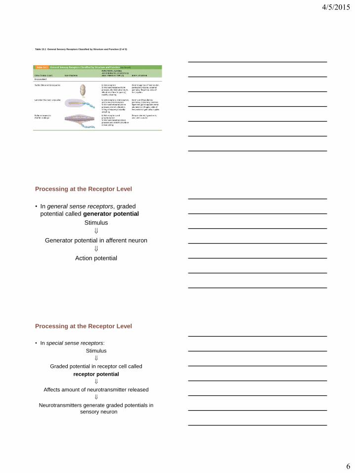
Table 13.1 General Sensory Receptors Classified by Structure and Function
4/5/2015
6
Table 13.1 General Sensory Receptors Classified by Structure and Function (2 of 3)
Processing at the Receptor Level
• In general sense receptors, graded
potential called generator potential
Stimulus
Generator potential in afferent neuron
Action potential
Processing at the Receptor Level
• In special sense receptors:
Stimulus
Graded potential in receptor cell called
receptor potential
Affects amount of neurotransmitter released
Neurotransmitters generate graded potentials in
sensory neuron

4/5/2015
7
Figure 13.2 Three basic levels of neural integration in sensory systems.
Perceptual level (processing in cortical sensory centers)
Motor cortex
Somatosensory cortex
Thalamus
Reticular formation
Cerebellum Pons
Medulla
Spinal cord
Circuit level (processing in ascending pathways)
Free nerve endings (pain, cold, warmth)
Muscle spindle
Receptor level
(sensory reception and transmission to CNS)
Joint kinesthetic receptor
3
2
1
Peripheral Nervous Function:
Basic Reflexes
• Basic functional unit of nervous system and simplest portion capable of receiving a stimulus and producing a response
• Automatic response to a stimulus that occurs without conscious thought.
• Quick, protective, homeostatic.
• Some integrated within spinal cord; some within brain
(those related to head) using cranial nerves
• Some involve excitatory neurons yielding a response;
some involve inhibitory neurons that prevent an action
• Higher brain centers can influence, suppress, or
exaggerate reflex responses
Variety of Reflexes
Functional classification
– Somatic reflexes
• Activate skeletal muscle
– Autonomic (visceral) reflexes
• Activate visceral effectors (smooth or cardiac muscle
or glands)

4/5/2015
8
Figure 13.15 The five basic components of all reflex arcs.
Stimulus
Skin
Receptor
Sensory neuron
Integration center
Motor neuron
Effector
1
2
3
4
5
Interneuron
Spinal cord (in cross scetion)
Receptor—site of stimulus action
Sensory neuron—transmits afferent impulses to CNS
Integration center—either monosynaptic or polysynaptic region within CNS
Motor neuron—conducts efferent impulses from integration center to effector organ
Effector—muscle fiber or gland cell that responds to efferent impulses by contracting or secreting
Stretch and Tendon Reflexes
• To smoothly coordinate skeletal muscle
nervous system must receive
proprioceptor input regarding
– Length of muscle
• From muscle spindles
– Amount of tension in muscle
• From tendon organs
Figure 13.16 Anatomy of the muscle spindle and tendon organ.
Flower spray endings
(secondary sensory
endings)
Efferent (motor)
fiber to muscle spindle
Efferent (motor) fiber to extrafusal muscle fibers
Extrafusal muscle fiber
Intrafusal muscle fibers
Sensory fiber
Tendon Tendon organ
Anulo-
spiral
endings
(primary
sensory
endings)
Capsule
(connective
tissue)
Muscle
spindle

4/5/2015
9
• Excited in two ways
1. External stretch of muscle and muscle spindle
2. Internal stretch of muscle spindle
• Stretch causes increased rate of impulses
to spinal cord
• Adjustment for moving/contracting muscles:
– Contracting muscle reduces tension on muscle
spindle
– Sensitivity lost unless muscle spindle shortened
by impulses in motor neurons
– – coactivation maintains tension and sensitivity
of spindle during muscle contraction
Figure 13.17a Operation of the muscle spindle.
How muscle stretch is detected
Muscle spindle
Intrafusal muscle fiber
Sensory fiber
Extrafusal muscle fiber
Time
Unstretched muscle. Action potentials (APs) are generated at a constant rate in the associated sensory fiber.
How muscle stretch is detected
Stretched muscle. Stretching activates the muscle spindle, increasing the rate of APs.
Time
Stretch Reflexes
• How stretch reflex works
– Stretch activates muscle spindle
– Sensory neurons synapse directly with
motor neurons in spinal cord
– motor neurons cause stretched muscle to
contract
• All stretch reflexes are monosynaptic and
ipsilateral

4/5/2015
10
Stretch Reflexes
• Reciprocal inhibition also occurs—IIa
fibers synapse with interneurons that
inhibit motor neurons of antagonistic
muscles
– Example: In patellar reflex, stretched muscle
(quadriceps) contracts and antagonists
(hamstrings) relax
Slide 6 Figure 13.18 Stretch Reflex (2 of 2)
Patellar ligament
Patella
Hamstrings (flexors)
Muscle spindle
Quadriceps (extensors)
Spinal cord (L2–L4)
Tapping the patellar ligament stretches the quadriceps and excites its muscle spindles. Afferent impulses (blue) travel to the spinal cord, where synapses occur with motor neurons and interneurons.
The motor neurons (red) send activating impulses to the quadriceps causing it to contract, extending the knee.
The interneurons (green) make inhibitory synapses with ventral horn neurons (purple) that prevent the antagonist muscles (hamstrings) from resisting the contraction of the quadriceps.
The patellar (knee-jerk) reflex—an example of a stretch reflex
1
2
3a 3b 3b
1
+
+
–
2
3a
3b
– Inhibitory synapse
+ Excitatory synapse
The Tendon Reflex
• Polysynaptic reflexes
• Helps prevent damage due to excessive
stretch
• Important for smooth onset and
termination of muscle contraction
• Produces muscle relaxation (lengthening)
in response to tension

4/5/2015
11
Slide 5 Figure 13.19 The tendon reflex.
Quadriceps strongly contracts.
Tendon organs are activated.
Afferent fibers synapse with
interneurons in the spinal cord.
Interneurons
Spinal cord
Quadriceps
(extensors)
Tendon organ
Hamstrings
(flexors)
Efferent
impulses to muscle
with stretched
tendon are damped.
Muscle relaxes,
reducing tension.
Efferent impulses
to antagonist muscle
cause it to contract.
3b
+ +
+ –
+ Excitatory synapse
– Inhibitory synapse
3a
2 1
The Flexor and Crossed-Extensor Reflexes
• Flexor (withdrawal) reflex
– Initiated by painful stimulus
– Causes automatic withdrawal of threatened
body part
– Ipsilateral and polysynaptic
– Protective; important
– Brain can override
• E.g., finger stick for blood test
Flexor and Crossed-Extensor Reflexes
• Crossed extensor reflex
– Occurs with flexor reflexes in weight-bearing
limbs to maintain balance
– Consists of ipsilateral withdrawal reflex and
contralateral extensor reflex
• Stimulated side withdrawn (flexed)
• Contralateral side extended
• e.g., step barefoot on broken glass

4/5/2015
12
Figure 13.20 The crossed-extensor reflex.
+ Excitatory synapse
– Inhibitory synapse
Afferent fiber
Efferent fibers
Arm movements
Extensor inhibited
Flexor stimulated
Interneurons
Efferent fibers
Flexor inhibited
Extensor stimulated
Site of reciprocal
activation: At the same time, the extensor muscles on the opposite side are activated.
Site of stimulus:
A noxious stimulus causes a flexor
reflex on the same side, withdrawing that limb.
+
+
+
–
–
+