Embed Size (px)
Citation preview

4+

Chapter 35
Plant Structure, Growth, and Development
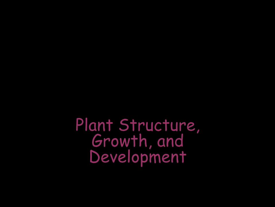
Overview: Plastic Plants?
• This plant, fanwort exhibits developmental plasticity, the ability to alter itself in response to its environment
• Developmental plasticity is more marked in plants than in animals

Fig. 35-1

• Plants, like muliticelluar animals, have organs composed of different tissues, which in turn are composed of cells

The Three Basic Plant Organs: Roots, Stems, and Leaves
• Basic morphology of vascular plants reflects that they are organisms that draw nutrients from below ground and above ground

• Three basic organs evolved:
roots, stems, and leaves
• They are organized into a root system and a shoot system
• Roots rely on sugar produced by photosynthesis in the shoot system, and shoots rely on water and minerals absorbed by the root system

Fig. 35-2
Reproductive shoot (flower)
Apical bud
Node
Internode
Apical bud
Shoot system
Vegetative shoot
Leaf Blade
Petiole
Axillary bud
Stem
Taproot
Lateral branch
roots
Root system

Roots
• Roots are multicellular organs with important functions:
–Anchoring the plant
–Absorbing minerals and water
–Storing organic nutrients
• A taproot system consists of one main vertical root that gives rise to lateral roots, or branch roots
• Adventitious roots arise from stems or leaves
• Seedless vascular plants and monocots have a fibrous root system characterized by thin lateral roots with no main root

• In most plants, absorption of water and minerals occurs near the root hairs, where vast numbers of tiny root hairs increase the surface area

Fig. 35-3

• Many plants have modified roots

Fig. 35-4a
Prop roots

Fig. 35-4b
Storage roots

Fig. 35-4d
Pneumatophores

Fig. 35-4e
Buttress roots

Fig. 35-4c
“Strangling” aerial roots
Roots: When plants go bad

Stems
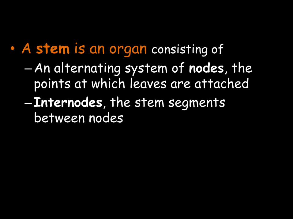
• A stem is an organ consisting of
–An alternating system of nodes, the points at which leaves are attached
–Internodes, the stem segments between nodes
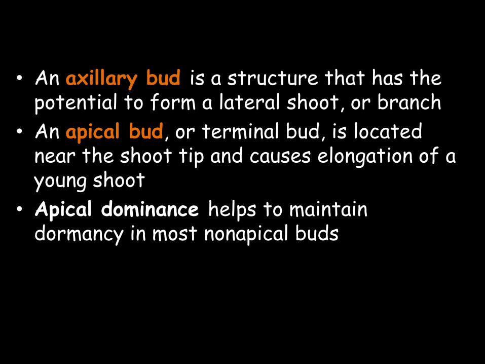
• An axillary bud is a structure that has the potential to form a lateral shoot, or branch
• An apical bud, or terminal bud, is located near the shoot tip and causes elongation of a young shoot
• Apical dominance helps to maintain dormancy in most nonapical buds

• Many plants have modified stems

Fig. 35-5 Rhizomes
Bulbs
Storage leaves
Stem
Stolons
Stolon
Tubers
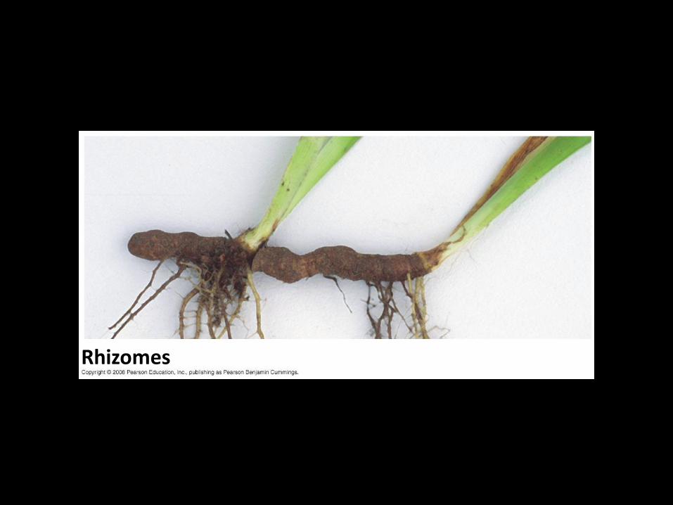
Fig. 35-5a
Rhizomes
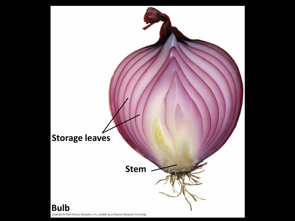
Fig. 35-5b
Bulb
Storage leaves
Stem

Fig. 35-5c
Stolons
Stolon

Fig. 35-5d
Tubers

Stems: When plants go bad


Leaves
• The leaf is the main photosynthetic organ of most vascular plants
• Leaves generally consist of a flattened blade and a stalk called the petiole, which joins the leaf to a node of the stem

• Monocots and dicots (or eudicots) differ in the arrangement of veins, the vascular tissue of leaves
–Most monocots have parallel veins
–Most dicots have branching veins

Fig. 35-6
(a) Simple leaf
Compound
leaf (b)
Doubly compound
leaf
(c)
Petiole Axillary bud
Leaflet
Petiole Axillary bud
Leaflet Petiole Axillary bud

Fig. 35-6a
(a) Simple leaf
Petiole
Axillary bud
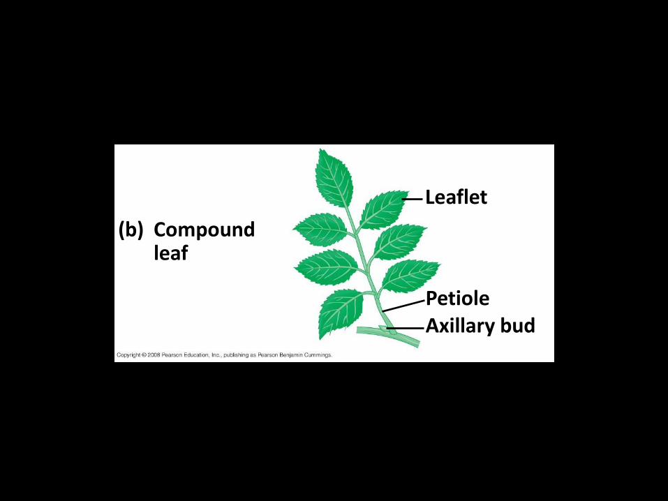
Fig. 35-6b
Compound leaf
(b)
Leaflet
Petiole Axillary bud
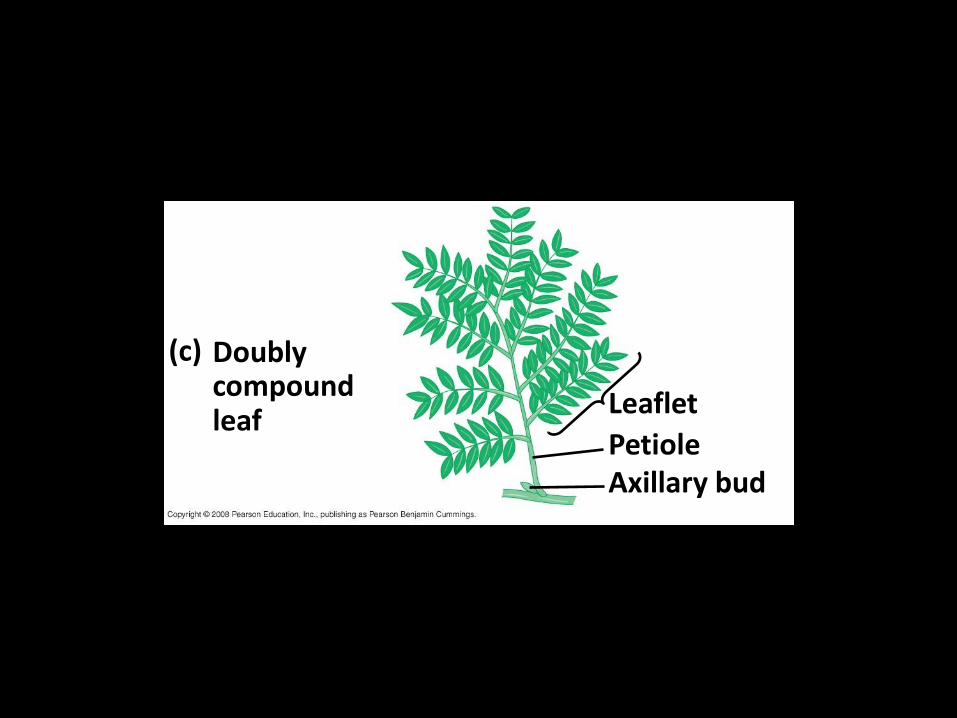
Fig. 35-6c
Doubly compound leaf
(c)
Leaflet
Petiole Axillary bud
• Some plant species have modified leaves that serve various functions

Fig. 35-7
Tendrils
Spines
Storage leaves
Reproductive leaves
Bracts
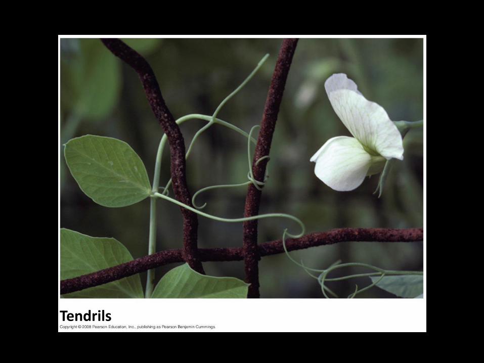
Fig. 35-7a
Tendrils
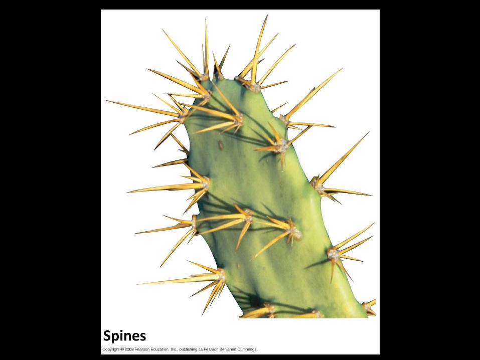
Fig. 35-7b
Spines

Fig. 35-7c
Storage leaves
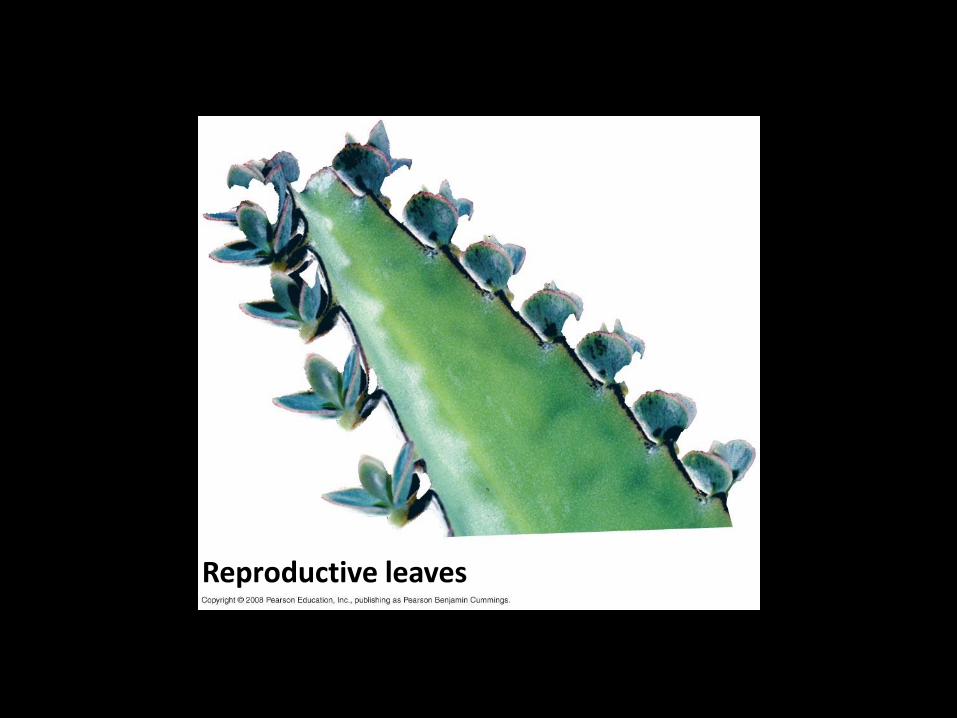
Fig. 35-7d
Reproductive leaves
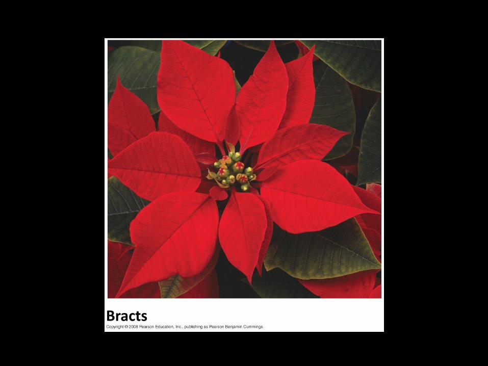
Fig. 35-7e
Bracts

Leaves: When plants go bad

Dermal, Vascular, and Ground Tissues
• Each plant organ has dermal, vascular, and ground tissues
• Each of these three categories forms a tissue system

Fig. 35-8
Dermal tissue
Ground tissue Vascular
tissue
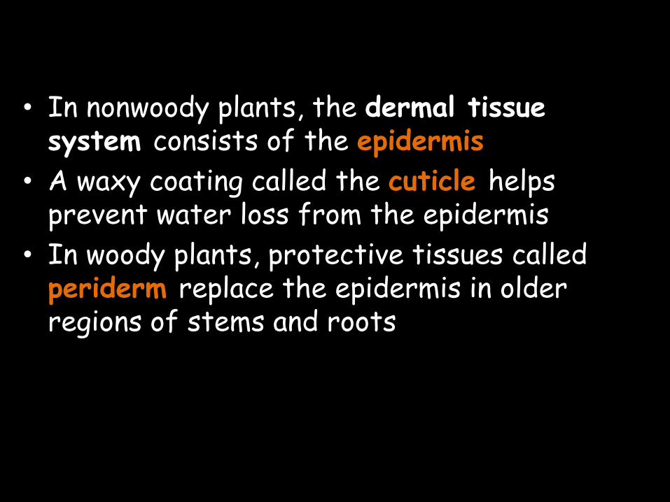
• In nonwoody plants, the dermal tissue system consists of the epidermis
• A waxy coating called the cuticle helps prevent water loss from the epidermis
• In woody plants, protective tissues called periderm replace the epidermis in older regions of stems and roots

• The vascular tissue system carries out long-distance transport of materials between roots and shoots
• The two vascular tissues are xylem and phloem
• Xylem conveys water and dissolved minerals upward from roots into the shoots
• Phloem transports organic nutrients from where they are made to where they are needed

• Tissues that are neither dermal nor vascular are the ground tissue system
• Ground tissue internal to the vascular tissue is pith; ground tissue external to the vascular tissue is cortex
• Ground tissue includes cells specialized for storage, photosynthesis, and support

Common Types of Plant Cells
• Like any multicellular organism, a plant is characterized by cellular differentiation, the specialization of cells in structure and function

• Some major types of plant cells:
–Parenchyma
–Collenchyma
–Sclerenchyma
–Water-conducting cells of the xylem
–Sugar-conducting cells of the phloem

Parenchyma Cells
• Mature parenchyma cells
– Have thin and flexible primary walls
– Lack secondary walls
– Are the least specialized
– Perform the most metabolic functions
– Retain the ability to divide and differentiate


Fig. 35-10a
Parenchyma cells in Elodea leaf, with chloroplasts (LM) 60 µm


Collenchyma Cells
• Collenchyma cells are grouped in strands and help support young parts of the plant shoot
• They have thicker and uneven cell walls
• They lack secondary walls
• These cells provide flexible support without restraining growth

Fig. 35-10b
Collenchyma cells (in Helianthus stem) (LM)
5 µm

Sclerenchyma Cells
• Sclerenchyma cells are rigid because of thick secondary walls strengthened with lignin
• They are dead at functional maturity
• There are two types:
–Sclereids are short and irregular in shape and have thick lignified secondary walls
– Fibers are long and slender and arranged in threads

Fig. 35-10c
5 µm
25 µm
Sclereid cells in pear (LM)
Fiber cells (cross section from ash tree) (LM)
Cell wall

Concept 35.2: Meristems generate cells for new organs
• A plant can grow throughout its life; this is called indeterminate growth
• Some plant organs cease to grow at a certain size; this is called determinate growth
• Annuals complete their life cycle in a year or less
• Biennials require two growing seasons
• Perennials live for many years

• Meristems are perpetually embryonic tissue and allow for indeterminate growth
• Apical meristems are located at the tips of roots and shoots and at the axillary buds of shoots
• Apical meristems elongate shoots and roots, a process called primary growth

• Lateral meristems add thickness to woody plants, a process called secondary growth
• There are two lateral meristems: the vascular cambium and the cork cambium
• The vascular cambium adds layers of vascular tissue called secondary xylem (wood) and secondary phloem
• The cork cambium replaces the epidermis with periderm, which is thicker and tougher

Fig. 35-11
Shoot tip (shoot apical meristem and young leaves)
Lateral meristems:
Axillary bud meristem
Vascular cambium
Cork cambium
Root apical meristems
Primary growth in stems
Epidermis
Cortex
Primary phloem
Primary xylem
Pith
Secondary growth in stems
Periderm
Cork cambium
Cortex
Primary phloem
Secondary phloem
Pith
Primary xylem
Secondary xylem
Vascular cambium
• Meristems give rise to initials, which remain in the meristem, and derivatives, which become specialized in developing tissues
• In woody plants, primary and secondary growth occur simultaneously but in different locations

Fig. 35-12 Apical bud
This year’s growth (one year old)
Bud scale
Axillary buds
Leaf scar
Bud scar
Node
Internode
One-year-old side branch formed from axillary bud near shoot tip
Last year’s growth (two years old)
Leaf scar
Stem
Bud scar left by apical bud scales of previous winters
Leaf scar
Growth of two years ago (three years old)

Concept 35.3: Primary growth lengthens roots and shoots
• Primary growth produces the primary plant body, the parts of the root and shoot systems produced by apical meristems

Primary Growth of Roots
• The root tip is covered by a root cap, which protects the apical meristem as the root pushes through soil
• Growth occurs just behind the root tip, in three zones of cells:
–Zone of cell division
–Zone of elongation
–Zone of maturation

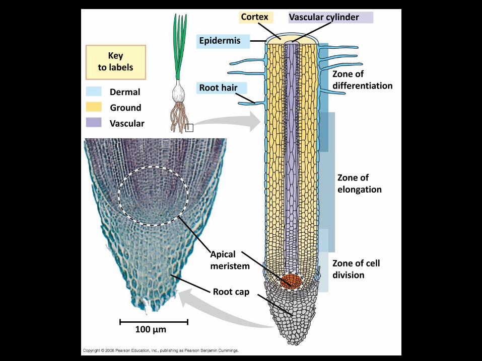
Fig. 35-13
Ground
Dermal
Key to labels
Vascular
Root hair
Epidermis
Cortex Vascular cylinder
Zone of differentiation
Zone of elongation
Zone of cell division
Apical meristem
Root cap
100 µm

Growth: Cell Division and Cell Expansion
• By increasing cell number, cell division in meristems increases the potential for growth
• Cell expansion accounts for the actual increase in plant size

The Plane and Symmetry of Cell Division
• The plane (direction) and symmetry of cell division are immensely important in determining plant form
• If the planes of division are parallel to the plane of the first division, a single file of cells is produced

Fig. 35-25
Plane of cell division
(a) Planes of cell division
Developing guard cells
Guard cell “mother cell”
Unspecialized epidermal cell
(b) Asymmetrical cell division

Fig. 35-25a
Plane of cell division
(a) Planes of cell division

• If the planes of division vary randomly, asymmetrical cell division occurs

Fig. 35-25b
Developing guard cells
Guard cell “mother cell”
Unspecialized epidermal cell
(b) Asymmetrical cell division
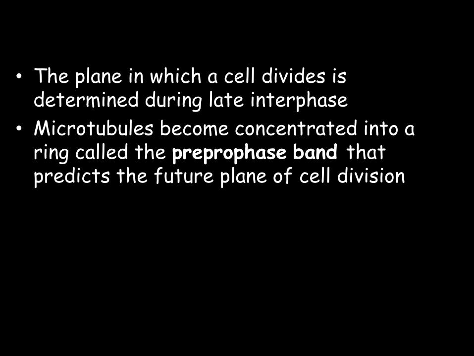
• The plane in which a cell divides is determined during late interphase
• Microtubules become concentrated into a ring called the preprophase band that predicts the future plane of cell division
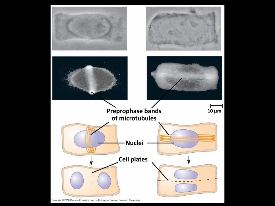
Fig. 35-26
Preprophase bands of microtubules
10 µm
Nuclei
Cell plates

Orientation of Cell Expansion
• Plant cells grow rapidly and “cheaply” by intake and storage of water in vacuoles
• Plant cells expand primarily along the plant’s main axis
• Cellulose microfibrils in the cell wall restrict the direction of cell elongation

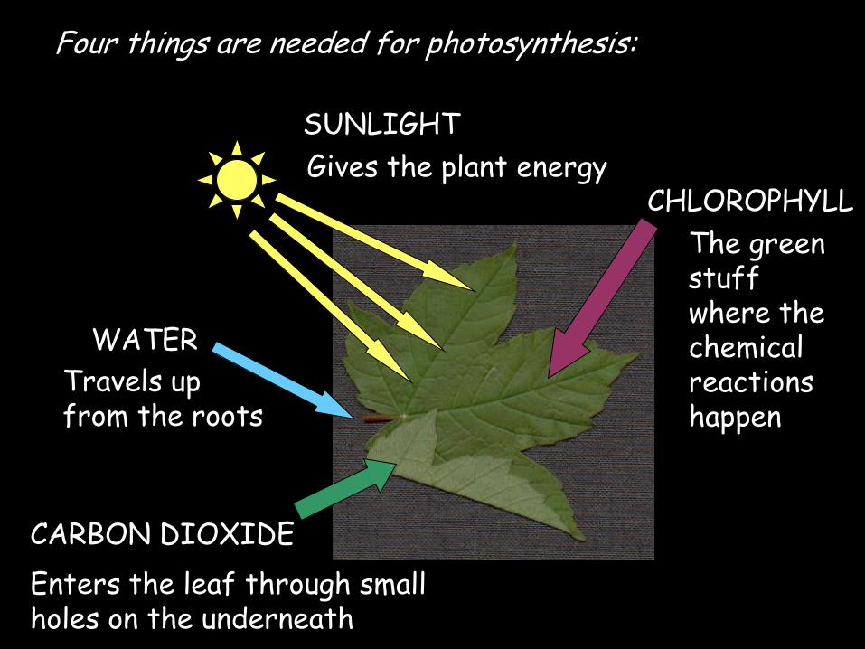
Four things are needed for photosynthesis:
Travels up from the roots
WATER
CARBON DIOXIDE
Enters the leaf through small holes on the underneath
SUNLIGHT
Gives the plant energy CHLOROPHYLL
The green stuff where the chemical reactions happen

The word and chemical equations for photosynthesis:
Carbon dioxide + water glucose + oxygen
6CO2 + 6H20 C6H12O6 + 6O2
Sunlight
Chlorophyll
Sunlight
Chlorophyll

Four factors affect photosynthesis:
1. Light – if there is more light photosynthesis happens faster
2. Water – if there is not enough water photosynthesis slows down
3. Temperature – the best temperature is about 300C – anything above 400C will slow photosynthesis right down
4. CO2 – if there is more carbon dioxide photosynthesis will happen quicker
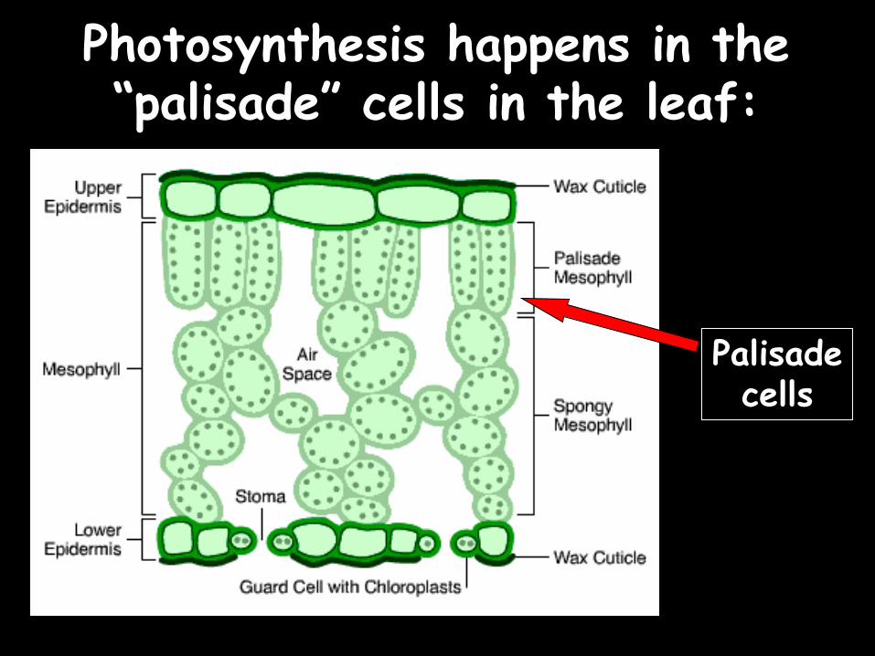
Photosynthesis happens in the “palisade” cells in the leaf:
Palisade cells

Close up on a palisade cell:
Cell wall
Cell membrane
Nucleus Large vacuole
Cytoplasm
Chloroplasts (containing chlorophyll)

Plant cells have three “extra” things than animal cells:
Both types of cell have these: Only plant cells have these:
4) Cell wall – provides support
Large Vacuole – contains sap
Chloroplasts – contain chlorophyll
1)
5)
6)
2)
3) Cell Membrane – holds the cell together
Cytoplasm - this is where the reactions happen
Nucleus – The “brain” of the cell

Plant growth
Plants grow using food they make through photosynthesis. So what else do they need?
Plants also need three important minerals to keep healthy. They absorb these through their roots.

Root hair cells
Plant roots are made of “root hair cells” which have a large surface area and a thin cell membrane to help absorb the minerals:
Thin cell membrane Large surface area
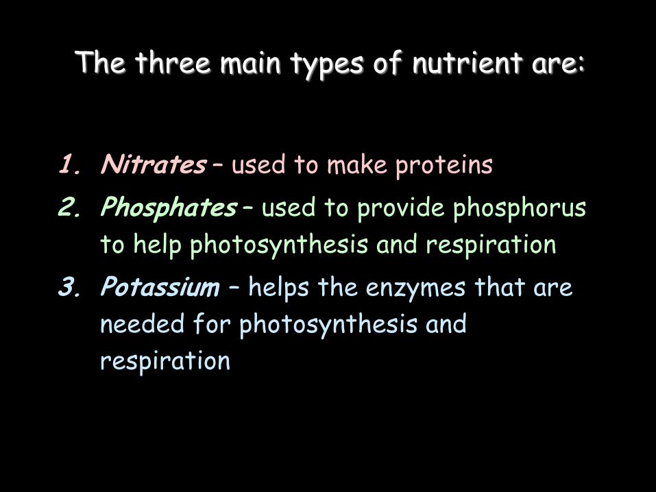
The three main types of nutrient are:
1. Nitrates – used to make proteins
2. Phosphates – used to provide phosphorus
to help photosynthesis and respiration
3. Potassium – helps the enzymes that are
needed for photosynthesis and
respiration

Lack of the three minerals would lead to a “Deficiency Symptom”:
Lack of nitrates: Small plant, yellow leaves
Lack of phosphates: Small roots and purple leaves
Lack of potassium: Yellow leaves with
dead parts of leaves

Chloroplasts: The Sites of Photosynthesis in Plants
• Leaves are the major locations of photosynthesis
• Their green color is from chlorophyll, the green pigment within chloroplasts
• Light energy absorbed by chlorophyll drives the synthesis of organic molecules in the chloroplast
• CO2 enters and O2 exits the leaf through microscopic pores called stomata
Copyright © 2008 Pearson Education, Inc., publishing as Pearson Benjamin Cummings

• Chloroplasts are found mainly in cells of the mesophyll, the interior tissue of the leaf
• A typical mesophyll cell has 30–40 chloroplasts
• The chlorophyll is in the membranes of thylakoids (connected sacs in the chloroplast); thylakoids may be stacked in columns called grana
• Chloroplasts also contain stroma, a dense fluid
Copyright © 2008 Pearson Education, Inc., publishing as Pearson Benjamin Cummings

Fig. 10-3 Leaf cross section
Vein
Mesophyll
Stomata CO2 O2
Chloroplast Mesophyll cell
Outer
membrane
Intermembrane
space
5 µm
Inner
membrane
Thylakoid
space
Thylakoid
Granum
Stroma
1 µm

Fig. 10-3a
5 µm
Mesophyll cell
Stomata CO2 O2
Chloroplast
Mesophyll
Vein
Leaf cross section

Fig. 10-3b
1 µm
Thylakoid
space
Chloroplast
Granum
Intermembrane
space
Inner
membrane
Outer
membrane
Stroma
Thylakoid

Tracking Atoms Through Photosynthesis: Scientific
Inquiry
• Photosynthesis can be summarized as the following equation:
6 CO2 + 12 H2O + Light energy C6H12O6 + 6 O2 + 6 H2O
Copyright © 2008 Pearson Education, Inc., publishing as Pearson Benjamin Cummings

The Splitting of Water
• Chloroplasts split H2O into hydrogen and oxygen, incorporating the electrons of hydrogen into sugar molecules
Copyright © 2008 Pearson Education, Inc., publishing as Pearson Benjamin Cummings
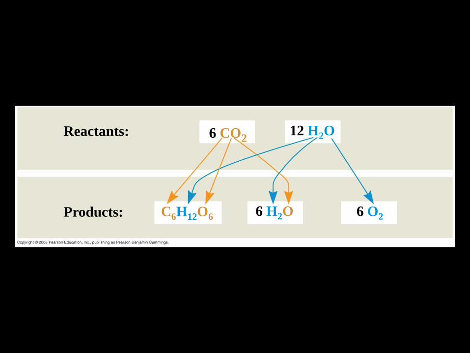
Reactants:
Fig. 10-4
6 CO2
Products:
12 H2O
6 O2 6 H2O C6H12O6


The Two Stages of Photosynthesis: A Preview
• Photosynthesis consists of the light reactions (the photo part) and Calvin cycle (the synthesis part)
• The light reactions (in the thylakoids): – Split H2O
– Release O2
– Reduce NADP+ to NADPH
– Generate ATP from ADP by photophosphorylation
Copyright © 2008 Pearson Education, Inc., publishing as Pearson Benjamin Cummings

• The Calvin cycle (in the stroma) forms sugar from CO2, using ATP and NADPH
• The Calvin cycle begins with carbon fixation, incorporating CO2 into organic molecules
Copyright © 2008 Pearson Education, Inc., publishing as Pearson Benjamin Cummings

Light
Fig. 10-5-1
H2O
Chloroplast
Light Reactions
NADP+
P
ADP
i +

Light
Fig. 10-5-2
H2O
Chloroplast
Light Reactions
NADP+
P
ADP
i +
ATP
NADPH
O2

Light
Fig. 10-5-3
H2O
Chloroplast
Light Reactions
NADP+
P
ADP
i +
ATP
NADPH
O2
Calvin Cycle
CO2

Light
Fig. 10-5-4
H2O
Chloroplast
Light Reactions
NADP+
P
ADP
i +
ATP
NADPH
O2
Calvin Cycle
CO2
[CH2O]
(sugar)
The light reactions convert solar energy to the chemical energy of ATP and NADPH
• Chloroplasts are solar-powered chemical factories
• Their thylakoids transform light energy into the chemical energy of ATP and NADPH
Copyright © 2008 Pearson Education, Inc., publishing as Pearson Benjamin Cummings
The Nature of Sunlight
• Light is a form of electromagnetic energy, also called electromagnetic radiation
• Like other electromagnetic energy, light travels in rhythmic waves
• Wavelength is the distance between crests of waves
• Wavelength determines the type of electromagnetic energy
Copyright © 2008 Pearson Education, Inc., publishing as Pearson Benjamin Cummings

• The electromagnetic spectrum is the entire range of electromagnetic energy, or radiation
• Visible light consists of wavelengths (including those that drive photosynthesis) that produce colors we can see
• Light also behaves as though it consists of discrete particles, called photons
Copyright © 2008 Pearson Education, Inc., publishing as Pearson Benjamin Cummings

UV
Fig. 10-6
Visible light
Infrared Micro- waves
Radio waves X-rays
Gamma
rays
103 m 1 m
(109 nm) 106 nm 103 nm 1 nm 10–3 nm 10–5 nm
380 450 500 550 600 650 700 750 nm
Longer wavelength
Lower energy Higher energy
Shorter wavelength
Photosynthetic Pigments: The Light Receptors
• Pigments are substances that absorb visible light
• Different pigments absorb different wavelengths
• Wavelengths that are not absorbed are reflected or transmitted
• Leaves appear green because chlorophyll reflects and transmits green light
Light and Pigments
Copyright © 2008 Pearson Education, Inc., publishing as Pearson Benjamin Cummings

Fig. 10-7
Reflected light
Absorbed light
Light
Chloroplast
Transmitted light
Granum

Fig. 10-8
Galvanometer
Slit moves to pass light of selected wavelength
White light
Green light
Blue light
The low transmittance (high absorption) reading indicates that chlorophyll absorbs most blue light.
The high transmittance (low absorption) reading indicates that chlorophyll absorbs very little green light.
Refracting prism
Photoelectric tube
Chlorophyll solution
TECHNIQUE
1
2 3
4
• An absorption spectrum is a graph plotting a pigment’s light absorption versus wavelength
• The absorption spectrum of chlorophyll a suggests that violet-blue and red light work best for photosynthesis
• An action spectrum profiles the relative effectiveness of different wavelengths of radiation in driving a process
Copyright © 2008 Pearson Education, Inc., publishing as Pearson Benjamin Cummings

Fig. 10-9
Wavelength of light (nm)
(b) Action spectrum
(a) Absorption spectra
(c) Engelmann’s experiment
Aerobic bacteria
RESULTS
Filament of alga
Chloro-
phyll a Chlorophyll b
Carotenoids
500 400 600 700
700 600 500 400
• Chlorophyll a is the main photosynthetic pigment
• Accessory pigments, such as chlorophyll b, broaden the spectrum used for photosynthesis
• Accessory pigments called carotenoids absorb excessive light that would damage chlorophyll
Copyright © 2008 Pearson Education, Inc., publishing as Pearson Benjamin Cummings
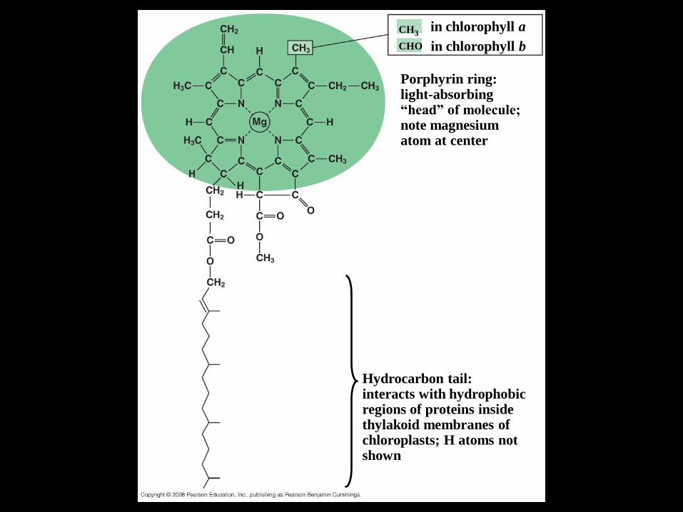
Fig. 10-10
Porphyrin ring: light-absorbing “head” of molecule; note magnesium atom at center
in chlorophyll a CH3
Hydrocarbon tail: interacts with hydrophobic regions of proteins inside thylakoid membranes of chloroplasts; H atoms not shown
CHO in chlorophyll b

Excitation of Chlorophyll by Light
• When a pigment absorbs light, it goes from a ground state to an excited state, which is unstable
• When excited electrons fall back to the ground state, photons are given off, an afterglow called fluorescence
• If illuminated, an isolated solution of chlorophyll will fluoresce, giving off light and heat
Copyright © 2008 Pearson Education, Inc., publishing as Pearson Benjamin Cummings

Fig. 10-11
(a) Excitation of isolated chlorophyll molecule
Heat
Excited state
(b) Fluorescence
Photon Ground state
Photon (fluorescence)
En
ergy o
f el
ectr
on
e–
Chlorophyll molecule
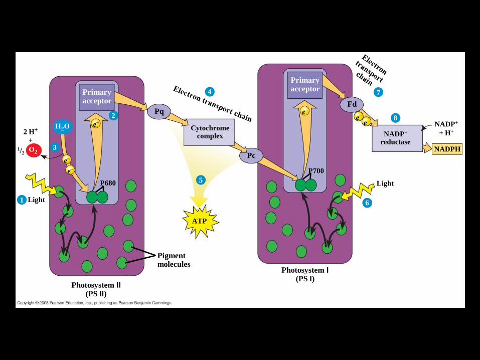
Pigment molecules
Light
P680
e–
Primary acceptor
2
1
e–
e–
2 H+
O2
+
3
H2O
1/2
4
Pq
Pc
Cytochrome complex
5
ATP
Photosystem I (PS I)
Light
Primary acceptor
e–
P700
6
Fd
NADP+ reductase
NADP+
+ H+
NADPH
8
7
e– e–
6
Fig. 10-13-5
Photosystem II (PS II)
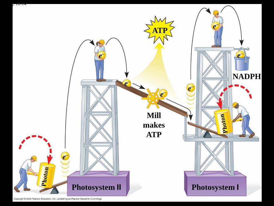
Fig. 10-14
Mill
makes
ATP
e–
NADPH
e– e–
e–
e–
e– ATP
Photosystem II Photosystem I
e–

Cyclic Electron Flow
• Cyclic electron flow uses only photosystem I and produces ATP, but not NADPH
• Cyclic electron flow generates surplus ATP, satisfying the higher demand in the Calvin cycle
Copyright © 2008 Pearson Education, Inc., publishing as Pearson Benjamin Cummings

Fig. 10-16
Key
Mitochondrion Chloroplast
CHLOROPLAST
STRUCTURE
MITOCHONDRION
STRUCTURE
Intermembrane
space
Inner
membrane
Electron transport
chain
H+ Diffusion
Matrix
Higher [H+]
Lower [H+]
Stroma
ATP
synthase
ADP + P i
H+ ATP
Thylakoid
space
Thylakoid
membrane

Fig. 10-17
Light
Fd
Cytochrome
complex
ADP
+
i H+
ATP
P
ATP synthase
To Calvin Cycle
STROMA (low H+ concentration)
Thylakoid membrane
THYLAKOID SPACE (high H+ concentration)
STROMA (low H+ concentration)
Photosystem II Photosystem I
4 H+
4 H+
Pq
Pc
Light NADP+
reductase
NADP+ + H+
NADPH
+2 H+
H2O O2
e– e–
1/2 1
2
3
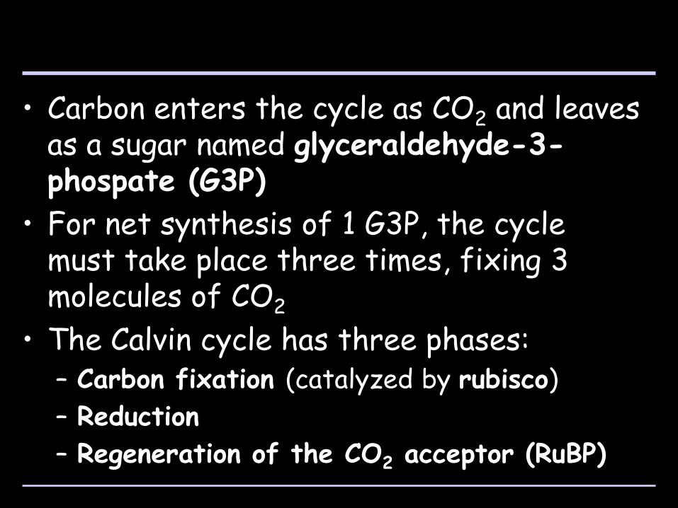
• Carbon enters the cycle as CO2 and leaves as a sugar named glyceraldehyde-3-phospate (G3P)
• For net synthesis of 1 G3P, the cycle must take place three times, fixing 3 molecules of CO2
• The Calvin cycle has three phases: – Carbon fixation (catalyzed by rubisco)
– Reduction
– Regeneration of the CO2 acceptor (RuBP)
Copyright © 2008 Pearson Education, Inc., publishing as Pearson Benjamin Cummings
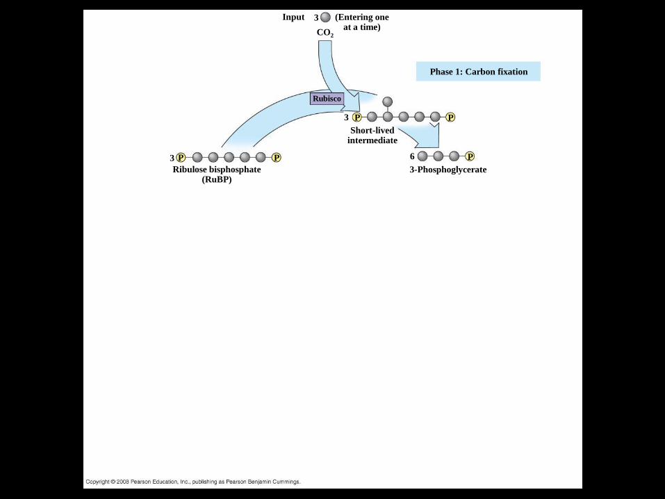
Fig. 10-18-1
Ribulose bisphosphate (RuBP)
3-Phosphoglycerate
Short-lived intermediate
Phase 1: Carbon fixation
(Entering one at a time)
Rubisco
Input
CO2
P
3 6
3
3
P
P P P
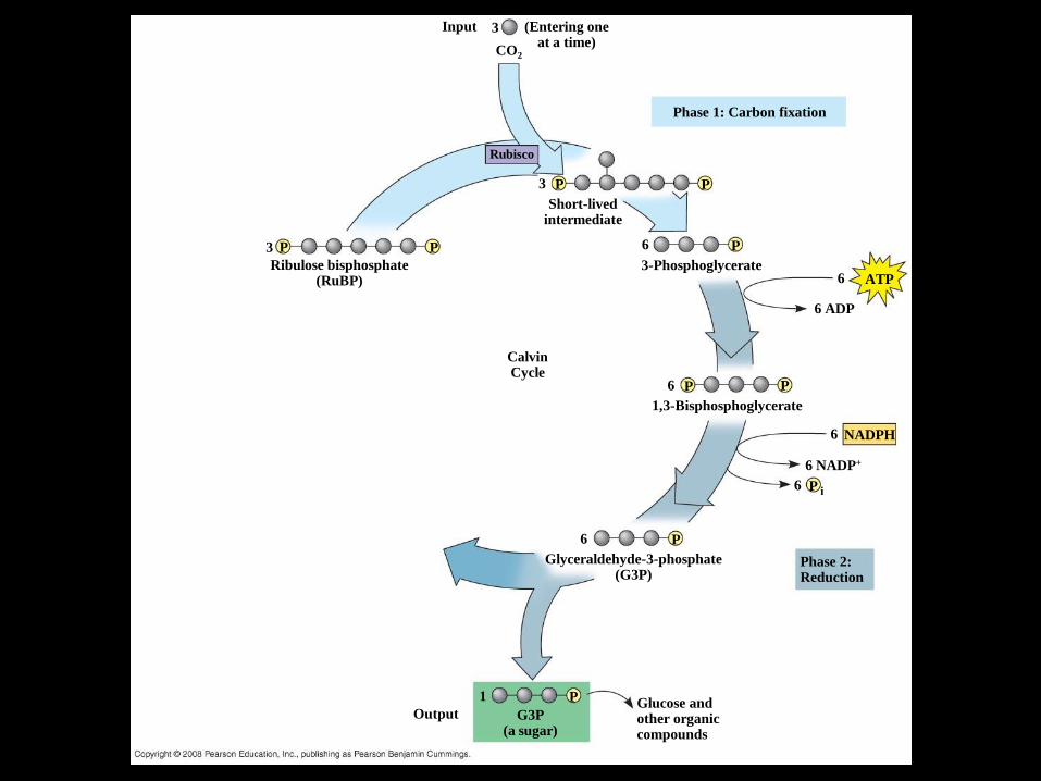
Fig. 10-18-2
Ribulose bisphosphate (RuBP)
3-Phosphoglycerate
Short-lived intermediate
Phase 1: Carbon fixation
(Entering one at a time)
Rubisco
Input
CO2
P
3 6
3
3
P
P P P
ATP 6
6 ADP
P P 6
1,3-Bisphosphoglycerate
6
P
P 6
6
6 NADP+
NADPH
i
Phase 2: Reduction
Glyceraldehyde-3-phosphate (G3P)
1 P
Output G3P (a sugar)
Glucose and other organic compounds
Calvin Cycle

Fig. 10-18-3
Ribulose bisphosphate (RuBP)
3-Phosphoglycerate
Short-lived intermediate
Phase 1: Carbon fixation
(Entering one at a time)
Rubisco
Input
CO2
P
3 6
3
3
P
P P P
ATP 6
6 ADP
P P 6
1,3-Bisphosphoglycerate
6
P
P 6
6
6 NADP+
NADPH
i
Phase 2: Reduction
Glyceraldehyde-3-phosphate (G3P)
1 P
Output G3P (a sugar)
Glucose and other organic compounds
Calvin Cycle
3
3 ADP
ATP
5 P
Phase 3: Regeneration of the CO2 acceptor (RuBP)
G3P