Embed Size (px)
Citation preview

7/30/2015
1
Pressure Ulcers: What do the Surveyors See
CAROL SIEM RN, MSN,GNP BCQIPMO Team Leader
4 goals listed by AHCPR guidelines for PUrisk and prevention.
• 1. ID of at risk individuals who need prevention, & specific factors placing them at risk.
• 2. Maintaining & improving tissue tolerance to pressure to prevent injury.
• 3. To protect against adverse effects of pressure, friction, & shear.
• 4. To reduce the incidence of pressure ulcers through education.
• Friction--Rubbing against an external object such as a sheet.
• Mechanical Load--Adverse affects of external mechanical forces such as a bed or chair.
• Shear--Conflicting forces applied to an object or area in opposite directions at the same time. Sliding down in bed causes pressure & pull from both the bed itself & from the internal bony prominences.
“Pressure ulcers develop when soft tissue is pressed between a bony prominence & a firm surface with the pressure causing the capillaries to collapse. This in turn interrupts the tissue’s supply of O2 & nutrients. If the capillaries remain closed the surrounding tissue dies, sometimes in a very short time.”
Vap, P, Dunaye,T., PU risk assessment
in LTC nursing. JOGN,6/2000.
Age related changes that contribute to pressure ulcer risk.
Thinning of the dermal epidermal junction. Leads to wrinkling, tearing, loss of elasticity, increased skin permeability, & alterations in barrier function of the skin.
An altered immune response & decreased dermal vascularity. Causes a greater potential for infection.
Areas of Risk for Formation of Pressure Ulcers
• Back of head
• Back of ears
• Shoulder blades
• Elbows
• Backbone
• Crest of pelvis
• Coccyx region
• Trochanter
• Buttocks
• Kneecaps
• Outsides of knees
• Insides of knees
• Outsides of feet
• Insides of feet
• Outsides of ankles
• Heels

7/30/2015
2
• Powell found a 129% higher death rate for pts admitted to LTC who developed a pressure ulcer than those who did not.
• Burd et. al reported a risk of death among geriatric patients who developed a pressure ulcer to be 4 times greater than the norm, & 6 times greater in those whose pressure ulcers did not heal.
• Lawsuit judgments can run as high as $312 million for a single case
• Voss AC, Bender SA, Ferguson ML, et al. Long-term care liability for pressure ulcers. J Am Geriatric Soc. 2005;53:1587-1592.
Latest figures shows the average cost for treating a pressure ulcer is over 1 billion annually and additional $2.2 million in Medicare Hospital Days
Cost for treatment $6,000 to $60,000 depending on size and stage
Some sources indicate cost per ulcer can be up to $90,000
Wake, W (2010, Summer). Pressure ulcers: What clinicians need to know. The Permanente Journal, 14 (2), 56-60.
Identification of Those At Risk
Use of a validated risk assessment tool.
Complete on admission & at periodic intervals.
AHCPR recommends that risk prediction be an integral part of pressure ulcer prevention programs.
Difference Between Risk Assessment & Skin Assessment
Risk Assessment
Determines if the resident is at risk for PUs or other skin problems.
Looks at areas like, mobility, activity, incontinence, nutrition, mental status, friction/shear.
Skin Assessment
Looks at the resident’s skin to determine if skin problems or pressure ulcers already exist, or have worsened.
Braden Scale
• Sensory Perception• Moisture• Activity• Mobility• Nutrition• Friction/Shear
• 23 points possible.• Lower score = more
risk.• Risk predicting score
of 18 or less.• 18 for individuals
with darker skin & those 75 & over.
• Direction for use in tool itself.

7/30/2015
3
Norton Scale
Risk factors
Physical Condition
4=good 3=Fair 2=poor 1=Bad
Mental State 4=Alert 3=Apathetic 2=Confused 2=Stupor
Activity 4=Ambulant 3=Walks with help
2=Chair bound
1=Bed rest
Mobility 4 =Full 3=Slightly limited
2=Very limited
1=Immobile
Incontinence 4=Not 3=Occasional 2=Usually Urine
1=Double incontinence
Norton Scale
Physical condition
Mental condition
Activity
Mobility
Continence
20 points possible.
Lower score=more risk.
Risk predicting score of 16 or less.
No directions or guidance for use.
Minimum Data Set (MDS)
• Bed Mobility
• Bladder Incontinence
• Bowel incontinence
• Unplanned weight loss
• Risk of PU
• Pressure ulcer/Unstageable
• Worsening PU
• Trunk restraint
• Any of these will trigger for risk in the CAAs.
• Recent retrospective studies show MDS may have better predictive value than Braden or Norton. (Study was done with MDS 2.0)
• Problem is timing.
Braden MDS
Fails to Address
pres. ulcer present
pres. in last 90 days
daily trunk restraint
PVD
Fails to Address
friction
shear
Many residents are at highest risk of developing pressure ulcers during 1st 2 weeks after admission. Esp.1st 48 hrs.
Prevention is based on eliminating or changing those risk factors that are amenable to intervention. In some situations not much can be done about sensory perception, activity, or mobility, but interventions must be put in place that lesson the effects of pressure because of these.
“There is no accurate predictive value of risk assessment scales because they measure residents & not staff.”
Kenneth Olshansky, MD

7/30/2015
4
Identify Specific Risk Factors
Immobility--requires assist with bed mobility & transfers
Incontinence of urine &/or bowel >daily
Impaired nutritional intake--eats 1/2 or less of food offered (rarely/never eats a complete meal)
Altered level of consciousness--unresponsive/sedated
Identify Risk Factors, cont’d
Impaired sensation--limited ability to feel pain or discomfort over 1/2 of body
Anemia--decreased oxygen carrying capacity of blood, affects circulation & wound healing
History of pressure ulcers--do conditions still exist?
Restraint use--can affect previously mentioned areas
Good - 30° Side-Lying Position
• Back pillow
• 30° head elevation
• No ankle/knees touching
Good - Heels Off Bed
Heel - very small surface for weight distribution
Recommended position for prevention of pressure on heels
Not Good - Heels On Bed Not Good - Chair Shearing
Sliding from a chair for a long period will cause shearing.
What would be the results?

7/30/2015
5
Prevention Strategies cont’d
Repositioning at least q2h when in bed, q1h when up in chair
– This is resident specific to their needs and skin condition
Donut/ring devices avoided
• “Data do not indicate how often patients should be turned to prevent ischemia of soft tissue, but two hours in a single position is the maximum duration of time recommended for patients with normal circulatory capacity.”
• “Chair bound residents should be repositioned at least hourly.”• NPUAP 2009
Skin Care
Cleanse
– Cleanse with no-rinse, non-irritating ph balanced cleanser
– Individualized bathing
Moisturize
Internal and external hydration
Use moisturizing cream if the skin is dry
Use moisturizing lotion to prevent dry skin
Protect
Incontinence
– Commercial cleansers
– Cleansing wipes
– Soap and water
Protect
Types of barriers
– Sealant
– Creams
Partially denuded skin with mixed incontinence
– Ointments
Cost effective

7/30/2015
6
Excessively Moist skin
Maceration
– MDS: New item Moisture Associated Skin Damage (MASD)
– M 1040 H
Denudation
Fungal Infections
Incontinence
Maceration
Denudation
Fungal Infection
Is this a Stage 1 Pressure Ulcer?Not a Stage 1 Pressure Ulcer
This is moisture associated skin damage from incontinence.
Moisture vs PressureAdapted from Defloor et al (2005), Nix (2005), Haugen (2010)
Moisture must be present
May be over bony prominence
Skin shiny, wet, appearance
Diffuse, multiple lesions
Irregular edges
Kissing ulcer
Anal cleft, linear
Partial thickness skin loss
No necrosis
Non-uniform redness, pink/white macerated periwound
Pressure and/or shear must be present
Most often over bony prominence, equipment related, skin folds
Regular, raised edges
Depth dependent on stage
Necrotic tissue depended on stage
Erythema, slough, necrosis, granulation, epithelial, infection
Isolated, individual lesions
Skin Tears Treatment
• New item on the MDS• M1040 G Skin Tears
• Gently cleanse area• Air dry or pat dry• Approximate the skin tear flap if possible• Apply moist non adherent wound dress• Avoid film dressing• Place an arrow to indicate the direction of
the skin tear on the dressing

7/30/2015
7
Assessment of the Pressure Ulcer: Parameters
• Location
• Staging
• Wound Measurement
• Undermining of tissue
• Tunneling of tissue
• Exudate
• Necrotic tissue
• Granulation tissue
• Epithelialization
• Periwound skin
• Pain
Assessment of the Pressure Ulcer
Location
Should refer to body landmarks or body diagrams
Assessment of the Pressure Ulcer: Measurement
Recorded Length by Width by Depth
– LXWXD
Recorded in Centimeters (cm)
Length
Measure the longest length from head to toe using a disposable device.
Head
Toe
Width
Measure widest width of the pressure ulcer side to side perpendicular (90° angle) to length.
The depth ofthis pressure ulcer is 3.7 cm.
Head
Toe

7/30/2015
8
Depth
Moisten a cotton-tipped applicator with 0.9% sodium chloride (NaCl) solution or sterile water.
Place applicator tip in deepest aspect of the wound and measure distance to the skin level.
Stages of a Pressure Ulcer
The following descriptions are from the National Pressure Ulcer Advisory Panel, 2014
Stages of a Pressure Ulcer
Suspected Deep Tissue Injury (SDTI)
– Purple or maroon localized area of discolored intact skin or blood filled blister due to damage of underlying soft tissue from pressure and or shear. The area may be preceded by tissue that is painful, firm, mushy, boggy, warmer or cooler as compared to adjacent tissue.
Further description
Deep tissue injury may be difficult to detect in individuals with dark skin tones. Evolution may include a thin blister over a dark ulcer bed. The ulcer may further evolve and become covered by thin eschar. Evolution may be rapid, exposing additional layers of tissue even with optimal treatment.
Deep Tissue InjuryStages of Pressure Ulcers
Stage I
– Intact skin with non-blanchable redness of a localized area, usually over a bony prominence. Darkly pigmented skin many not have visible blanching; its color may differ from the surrounding area.

7/30/2015
9
Stage I
Further description:
– The area may be painful, firm, soft, warmer, or cooler as compared to adjacent tissue. Stage I may be difficult to detect in individuals with dark skin tones. May indicate “at risk” persons (a heralding sign of risk).
Stage 1
Category/ Stage 1 Pressure Ulcer
Intact skin with non-blanchable redness of a localized area usually over a bony prominence.
Darkly pigmented skin may not have visible blanching.
Color may differ from the surrounding area.
Stages of Pressure Ulcers
Stage II
– Partial thickness loss of dermis presenting as a shallow open ulcer with a red pink ulcer bed, without slough. May also present as an intact or open/ruptured serum filled blister.
Stage II
Further description
– Presents as a shiny or dry shallow ulcer without slough or bruising (Bruising indicates suspected deep tissue injury). This stage should not be used to describe skin tears, tape burns, perineal dermatitis, maceration or excoriation.

7/30/2015
10
Stage II
Category/ Stage 2 Pressure Ulcer
Partial thickness loss of dermis presenting as:
– Shallow open ulcer
– Red or pink wound bed
– Without slough
Category/ Stage 2 Pressure Ulcer
May also present as an intact or open/ ruptured blister.
Stages of Pressure Ulcer
Stage III
– Full thickness tissue loss. Subcutaneous fat may be visible but bone, tendon, or muscle are not exposed. Slough may be present but does not obscure the depth of tissue. May include undermining and tunneling.
Stage III
Further description
– The depth of a Stage III pressure ulcer varies by anatomical location. The bridge of the nose, ear, occiput and malleolus do not have subcutaneous tissue and stage III ulcers can be shallow. In contrast, areas of significant adiposity can develop extremely deep Stage III pressure ulcers. Bone/tendon is not visible or directly palpable.
Category/ Stage 3 Pressure Ulcer
• Full thickness tissue loss.
• Subcutaneous fat may be visible but bone, tendon or muscle are not exposed.
• Slough may be present but does not obscure the depth of tissue loss.
• May include undermining and tunneling.

7/30/2015
11
Stage III Stages of Pressure Ulcer
Stage IV
– Full thickness tissue loss with exposed bone, tendon, or muscle. Slough or eschar may be present on some parts of the ulcer bed. Often include, undermining and tunneling.
Stage IV
Further description– The depth of a Stage 4 pressure ulcer varies by
anatomical location. The bridge of the nose, ear, occiput and malleolus do not have subcutaneous tissue and these ulcers can be shallow. Stage 4 ulcers can extend into muscle and/or supporting structures (e.g., fascia, tendon or joint capsule making osteomlyelitis possible. Exposed bone, tendon is visible or directly palpable.
Category/ Stage 4 Pressure Ulcer
• Full thickness tissue loss with exposed bone, tendon or muscle.
• Slough or eschar may be present on some parts of the wound bed.
• Often includes undermining and tunneling.
• Depth varies by anatomical location (bridge of nose, ear, occiput, and malleous ulcers can be shallow).
STAGE IVExtensive destruction tissue
necrosis or damage to muscle,
bone, or supporting structures
with or without full thickness
skin loss
The ulcer has eroded deeply,
causing damage to body
tissue, bone, muscle, tendons
and joints. The risk of infection
is much higher at this stage
Stages of Pressure Ulcer
Unstageable
– Full thickness tissue loss in which the base of the ulcer is covered by slough (yellow, tan, gray, green or brown) and or eschar (tan, brown or black) in the ulcer bed.

7/30/2015
12
Unstageable
Further description
– Until enough slough and or eschar is removed to expose the base of the ulcer, the true depth, and therefore stage, cannot be determined. Stable (dry, adherent, intact without erythema, or fluctuance) eschar on the heels serves as “the body’s natural (biological) cover” and should not be removed.
UnstageableNon-Removable Device
Ulcer covered with eschar under plaster cast
Known but not stageable because of the non-removable device
Un-stageableSlough and/ or Eschar
• Known but not stageable related to coverage of wound bed by slough and/ or eschar
• Full thickness tissueloss
• Base of ulcer covered by slough (yellow, tan, gray, green or brown) and/ or eschar (tan, brown or black) in the wound bed
Slough Eschar
M0300G UnstageableSuspected Deep Tissue Injury
Purple or maroon area of discolored intact skin due to damage of underlying soft tissue. The area may be preceded by tissue that is painful, firm, mushy, boggy, warmer or cooler as compared to adjacent tissue.
M0300G UnstageableSuspected Deep Tissue Injury
• Localized area of discolored (darker than surrounding tissue) intact skin.
• Related to damage of underlying soft tissue from pressure and/ or shear.
• Area of discoloration may be preceded by tissue that is painful, firm, mushy, boggy, warmer or cooler as compared to adjacent tissue.
• Deep tissue injury may be difficult to detect in individuals with dark skin tones.

7/30/2015
13
Eschar vs. Scab
• Lengthy discussion on the differences between scabs and eschar is now on page M-5• Eschar: collection of dead tissue within the
wound that is flush with the surface of the wound• Scab: dried blood cells and serum, sits on top of
the skin, and forms over exposed wounds, such as wounds with granulating surfaces (like pressure ulcers, lacerations, evulsions.
• A PU that was staged as a 2 and now has a scab indicates it is a healing stage 2 & therefore, staging should NOT change (M-5)
Slough Eschar
Assessment of the Pressure Ulcer: Undermining
Generally appears as an area of skin ulceration at the margins of the ulcer
Usually an indication of regression
Measured in cm described according to a clockface
– “0.7cm undermining from 9:00 to 12:00”
Assessment of the Pressure Ulcer: Tunneling
A passageway under the surface of the skin
Usually an indication of regression
Measured in cm, described according to a clock face
– “1.5cm tunneling at 10:00”

7/30/2015
14
Assessment of the Pressure Ulcer: Exudate
Often called drainage
Fluid extruded from a wound bed
Assessment of the Pressure Ulcer: Epithelialization
Migration of cells across the top of the wound bed
Necessary for wound closure
Assessment of the Pressure Ulcer: Granulation Tissue
Pink/red moist tissue that contains new blood vessels and essential components to promote growth.
Healthy components of a wound bed, presents like a “good beef steak”
Assessment of the Pressure Ulcer: Necrotic Tissue
“Dead tissue”
May present as gray, brown, yellow slough or leathery brown, black eschar
Slough Necrotic Tissue (Eschar)

7/30/2015
15
Assessment of the Pressure Ulcer: Peri-wound skin
Skin surrounding the wound
– Erythema-redness of the intact skin
some redness is normal response to healing
– Maceration-dampness of the skin
skin will look white, wrinkled
– Induration-hardness of the skin
Assessment of the Pressure Ulcer: Pain
Routine assessment/management of pain should occur ongoing, specifically with each dressing change and with any invasive procedure
Increasing pain may indicate regression or worsening of a wound
Reassessment of the Pressure Ulcer
Wound bed and periwound skin should be reassessed daily or with every dressing change if less than daily
Documentation of wound progress should occur weekly unless there is evidence of worsening
Reassessment of the Pressure Ulcer
Evidence of wound healing is expected within 2-4 weeks
Increases in exudate, edema, necrosis, pain, and/or loss of granulation tissue indicate wound regression

7/30/2015
16
Treatment of Pressure Ulcers
“Goals of pressure ulcer treatment should not only include assessment and management of the wound, but overall assessment and management of the individual.”
AHCPR Clinical Practice Guideline: Treatment of Pressure Ulcers (1994)
Treatment of Pressure Ulcers
“Ulcer healing may not be achievable in all cases; however, in the absence of complications, some improvement in ulcer characteristics should be expected in most patients.”
AMDA Practice Guideline for Pressure Ulcers (2008)
Treatment Factors
Ulcer location, size, and depth (full or partial thickness)
Presence of undermining or tunneling Presence of necrotic tissue Type & amount of drainage Presence of granulation or
epithelialization Presence of surrounding skin erythema,
edema or induration Presence & severity of ulcer related pain
Treatment of the Pressure Ulcer: Wound Cleansing
Cleanse initially and with each dressing change
– Use normal saline or approved wound cleanser
– Avoid skin cleansers or antiseptic agents i.e., betadine, alcohol, hydrogen peroxide, acetic acid
– Avoid aggressive cleansing/scrubbing of the wound bed.
Cleansing
Only use safe and effective ulcer irrigation pressures (4 to 15 pounds per square inch [PSI]).
– Normal saline in a 35cc syringe with an 18 gauge needle delivers 8 psi
– Water pik at lowest setting delivers 6 psi, mid setting 42 psi, high setting > 50 psi
– Pressure settings too low are ineffective, too high can drive bacteria back into the wound bed and cause serious problems.
Cleansing
Whirlpool treatments are appropriate for cleansing ulcers that have thick exudate or necrosis.
– Clean wounds tend to dry out and are not appropriate for WP treatments

7/30/2015
17
Treatment of Pressure UlcersDressing Selection
Stage I-”Intact Skin”
– Goal is to provide pressure relief, pressure relief, pressure relief
– Only appropriate dressing would be hydrocolloid (i.e., duoderm) if friction is a factor
– May not have any dressing at all
Dressing Selection
Stage II-IV clean wound covered with granulation tissue
– Goal is a moist, clean wound bed
– If the wound is a shallow stage II, may only use moisture barrier or hydrocolloid.
– May use hydrogel (water like jelly) covered with gauze.
– Loosely pack with gauze if depth is present
Dressing Selection
Stage III or IV exudating wounds
– Goal to absorb exudate, prevent breakdown of periwound skin and prevent drying out of wound bed
– Should use absorptive dressing (i.e., calcium alginate) with moisture barrier applied to periwound skin
– Whirlpool treatments daily to twice daily
Dressing Selection
Stage III or IV necrotic wound
– Goal to debride necrotic tissue to promote healing
– EXCEPTION: stable heel ulcers with dry eschar (no edema, erythema, exudate (drainage)…aggressive pressure relief only
Not all ulcers require debridement
Debridement
Types of debridement
– Sharp (knife, scalpel, scissors…must be done by a physician or licensed qualified staff i.e. therapist, RN)
– Mechanical (wet to dry, whirlpool, wound irrigation)
– Enzymatic (collagenase – FDA approved)
– Autolytic (bodies own mechanism of fighting – self digest…may use hydrocolloid)

7/30/2015
18
Eschar
Sharp Debridement
Sterile instruments
Sharp Debridement
Sharp Debridement
Dressing Selection
Cardinal rule-Keep wound bed moist and surrounding intact skin dry

7/30/2015
19
What’s wrong with this wound???What’s is wrong with the dressing that is being used?
Treatment Categories
Polyurethane Film (Tegaderm™, Op-Site◊)
– Adhesive and transparent
– Stages 1-2
– Occlusive and waterproof
– Impermeable to bacteria & contamination
– Change every 3-7 days
Treatment Categories
Hydrocolloid (Duoderm®, Replicare®)
– Adhesive wafers composed of gelatin, pectin and carbocymethyl-cellulose
– Stages 1-4
– Occlusive and waterproof
– Moderately absorbent
Treatment Categories
Hydrogels (Hypergel®, SoloSite◊)
– Glycerin or water based gels, wafers, sheets & impregnated gauze with or without adhesive border
– Stages 2-4
– Non-adherent
– Fills dead space
– Easy to apply and remove
Treatment Categories
Foams (PolyMem®, Allevyn◊)
– Hydrophillic polyurethane foam, available in wafers, sheets and pillow with foam covering
– Stages 2-4
– Non adherent
– Easy to apply and remove
– Highly absorbent

7/30/2015
20
Treatment Categories
Alginates (Sorbsan™, Kaltostat®)
– Non woven fibers containing calcium sodium slats of alginic acid, available in pads or ropes
– Stage 2 wounds with a lot of exudate
– Stages 3-4
– Non-adherent
– Promotes moist wound healing
– Can be used on infected wounds
Treatment categories
Antimicrobial (ACTICOAT◊, ALLEVYN Ag◊)
– Ionic silver & cadxomer idenit that probides sustain antimicrogial barrier to bacteria include MRSA and VRE
– Can be found in alginates, gels and polyurethane film
– Stage 2 wound if antimicrobial is needed
– Stages 3-4
– Manages bacterial burden
– Non-cytotoxic
Treatment Categories
Collagen (Biostep◊, Prisma®)
– Provides the matrix for the body’s tissue structure. Stimulates wound healing
– Can be found as dried collagen matrix, hydrogel with collagen, hydrogel base)
– Wounds that have stalled in healing
– Chronic wounds
– Pulls wound edges together
Treatment Categories
Gauze, Dry or wet
– Woven natural cotton fibers, available in pads, and rolls, sterile and non sterile
– Stages 2-4 especially if wound is deep or has tissue that needs debridement
– Facilitates moist to dry debridement
Related Treatment Options
Wound Vacs (KCI Vac®, V1STA ◊)
– Controlled negative pressure to promote wound healing
– Pulls infectious materials and excess interstitial fluid from the wound
– Pressure Ulcers, traumatic wounds, post op dehisced and surgical wounds
– FDA warning has been posted related to deaths
7/30/2015
21
Surgical intervention – skin flap
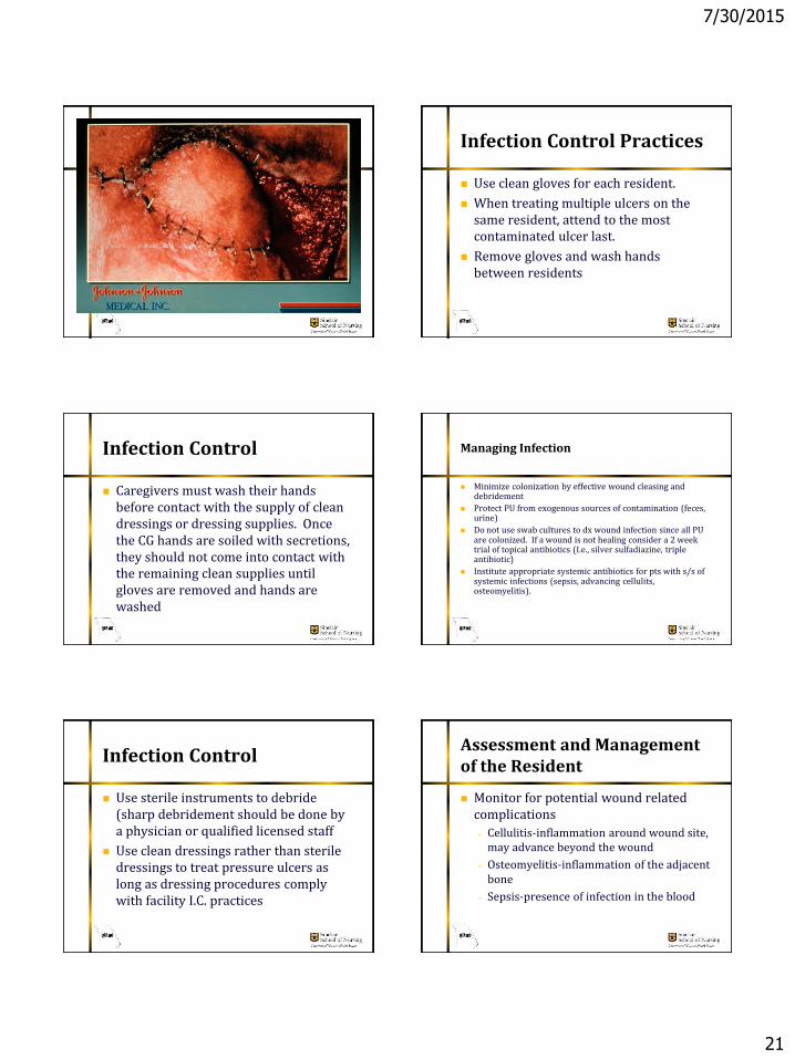
Infection Control Practices
Use clean gloves for each resident.
When treating multiple ulcers on the same resident, attend to the most contaminated ulcer last.
Remove gloves and wash hands between residents
Infection Control
Caregivers must wash their hands before contact with the supply of clean dressings or dressing supplies. Once the CG hands are soiled with secretions, they should not come into contact with the remaining clean supplies until gloves are removed and hands are washed
Managing Infection
Minimize colonization by effective wound cleasing and debridement
Protect PU from exogenous sources of contamination (feces, urine)
Do not use swab cultures to dx wound infection since all PU are colonized. If a wound is not healing consider a 2 week trial of topical antibiotics (I.e., silver sulfadiazine, triple antibiotic)
Institute appropriate systemic antibiotics for pts with s/s of systemic infections (sepsis, advancing cellulits, osteomyelitis).
Infection Control
Use sterile instruments to debride (sharp debridement should be done by a physician or qualified licensed staff
Use clean dressings rather than sterile dressings to treat pressure ulcers as long as dressing procedures comply with facility I.C. practices
Assessment and Management of the Resident
Monitor for potential wound related complications
– Cellulitis-inflammation around wound site, may advance beyond the wound
– Osteomyelitis-inflammation of the adjacent bone
– Sepsis-presence of infection in the blood

7/30/2015
22
Assessment and Management of the Resident
Nutrition/Hydration
– Monitor and document intake
– Offer assistance as necessary to ensure adequate intake
– Protein, vitamin, mineral supplements as appropriate
Vitamin C and Zinc most commonly used
Assessment and Management of the Resident
Pain
– Routine assessment every 4 hours, prior to dressing change, and any invasive procedures
– Assessment of cause of pain, worsening pain may indicate worsening of the wound
– Interventions to include pharmacologic and non pharmacologic measures
Assessment and Management of the Resident
Psychosocial
– Potential for depression and/or problem behavior due to wound presence, pain or change in function
– Appropriate management of depression symptoms and problem behaviors should be occurring
Documentation
Initial and ongoing risk assessments
Weekly wound record to include assessment of wound bed and periwound skin
Record of changes to treatment plan as wound changes (heals or regresses)
Documentation
CAA that addresses:
– Review using Clinical Practice Guidelines or the Information CMS has provided in Appendix C
Documentation
Care Plan to address– Problem statement including resident
specific risks and any actual wounds
– Appropriate, realistic goals determined with interdisciplinary input
– Interventions for prevention and/or treatment as appropriate
– Interventions for management of the resident (nutrition, hydration, mobility, etc)

7/30/2015
23
Types of Leg Ulcers
Venous Insufficiency
Arterial
Diabetic Neuropathic
Venous Insufficiency Ulcer
Increased
venous
pressure
induced by
incompetent
valves
Induces
inflammation
Chronic stasis
dermatitis Arterial Ulcer
Diabetic Foot Ulcers

7/30/2015
24
Diabetic Neuropathic Ulcer References:
American Medical Directors Association. Pressure Ulcers in the Long Term Care Setting Clinical Practice Guidelines. Columbia, MD:AMDA 2008.
Berstrom, N., Demuth, P., & Braden, B. (1987). A clinical trial of the Braden scale for predicting pressure sore risk. Nursing clinics of North America, 22(2), 417-424.
National Pressure Ulcer Advisory Panel. (1995). Position on reverse staging of pressure ulcers. NPUAP Report 4, no. 2:32-33.
National Pressure Ulcer Advisory Panel. (2007)Pressure Ulcer stages Revised by NPAUP
References:
U.S. Department of Health and Human Services: Agency for Health Care Policy and Research. (1992). Clinical practice guidelines: Pressure ulcers in adults: Prediction and prevention. (AHCPR 92-0050). Rockville, MD
U.S. Department of Health and Human Services: Agency for Health Care Policy and Research. (1994). Clinical practice guidelines: Pressure ulcer treatment. (AHCPR 95-0653). Rockville, MD
Guideline for Prevention & Management of Pressure Ulcers. (2010). WOCN. Mount Laurel, NJ
http://www.globalwoundacademy.com/