Embed Size (px)
Citation preview

551
A Chinese Herbal Medicine, Fu-Ling,Regulates Interleukin-10 Production
by Murine Spleen Cells
Chian-Jiun Liou and Jerming TsengDepartment of Biology, National Taiwan Normal University
88 Ting-Chou Road, Section 4, Taipei 116, Taiwan
Abstract: Fu-Ling is one of the most widely used Chinese herbal medicines. In this study, weinvestigated the regulatory effect of Fu-Ling on interleukin-10 (IL-10) production in vivo.Mice were i.p. administered 0.1 mg to 1.0 mg Fu-Ling per gram body weight daily for threedays. The spleen cells were isolated and assayed for both IL-10 and immunoglobulin (Ig)production. Results indicated that the mice treated with Fu-Ling had significantly increasedspleen cell ability to secrete IL-10. Spleen cells isolated from the mice injected with either0.1 mg or 1.0 mg Fu-Ling per gram body weight also showed an increase in IL-10 mRNAexpression. As IL-10 is a potent differentiation factor of B-lymphocytes, the possible effectof Fu-Ling on Ig production was also studied. Results indicated that Fu-Ling significantlyinduced an increase in IgG and IgA secretion by spleen cells but showed no effect on IgMsecretion. Thus, Fu-Ling may affect the function of B-lymphocytes via stimulating IL-10production.
Keywords: Fu-Ling; IL-10; Immunoglobulin.
Introduction
Tonic recipe is one of the major groups of Chinese herbal medicines believed to be highlybeneficial to the immune and gastrointestinal systems in chronic disease patients. Althoughclinical trials have been performed in Asian countries for hundreds of years, detailedpharmacological studies of traditional tonic recipes are still in the early stages. Si-Jun-Zi-Tang is one of the most widely used tonic recipes in Chinese herbal medicine. Si-Jun-Zi-Tang consists of four major herbs, Ren-Shen (Panax ginseng C. A. Mey), Fu-Ling (Poriacocos (Schw.) Wolf), Bai-Zhu (Atractylodes macrocephala Koidzumi) and Gan-Cao(Glycyrrhiza uralensis Fischer). Our previous studies demonstrated that Si-Jun-Zi-Tang
Correspondence to: Dr. Jerming Tseng, Department of Biology, National Taiwan Normal University, 88 Ting-Chou Road, Sec. 4, Taipei 116, Taiwan. Fax: (+886) 2-2931-2904, E-mail: [email protected]
The American Journal of Chinese Medicine, Vol. 30, No. 4, 551–560© 2002 World Scientific Publishing Company
& Institute for Advanced Research in Asian Science and Medicine

C.-J LIOU & J. TSENG552
regulated immunoglobulin A (IgA) production by human peripheral blood mononuclearcells under in vitro conditions (Lu et al., 1994). Si-Jun-Zi-Tang enhanced granulocytemacrophage colony-stimulating factor (GM-CSF) secretion by human peripheral bloodmononuclear cells (Tseng and Li, 1996). Among the components in Si-Jun-Zi-Tang, Fu-Ling may be the active ingredient that stimulates IgA production and augments GM-CSFsecretion. Fu-Ling significantly enhanced the secretion of interleukin-1 (IL-1), tumor necrosisfactor-α (TNF-α), interleukin-6 (IL-6), but suppressed the secretion of transforming growthfactor-β (TGF-β) in vitro (Yu and Tseng, 1996). Fu-Ling is another one of the most widelyused Chinese herbal drugs. It occurs naturally as the sclederma of Poria coccus (Schw.), afungus which grows on the roots of pine trees. Based on its major pharmacological effects,Fu-Ling has been classified as a sedative and diuretic. In addition, Fu-Ling has beenconsidered as one of the major ingredients for various tonic recipes.
This study focused on the possible effect of Fu-Ling on interleukin-10 (lL-10) production.IL-10 is a cytokine that shows multiple functions in a wide range of immune cells (Mooreet al., 1993). mIL-10 was originally found as a cytokine product of Th2 clones. It significantlyinhibits the synthesis of interferon-γ (IFN-γ) and other cytokines produced by stimulatedTh1 clones (Moore et al., 1990). hIL-10, however, is produced by both Th1 and Th2 cells,and subsequently down-regulates the functions of both Th1 and Th2 cells (del Prete et al.,1993). Monocytes and macrophages are another major source of IL-10 (Wanidworanunand Strober, 1993). For monocytes and macrophages, IL-10 is an autocrine that down-regulates the expression of MHC class II, inhibits the production of proinflammatorycytokines (e.g. IL-6, TNF-α, GM-CSF, G-CSF and IL-8), suppresses NO and superoxideproduction and diminishes the antigen-presenting capacity (Moore et al., 1993; de WaalMalefyt et al., 1991). However, IL-10 also displays some positive effects on immune-relatedcells. For example, IL-10 has been demonstrated to be a differentiation factor of B-lymphocytes. Together with CD40 and other cytokines, IL-10 induces an increase inimmunoglobulin production (Rousset et al., 1992). In IL-10-deficient mice, lymphocytedevelopment and antibody response were normal, but most animals were growth retarded,anemic and suffered from chronic enterocolitis (Kuhn et al., 1993).
In this study, we observed the induction of IL-10 production by administration of Fu-Ling intraperitoneally. Using a similar protocol, we found that the immunoglobulinproduction by spleen cells was also regulated by Fu-Ling. Therefore, we propose that Fu-Ling might affect the function of B-lymphocytes by stimulating IL-10 production.
Materials and Method
Preparation of Herbal Drug Extract
A batch of herbal drugs was purchased from the Lao-Chen-Ge Chinese herbal drugstore,De-Hua Street, Taipei. The herbs were ground into dried powder. The Fu-Ling (5 g) powderwas mixed and suspended in 100 ml of 50% ethanol. The drug suspension was boiled untilhalf the volume of liquid remained. The suspension was then spun at 10,000 g for 30 minutes

FU-LING REGULATES IL-10 PRODUCTION 553
and the supernatant was collected and dried with a Speed Vac. The dried extracts werereconstituted using phosphate buffered saline (PBS) to make a stock of 100 mg drug/ml andsterilized with a 0.2 mm Millipore filter before use.
Animal Treatment
For the drug treatment, the animals were divided into four groups and injected peritoneally(i.p.) with 1 ml of Fu-Ling extract, ranging from 0.1 mg, 0.5 mg to 1 mg/g body weight(mg/gw) daily for 3 days. The control group was composed of mice injected with an equalvolume of PBS. Mice were sacrificed at day 4 and the spleen cells were isolated. The spleencells (5×10⁵ cells/ml) were cultured in a medium containing RPMI 1640 supplementedwith 10% fetal bovine serum (FBS), 2 mM L-glutamine, antibiotics and 1 µg/ml concanavalinA (Con A) for 3 days. Culture spleen cell supernatants were collected, and the IL-10concentrations were measured using the ELISA technique. For investigating the effects ofFu-Ling on the Ig production, the mice were treated as described. However, the spleen cellswere cultured in the presence of lipopolysaccharide (LPS; 1 µg/105 cells) for 5 days. Theimmunoglobulin concentrations in the culture supernatants were estimated using the ELISAtechnique.
ELISA
For the quantitative analysis of IL-10, a cytokine ELISA set purchased from R&D Systems(MN, USA) was used. The capture antibody was a rat monoclonal antibody to mouse IL-10and the detection antibody was a biotinylated goat anti-IL-10 polyclonal antibody. Thecolor was developed by incubating the plate with a HRP-conjugated streptoavidin (Zymed,CA, USA), followed by a substrate solution containing hydrogen peroxide andtetramethylbenezidine (TMB). The reaction continued for 30 minutes at room temperature,and was stopped by adding 100 µl of 2N of sulfuric acid. The absorbence at 450 nm in eachwell was read using an ELISA reader (EL311, BioTek, Winooski, VT), and the data wasanalyzed using log-logit model.
IgA, IgG and IgM concentrations were measured using a sandwich ELISA technique.The capture antibody for the assay was a rabbit anti-mouse IgG + IgA + IgM antibody(Zymed Laboratory, CA). The secondary antibody for the assay was the horseradishperoxidase (HRP)-conjugated goat anti-mouse IgA (1:500 diluted; Zymed Laboratory, CA),HRP-conjugated goat anti-mouse IgG (1:10,000 diluted; whole IgG molecule; JacksonImmunoresearch, PA) or HRP-conjugated goat anti-mouse IgM (1:2000 diluted; heavychain-specific; Jackson Immunoresearch, PA). Briefly, a 96-well microtiter plate (Nunc-Immuno Plate, MaxiSorp, Nunc, Denmark) was precoated with 100 ng/well of captureantibodies at 4oC overnight. The plate was washed with PBS-0.05% Tween 20 solution andblocked with PBS-1% gelatin. After the blocking, the properly diluted samples and standardIgG, IgA or IgM (ranged from 1 µg/ml to 0.03125 µg/ml) were added (100 µl/well). Theplate was then incubated at 37oC for 2 hours. At the end of incubation, a HRP-conjugated

C.-J LIOU & J. TSENG554
secondary antibody was added (100 µl/well). After 1 hour of incubation at 37oC, the colorwas developed using a substrate solution containing 0.1 M citrate buffer, pH 4.5, 0.03%H2O2 and 0.1% of o-phenylenediamine. The absorbence at 490 nm in each well was readusing an ELISA reader (EL311, BioTek, VT), and the data was analyzed using log-logitmodel.
Synthesis of cDNA
The level of IL-10 mRNA in the spleen cells was estimated using RT-PCR analysis 24hours after culture without Con A. Spleen cells (1 × 10⁷ cells) isolated from Fu-Ling-treatedanimals were washed twice with 1 × RNase-free PBS. The cell pellet was mixed with 1 mlof TRIzol reagent (GIBCO-BRL, MD), and the mixture was forced to pass through a 25Gneedle five times to release RNA from the cells. This homogenate was then vigorouslymixed with 0.2 ml chloroform. After sitting at room temperature for 10 minutes, the mixturewas spun at 4oC for 15 minutes to separate the organic from the aqueous layers. The aqueouslayers were removed into a new tube and RNA was precipitated with 0.5 ml of isopropanol.The precipitate was then resuspended in 30 µl of RNase-free water, and a 5 µl aliquot wasremoved for RNA quantification using GeneQuant II (Pharmacia Biotech, Piscataway, NJ).RNA in RNase-free water (2 µg in 10 µl) was mixed with 2.5 µl of oligo (dT)15 solution (40pM; Promega, WI). The solution was heated at 70oC for 10 minutes, followed by cooling atroom temperature for 10 minutes and then transferred onto ice. A reaction mixture containingfour dNTPs (Boehringer Mannheim, Germany), DTT (GIBCO/BRL, MD), reversetranscriptase (M-MLV; Promega, WI) and RNasin (Promega, WI) was subsequently mixedwith RNA. The reaction was carried out at 37oC for 60 minutes to synthesize the cDNA.
PCR
cDNA (5 µl) was mixed with 0.5 µl 4d NTP, 10 µl primer mix (2.5 µM each), 0.5 µl Tagpolymerase, 2 µl MgCl2 (2 mM) and 5 µl PCR buffer. The DEPC-treated water was thenadded to make up a total volume of 50 µl. The primers (Promega, WI) used for PCR ampli-fication were as follows: IL-10 sense primer 5′-ATGCAGGACTTTAAGGGTTACTTG-3′; IL-10 antisense primer 5′-TAGACACCTTGGTCTTGGAGCTTA-3′; β2 microglobulin(internal control) sense primer 5′-TGACCGGCTTGTATGCTATC-3′ and β2 microglobulinantisense primer 5′-CAGTGTGAGCCAGGATATAG-3′. The PCR conditions were dena-turation at 94oC for 50 seconds, annealing at 60oC for 45 seconds and primer extension at72oC for 45 seconds. After 40 cycles (35 cycles for β2-microglobulin) of amplification, thePCR products were subjected to gel electrophoresis through 1.5% agarose (Sigma, MO)containing ethidium bromide at 80 V. The amplicions were visualized under UV light.

FU-LING REGULATES IL-10 PRODUCTION 555
Statistics Analysis
Data from the control or drug-treatment groups were tested by ANOVA. The differencebetween the two means was assessed using the Student’s t-test. Probability values of < 0.05were considered to be significant.
Results
Fu-Ling Induced Increased IL-10 Production
Spleen cells isolated from the mice injected with various doses of Fu-Ling or the samevolume of PBS (control group) were cultured in vitro for three days. The daily amount ofIL-10 secretion by the spleen each was measured using ELISA. The results indicated thatthe spleen cells isolated from the mice injected with 0.5 mg and 1.0 mg/gw of Fu-Lingsignificantly increased levels of IL-10 secretion (Fig. 1). However, the level of IL-10 secretioninduced by 1.0 mg/gw body weight was less than that induced by 0.5 mg/gw (Fig. 1).
Figure 1. Fu-Ling induced an increase in IL-10 secretion by spleen cells in vitro. BALB/c mice were peritoneallyinjected with various doses of Fu-Ling, ranging from 0.1 mg/gw to 1.0 mg/gw of body weight, for 3 consecutivedays. The control group consisted of mice injected with an equal volume of PBS. Spleen cells were isolated 48hours after treatment and then incubated for three days with 1 µg/ml of Con A. Supernatants were harvested andassayed for IL-10 by ELISA. ANOVA indicated a significant effect by Fu-Ling on the spleen cells to secrete IL-10. Data were mean ± SEM of five similar experiments.
C.-J LIOU & J. TSENG556
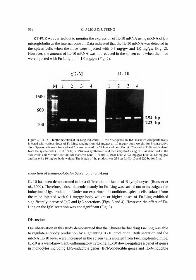
RT-PCR was carried out to monitor the expression of IL-10 mRNA using mRNA of β2-microglobulin as the internal control. Data indicated that the IL-10 mRNA was detected inthe spleen cells when the mice were injected with 0.5 mg/gw and 1.0 mg/gw (Fig. 2).However, the amount of IL-10 mRNA was not reduced in the spleen cells when the micewere injected with Fu-Ling up to 1.0 mg/gw (Fig. 2).
Figure 2. RT-PCR for the detection of Fu-Ling-induced IL-10 mRNA expression. BALB/c mice were peritoneallyinjected with various doses of Fu-Ling, ranging from 0.1 mg/gw to 1.0 mg/gw body weight, for 3 consecutivedays. Spleen cells were isolated and in vitro cultured for 24 hours without Con A. The total mRNA was isolatedfrom the spleen cells (1 × 10⁷ cells). cDNA was synthesized and then amplified using PCR as described in the“Materials and Method” section. M: markers; Lane 1: control (PBS); Lane 2: 0.1 mg/gw; Lane 3: 1.0 mg/gw;and Lane 4 : 10 mg/gw body weight. The length of the product was 254 bp for IL-10 and 222 bp for β2m.
Induction of Immunoglobulin Secretion by Fu-Ling
IL-10 has been demonstrated to be a differentiation factor of B-lymphocytes (Rousset etal., 1992). Therefore, a dose-dependent study for Fu-Ling was carried out to investigate theinduction of Igs production. Under our experimental conditions, spleen cells isolated fromthe mice injected with 0.1 mg/gw body weight or higher doses of Fu-Ling exhibitedsignificantly increased IgG and IgA secretions (Figs. 3 and 4). However, the effect of Fu-Ling on the IgM secretion was not significant (Fig. 5).
Discussion
Our observation in this study demonstrated that the Chinese herbal drug Fu-Ling was ableto regulate antibody production by augmenting IL-10 production. Both secretion and themRNA IL-10 level were increased in the spleen cells isolated from Fu-Ling-treated mice.IL-10 is a well-known anti-inflammatory cytokine. IL-10 down-regulates a panel of genesin monocytes including LPS-inducible genes, IFN-γ-inducible genes and IL-4-inducible

FU-LING REGULATES IL-10 PRODUCTION 557
genes (Donnelly et al., 1999). Among those LPS-inducible genes, IL-10 significantly reducedTNF-α, IL-1, IL-6, IL-8, IL-12, GM-CSF and even IL-10 gene expression by itself.
Figure 3. Fu-Ling induced an increase in immunoglobulin secretion by spleen cells in vitro. BALB/c mice wereperitoneally injected with various doses of Fu-Ling, ranging from 0.1 mg/gw to 1.0 mg/gw body weight, for 3consecutive days. The control group consisted of mice injected with an equal volume of PBS. Spleen cells wereisolated after the treatment and then incubated for 5 days with 1 µg/ml of LPS. Supernatants were harvested andassayed for IgG concentrations at days 3, 4 and 5. Data were mean ± SEM of five similar experiments. * indicatesp < 0.05 from the control group.
However, our previous studies indicated that Fu-Ling modulated cytokine productionby human monocytes. Fu-Ling significantly enhanced the secretion of cytokines producedby monocytes, i.e. IL-1β, TNF-α and IL-6 but suppressed TGF-β production under in vitroconditions (Yu and Tseng, 1996). Under similar experimental conditions, our data alsoindicated that Fu-Ling significantly augmented GM-CSF secretions using human peripheralblood mononuclear cells (Tseng and Li, 1996).

C.-J LIOU & J. TSENG558
The controversial data obtained from in vivo and in vitro trials may result from thetarget cells of the drug. Under in vitro conditions, the crude extract of Fu-Ling directly actson the monocytes by either interacting with intracellular molecules or going through areceptor-mediated pathway. Under in vivo conditions, the components of Fu-Ling may notbe able to directly affect the monocyte functions. Instead, Fu-Ling may act on the Th2 cellsand monocytes to induce IL-10 production. The Fu-Ling-induced IL-10 subsequently reducesthe production of monocyte-derived cytokines. Whether administration of Fu-Ling extractcan inhibit the synthesis of monocyte-derived cytokines remains for further investigation.
In terms of immunoglobulin production, data obtained from this study were consistentwith the IL-10 production. IL-10 is best known for its ability to induce B-lymphocytedifferentiation (Rousset et al., 1992). An increase in IL-10 implied that more B-lymphocytes
Figure 4. Fu-Ling induced an increase in immunoglobulin secretion by spleen cells in vitro. BALB/c mice wereperitoneally injected with various doses of Fu-Ling, ranging from 0.1 mg/gw to 1.0 mg/gw body weight, for 3consecutive days. The control group consisted of mice injected with an equal volume of PBS. Spleen cells wereisolated after the treatment and then incubated for 5 days with 1 µg/ml of LPS. Supernatants were harvested andassayed for IgA concentrations using ELISA at days 3, 4 and 5. Data were mean ± SEM of five similar experiments.* indicates p < 0.05 from the control group.

FU-LING REGULATES IL-10 PRODUCTION 559
were differentiated from the IgM producer to the IgG or IgA producers. Our data supportedthe hypothesis that spleen cells isolated from Fu-Ling-treated mice did show an increase inboth IgG and IgA secretions but no increase in IgM.
Figure 5. Fu-Ling induced an increase in immunoglobulin secretion by spleen cells in vitro. BALB/c mice wereperitoneally injected with various doses of Fu-Ling, ranging from 0.1 mg/g to 1.0 mg/g body weight, for 3consecutive days. The control group consisted of mice injected with an equal volume of PBS. Spleen cells wereisolated after the treatment and then incubated for 5 days with 1 mg/ml of LPS. Supernatants were harvested andassayed for IgM concentrations using ELISA at days 3, 4 and 5. Data were mean ± SEM of five similar experiments.* indicates p < 0.05 from the control group.
This observation was also inconsistent with the data obtained from the in vitro study onthe human blood cells (Lu et al., 1994). Under in vitro conditions, Fu-Ling significantlyinduced an increase in IgM secretions using human peripheral blood mononuclear cells buthad less effect on IgA and IgM secretions. This might result from the failure of Fu-Ling

C.-J LIOU & J. TSENG560
alone to induce B-cells in human blood to undergo differentiation, and most of the B-cellsin peripheral blood were resting cells.
IL-10 is one of the potent inhibitors of inflammatory response. Therefore, our observationmay provide a convincing interpretation of why Fu-Ling acts as a powerful drug to reduceedema and to enhance the immune response. Our previous report (Yu and Tseng, 1996) andcurrent data suggested that a steroid-type compound isolated from Fu-Ling extract may bethe biologically active ingredient that regulates cytokine production. It will therefore bepossible to isolate a therapeutic compound from Fu-Ling extract.
References
Donnelly, R.P., H. Dickensheets and D.S. Finbloom. The interleukin-10 signal transduction pathwayand regulation of gene expression in mononuclear phagocytes. J. Interferon Cytokine Res. 19:563–573, 1999.
Kuhn, R., J. Lohler, D. Rennick, K. Rajewsky and W. Muller. Interleukin-10-deficient mice developchronic enterocolitis. Cell. 75: 263–270, 1993.
Lu, T.-N., S.-J. Yu and J. Tseng. Effect of Fu-ling on immunoglobulin secreted by lymphocytes inhuman blood. Biol. Bull. NTNU 29: 43–51, 1994 (in Chinese with English abstract).
Moore, K.W., P. Vieira, D.F. Fiorentino, M.L. Trounstine, T.A. Khan and T.R. Mosmann. Homologyof cytokines synthesis inhibitory factor (IL-10) to the Epstein-Barr virus gene BCRFI. Science248: 1230–1234, 1990.
Moore, K.W., A. O’garra, R. del Waal Malefyt, P.N. Vieira and T.R. Mosmann. Interleukin-10.Annu. Rev. Immunol. 11: 165–190, 1993.
del Prete, G., M. De Carli, F. Almerigogna, M.G. Giudizi, R. Biagiotti and S. Romagnani. Human IL-10 is produced by both Type 1 helper (TH1) and Type 2 helper (TH2) T cell clones and inhibitstheir antigen-specific proliferation and cytokine production. J. Immunol. 150: 353–360, 1993.
Rousset, F., E. Garcia, T. Defrance, C. Peronne, N. Vezzio, D.H. Hsu, R. Kastelein, K.W. Moore andJ. Banchereau. Interleukin-10 is a potent growth and differentiation factor for activated humanB lymphocytes. Proc. Natl. Acad. Sci. USA. 89: 1890–1893, 1992.
Thompson-Snipes, L., V. Dhar, M.W. Bond, T.R. Mosmann, K.W. Moore and D. Rennick. Interleukin-10: a novel stimulatory factor for mast cells and their progenitors. J. Exp. Med. 173: 507–510,1991.
Tseng, J. and T.L. Li. Si-Jun-Zi-Tang regulates granulocyte macrophage colony-stimulating factorsecretion by human peripheral blood mononuclear cells. Am. J. Chin. Med. 24: 45–52, 1996.
de Waal Malefyt, R., J. Abrams, B. Bennett, C.G. Figdor and J.E. de Vries. Interkeukin-10 (IL-10)inhibits cytokine synthesis by human monocytes: an autoregulatory role of IL-10 produced bymonocytes. J. Exp. Med. 174: 1209–1220, 1991.
Wanidworanun, C. and W. Strober. Predominant role of tumor necrosis factor-α in human monocyteIL-10 synthesis. J. Immunol. 151: 6853–6861, 1993.
Yu, S.J., J. Tseng. Fu-Ling, a Chinese herbal drug, modulates cytokine secretion by human peripheralblood monocytes. Int. J. Immunopharmac. 18: 37–44, 1996.