Embed Size (px)
Citation preview

7/30/2019 358 Multifunctional Actions of Approved and Candidate Stroke Drugs.
http://slidepdf.com/reader/full/358-multifunctional-actions-of-approved-and-candidate-stroke-drugs 1/10
Multifunctional Actions of Approved and Candidate
Stroke Drugs
Jens Minnerup and Wolf-Rüdiger Schäbitz
Department of Neurology, University of Münster, Albert-Schweitzer–Straße 33, 48149 Münster, Germany
Summary: Ischemic stroke causes brain damage by multiplepathways. Previous stroke trials have demonstrated that drugstargeting one or only a few of these pathways fail to improveclinical outcome after stroke. Drugs with multimodal actions
have been suggested to overcome this challenge. In this review,we describe the mechanisms of action of agents approved forsecondary prevention of ischemic stroke, such as antiplatelet,antihypertensive, and lipid-lowering drugs. These drugs exhibitconsiderable properties beyond their classical mechanisms, in-
cluding neuroprotective and neuroregenerative properties. Inaddition, candidate stroke drugs currently studied in clinicalphase III trials are described. Among these, albumin, hemato-poietic growth factors, and citicoline have been identified as
promising agents with multiple mechanisms. These drugs offerhope that additional treatment options for the acute phase aftera stroke will become available in the near future. Key Words:Stroke, drug therapy, mechanisms, neuroprotection, neurore-generation.
INTRODUCTION
Despite the tremendous mortality and morbidity of
stroke, treatment options remain limited. Many patho-
physiological key mechanisms of cerebral ischemia have
been identified in recent years, but drug treatment tar-
geting one or a few of these mechanisms has failed to
improve clinical outcome after stroke. The most plausi-
ble reason for this failure is the multiplicity of mecha-
nisms involved in causing neuronal damage during ische-
mia. Drugs targeting a multimodal mode of action could
potentially overcome this dilemma, and have recently
been shown to provide a remarkable benefit in preclinical
studies.
The only drug approved for the acute phase of ische-
mic stroke is recombinant tissue plasminogen activator
(rtPA)—which, however, predominantly acts by target-
ing a single mechanism, the lysis of the intravascular
clot. Antiplatelet, antihypertensive, and lipid-lowering
therapies are approved for secondary stroke prevention.
In contrast to rtPA, additional effects of these drugs
beyond their classical mechanisms are suggested, partic-
ularly because prestroke use of drugs for secondary
stroke prevention was found to be associated with less
severe deficits when a stroke occurred. Moreover, animal
experimental studies confirmed that antiplatelet, antihy-
pertensive, and lipid-lowering drugs exert multimodal
actions including neuroprotective and neuroregenerative
properties. We review these approved stroke drugs witha focus on their multimodal modes of action. In addition
to drugs for secondary prevention of stroke, further
agents were identified that interfere with various patho-
physiological mechanisms in cerebral ischemia. Some of
these drugs were extensively tested in preclinical stroke
studies and showed promising results regarding infarct
size reduction and functional recovery enhancement.
Those candidate stroke drugs with multiple mechanisms
in acute or chronic stroke that were successfully trans-
lated into clinical phase III trials are also described in this
review.
APPROVED DRUGS FOR SECONDARY
STROKE PREVENTION
Antiplatelet agents
Platelet aggregation is important in stroke develop-
ment, both in the pathogenesis of atherosclerosis and in
the occurrence of acute cerebral artery occlusions.1 The
initial step is platelet adhesion to the arterial wall, a result
of endothelial damage.2 Platelet adhesion is promoted by
several factors, including as von Willebrand factor, fi-
brinogen, and subendothelial collagen.2 Afterwards, ad-
hesion platelets are activated mainly by thromboxane
Address correspondence and reprint requests to: Jens Minnerup, Uni-
versity of Münster, Department of Neurology, Albert-Schweitzer-Straße33, 48149 Münster, Germany. E-mail: [email protected]. OrWolf-Rüdiger Schäbitz; e-mail: [email protected].
Neurotherapeutics: The Journal of the American Society for Experimental NeuroTherapeutics
Vol. 6, 43–52, January 2009 © The American Society for Experimental NeuroTherapeutics, Inc. 43
7/30/2019 358 Multifunctional Actions of Approved and Candidate Stroke Drugs.
http://slidepdf.com/reader/full/358-multifunctional-actions-of-approved-and-candidate-stroke-drugs 2/10
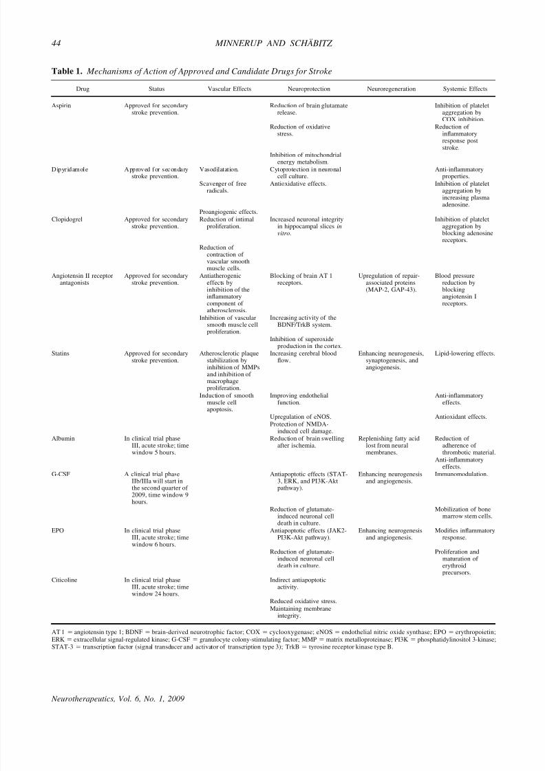
Table 1. Mechanisms of Action of Approved and Candidate Drugs for Stroke
Drug Status Vascular Effects Neuroprotection Neuroregeneration Systemic Effects
Aspirin Approved for secondarystroke prevention.
Reduction of brain glutamaterelease.
Inhibition of plateletaggregation byCOX inhibition.
Reduction of oxidative
stress.
Reduction of
inflammatoryresponse poststroke.
Inhibition of mitochondrialenergy metabolism.
D ip yr id am ol e A pp rov ed f or s ec on da rystroke prevention.
Vasodilatation. Cytoprotection in neuronalcell culture.
Anti-inflammatoryproperties.
Scavenger of freeradicals.
Antioxidative effects. Inhibition of plateletaggregation byincreasing plasmaadenosine.
Proangiogenic effects.
Clopidogrel Approved for secondarystroke prevention.
Reduction of intimalproliferation.
Increased neuronal integrityin hippocampal slices invitro.
Inhibition of plateletaggregation byblocking adenosinereceptors.
Reduction of contraction of vascular smooth
muscle cells.Angiotensin II receptor
antagonistsApproved for secondary
stroke prevention.Antiatherogenic
effects byinhibition of theinflammatorycomponent of atherosclerosis.
Blocking of brain AT 1receptors.
Upregulation of repair-associated proteins(MAP-2, GAP-43).
Blood pressurereduction byblockingangiotensin Ireceptors.
Inhibition of vascularsmooth muscle cellproliferation.
Increasing activity of theBDNF/TrkB system.
Inhibition of superoxideproduction in the cortex.
Statins Approved for secondarystroke prevention.
Atherosclerotic plaquestabilization byinhibition of MMPsand inhibition of macrophageproliferation.
Increasing cerebral bloodflow.
Enhancing neurogenesis,synaptogenesis, andangiogenesis.
Lipid-lowering effects.
Induction of smooth
muscle cellapoptosis.
Improving endothelial
function.
Anti-inflammatory
effects.
Upregulation of eNOS. Antioxidant effects.
Protection of NMDA-induced cell damage.
Albumin In clinical trial phaseIII, acute stroke; timewindow 5 hours.
Reduction of brain swellingafter ischemia.
Replenishing fatty acidlost from neuralmembranes.
Reduction of adherence of thrombotic material.
Anti-inflammatoryeffects.
G-CSF A clinical trial phaseIIb/IIIa will start inthe second quarter of 2009, time window 9hours.
Antiapoptotic effects (STAT-3, ERK, and PI3K-Aktpathway).
Enhancing neurogenesisand angiogenesis.
Immunomodulation.
Reduction of glutamate-induced neuronal celldeath in culture.
Mobilization of bonemarrow stem cells.
EPO In clinical trial phase
III, acute stroke; timewindow 6 hours.
Antiapoptotic effects (JAK2-
PI3K-Akt pathway).
Enhancing neurogenesis
and angiogenesis.
Modifies inflammatory
response.
Reduction of glutamate-induced neuronal celldeath in culture.
Proliferation andmaturation of erythroidprecursors.
Citicoline In clinical trial phaseIII, acute stroke; timewindow 24 hours.
Indirect antiapoptoticactivity.
Reduced oxidative stress.
Maintaining membraneintegrity.
AT 1 ϭ angiotensin type 1; BDNF ϭ brain-derived neurotrophic factor; COX ϭ cyclooxygenase; eNOS ϭ endothelial nitric oxide synthase; EPO ϭ erythropoietin;
ERK ϭ extracellular signal-regulated kinase; G-CSF ϭ granulocyte colony-stimulating factor; MMP ϭ matrix metalloproteinase; PI3K ϭ phosphatidylinositol 3-kinase;STAT-3 ϭ transcription factor (signal transducer and activator of transcription type 3); TrkB ϭ tyrosine receptor kinase type B.
MINNERUP AND SCHA BITZ 44
Neurotherapeutics, Vol. 6, No. 1, 2009

7/30/2019 358 Multifunctional Actions of Approved and Candidate Stroke Drugs.
http://slidepdf.com/reader/full/358-multifunctional-actions-of-approved-and-candidate-stroke-drugs 3/10
A2, thrombin, and adenosine diphosphate.3 This process
requires the cyclooxygenase (COX)–mediated metabo-
lism of arachidonic acid to prostaglandin H2 (PGH2),
which in turn is processed to thromboxane A2.3 Platelet
activation results in platelet aggregation due to the con-
version of the glycoprotein IIb/IIIa receptor to a form
that binds fibrinogen and other adhesion molecules.1
Based on the relevance of platelet aggregation in stroke
pathogenesis, its inhibition became a target for therapeu-
tic intervention. The results of many trials that evaluated
antiplatelet agents in stroke led to recommendations that
all stroke patients not requiring anticoagulation should
receive antiplatelet therapy.4 Aspirin, clopidogrel, and
the combination of aspirin and extended-release dipyrid-
amole are accepted treatment options.4
Aspirin. Aspirin was the first antiplatelet drug used
for secondary prevention in ischemic stroke.5 Within the
last 20 years, the effectiveness of aspirin for recurrent
ischemic stroke prevention has been shown in several
trials.6,7 The best-characterized effects of aspirin in the
prevention of cardiovascular diseases are based on its
platelet inhibitory functions.8 After diffusion through the
cell membranes of platelets, aspirin first binds and then
acetylates a specific serine residue of COX, thereby pre-
venting the conversion of arachidonic acid to PGH2 and
thus also preventing the formation of thromboxane A2.
Apart from the antiplatelet effects, there is growing
evidence for direct neuroprotective properties of aspirin
(Table 1).9–14 In animal models of focal cerebral ische-
mia, both pretreatment and administration of aspirin
after the onset of ischemia is neuroprotective, as indi-
cated by infarct size reduction for aspirin doses between
15 mg/kg and 80 mg/kg body weight.9,13,15 Reduction of
brain glutamate release after ischemia appears to be an
important mechanism by both NF-B dependent and
independent pathways.9,11,12 Glutamate toxicity may ad-
ditionally be reduced by aspirin-mediated inhibition of
prostaglandin (PG) formation. Prostaglandins synthe-
sized by COX enhance the release of glutamate from
astrocytes. Its decreased composition may therefore
abate glutamatergic neurotoxicity.16 In vitro experiments
showed that aspirin also prevents neurotoxicity by inhi-bition of the atypical protein kinase C , which is a down-
stream signal in NMDA-induced neuronal cell death.17
Moreover, aspirin exerts neuroprotective properties
through reduction of oxidative stress and by inhibitory
effects on mitochondrial energy metabolism.18,19 Neu-
roprotection by aspirin was also suggested by clinical
studies showing that prestroke use of aspirin reduced
stroke severity.20,21 Increasing evidence suggests that
inflammation plays a role both in atherosclerosis with
subsequent stroke and in poststroke morbidity and
mortality.22–24 Aspirin was found to be associated
with lower values of inflammatory parameters after
stroke.25,26 To date, however, the relevance of these
anti-inflammatory effects for stroke prevention re-
mains unknown.
Dipyridamole. Dipyridamole is available in imme-
diate-release form, usually given in doses between 50
and 100 mg three times per day, or as a fixed combina-
tion of 25 mg aspirin and 200 mg extended-release di-pyridamole (ER-dipyridamole), given two times per day.
In comparison of placebo to three active treatment
groups (aspirin alone, ER-dipyridamole alone, and the
combination of aspirin and ER-dipyridamole) in second-
ary prevention of ischemic stroke, the single use of either
aspirin or ER-dipyridamole was superior to placebo, and
the combination of aspirin and ER-dipyridamole was
superior to either drug alone.27 A meta-analysis of six
trials comparing the combination of dipyridamole and
aspirin (also including the immediate-release formula-
tion) to aspirin alone confirmed these findings, revealing
a significantly lower risk of serious vascular events whenaspirin was combined with dipyridamole.28
Dipyridamole exerts antiplatelet effects by inhibition
of the adenosine uptake in erythrocytes and the attenua-
tion of adenosine catabolism, both of which result in
increasing plasma concentrations of adenosine, an inhib-
itor of platelet aggregation. Dipyridamole is also known
to act via adenosine as a potent vasodilator, mainly in
coronary arteries.29 The effect of dipyridamole on cere-
bral blood flow is, however, not fully understood.30,31
Dipyridamole possesses antithrombotic properties in the
vessel wall by enhancing the production of the endoge-
nous platelet inhibitor prostacyclin, thereby preven-ting platelet adhesion to the endothelium.32,33 Damage
caused by hydrophilic and hydrophobic radicals can be
prevented by the scavenger properties of dipyridamole.34
Dipyridamole inhibits oxidation of low density lipopro-
tein (LDL) and may prevent the development of athero-
sclerosis.35 In various neuronal cell culture models
dipyridamole was cytoprotective.36,37 Dipyridamole also
exerted neuroprotective properties in an embolic stroke
model.38 The underlying mechanisms of neuroprotection
remain unknown. Both adenosine effects and antioxida-
tive effects are assumed to be relevant.36,39 Further
potential mechanisms of dipyridamole in stroke patho-physiology include anti-inflammatory actions and proan-
giogenic effects.40,41
Clopidogrel. In a large clinical trial, clopidogrel
reduced the composite endpoint of stroke, myocardial
infarction, or vascular death compared with aspirin in
high-risk patients.42 In patients with peripheral arterial
disease, clopidogrel is superior to aspirin in terms of
stroke prevention.42 As recently shown, there were no
significant differences in secondary stroke prevention
when clopidogrel was compared with the combination of
aspirin and ER-dipyridamole.43
Clopidogrel is a second-generation thienopyridine.
MULTIFUNCTIONAL STROKE DRUGS 45
Neurotherapeutics, Vol. 6, No. 1, 2009

7/30/2019 358 Multifunctional Actions of Approved and Candidate Stroke Drugs.
http://slidepdf.com/reader/full/358-multifunctional-actions-of-approved-and-candidate-stroke-drugs 4/10
Clopidogrel irreversibly blocks the P2Y12 receptor, an
adenosine receptor located on the surface of platelets,
thereby preventing platelet aggregation by inhibition of
the glycoprotein IIb/IIIa complex.44 Clopidogrel itself
has no antiplatelet activity and needs to be oxidated by
the hepatic cytochrome P-450 system to an active me-
tabolite.45 Beyond the inhibition of thrombus formation,
animal experimental studies suggested direct effects of
clopidogrel on arterial vessels, such as a reduced intimal
proliferation after arterial injury and reduced seroto-
nin and endothelin-1-mediated contraction of vascular
smooth muscle cells (Table 1).46–48 In vitro studies mea-
suring the population spike amplitude in murine hip-
pocampal slices after a hypoxic episode found that pre-
treatment with clopidogrel increased neuronal integrity,
compared with control treatment, taken as indication of a
neuroprotective capacity of clopidogrel.49
Antihypertensive drugsHigh blood pressure is well known as an important risk
factor for ischemic stroke. Lowering an elevated blood
pressure is recommended for primary prevention, as well
as for prevention of recurrent stroke.4 Several classes of
antihypertensive drugs were found to be effective in
secondary stroke prevention, and to date there is no
recommendation clearly favoring a special antihyperten-
sive drug.4,50 Angiotensin II receptor antagonists were,
however, found to be superior to a beta-blocker (losartan
vs atenolol) in primary prevention and to a calcium chan-
nel blocker (eprosartan vs nitrendipine) in secondary pre-
vention of ischemic stroke.51,52
Because blood pressurereduction was similar in both comparisons, other mech-
anisms were assumed. Angiotensin II receptor antago-
nists reduce blood pressure by inhibition of the renin
angiotensin system. Renin catalyzes the cleavage of an-
giotensinogen to angiotensin I, which in turn is cleaved
by the angiotensin converting enzyme to its active form
angiotensin II.53,54 The appropriate stimulus for renin
production by the kidney is glomerular hypoperfusion.53
Angiotensin II acts via the specific angiotensin II recep-
tors AT1 and AT2.55 The latter is less well characterized,
whereas AT1 is well known to mediate vasoconstriction,
aldosterone-mediated sodium and water retention, leftventricular hypertrophy, and growth in the arterial
wall.56 Angiotensin II receptor antagonists counteract
these mechanisms by blocking the angiotensin I receptor,
which results in a lowered blood pressure.57
Beyond lowering blood pressure, other mechanisms of
AT1 receptor blockers in stroke pathophysiology have
been suggested (Table 1). In various animal experimental
studies of focal cerebral ischemia, neuroprotective prop-
erties have been demonstrated.58–60 Effects of AT1 re-
ceptor blockers on brain AT1 receptors were suggested
to mediate these neuroprotective characteristics.58,61 The
ability of different AT1 receptor blockers to cross the
blood–brain barrier is controversial, however.62,63 Can-
desartan was shown to enhance the TrkB receptor of
brain-derived neurotrophic factor (BDNF), a neurotro-
phin well known for its neuroprotective effects.64 Addi-
tionally, candesartan inhibited the increase of superoxide
production in the cerebral cortex after cerebral ischemia
in mice.65 Expression of the proinflammatory cytokine
tumor necrosis factor ␣ (TNF␣) and the proinflammatory
marker gene CXC ligand 1 were downregulated by can-
desartan, thereby decreasing infarct size.60
In addition to neuroprotection, candesartan exhibits
poststroke neuroregenerative properties, as indicated by
an upregulated expression of repair-associated proteins,
such as MAP-2 and GAP-43.65 Because a substantial
role of AT1 receptors in atherogenesis was uncovered,
vasculoprotective properties of angiotensin II receptor
antagonists independent of blood pressure effects are
suggested.66 Indeed, the AT1 blocker losartan was found
to exhibit antiatherogenic effects in nonhuman primates,as indicated by the inhibition of fatty-streak formation.67
Assumed underlying mechanisms gained from animal
model and in vitro studies include a reduction of oxida-
tive stress, inhibition of the inflammatory component of
atherosclerotic lesion formation, inhibition of vascular
smooth muscle cell proliferation, and a reduced intimal
thickening.68–71
Lipid-lowering drugs
Hyperlipidemia is a well-established risk factor for
myocardial infarction, whereas the effect of high serum
cholesterol levels on stroke incidence is less clear.72,73
This might explain why statins, in contrast to other lipid-
lowering drugs, clearly prevent stroke recurrence in pa-
tients with prior stroke or other vascular diseases, irre-
spective of blood lipid levels at treatment initiation.74,75
These findings strongly suggest a multimodal mode of
action.
Statins, or hydroxymethylglutaryl coenzyme A (HMG-
CoA) reductase inhibitors, are similar to HMG-CoA, a
precursor of cholesterol. Statins competitively inhibit
HMG-CoA reductase, the rate-limiting step in the syn-
thesis of cholesterol.76 Reduced intrahepatic cholesterol
leads to an upregulated low-density lipoprotein (LDL)receptor activity with an increased clearance of LDL
from the circulation.
Statins non–lipid-lowering effects, which are relevant
in stroke pathophysiology, can be divided into two main
groups: effects on the arterial vessels (including anti-
atherothrombotic and anti-inflammatory actions, as well
as improvement of endothelial function) and direct neu-
roprotective and neuroregenerative effects on the brain
(Table 1).
Atherosclerotic plaque-stabilizing effects of statins
have been shown in both animal and human studies.77,78
Statins were found to mediate plaque stabilization by
MINNERUP AND SCHA BITZ 46
Neurotherapeutics, Vol. 6, No. 1, 2009

7/30/2019 358 Multifunctional Actions of Approved and Candidate Stroke Drugs.
http://slidepdf.com/reader/full/358-multifunctional-actions-of-approved-and-candidate-stroke-drugs 5/10
various mechanisms, including inhibitory effects on
matrix metalloproteinases, inhibition of macrophage
proliferation in atherosclerotic plaque, and induction of
smooth muscle cell apoptosis.78,79 The so-called pleio-
tropic effects of statins include additional anti-infla-
mmatory and antioxidant properties. C-reactive protein
(CRP), which is a marker for inflammation associatedwith atherosclerosis, is reduced by statin therapy.80 Un-
derlying mechanisms of the anti-inflammatory effects of
statins include reduced leukocyte adhesion by integrin
inhibition and inhibition of MHC-II-mediated T-cell ac-
tivation.81,82 In addition, statins promote systemic anti-
oxidant effects through suppression of distinct oxidation
pathways, such as the formation of myeloperoxidase-
derived and nitric oxide–derived oxidants.83
The neuroprotective properties of statins have been
shown in animal stroke models, in which statin therapy
leads to reduced infarct volumes.84 The mechanisms of
neuroprotection are presumably indirect effects, relatedto increased cerebral blood flow and improved endothe-
lial function rather than to direct neuronal effects. The
upregulation of brain endothelial nitric oxide synthase
(eNOS) by statins with subsequent NO synthesis appears
to be of particular importance.85,86 Statins suppress the
isoprenoid production by HMG-CoA reductase inhibi-
tion and thereby prevent the isoprenylation of the small
GTPase- , a negative regulator of eNOS expression.87 In
addition, statins act via post-translational modification on
eNOS activity by activation of the phosphatidylinositol
3-kinase–protein kinase Akt (PI3K–Akt) pathway.88 St-
atins reduced the association of NMDA receptors to lipidrafts of neuronal cells in vitro, thus suggesting neuropro-
tective effects of statins mediated by protection from
NMDA-induced neuronal damage.89
In addition to their neuroprotective effects, statins
appear to have neuroregenerative properties post
stroke.90,91 Atorvastatin improved functional recovery in
a rat stroke model independent from infarct size reduc-
tion by enhancing neurogenesis, synaptogenesis, and an-
giogenesis.91
CANDIDATE DRUGS FOR ACUTE AND
CHRONIC STROKE TREATMENT
Albumin
Human albumin is in widespread use for fluid resus-
citation in shock patients, although convincing evidence
for this indication is lacking.92 Recently, high-dose hu-
man albumin (doses between 0.63 g/kg and 2.5 g/kg) was
studied in animal models of focal cerebral ischemia.93–96
Albumin reduced infarct size and improved functional
recovery after permanent and temporary cerebral ische-
mia.94,97 Preclinical studies included a dose–response
relationship evaluation and a time window investigation
(up to 4 hours).93,94 Despite a reasonable number of
preclinical albumin studies, the significance of the results
is in some aspects weak. The studies showing efficacy of
albumin in stroke animals were conducted in only one
laboratory, only one species (rat) was tested, and only
animals without comorbidities (e.g., diabetes or hyper-
tension due to age) were included in the studies.98
Based on the preclinical results, a phase I dose-
escalation study, the Albumin in Acute Stroke (ALIAS)
Pilot Trial, was conducted with 82 stroke patients. The
results showed that albumin is safe in stroke patients. The
authors of that study interpreted the results as a sign for
efficacy, based on comparison of higher doses with lower
doses and in comparison with a historical control group.99,100
A randomized, multicenter, double-blind, placebo-
controlled trial (ALIAS Phase III Trial, www.clinicaltrials.
gov; NCT00235495) is currently being conducted.
Animal stroke studies have suggested various mecha-
nisms by which albumin provides an improved outcome
after stroke. After middle cerebral artery occlusion
(MCAO), albumin significantly reduced brain swelling
in rats.94,101 Intravascular mechanisms (e.g., reduced ad-
herence of thrombotic material and an improved eryth-
rocyte perfusion) may also contribute to a beneficial
outcome.95,102 Albumin mobilizes systemic fatty acids
and may contribute to neural cell integrity post stroke by
replenishing fatty acid lost from neural membranes dur-
ing ischemia.103,104 In addition, albumin was sugges-
ted to exert anti-inflammatory effects by binding the
proinflammato ry lysolipid lysophosphatidylcholine.105
To date, however, convincing findings on the neuropro-
tective mode of action of albumin are lacking. The
ALIAS pilot trial suggested a favorable outcome when
thrombolysis was combined with albumin.100 Synergistic
effects of albumin with thrombolysis were also demon-
strated in a study using a rat stroke model in which the
combination therapy improved microvascular hemody-
namics.96
G-CSF
The granulocyte colony-stimulating factor (G-CSF) is
approved for the prevention and treatment of chemother-
apy-induced neutropenia.106 Within the last several
years, a number of reports showed efficacy of G-CSF in
animal models of focal cerebral ischemia.107–111 To ob-
tain an overall impression of the efficacy of G-CSF in
preclinical studies and to evaluate conditions under
which maximum efficacy can be achieved, we performed
a meta-analysis and meta-regression analysis of G-CSF
in animal stroke models.112 We showed that G-CSF re-
duced both infarct volumes and sensorimotor deficits.112
Our meta-regression analysis revealed a clear dose–
response relationship of the efficacy of G-CSF.112 When
administered within the first 6 hours after the induction
of ischemia, G-CSF reduced infarct sizes by 0.8% per 1
MULTIFUNCTIONAL STROKE DRUGS 47
Neurotherapeutics, Vol. 6, No. 1, 2009

7/30/2019 358 Multifunctional Actions of Approved and Candidate Stroke Drugs.
http://slidepdf.com/reader/full/358-multifunctional-actions-of-approved-and-candidate-stroke-drugs 6/10
g/kg body weight and by 2.1% per 1 g/kg body
weight when applied later than 6 hours.112
Based on these promising preclinical studies, a number
of clinical studies were initiated to test the efficacy of
G-CSF in stroke patients.113 We conducted a multi-
center, randomized, double-blind, placebo-controlled,
dose-escalating phase IIa trial (AXIS), which was re-
cently completed.114 In this trial, which included 45 pa-
tients who had suffered an acute stroke, G-CSF was
shown to be safe and well-tolerated. In addition, G-CSF
showed signs of clinical efficacy in patients with larger
baseline lesions evidenced with diffusion MRI.114 A
phase IIb/IIIa trial will start in the second quarter of
2009.
It cast doubt on the perception that G-CSF’s natural
function of mobilizing bone marrow stem cells is the
most important mechanism, particularly since both G-
CSF and its receptor were found to be expressed in the
brain (Table 1).107 In vitro and in vivo experiments con-
firmed that G-CSF acts antiapoptotically on neurons by
at least three different antiapoptotic pathways: the signal
transducer and activation of transcription 3 pathway
(STAT-3), the extracellular signal-regulated kinase path-
way (ERK), and the phophatidylinositol 3-kinase–Akt
pathway (PI3K-Akt).107,108,111
Beyond the acute phase of stroke, G-CSF facilitates
regeneration by enhancing endogenous neurogenesis.107
Another mechanism underlying long-term functional re-
covery appears to be angiogenesis, which was increased
by G-CSF post stroke even when administered as late as
7 days after the induction of ischemia.109 G-CSF is wellknown for its immunomodulative properties.115 Al-
though the exact effect of the G-CSF–mediated systemic
immunomodulation on stroke outcome remains un-
known, G-CSF obviously affects the local brain immune
response post stroke. G-CSF treatment reduced infiltra-
tion of neutrophils and microglia in the ischemic hemi-
sphere, and G-CSF lowered interleukin-1 upregula-
tion.108,110,115
Erythropoietin
Erythropoietin (EPO) is in broad clinical use for thetreatment of cancer-related anemia.116 EPO is the second
hematopoietic growth factor besides G-CSF to be exten-
sively tested in preclinical stroke studies.117,118 A small
clinical trial showed that EPO is safe and might be ben-
eficial in acute ischemic stroke.119 The results of a ran-
domized, double-blind, placebo-controlled multicenter
study of EPO treatment in acute stroke has been com-
pleted, and results are expected in the near future (http://
www.clinicaltrials.gov; NCT00604630).
Erythropoietin parallels G-CSF in many respects with
regard to the mode of action in ischemic stroke. Like
G-CSF, EPO and its receptor are expressed in the
brain.120 Notably, the EPO receptor that mediates neu-
roprotection seems to be distinct from those expressed by
erythroid precursors.121 EPO also exerts antiapoptotic
properties in vitro and in vivo. The JAK2–PI3K pathway
was shown to be a key regulator in EPO-mediated neu-
roprotection.122 In addition to its neuroprotective effects,
EPO has neuroregenerative properties by stimulating
neuronal differentiation and neurogenesis, as evidenced
in vitro and in vivo.117 After experimental stroke in mice,
EPO restored local cerebral blood flow as measured by
laser scanning imaging.123 Concordantly, histological
examination showed enlarged vascular perimeters and an
increased vascular density around the ischemic lesion,
indicating an enhancement of angiogenesis after EPO
treatment.117 EPO also modifies the inflammatory re-
sponse after cerebral injury, as shown by reduced leuko-
cyte infiltration after neonatal cerebral ischemia and re-
duced production of TNF␣ and IL-6.118,124
Citicoline
Citicoline is a dietary supplement. In some countries, it
is in clinical use for the treatment of acute ischemic
stroke. A meta-analysis of four clinical trials found that
citicoline administration in stroke patients within the first
24 hours after symptom onset increases the probability of
functional recovery at 3 months.125 A randomized, dou-
ble-blind, placebo-controlled, multicenter phase III trial
is currently recruiting patients to support this evidence
(http://www.clinicaltrials.gov; NCT00331890).
Citicoline is an essential intermediate in the synthesis
of the key membrane phospholipid phosphatidylcho-
line.126 Its role in maintaining membrane integrity was
suggested as being a relevant mechanism in post-stroke
recovery. After experimental stroke in rats, phospho-
lipases (phosphatidylcholine degrading enzymes) are
activated and CTP:phosphocholine cytidylyltransferase
(CCT), the rate-limiting enzyme in phosphatidylcholine
synthesis, is inactivated, both of which lead to neuronal
cell death due to reduced phosphatidylcholine concen-
trations.127 Citicoline restores phosphatidylcholine levels
by attenuating phospholipase activity and by enhancing
CCT activity.127 Similar mechanisms were suggested for
the preserving effects of citicoline on sphingomyelin and
cardiolipin.128 Excessive neuronal stimulation with ace-
tylcholine release after cerebral ischemia leads to choline
depletion, which in turn induces apoptosis.129 Citicoline
provides choline for the synthesis of acetylcholine and
may thereby prevent apoptotic cell death in cholinergic
neurons. The relevance of this mechanism was chal-
lenged, however, because acetylcholine concentrations
were not significantly increased by citicoline treat-
ment.130 In addition, citicoline was shown to reduce
oxidative stress.128,131
MINNERUP AND SCHA BITZ 48
Neurotherapeutics, Vol. 6, No. 1, 2009
7/30/2019 358 Multifunctional Actions of Approved and Candidate Stroke Drugs.
http://slidepdf.com/reader/full/358-multifunctional-actions-of-approved-and-candidate-stroke-drugs 7/10
CONCLUSION
Currently, multifunctional actions of stroke thera-
pies are limited to drugs used for secondary stroke
prevention. These drugs exhibit considerable proper-
ties beyond their classical mechanisms. In particular,
statins show a broad range of pleiotropic effects thatmay be as important in stroke pathophysiology and
recurrent stroke prevention as their lipid-lowering ef-
fects. In contrast to several drugs for secondary pre-
vention of ischemic stroke, treatment options for the
acute phase of stroke are limited to thrombolysis. On-
going phase III clinical trials raise hope that additional
treatment options for this acute phase will be available
in the near future. This includes, in particular, drugs
with multimodal modes of action that act on the brain
itself, as well as on other systems such as the vascu-
lature or the immune system.
REFERENCES
1. Davi G, Patrono C. Platelet activation and atherothrombosis.
N Engl J Med 2007;357:2482–2494.2. Savage B, Cattaneo M, Ruggeri ZM. Mechanisms of platelet
aggregation. Curr Opin Hematol 2001;8:270–276.
3. Ruggeri ZM. Platelets in atherothrombosis. Nat Med 2002;8:1227–1234.
4. European Stroke Organisation (ESO) Executive Committee; ESO
Writing Committee. Guidelines for management of ischaemicstroke and transient ischaemic attack 2008. Cerebrovasc Dis
2008;25:457–507.5. Craven LL. Prevention of coronary and cerebral thrombosis. Miss
Valley Med J 1956;78:213–215.
6. The Dutch TIA Trial Study Group. A comparison of two doses of
aspirin (30 mg vs. 283 mg a day) in patients after a transientischemic attack or minor ischemic stroke. N Engl J Med 1991;325:1261–1266.
7. International Stroke Trial Collaborative Group. The International
Stroke Trial (IST): a randomised trial of aspirin, subcutaneousheparin, both, or neither among 19435 patients with acute ischae-mic stroke. Lancet 1997;349:1569–1581.
8. Patrono C, García Rodriguez LA, Landolfi R, Baigent C. Low-dose aspirin for the prevention of atherothrombosis. N Engl
J Med 2005;353:2373–2383.9. De Cristóbal J, Moro MA, Dávalos, et al. Neuroprotective effect
of aspirin by inhibition of glutamate release after permanent focal
cerebral ischaemia in rats. J Neurochem 2001;79:456–459.10. Berger C, Xia F, Schäbitz WR, Schwab S, Grau A. High-dose
aspirin is neuroprotective in a rat focal ischemia model. Brain Res2004;998:237–242.
11. Grilli M, Pizzi M, Memo M, Spano P. Neuroprotection by aspirinand sodium salicylate through blockade of NF-B activation.Science 1996;274:1383–1385.
12. Berger C, Stauder A, Xia F, Sommer C, Schwab S. Neuropro-
tection and glutamate attenuation by acetylsalicylic acid in tem-porary but not in permanent cerebral ischemia. Exp Neurol 2008;
210:543–548.13. Zheng Z, Schwab S, Grau A, Berger C. Neuroprotection by early
and delayed treatment of acute stroke with high dose aspirin.
Brain Res 2007;1186:275–280.14. Whitehead SN, Bayona NA, Cheng G, Allen GV, Hachinski VC,
Cechetto DF. Effects of triflusal and aspirin in a rat model of cerebral ischemia. Stroke 2007;38:381–387.
15. Khayyam N, Thavendiranathan P, Carmichael FJ, Kus B, Jay V,
Burnham WM. Neuroprotective effects of acetylsalicylic acid inan animal model of focal brain ischemia. Neuroreport 1999;10:371–374.
16. Bezzi P, Carmignoto G, Pasti L, et al. Prostaglandins stimulatecalcium-dependent glutamate release in astrocytes. Nature 1998;
391:281–285.17. Crisanti P, Leon A, Lim DM, Omri B. Aspirin prevention of
NMDA-induced neuronal death by direct protein kinase C inhi-bition. J Neurochem 2005;93:1587–1593.
18. Maharaj H, Maharaj DS, Daya S. Acetylsalicylic acid and acet-
aminophen protect against oxidative neurotoxicity. Metab Brain
Dis 2006;21:189–199.19. Riepe MW, Kasischke K, Raupach A. Acetylsalicylic acid in-
creases tolerance against hypoxic and chemical hypoxia. Stroke1997;28:2006–2011.
20. Sanossian N, Saver JL, Rajajee V, et al. Premorbid antiplateletuse and ischemic stroke outcomes. Neurology 2006;66:319–323.
21. Wilterdink JL, Bendixen B, Adams HP Jr, Woolson RF, ClarkeWR, Hansen MD. Effect of prior aspirin use on stroke severity inthe trial of Org 10172 in acute stroke treatment (TOAST). Stroke
2001;32:2836–2840.22. Lindsberg PJ, Grau AJ. Inflammation and infections as risk fac-
tors for ischemic stroke. Stroke 2003;34:2518–2532.
23. Kammersgaard LP, Jørgensen HS, Reith J, et al. Early infectionand prognosis after acute stroke: the Copenhagen Stroke Study. J
Stroke Cerebrovasc Dis 2001;10:217–221.24. Paoletti R, Gotto AM Jr, Hajjar DP. Inflammation in atheroscle-
rosis and implications for therapy. Circulation 2004;109(23 Suppl1):III20–III26.
25. Marquardt L, Ruf A, Mansmann U, et al. Inflammatory response
after acute ischemic stroke. J Neurol Sci 2005;236:65–71.26. Ridker PM, Cushman M, Stampfer MJ, Tracy RP, Hennekens
CH. Inflammation, aspirin, and the risk of cardiovascular disease
in apparently healthy men. N Engl J Med 1997;336:973–979.27. Diener HC, Cunha L, Forbes C, Sivenius J, Smets P, Lowenthal
A. European Stroke Prevention Study: 2. Dipyridamole and ace-
tylsalicylic acid in the secondary prevention of stroke. J NeurolSci 1996;143:1–13.
28. Halkes PH, van Gijn J, Kappelle LJ, Koudstaal PJ, Algra A.Aspirin plus dipyridamole versus aspirin alone after cerebral isch-aemia of arterial origin (ESPRIT): randomised controlled trial
[Erratum in: Lancet 2007;369:274]. Lancet 2006;367:1665–1673.29. Gould KL, Westcott RJ, Albro PC, Hamilton GW. Noninvasive
assessment of coronary stenoses by myocardial imaging duringpharmacologic coronary vasodilatation: II. Clinical methodologyand feasibility. Am J Cardiol 1978;41:279–287.
30. Ito H, Kinoshita T, Tamura Y, Yokoyama I, Iida H. Effect of intravenous dipyridamole on cerebral blood flow in humans: aPET study. Stroke 1999;30:1616 –1620.
31. Heistad DD, Marcus ML, Gourley JK, Busija DW. Effect of adenosine and dipyridamole on cerebral blood flow. Am J Physiol
1981;240:H775–H780.32. Costantini V, Talpacci A, Bastiano ML, et al. Increased prosta-
cyclin production from human veins by dipyridamole: an in vitro
and ex vivo study. Biomed Biochim Acta 1990;49:263–271.33. Neri Serneri GG, Masotti G, Poggesi L, Galanti G, Morettini A.
Enhanced prostacyclin production by dipyridamole in man. EurJ Clin Pharmacol 1981;21:9–15.
34. Iuliano L, Pedersen JZ, Rotilio G, Ferro D, Violi F. A potent
chain-breaking antioxidant activity of the cardiovascular drugdipyridamole. Free Radic Biol Med 1995;18:239–247.
35. Iuliano L, Colavita AR, Camastra C, et al. Protection of low
density lipoprotein oxidation at chemical and cellular level by theantioxidant drug dipyridamole. Br J Pharmacol 1996;119:1438 –1446.
36. Farinelli SE, Greene LA, Friedman WJ. Neuroprotective actionsof dipyridamole on cultured CNS neurons. J Neurosci 1998;18:
5112–5123.
37. Blake AD. Dipyridamole is neuroprotective for cultured rat em-bryonic cortical neurons. Biochem Biophys Res Commun 2004;
314:501–504.
38. Aldandashi S, Noor R, Wang CX, Uddin G, Shuaib A. Combi-nation treatment with dipyridamole, aspirin, and tPA in an em-
bolic model of stroke in rats. Exp Neurol 2007;205:563–568.
39. Picano E, Abbracchio MP. European Stroke Prevention Study-2results: serendipitous demonstration of neuroprotection induced
MULTIFUNCTIONAL STROKE DRUGS 49
Neurotherapeutics, Vol. 6, No. 1, 2009

7/30/2019 358 Multifunctional Actions of Approved and Candidate Stroke Drugs.
http://slidepdf.com/reader/full/358-multifunctional-actions-of-approved-and-candidate-stroke-drugs 8/10
by endogenous adenosine accumulation? Trends Pharmacol Sci
1998;19:14 –16.
40. Weyrich AS, Denis MM, Kuhlmann-Eyre JR, et al. Dipyridamoleselectively inhibits inflammatory gene expression in platelet–
monocyte aggregates. Circulation 2005;111:633– 642.
41. Torry RJ, O’Brien DM, Connell PM, Tomanek RJ. Dipyridam-ole-induced capillary growth in normal and hypertrophic hearts.Am J Physiol 1992;262:H980–H986.
42. CAPRIE Steering Committee. A randomised, blinded, trial of clopidogrel versus aspirin in patients at risk of ischaemic events(CAPRIE). Lancet 1996;348:1329–1339.
43. Sacco RL, Diener HC, Yusuf S, et al. Aspirin and extended-release dipyridamole versus clopidogrel for recurrent stroke.N Engl J Med 2008;359:1238–1251.
44. Foster CJ, Prosser DM, Agans JM, et al. Molecular identificationand characterization of the platelet ADP receptor targeted bythienopyridine antithrombotic drugs. J Clin Invest 2001;107:
1591–1598.
45. Savi P, Combalbert J, Gaich C, et al. The antiaggregating activityof clopidogrel is due to a metabolic activation by the hepatic
cytochrome P450-1A. Thromb Haemost 1994;72:313–317.
46. Herbert JM, Tissinier A, Defreyn G, Maffrand JP. Inhibitoryeffect of clopidogrel on platelet adhesion and intimal proliferation
after arterial injury in rabbits. Arterioscler Thromb 1993;13:1171–1179.
47. Cortelekoglu T, Bozkurt AK, Ustundag N, Koksal C, Sayin AG.
The effects of clopidogrel and calcium dobesilate on intimalhyperplasia following vascular injury. Acta Chir Belg 2006;106:
206–210.
48. Yang LH, Fareed J. Vasomodulatory action of clopidogrel andticlopidine. Thromb Res 1997;86:479– 491.
49. Huber R, Riepe MW. Improved posthypoxic recovery in vitro on
treatment with drugs used for secondary stroke prevention. Neu-ropharmacology 2005;48:558–565.
50. PATS Collaborating Group. Post-stroke antihypertensive treat-
ment study: a preliminary result. Chin Med J (Engl) 1995;108:710–717.
51. Schrader J, Lüders S, Kulschewski A, et al. Morbidity and mor-
tality after stroke, eprosartan compared with nitrendipine for sec-ondary prevention: principal results of a prospective randomizedcontrolled study (MOSES). Stroke 2005;36:1218–1226.
52. Kizer JR, Dahlof B, Kjeldsen SE, et al. Stroke reduction inhypertensive adults with cardiac hypertrophy randomized to lo-sartan versus atenolol: the Losartan Intervention for Endpoint
Reduction in Hypertension study. Hypertension 2005;45:46 –52.
53. Toffelmire EB, Slater K, Corvol P, Menard J, Schambelan M.Response of plasma prorenin and active renin to chronic and
acute alterations of renin secretion in normal humans: studiesusing a direct immunoradiometric assay. J Clin Invest 1989;83:
679–687.
54. Naftilan AJ, Zuo WM, Inglefinger J, Ryan TJ Jr, Pratt RE, DzauVJ. Localization and differential regulation of angiotensinogenmRNA expression in the vessel wall. J Clin Invest 1991;87:
1300–1311.
55. Griendling KK, Lassègue B, Alexander RW. Angiotensin recep-tors and their therapeutic implications. Annu Rev Pharmacol
Toxicol 1996;36:281–306.
56. Goodfriend TL, Elliott ME, Catt KJ. Angiotensin receptors andtheir antagonists. N Engl J Med 1996;334:1649–1654.
57. Burnier M, Brunner HR. Angiotensin II receptor antagonists.Lancet 2000;355:637–645.
58. Dai WJ, Funk A, Herdegen T, Unger T, Culman J. Blockade of
central angiotensin AT receptors improves neurological outcomeand reduces expression of AP-1 transcription factors after focalbrain ischemia in rats. Stroke 1999;30:2391–2398.
59. Lu Q, Zhu YZ, Wong PT. Neuroprotective effects of candesartanagainst cerebral ischemia in spontaneously hypertensive rats.Neuroreport 2005;16:1963–1967.
60. Schmerbach K, Schefe JH, Krikov M, et al. Comparison betweensingle and combined treatment with candesartan and pioglitazonefollowing transient focal ischemia in rat brain. Brain Res 2008;
1208:225–233.
61. Hosomi N, Nishiyama A, Ban CR, et al. Angiotensin type 1
receptor blockage improves ischemic injury following transientfocal cerebral ischemia. Neuroscience 2005;134:225–231.
62. Polidori C, Ciccocioppo R, Pompei P, Cirillo R, Massi M. Func-tional evidence for the ability of angiotensin AT1 receptor antag-onists to cross the blood-brain barrier in rats. Eur J Pharmacol
1996;307:259–267.63. Bui JD, Kimura B, Phillips MI. Losartan potassium, a nonpeptide
antagonist of angiotensin II, chronically administered p.o. doesnot readily cross the blood–brain barrier. Eur J Pharmacol 1992;219:147–151.
64. Krikov M, Thone-Reineke C, Müller S, Villringer A, Unger T.Candesartan but not ramipril pretreatment improves outcome af-ter stroke and stimulates neurotrophin BNDF/TrkB system in rats.
J Hypertens 2008;26:544–552.65. Liu H, Kitazato KT, Uno M, et al. Protective mechanisms of the
angiotensin II type 1 receptor blocker candesartan against cere-bral ischemia: in-vivo and in-vitro studies. J Hypertens 2008;26:1435–1445.
66. Wassmann S, Czech T, van Eickels M, Fleming I, Bohm M,Nickenig G. Inhibition of diet-induced atherosclerosis and endo-
thelial dysfunction in apolipoprotein E/angiotensin II type 1Areceptor double-knockout mice. Circulation 2004;110:3062–3067.
67. Strawn WB, Chappell MC, Dean RH, Kivlighn S, Ferrario CM.Inhibition of early atherogenesis by losartan in monkeys withdiet-induced hypercholesterolemia. Circulation 2000;101:1586–
1593.68. Oubiña MP, de Las Heras N, Vázquez-Pérez S, et al. Valsartan
improves fibrinolytic balance in atherosclerotic rabbits. J Hyper-tens 2002;20:303–310.
69. Wolf SC, Sauter G, Rodemann HP, Risler T, Brehm BR. Influ-
ence of growth factors on the proliferation of vascular smoothmuscle cells isolated from subtotally nephrectomized rats after
endothelin or angiotensin II antagonism. Nephrol Dial Transplant2005;20:312–318.
70. Doran DE, Weiss D, Zhang Y, Griendling KK, Taylor WR.
Differential effects of AT1 receptor and Ca2ϩ channel blockade
on atherosclerosis, inflammatory gene expression, and productionof reactive oxygen species. Atherosclerosis 2007;195:39 – 47.
71. Tsuda M, Iwai M, Li JM, et al. Inhibitory effects of AT1 receptorblocker, olmesartan, and estrogen on atherosclerosis via anti-oxidative stress. Hypertension 2005;45:545–551.
72. Piechowski-Jozwiak B, Bogousslavsky J. Cholesterol as a risk factor for stroke: the fugitive? Stroke 2004;35:1523–1524.
73. Thrift AG. Cholesterol is associated with stroke, but is not a risk
factor. Stroke 2004;35:1524 –1525.74. Amarenco P, Bogousslavsky J, Callahan A III, et al. High-dose
atorvastatin after stroke or transient ischemic attack. N EnglJ Med 2006;355:549–559.
75. Corvol JC, Bouzamondo A, Sirol M, Hulot JS, Sanchez P, Lechat
P. Differential effects of lipid-lowering therapies on stroke pre-vention: a meta-analysis of randomized trials. Arch Intern Med2003;163:669–676.
76. Istvan ES, Deisenhofer J. Structural mechanism for statin inhibi-tion of HMG-CoA reductase. Science 2001;292:1160–1164.
77. Schartl M, Bocksch W, Koschyk DH, et al. Use of intravascularultrasound to compare effects of different strategies of lipid-lowering therapy on plaque volume and composition in patients
with coronary artery disease. Circulation 2001;104:387–392.
78. Crisby M, Nordin-Fredriksson G, Shah PK, Yano J, Zhu J, Nil-sson J. Pravastatin treatment increases collagen content and de-
creases lipid content, inflammation, metalloproteinases, and celldeath in human carotid plaques: implications for plaque stabili-zation. Circulation 2001;103:926–933.
79. Knapp AC, Huang J, Starling G, Kiener PA. Inhibitors of HMG-CoA reductase sensitize human smooth muscle cells to Fas-ligand
and cytokine-induced cell death. Atherosclerosis 2000;152:217–227.
80. Plenge JK, Hernandez TL, Weil KM, et al. Simvastatin lowers
C-reactive protein within 14 days: an effect independent of low-density lipoprotein cholesterol reduction. Circulation 2002;106:1447–1452.
MINNERUP AND SCHA BITZ 50
Neurotherapeutics, Vol. 6, No. 1, 2009
7/30/2019 358 Multifunctional Actions of Approved and Candidate Stroke Drugs.
http://slidepdf.com/reader/full/358-multifunctional-actions-of-approved-and-candidate-stroke-drugs 9/10
81. Weitz-Schmidt G, Welzenbach K, Brinkmann V, et al. Statins
selectively inhibit leukocyte function antigen-1 by binding to a
novel regulatory integrin site. Nat Med 2001;7:687–692.
82. Kwak B, Mulhaupt F, Myit S, Mach F. Statins as a newly rec-ognized type of immunomodulator. Nat Med 2000;6:1399–1402.
83. Shishehbor MH, Brennan ML, Aviles RJ, et al. Statins promotepotent systemic antioxidant effects through specific inflammatorypathways. Circulation 2003;108:426– 431.
84. Sironi L, Cimino M, Guerrini U, et al. Treatment with statins afterinduction of focal ischemia in rats reduces the extent of braindamage. Arterioscler Thromb Vasc Biol 2003;23:322–327.
85. Asahi M, Huang Z, Thomas S, et al. Protective effects of statins
involving both eNOS and tPA in focal cerebral ischemia. J CerebBlood Flow Metab 2005;25:722–729.
86. Amin-Hanjani S, Stagliano NE, Yamada M, Huang PL, Liao JK,
Moskowitz MA. Mevastatin, an HMG-CoA reductase inhibitor,reduces stroke damage and upregulates endothelial nitric oxide
synthase in mice. Stroke 2001;32:980–986.
87. Laufs U, Liao JK. Post-transcriptional regulation of endothelialnitric oxide synthase mRNA stability by Rho GTPase. J Biol
Chem 1998;273:24266–24271.
88. Kureishi Y, Luo Z, Shiojima I, et al. The HMG-CoA reductaseinhibitor simvastatin activates the protein kinase Akt and pro-
motes angiogenesis in normocholesterolemic animals. Nat Med2000;6:1004–1010.
89. Ponce J, de la Ossa NP, Hurtado O, et al. Simvastatin reduces the
association of NMDA receptors to lipid rafts: a cholesterol-me-diated effect in neuroprotection. Stroke 2008;39:1269 –1275.
90. Zheng Z, Chen B. Effects of pravastatin on neuroprotection and
neurogenesis after cerebral ischemia in rats. Neurosci Bull 2007;23:189–197.
91. Chen J, Zhang ZG, Li Y, et al. Statins induce angiogenesis,
neurogenesis, and synaptogenesis after stroke. Ann Neurol 2003;53:743–751.
92. Finfer S, Bellomo R, Boyce N, French J, Myburgh J, Norton R.A comparison of albumin and saline for fluid resuscitation in the
intensive care unit. N Engl J Med 2004;350:2247–2256.
93. Belayev L, Khoutorova L, Belayev A, et al. Delayed post-isch-emic albumin treatment neither improves nor worsens the out-
come of transient focal cerebral ischemia in rats. Brain Res 2004;998:243–246.
94. Belayev L, Liu Y, Zhao W, Busto R, Ginsberg MD. Human
albumin therapy of acute ischemic stroke: marked neuroprotec-tive efficacy at moderate doses and with a broad therapeutic
window. Stroke 2001;32:553–560.
95. Belayev L, Pinard E, Nallet H, et al. Albumin therapy of transientfocal cerebral ischemia: in vivo analysis of dynamic microvas-
cular responses. Stroke 2002;33:1077–1084.
96. Park HP, Nimmagadda A, DeFazio RA, Busto R, Prado R, Gins-berg MD. Albumin therapy augments the effect of thrombolysis
on local vascular dynamics in a rat model of arteriolar thrombo-sis: a two-photon laser-scanning microscopy study. Stroke 2008;39:1556 –1562.
97. Liu Y, Belayev L, Zhao W, Busto R, Belayev A, Ginsberg MD.Neuroprotective effect of treatment with human albumin in per-manent focal cerebral ischemia: histopathology and cortical per-
fusion studies. Eur J Pharmacol 2001;428:193–201.
98. Ginsberg MD. Life after cerovive: a personal perspective onischemic neuroprotection in the post-NXY-059 era. Stroke 2007;
38:1967–1972.
99. Ginsberg MD, Hill MD, Palesch YY, Ryckborst KJ, Tamariz D.The ALIAS Pilot Trial: a dose-escalation and safety study of
albumin therapy for acute ischemic stroke: I. Physiological re-sponses and safety results. Stroke 2006;37:2100–2106.
100. Palesch YY, Hill MD, Ryckborst KJ, Tamariz D, Ginsberg MD.
The ALIAS Pilot Trial: a dose-escalation and safety study of albumin therapy for acute ischemic stroke: II. neurologic outcomeand efficacy analysis. Stroke 2006;37:2107–2114.
101. Matsui T, Sinyama H, Asano T. Beneficial effect of prolongedadministration of albumin on ischemic cerebral edema and in-farction after occlusion of middle cerebral artery in rats. Neuro-
surgery 1993;33:293–300.
102. Nimmagadda A, Park HP, Prado R, Ginsberg MD. Albumin
therapy improves local vascular dynamics in a rat model of pri-
mary microvascular thrombosis: a two-photon laser-scanning mi-
croscopy study. Stroke 2008;39:198–204.
103. Rodriguez de Turco EB, Belayev L, Liu Y, et al. Systemic fatty
acid responses to transient focal cerebral ischemia: influence of
neuroprotectant therapy with human albumin. J Neurochem 2002;
83:515–524.
104. Belayev L, Marcheselli VL, Khoutorova L, et al. Docosahexae-
noic acid complexed to albumin elicits high-grade ischemic neu-
roprotection. Stroke 2005;36:118–123.
105. Parkkinen J, Ojala P, Niiranen J, Jolkkonen J. Molecular mech-
anisms underlying neuroprotective effects of albumin after ische-
mic stroke. Stroke 2007;38:255.
106. Frampton JE, Lee CR, Faulds D. Filgrastim: a review of its
pharmacological properties and therapeutic efficacy in neutrope-
nia. Drugs 1994;48:731–760.
107. Schneider A, Krüger C, Steigleder T, et al. The hematopoietic
factor G-CSF is a neuronal ligand that counteracts programmed
cell death and drives neurogenesis. J Clin Invest 2005;115:
2083–2098.
108. Komine-Kobayashi M, Zhang N, Liu M, et al. Neuroprotective
effect of recombinant human granulocyte colony-stimulating fac-
tor in transient focal ischemia of mice. J Cereb Blood Flow Metab2006;26:402–413.
109. Lee ST, Chu K, Jung KH, et al. Granulocyte colony-stimulating
factor enhances angiogenesis after focal cerebral ischemia. Brain
Res 2005;1058:120–128.
110. Gibson CL, Jones NC, Prior MJ, Bath PM, Murphy SP. G-CSF
suppresses edema formation and reduces interleukin-1 expres-
sion after cerebral ischemia in mice. J Neuropathol Exp Neurol
2005;64:763–769.
111. Schäbitz WR, Kollmar R, Schwaninger M, et al. Neuroprotective
effect of granulocyte colony-stimulating factor after focal cere-
bral ischemia. Stroke 2003;34:745–751.
112. Minnerup J, Heidrich J, Wellmann J, Rogalewski A, Schneider A,
Schäbitz WR. Meta-analysis of the efficacy of granulocyte-col-
ony stimulating factor in animal models of focal cerebral ische-
mia. Stroke 2008;39:1855–1861.
113. Bath PM, Sprigg N. Colony stimulating factors (including eryth-
ropoietin, granulocyte colony stimulating factor and analogues)for stroke. Cochrane Database Syst Rev 2007;(2):CD005207.
114. Schäbitz WR, Laage R, Schwab S, et al. AX 200 (G-CSF) for thetreatment of acute ischemic stroke (AXIS). 17th European StrokeConference, Nice, France, May 13–16, 2008. Abstracts. Basel:
Karger, 2008:62.
115. Hartung T, Von Aulock S, Schneider C, Faist E. How to leveragean endogenous immune defense mechanism: the example of gran-
ulocyte colony-stimulating factor. Crit Care Med 2003;31(1Suppl):S65–S75.
116. Glaspy J, Beguin Y. Anaemia management strategies: optimising
treatment using epoetin  (NeoRecormon). Oncology 2005;69Suppl 2:8–16.
117. Wang L, Zhang Z, Wang Y, Zhang R, Chopp M. Treatment of
stroke with erythropoietin enhances neurogenesis and angiogen-esis and improves neurological function in rats. Stroke 2004;35:
1732–1737.
118. Villa P, Bigini P, Mennini T, et al. Erythropoietin selectivelyattenuates cytokine production and inflammation in cerebral isch-
emia by targeting neuronal apoptosis. J Exp Med 2003;198:971–975.
119. Ehrenreich H, Hasselblatt M, Dembowski C, et al. Erythropoietin
therapy for acute stroke is both safe and beneficial. Mol Med2002;8:495–505.
120. Sirén AL, Knerlich F, Poser W, Gleiter CH, Brück W, Ehrenreich
H. Erythropoietin and erythropoietin receptor in human ischemic/ hypoxic brain. Acta Neuropathol 2001;101:271–276.
121. Brines M, Grasso G, Fiordaliso F, et al. Erythropoietin mediatestissue protection through an erythropoietin and common -sub-unit heteroreceptor. Proc Natl Acad Sci U S A 2004;101:14907–14912.
MULTIFUNCTIONAL STROKE DRUGS 51
Neurotherapeutics, Vol. 6, No. 1, 2009

7/30/2019 358 Multifunctional Actions of Approved and Candidate Stroke Drugs.
http://slidepdf.com/reader/full/358-multifunctional-actions-of-approved-and-candidate-stroke-drugs 10/10
122. Ruscher K, Freyer D, Karsch M, et al. Erythropoietin is a para-
crine mediator of ischemic tolerance in the brain: evidence froman in vitro model. J Neurosci 2002;22:10291–10301.
123. Li Y, Lu Z, Keogh CL, Yu SP, Wei L. Erythropoietin-induced
neurovascular protection, angiogenesis, and cerebral blood flowrestoration after focal ischemia in mice. J Cereb Blood FlowMetab 2007;27:1043–1054.
124. Sun Y, Calvert JW, Zhang JH. Neonatal hypoxia/ischemia is
associated with decreased inflammatory mediators after erythro-poietin administration. Stroke 2005;36:1672–1678.
125. Dávalos A, Castillo J, Alvarez-Sabín J, et al. Oral citicoline inacute ischemic stroke: an individual patient data pooling analysis
of clinical trials. Stroke 2002;33:2850–2857.126. Agut J, Font E, Sacristán A, Ortiz JA. Radioactivity incorporation
into different cerebral phospholipids after oral administration of 14C methyl CDP-choline. Arzneimittelforschung 1983;33:1048–1050.
127. Adibhatla RM, Hatcher JF, Larsen EC, Chen X, Sun D, Tsao
FH. CDP-choline significantly restores phosphatidylcholine
levels by differentially affecting phospholipase A2 and CTP:phosphocholine cytidylyltransferase after stroke. J Biol Chem2006;281:6718– 6725.
128. Adibhatla RM, Hatcher JF, Dempsey RJ. Effects of citicoline onphospholipid and glutathione levels in transient cerebral ische-mia. Stroke 2001;32:2376–2381.
129. Yen CL, Mar MH, Zeisel SH. Choline deficiency-induced apo-
ptosis in PC12 cells is associated with diminished membranephosphatidylcholine and sphingomyelin, accumulation of cer-amide and diacylglycerol, and activation of a caspase. FASEB J1999;13:135–142.
130. Kakihana M, Fukuda N, Suno M, Nagaoka A. Effects of CDP-choline on neurologic deficits and cerebral glucose metabolism in
a rat model of cerebral ischemia. Stroke 1988;19:217–222.131. Fresta M, Puglisi G, Di Giacomo C, Russo A. Liposomes as
in-vivo carriers for citicoline: effects on rat cerebral post-ischae-
mic reperfusion. J Pharm Pharmacol 1994;46:974–981.
MINNERUP AND SCHA BITZ 52
Neurotherapeutics, Vol. 6, No. 1, 2009