Embed Size (px)
Citation preview
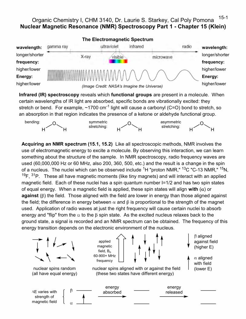
Acquiring an NMR spectrum (15.1, 15.2) Like all spectroscopic methods, NMR involves theuse of electromagnetic energy to excite a molecule. By observing this interaction, we can learnsomething about the structure of the sample. In NMR spectroscopy, radio frequency waves areused (60,000,000 Hz or 60 MHz, also 200, 360, 500, etc.) and the result is a change in the spinof a nucleus. The nuclei which can be observed include 1H "proton NMR," 13C "C-13 NMR," 15N,19F, 31P. These all have magnetic moments (like tiny magnets) and will interact with an appliedmagnetic field. Each of these nuclei has a spin quantum number I=1/2 and has two spin statesof equal energy. When a magnetic field is applied, these spin states will align with ( ) oragainst ( ) the field. Those aligned with the field are lower in energy than those aligned againstthe field; the difference in energy between and is proportional to the strength of the magnetused. Application of radio waves at just the right frequency will cause certain nuclei to absorbenergy and "flip" from the to the spin state. As the excited nucleus relaxes back to theground state, a signal is recorded and an NMR spectrum can be obtained. The frequency of thisenergy transition depends on the electronic environment of the nucleus.
nuclear spins random(all have equal energy)
appliedmagneticfield, Bo
60-900+ MHzfrequency
nuclear spins aligned with or against the field(these two states have different energy)
alignedwith field(lower E)
alignedagainst field(higher E)
energyabsorbed
energyreleased
Organic Chemistry I, CHM 3140, Dr. Laurie S. Starkey, Cal Poly PomonaNuclear Magnetic Resonance (NMR) Spectroscopy Part 1 - Chapter 15 (Klein)
15-1
E varies withstrength of
magnetic field
(Image Credit: NASA's Imagine the Universe)
wavelength:
longer/shorter
frequency:
higher/lower
Energy:
higher/lower
wavelength:
longer/shorter
frequency:
higher/lower
Energy:
higher/lower
The Electromagnetic Spectrum
Infrared (IR) spectroscopy reveals which functional groups are present in a molecule. Whencertain wavelengths of IR light are absorbed, specific bonds are vibrationally excited: theystretch or bend. For example, ~1700 cm-1 light will cause a carbonyl (C=O) bond to stretch, soan absorption in that region indicates the presence of a ketone or aldehyde functional group.
HO
H HO
H HO
H
bending: symmetricstretching:
asymmetricstretching:

Information obtained from a 1H "Proton" NMR spectrum (15.3):
1) # of signals indicates the number of different types of hydrogens (chemical equivalence).
2) Integration or peak area indicates how many hydrogens are in each signal. It is given as a ratio.
3) Chemical shifts are given as (delta) values, in ppm (~0-10). The chemical shift indicates the
electronic environment of the hydrogens (electron-rich/shielded or electron-poor/deshielded).
4) Splitting patterns indicate the # of neighboring hydrogens. The magnitude of the coupling
constants (given as J values) depend on the spatial relationship (dihedral angle) of the two protons.
15-2
050100150200PPM
IR spectrum showsfunctional groups
(see Klein Chapter 14& CHM 3140L lab)
13C NMR spectrum shows how many different types of carbons are in a molecule,and whether each carbon is in an electron-rich or electron-deficient environment.
0.0 PPM
Si CH3
CH3
CH3
CH3
tetramethylsilane(TMS) serves as areference, = 0
1H NMR (Proton NMR) spectrum
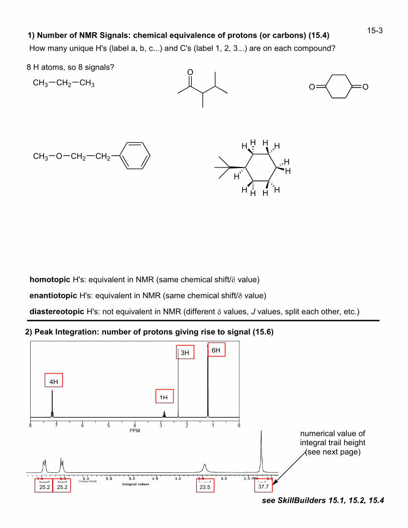
15-31) Number of NMR Signals: chemical equivalence of protons (or carbons) (15.4)
How many unique H's (label a, b, c...) and C's (label 1, 2, 3...) are on each compound?
CH2
O
CH2O
CH3 CH2 CH3
8 H atoms, so 8 signals?
homotopic H's: equivalent in NMR (same chemical shift/ value)
enantiotopic H's: equivalent in NMR (same chemical shift/ value)
diastereotopic H's: not equivalent in NMR (different values, J values, split each other, etc.)
2) Peak Integration: number of protons giving rise to signal (15.6)
H H
HH
HH
H H
H
HH
25.2 25.2 23.5 37.7
4H
1H
3H 6H
CH3
OO
see SkillBuilders 15.1, 15.2, 15.4
numerical value ofintegral trail height(see next page)
15.1, 15.2,
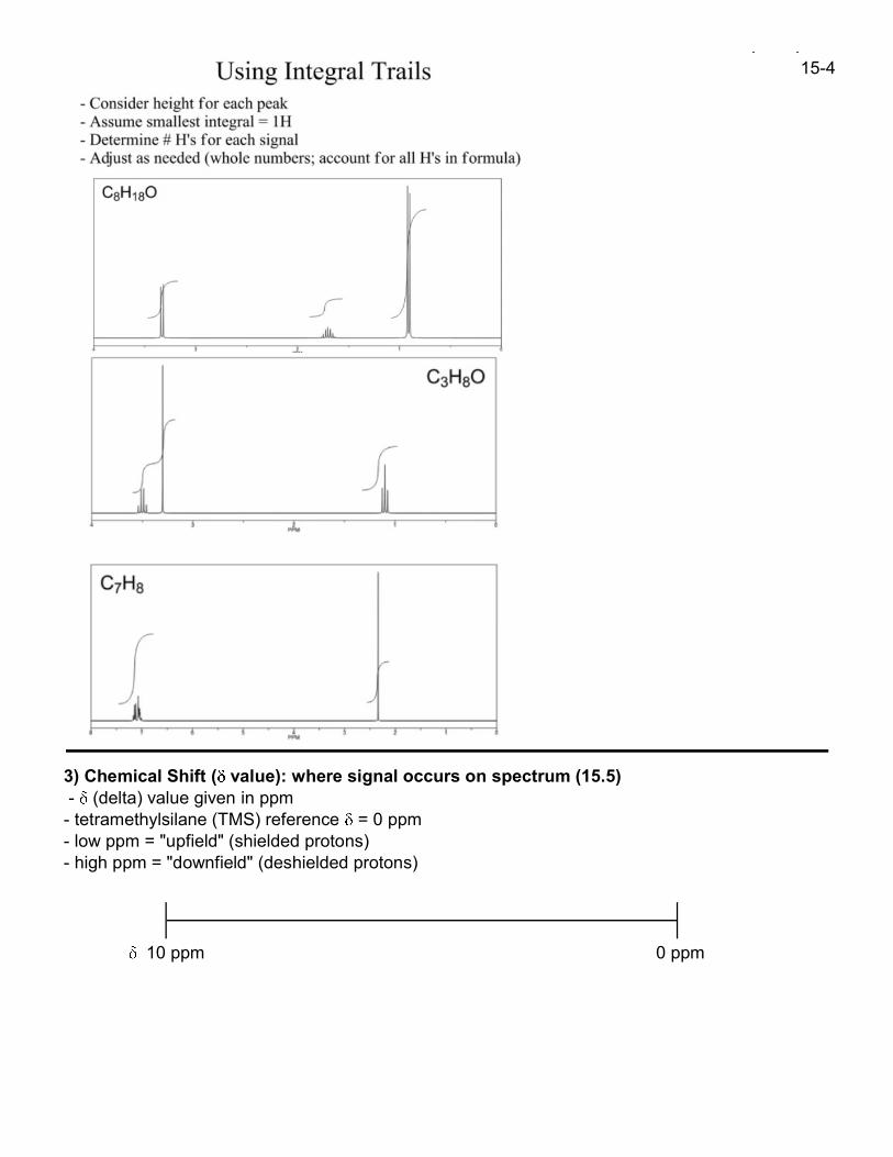
3) Chemical Shift ( value): where signal occurs on spectrum (15.5)- (delta) value given in ppm- tetramethylsilane (TMS) reference = 0 ppm- low ppm = "upfield" (shielded protons)- high ppm = "downfield" (deshielded protons)
10 ppm 0 ppm
15-4

15-5Delta values are given in parts per million (ppm) relative to the reference compoundtetramethylsilane (TMS) which resonates at = 0.0 ppm.
11 10 9 8 7 6 5 4 3 2 1 0
R H
OH R2C CR
H
ROCH3CH3
R CH3
O
RH
CH3
C H
N H
O H
R NH2
ONH2 RNH2
R OH
OOH ROH
(ppm)
Type of C-H (ppm) Description of Proton
0.9 alkyl (methyl)
1.3 alkyl (methylene)
1.5-2 alkyl (methine)
1.8 allylic (C is next to a pi bond)
2-2.3 to carbonyl (C is next to C=O)
2.3 benzylic (C is next to Ph)
2.5 alkynyl
2-3 to nitrogen (C is attached to N)
3-3.5 to halogen (C attached to Cl/Br/I)
3.8 to oxygen (C is attached to O)
5-5.3 vinylic (H is attached to alkene C)
7.3 aromatic (H is on phenyl ring)
9.7 aldehyde (H is on C=O)
R CH3
R CH2 R
R3C H
R2C CRH
Ar CH3
RC C H
R2N CH3
R CH2 X
RO CH3
Ar H
R C HO
R C CH3
O
CH3
Protons on Carbon
Type of H (ppm) Description
ROH
ArOH
R C OH
O
RNH2
R C NHR
O
ArNH2
R CH2 F 4.5 to fluorine (C is attached to F)
0.5-5 alcohol
4-7 phenol
10-13 carb. acid
0.5-5 amine
3-5 aniline
5-9 amide
Protons on Oxygen/Nitrogen*
*Protons on N or O typically have wide ranges
of expected chemical shifts; the actual value
depends on the solvent used, the concentration,
temp., etc. Because these protons are acidic and,
therefore, exchangeable, they may be broad
peaks and usually do not couple with
neighboring protons (typically they are broad
singlets). If a protic deuterated solvent is used
(e.g., D2O or CD3OD), then the NH and OH
protons will exchange with the deuterium and the
peaks will shrink or disappear entirely, since D
(2H) does not show up in the 1H NMR spectrum.
R = alkyl group
Ar = aromatic ring, such as phenyl (Ph)Note: aldehyde proton (-CHO) has small coupling withneighboring H's, so it usually appears as a singlet
7.3 aromatic (H is on phenyl ring)Ar H
15-6
see SkillBuilder 15.3
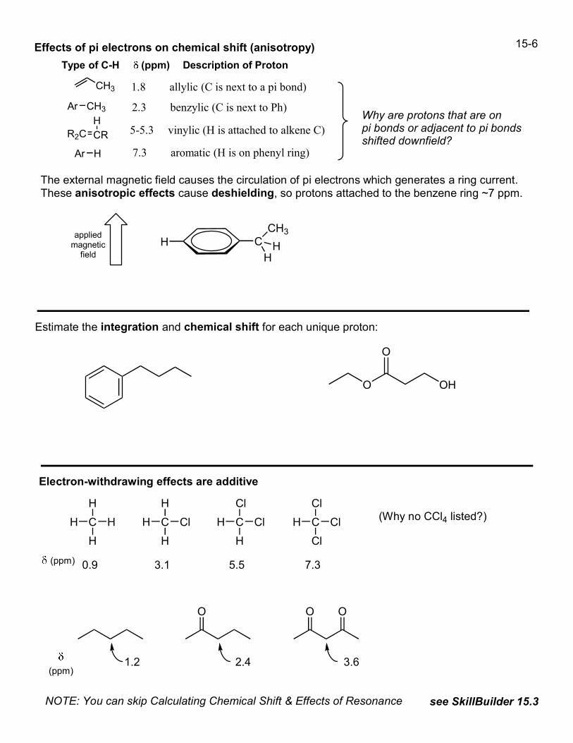
Effects of pi electrons on chemical shift (anisotropy)
Type of C-H (ppm) Description of Proton
The external magnetic field causes the circulation of pi electrons which generates a ring current.These anisotropic effects cause deshielding, so protons attached to the benzene ring ~7 ppm.
CHappliedmagneticfield
1.8 allylic (C is next to a pi bond)CH3
5-5.3 vinylic (H is attached to alkene C)R2C CRH Why are protons that are on
pi bonds or adjacent to pi bondsshifted downfield?
CH3
HH
2.3 benzylic (C is next to Ph)Ar CH3
Estimate the integration and chemical shift for each unique proton:
O OH
O
Electron-withdrawing effects are additive
C
H
HH
H
(ppm) 0.9
C
H
ClH
H
3.1
C
Cl
ClH
H
5.5
C
Cl
ClH
Cl
7.3
(Why no CCl4 listed?)
(ppm)1.2 2.4 3.6
O O O
NOTE: You can skip Calculating Chemical Shift & Effects of Resonance
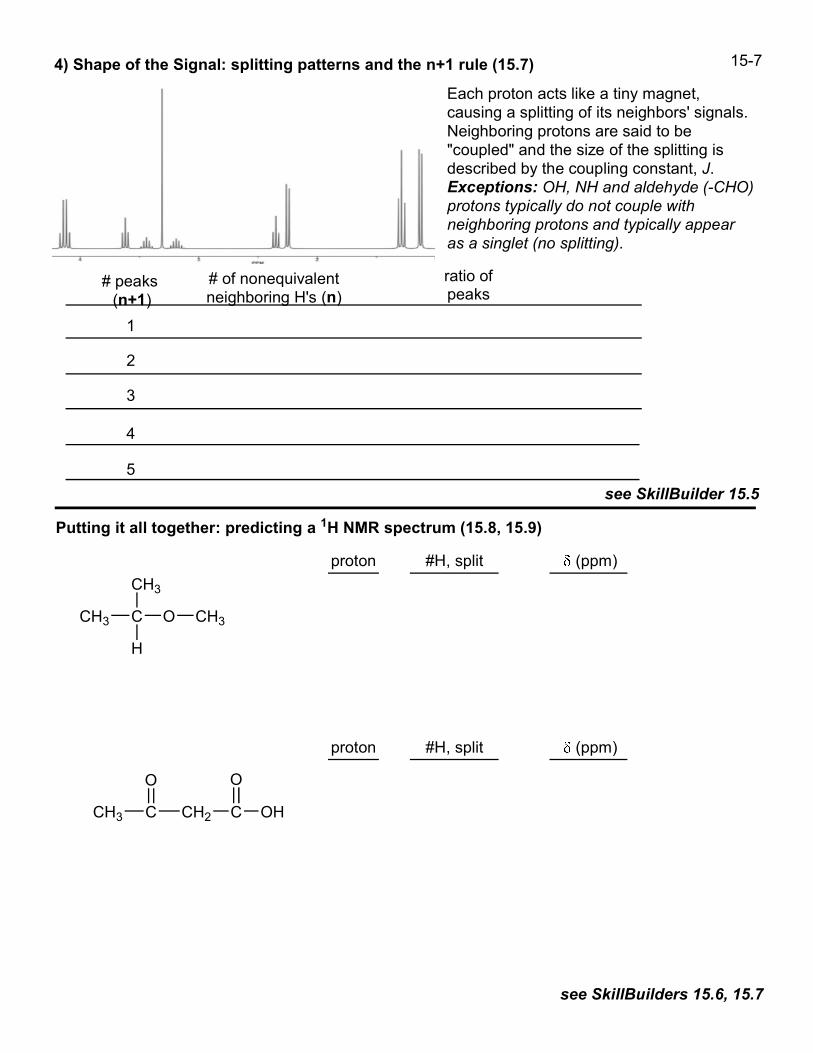
4) Shape of the Signal: splitting patterns and the n+1 rule (15.7)
see SkillBuilder 15.5
Putting it all together: predicting a 1H NMR spectrum (15.8, 15.9)
see SkillBuilders 15.6, 15.7
Each proton acts like a tiny magnet,causing a splitting of its neighbors' signals.Neighboring protons are said to be"coupled" and the size of the splitting isdescribed by the coupling constant, J.Exceptions: OH, NH and aldehyde (-CHO)protons typically do not couple withneighboring protons and typically appearas a singlet (no splitting).
# peaks(n+1)
ratio ofpeaks
1
2
3
4
5
C
CH3
CH3
H
O CH3
proton #H, split (ppm)
proton #H, split (ppm)
C CH2 C
O
OH
15-7
# of nonequivalentneighboring H's (n)
O
CH3
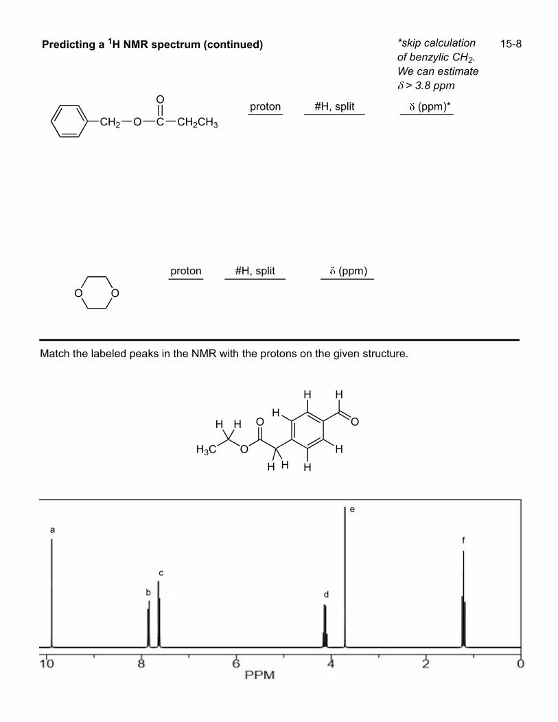
a
b
c
d
e
f
OH3C
H
H
HHH
HH
H
O
H
O
15-8
Match the labeled peaks in the NMR with the protons on the given structure.
Predicting a 1H NMR spectrum (continued)
proton #H, split (ppm)*
proton #H, split (ppm)
CH2 O C
O
CH2CH3
O O
*skip calculationof benzylic CH2.We can estimate> 3.8 ppm

15-9
O
OCH3
a
b
c
d
e
050100150200PPM
Interpreting 13C NMR Spectra (15.12) Educator lecture: NMR, Part II (1:44:49 - 1:50:16)
- one signal for each unique carbon type
- chemical shifts ~ 0 to 220 ppm
- signals are typically all singlets ("proton-decoupled" or "broadband decoupled")
- # of hydrogens attached to each carbon can be determined by DEPT experiment (15.13)
- 13C isotope ~1% of carbon atoms, so 13C NMR requires more sample and/or more scans
Match the labeled peaks in the NMR with the protons on the given structure.

15-10
13C NMR Chemical Shifts
220 200 180 160 140 120 110 100 80 60 40 20
alkane
R H
O
(ppm)
Type of carbon (ppm) Description of carbon
10-30 primary alkyl (methyl)
15-55 secondary alkyl (methylene)
20-60 tertiary or quaternary alkyl
0-40 attached to iodine
25-65 attached to bromine
40-60 attached to nitrogen
35-80 attached to chlorine
40-80 attached to oxygen
65-90
110-170 aromatic (phenyl ring C)
185-220 C=O, ketone or aldehyde
R CH3
R CH2 R
R3C H
R2C CR2
R C H
O
0
R R
O
R NH2
O
R OH/OR
O
C O C CC C
aromatic
C O
C X
C N
C I
C Br
R C R
O
C N
alkynylRC CR
C Cl
C O
C
R
R R
R
100-150 alkenyl
165-185 C=O, carboxylic acid, ester, amideR C NH2
O
R C OH
O
R C OR
O
carbonyls sp2 carbons sp3 carbons
Interpreting 13C NMR Spectra (continued)
While it is possible to acquire a "proton coupled" spectrum that shows the splitting of each carbonby its attached hydrogens (e.g., a CH3 would appear as a quartet), such spectra are rarely used and"broadband-decoupled" 13C spectra are far more common. Proton-decoupled 13C spectra give asinglet peak for each unique carbon. In order to determine the number of hydrogens on eachcarbon, a series of experiments with varying pulse sequences, known as DEPT experiments, areemployed (see Klein 15.13 and SkillBuilder 15.10, but we will not be covering DEPT in CHM 3140).

15-11
see SkillBuilder 15.9
How could you use 13C NMR to distinguish between the three isomers of dimethylbenzene?
O
O
H
O
O
050100150200PPM
Each of the compounds shown has seven signals in its 13C NMR spectrum.
Which structure matches the spectrum provided? Explain
Chapter 15 textbook problems for Exam II:
SkillBuilders 15.1-15.7 (1H NMR) and 15.9 (13C)
Do the following problems: 1-22, 26, 35-39, 41, 42, 45, 47, 48, 50, 63-71.
Exam III (interpreting 1H NMR spectra): SkillBuilder 15.8 and problems: 23-25, 57-59, 64.
Which would be better to distinguish the following
compounds, 1H or 13C NMR (or are they equally
suitable)? Explain, and describe the peak(s) to look for.
CO
O
CH3CH2CH3 CCH2
O
OCH3 CH3
CH3H3C H3C CH3
CH3CH3
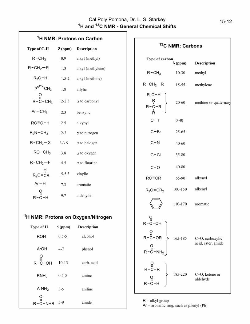
15-12Cal Poly Pomona, Dr. L. S. Starkey1H and 13C NMR - General Chemical Shifts
Type of C-H (ppm) Description
0.9 alkyl (methyl)
1.3 alkyl (methylene)
1.5-2 alkyl (methine)
1.8 allylic
2-2.3 to carbonyl
2.3 benzylic
2.5 alkynyl
2-3 to nitrogen
3-3.5 to halogen
3.8 to oxygen
5-5.3 vinylic
7.3 aromatic
9.7 aldehyde
R CH3
R CH2 R
R3C H
R2C CR
H
Ar CH3
RC C H
R2N CH3
R CH2 X
RO CH3
Ar H
R C H
O
R C CH3
O
CH3
1H NMR: Protons on Carbon
Type of H (ppm) Description
ROH
ArOH
R C OH
O
RNH2
R C NHR
O
ArNH2
R CH2 F 4.5 to fluorine
0.5-5 alcohol
4-7 phenol
10-13 carb. acid
0.5-5 amine
3-5 aniline
5-9 amide
1H NMR: Protons on Oxygen/Nitrogen
R = alkyl groupAr = aromatic ring, such as phenyl (Ph)
Type of carbon(ppm) Description
10-30 methyl
15-55 methylene
20-60 methine or quaternary
0-40
25-65
40-60
35-80
40-80
65-90
110-170 aromatic
185-220 C=O, ketone oraldehyde
R CH3
R CH2 R
R3C H
R2C CR2
R C H
O
C I
C Br
R C R
O
C N
alkynylRC CR
C Cl
C O
C
R
R R
R
100-150 alkenyl
165-185 C=O, carboxylicacid, ester, amide
R C NH2
O
R C OH
O
R C OR
O
13C NMR: Carbons

NMR practice problems (CHM 3140, Dr. Starkey)
How many signals are expected for each compound? Label each unique proton type (a/b/c).
CH3
CH3
HO
Draw the dichloropentane isomer that has exactly two 1H NMR signals.
Describe the relationship of the indicated protons:a) enantiotopic (one signal in NMR)b) enantiotopic (separate signals in NMR)c) diastereotopic (one signal in NMR)d) diastereotopic (separate signals in NMR)e) homotopic (one signal in NMR)
Br
1)
2)
3)
H
H
4)
H
O
H
OCH3
CH2 CH2 CH3
H
Match each highlighted proton with itsapproximate chemical shift:
1, 2, 4, 5, 7, 10 ppm
Also, predict the splitting patternfor each highlighted proton.Note: aldehyde protons typically donot couple with neighboring protons.
15-13
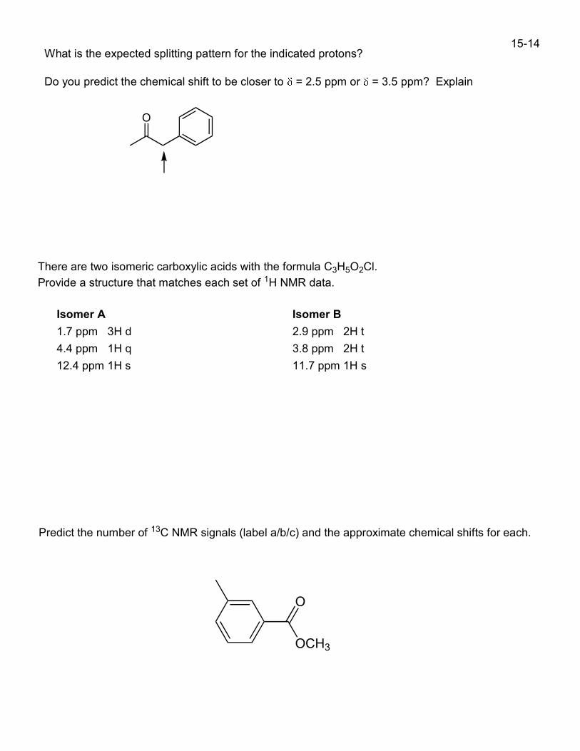
What is the expected splitting pattern for the indicated protons?
Do you predict the chemical shift to be closer to = 2.5 ppm or = 3.5 ppm? Explain
O
There are two isomeric carboxylic acids with the formula C3H5O2Cl.
Provide a structure that matches each set of 1H NMR data.
Isomer A
1.7 ppm 3H d
4.4 ppm 1H q
12.4 ppm 1H s
Isomer B
2.9 ppm 2H t
3.8 ppm 2H t
11.7 ppm 1H s
Predict the number of 13C NMR signals (label a/b/c) and the approximate chemical shifts for each.
O
OCH3
15-14