Embed Size (px)
Citation preview

1
RAD I – NORMAL ANATOMY
Cervical Spine
Lateral (1st)AP Open MouthAP LowerSwimmersObliquesFlexion/ExtensionOthers:
PillarsFuchs
Lateral Cervical
P&T72” FFDmust include top of T1 otherwise do a Swimmersfirst film in trauma cases
Cervical Laterala. sella turcica
a. sinus esb. clinoids
b. vertebra c. pedicle d. facets e spinolaminar line
a
g
j
k
l
e. spinolaminar line f. disc g. ADI h. pre‐vertebral soft tissues
a. nasopharynxb. larynx
i. hyoid j. pinna (ear) k. mastoids l. lambdoid suture
b
c
d e
f h
I
Cervical Spine Data
ADI < 3mm (4 mm child) AP and lateralDisk height C2< C3 < C4 < C5 < C6 > C7 Vertebral body should fit in adjacent canal Retro pharyngeal space < 6mm at C2 Retro tracheal space < 22mm at C6
AP Lower Cervical
P&T40” FFD15 degree tube tiltI l d C TIncludes C2‐T1

2
Cervical AP a. articular pillar\facetb. uncinate process c. spinous processd. pedicle e. lung apex
R a
b
cd
k
f. tracheaa. region of vocal cords
g. T1 with upward slanted transverse processes
h. ribs i. claviclesj. sternum k. mandible
c
e
f
g h
I
j
AP Open Mouth
P&T40” FFD5o tube tilt helpfulevaluates occiput though C3 evaluates occiput though C3
Cervical AP Open Mouth or Odontoida. occipital condyle b. C1 atlas
a. lateral mass b. transverse processes c. anterior arch
C i
a
b d
h
c. C2 axisd. odontoide. C2 pediclesf. lamina g. spinous h. mastoid i. teeth j. mandible
c e f g
I j
Cervical Oblique
P&T72” FFD15 degree cephalic tube tilt Exam includes right and left Exam includes right and left oblique studies
Cervical Oblique
a. body b. right pediclec left pedicle
R
a
b
c g
j
c. left pedicle d. uncinate process e. neuroforamen f. facetg. spinoush. ribs i. tracheaj. mastoid
d e
f
h
I
Understanding the oblique cervical
set your patients in the oblique position.
l th k th LLplace the marker on the corresponding side of the patient i.e. right on right, or left on left, just like on the AP view.
LL
3
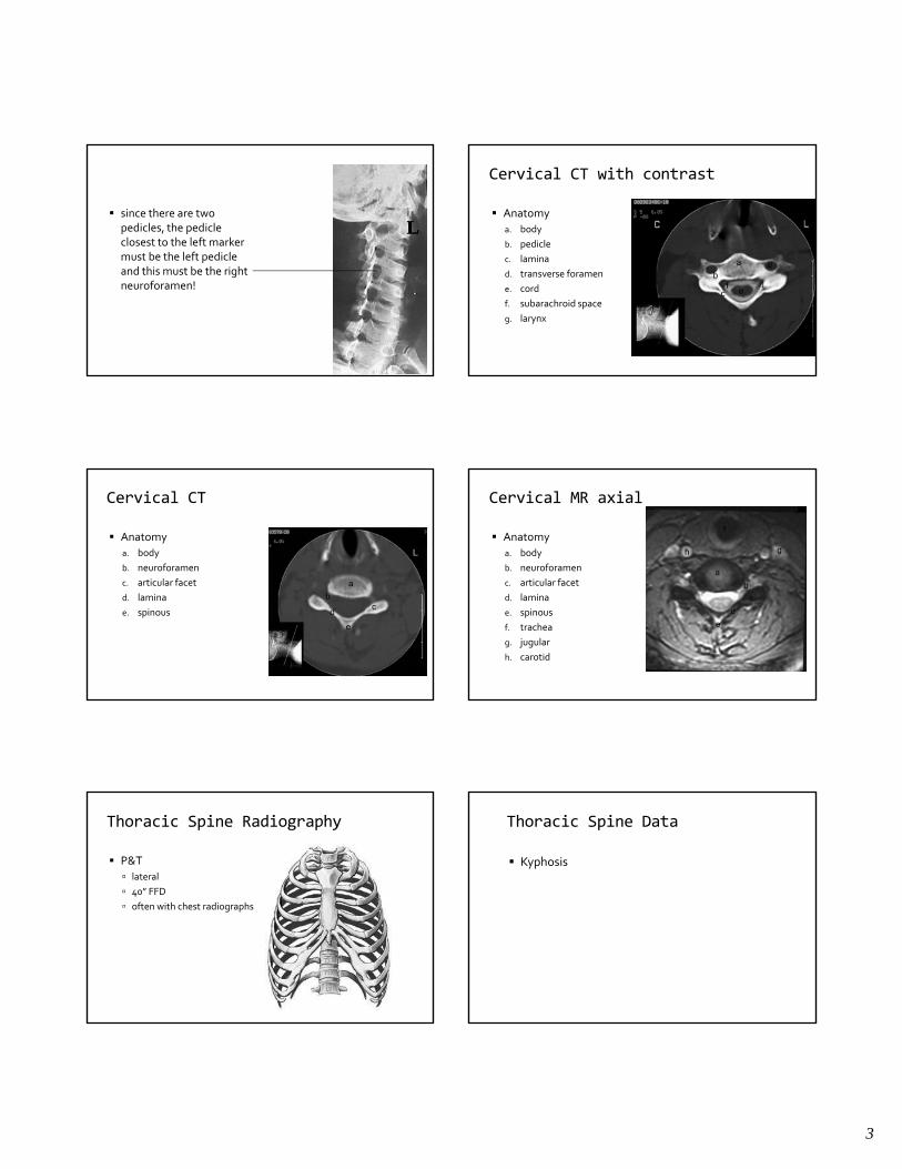
since there are two pedicles, the pedicle closest to the left marker must be the left pedicle
LLmust be the left pedicle and this must be the right neuroforamen!
Cervical CT with contrast
Anatomy a. body b. pedicle c lamina
g
c. lamina d. transverse foramen e. cord f. subarachroid space g. larynx
a
b
c
d
e f j
Cervical CT
Anatomy a. body b. neuroforamen c articular facet ac. articular facet d. lamina e. spinous
a
b c
d
e
Cervical MR axial
Anatomy a. body b. neuroforamen c articular facet
a
b
f
g h
c. articular facet d. lamina e. spinous f. trachea g. jugular h. carotid
b
c
d
e
Thoracic Spine Radiography
P&T lateral 40” FFD often with chest radiographs often with chest radiographs
Thoracic Spine Data
Kyphosis

4
Thoracic Spine AP vertebral body
pedicle spinous process
rib costal cartilage (calcified)
trachea carina
heart right atrium left ventricle
aorta paraspinal line(s) hili meganblase
Soft Tissue Lines Thoracic Spine
descending aorta
paraesophageal line
Thoracic Spine Lateral vertebral body
pedicle
ribs scapula and shoulder heart
retrosternal clear space retrosternal clear space left atrium aortainferior vena cava
trachea left and right bronchus
posterior gutter hili meganblase
Lumbar Spine Radiography
P&T40” FFD
AnatomyT12‐S1
ViewsAP, lateralobliquesrecumbents
Lumbar AP vertebral anatomy sacrum
sacral crest anterior sacral foramina sacroiliac joints
pelvis anatomy femora ribs abdominal/pelvic viscera
spleen liver bowel
Pelvis APilium
crest ASIS AIIS
pubis ischium
ischial spines superior and inferior ischiopubic rami
obturator foramen acetabulum
acetabula fossa
femoral head trochanters viscera
bladder bowel

5
Lumbar Spine Lateral
P&T40” FFDrecumbent on large patients
Lumbar Spine Lateral
Anatomyvertebral bodies pedicles superior and inferior ti l articular processes
pars interarticularesfacets spinous processes meganblase bowel gas posterior gutter/lung
Lumbar Spine Oblique
P&T40” FFD 350 obliquity
i i anterior or posterior
Lumbar Spine Oblique
Anatomyvertebral body pedicle pars pars articular processeslamina spinous process does not image foramen!
Understanding the oblique lumbar
Place the left or right marker on the corresponding side of the patientpat e t
Understanding the oblique cervical
this time, use the spinous process as the reference point.
in this instance, the marker indicates that this oblique shows the right pars to best advantage

6
Lumbar Obliques
spinous processright parsright pedicle
Lumbar CT with contrast
vertebral body pedicle transverse process lamina spinous IVC aorta psoas m. erector spinae m.
Lumbar MR sagittal
vertebral body disc epidural space cauda equina spinous
Shoulder
P&T40”FFDboomerang filter
AnatomyAC/GH joints
ViewsAP Internal External GrasheyStress Views
AP Shoulder
P&TExternal rotationInternal rotation Grashey position (preferred)Grashey position (preferred)
Shoulder AP (external rotation)Anatomy
glenohumeral jointacromioclavicular joint greater tuberosity lesser tuberositylesser tuberosityintertubercular groove acromion process coracoid processglenoid ribs clavicle

7
Shoulder AP (internal rotation)
Anatomyglenohumeral jointacromioclavicular joint greater tuberosity greater tuberosity lesser tuberosityintertubercular groove acromion process coracoid process
Shoulder Dislocation
Elbow
P&T40” FFDextremity cassettes
ViewsViewsAPlateralrotation (external)
Elbow AP
Anatomy humerus
trochlea capitellum epicondyles
ulna olecranon
radius radial tuberosity
Lateral Elbow
P&T40” FFDextremity cassettespositioning importantpositioning important
Elbow Lateral
Anatomyhumerus
distal humeral fat pads
ulna olecranon coranoid process
radial head

8
Wrist/Hand
P&T40” FFDExtremity Cassettes
ViewsPALatObliqueUlna deviated
PA Wrist/Hand
P&Twrist flat on cassetteextremity film
Anatomycarpus
Wrist PA
Anatomy carpals radius ulna ulna
Oblique Wrist/Hand
P&T45o
Wrist Oblique
Anatomy Evaluates scaphoid well.
Lateral Wrist
P&T‐ Positioning is criticalWrist “flat” and perpendicular to cassetteperpendicular to cassette.

9
Lateral Wrist
Anatomy radius ulna carpus carpus pronator fat pad.
POIradius‐lunate–capitate alignment.
Knee
ViewsAP Lateral O j iOpen jointMerchantTangential
AP Knee
P&Textremity cassette40”FFD CTT5o CTT
Knee AP
Anatomy femur
popliteal notch intercondylar notch
tibia interc0ndylar spines
fibula
POI epiphyseal scar
Knee Lateral
P&Ttrue lateral needed60o flexionl l id d lateral side down
Knee Lateral
Anatomy femur
quad tendon Hoffa fat pad
tibiatuberosity
fibulapatella
patella tendon

10
Knee Open Joint
Anatomy femur
intercondylar fossav. medialis v. lateralis
tibia tibia spines
fibula
Knee MRI sagittal
Ankle
P&TExtremity cassettes
ViewsAPLateralObliqueStress views
AP Ankle
P&T40” FFDtrue AP needed
Ankle AP
Anatomyf ibula.tibia.talustalus.
POIsubchondral fxalignmentswelling
Oblique Ankle
P&TInternal rotation 35 degrees.Positioned to view the tibio‐fibular joint space.j p

11
Ankle Oblique
Fibula.Tibia.Talus.
lNavicula.Cuboid.Tibia‐fibula joint.Talar dome.
Lateral Ankle
P&Tlateral side down
Anatomytalotibtalotibmidtalar jointscalcaneus
POIAVNstress fxs
Ankle lateral
Fibula.Tibia.Talus.Calcaneus.Navicula.Cuneiforms.Cuboid.5th metatarsal.Achilles tendon.
Ankle lateral
Fibula.Tibia.Talus.Calcaneus.Navicula.Cuneiforms.Cuboid.5th metatarsal.Achilles tendon.
Foot
P&Textremity cassettesfiltration helpful
ViewsAP/DPlateralobliquetangential

12
Foot dorsoplantar and oblique Foot dorsoplantar and oblique
Foot lateral Foot lateral
Ankle/Foot Fractures Chest
P‐ALateral

13
ChestP & T
72’’ FFDPA radiographChest film
POILungsTracheaDiaphragmHeartAortaHiliCostophrenic and cardiophrenic anglesGastric air bubbleBones (ribs, clavicles, etc.)
PA Chest
Clavicle
First Rib
Posterior rib
Aortic arch/knob
Trachea
carinart main bronchus
Ascending aorta
Anterior rib
cardiophrenic angleGastric air bubblecostophrenic angle
HilaSuperior
vena cava
Pulmonary artery branches
Breast shadow
Diaphragm
PA Chest
Lateral Chest