Embed Size (px)
Citation preview


=3
i ,tVILLINOIS t
U URBAHA-CHAMPAIGNLXLOGY


FIELDIANA
GeologyPublished by Field Museum of Natural History
Volume 33, No. 26 June 23, 1977
This volume is dedicated to Dr. Rainer Zangerl
New Agnathous Fishes
from the Pennsylvanian of Illinois
David BardackDepartment of Biological Sciences
University of Illinois at Chicago Circle
AND
Research AssociateField Museum of Natural History
AND
Eugene S. Richardson, Jr.
Curator. Fossil InvertebratesField Museum of Natural History
Among the several well-preserved but perplexing animals in the
Middle Pennsylvanian (early Westphalian D) Essex fauna of north-
ern Illinois (Richardson and Johnson, 1971) are two which appear to
be chordates at the agnathan level of organization. Both show elab-
orate oral and pharyngeal structures without analog among knownchordates. Some cranial elements are indicated by darkly stained
patches on the plane of the concretions, but, if cartilaginous
skeletons were developed, they were uncalcified and less substantial
than those of Mayomyzon, a lamprey (Bardack and Zangerl, 1971)
found in the same fauna. It is quite possible that the two animals
described here are larvae with a still unknown adult phase.
The Essex fauna lived on a delta at the end of an embayment in
the northeastern corner of Illinois Basin. Fine clastic material, with
occasional increments of sand, was brought in by streams that
flowed for long distances through a coal swamp; in and adjacent to
the streams lived animals of the Braidwood fauna. The vertebrates
of both faunas are represented almost entirely by small individuals;
the presence of large and presumably mature individuals is indi-
cated by scattered scales and bones of large lungfishes and rhipidis-
tians plus large coprolites and rare teeth and cartilage elements of
Library of Congress Catalog Card Number 77- 76919 The Library of thB
Publication 1261 489 QEQtOG^ MAR 1 4 ^8university 61 Illinois
at Urbana-ChampaW

490 FIELDIANA: GEOLOGY, VOLUME 33
sharks. The delta environment, both subaerially and subaqueously,
provided suitable habitat for young animals. The specimens here
described occur in Pit Eleven of the Peabody Coal Company in Will
and Kankakee counties. Within this pit, one traverses a sequence of
ancient environments from shallow marine at the south to a margin-al shore zone at the north, and in the band of other pits farther north
a subaerial zone. Local concentrations of various members of the
fauna indicate that local habitats must have varied in depth, salin-
ity, warmth, vegetational cover, turbidity, and other environmental
parameters. The preponderance of small forms among the fishes (as
well as the tetrapods, eurypterids, and xiphosures) suggests that
this portion of the delta provided a modicum of protection from
rapacious adults.
Phylum Chordata
Class Agnatha
Pipiscius, new genus
Diagnosis. —Small agnathous fishes with oral structure compris-
ing 23 collar lamellae and 23 plates surrounding a circular buccal
cavity. At least 4 gill clefts. Post-anal dorsal fin.
Derivation of name. —From pise, L. for fish + pi as a reduplicated
syllable.
Pipiscius zangerli, new species. Figures 1-5.
Diagnosis. —Same as for genus.
Holotype. -Field Museum of Natural History (FMNH) PF 8344
part and counterpart, Pit Eleven, Peabody Coal Co. Collected byMr. and Mrs. F.A.Wolff.
Fig. 1. Pipiscius zangerli, n. g., n. sp. Diagrammatic restoration of the whole ani-
mal in lateral view, including internal structures. Dt, digestive tract; E, eye; F, fin,
G, gill pouches; O, otic capsule; P, pharyngeal chamber.

=3O
bO3Oa
d
C
bb
C
491

492 FIELDIANA: GEOLOGY, VOLUME 33
Referred specimens.—FMNH: PF 8345, one piece; PF 8346, part
and counterpart; PF 7514, part and counterpart. Sobolik Collection,
P 33, part and counterpart. Piecko Collection: HTP 153, one piece;
HTP 5099, part and counterpart; HTP 5431, one piece. Kimball
Collection, part and counterpart. Sherman Collection, 1180, partand counterpart; Douglass Collection 604, part and counterpart.
Geologic horizon and locality.— Pit Eleven, Peabody Coal Co.,
Will and Kankakee counties, 111., Francis Creek Shale, Carbondale
Formation ( Westphalian D).
Derivation ofname. — In honor of Dr. Rainer Zangerl.
Description. —This account is based on all 10 known individuals
(fig. 1 ). Specimens range in length from about 4 to 6.5 cm. and bodydepth varies from 0.7 to 1.4 cm. About half of the specimens, pre-
dominantly the shorter individuals, exhibit a plump ventral dis-
tension of the body. This ventral expansion may be a remnant of a
yolk sac similar in form to that of a late stage of anuran embryonic
development (fig. 2). Larger individuals are elongate and possiblycircular in cross-section. Most individuals are preserved in lateral
aspect, although the head end is often twisted so that the character-
istic mouth structure is viewed dorsally or ventrally (fig. 3). Al-
though no distinct hard parts are preserved, certain consistent pig-
mentation patterns, depressions and ridges, particularly in the
mouth region, permit tentative reconstruction of some internal
anatomy.
The head as a whole is not topographically distinguished from the
body. Anteriorly, between the mouth and the eye, the head appearsrather flexible, as suggested by the rotation of the mouth parts in
laterally preserved individuals. A notch in the dorsal and ventral
margins of the head anterior to the eye, ventrally extended profile of
the oral part of the head and some darkly stained areas above the
mouth all suggest the presence of an oral hood analogous to that of
modern adult lampreys.
The mouth margin and the wall of the mouth cavity were sur-
rounded by a firm substance that has not been preserved but has
left a distinct impression (figs. 4, 5). Proximally, the mouth struc-
ture comprises a collar in the shape of a thin truncated cone, its
wall composed of 23 rectangular lamellae (collar lamellae), with thin
radial vanes (collar vanes) projecting into the lumen of the oral
cavity at the junctions between lamellae. Distally the collar vanes
terminate in a knob-like thickening. The lamellae and vanes on the

493

494 FIELDIANA: GEOLOGY, VOLUME 33
Fig. 4. Pipiscius zangerli, n. g., n. sp. Impression of inner surface of mouth, as
viewed from within the animal, anterior to upper left. FMNH PF 8344, holotype,X12.5
anterior aspect of the collar are somewhat longer and wider than
those on the posterior. A deep pit (collar pit) is excavated into the
junction of each pair of collar lamellae, on the outer surface, oppo-site each vane; these pits are very pronounced in the anterior aspectand become faint or absent toward the posterior aspect. An irregu-
lar system of polygonal ridges on the outer surface of the lamellae
suggests that either the substance was cracked by volume loss dur-
ing decay or the lamellae were compound.
Distally, the collar articulates at about a right angle to a more
elongated and distally expanded set of 23 plates, forming a circle
that is gently inflected to embrace the margin of the mouth. Dis-
tally, the diameter of the circle is about twice the proximal diameter
of the collar. The circle plates, like the structures of the collar, are
larger in the anterior aspect. A plate lies opposite each collar vane.
Each plate is roughly triangular in outline, thickened just beyondits junction with the collar, then thinning and narrowing distally.
The thickened regions of neighboring circle plates abut against each
other and probably could pivot on this abutment. Each plate also
bears on its midline a radial vane (circle plate vane) projecting into

BARDACK & RICHARDSON: AGNATHOUS FISHES 495
Fig. 5. Pipiscius zangerli, n. g., n. sp. Impression of outer surface of mouth, as
viewed from outside the animal, anterior toward left. FMNH PF 8344, holotype,
X12.5
the lumen of the oral cavity. The vane terminates in a thickening ad-
jacent to that of the corresponding collar vane. A triangular area
with its broad base at the distal end of the mouth lies between each
pair of circle plates. It, too, has a central vane on the buccal surface.
This complex mouth structure must have been operated by a set
of muscles. The collar formed a fixed diameter, with little or no po-
tential for expansion or contraction; however, a set of circumferen-
tial collar muscles might have provided for a limited amount of ex-
pansion. Most of the movement of the mouth structure was con-
fined to the circle of plates. Muscles running between the terminal
knob-like thickenings of the collar vanes and plate vanes could draw
the plates inward. Circumferential muscles between adjacent circle
plates assisted in constricting the distal opening of the mouth.
Restoration of an open position could have been achieved bymuscles or ligaments extending between the collar pits and the
thickenings of the circle plates.
Behind the mouth, several specimens exhibit an impression of a
slender tube followed by an expanded cavity with a series of ridges

496 FIELDIANA: GEOLOGY, VOLUME 33
and grooves. We interpret this as a pharyngeal tube and pharyngeal
pouch surrounded by muscles and capable of pumping nutritive
material into the animal. Coupled with the mouth structure, it
provided a strong feeding mechanism capable of ingesting detritus
or small invertebrates.
A pair of eyes represented by discs of pigmented material with a
small, clear central area doubtlessly occupied in life by a lens, lies
above the pharyngeal pouch. Behind the eyes a slightly anteropos-
teriorly expanded bulbous structure may represent the otic capsule.
Beginning behind the pharyngeal pouch and extending below andbehind the otic capsule lies a series of at least four dorsoventrally
expanded clefts (clear, unpigmented areas). Each is surrounded bydarkly stained areas which are most emphatically marked ventrally.
In some specimens these pouches are small and closely packed,while in others they occupy a broader area. These clefts and sur-
rounding tissues are probably the gill pouches. We are somewhat
puzzled by the limited number of such respiratory structures in an
agnathan. Possibly the entire set is not well preserved or more maybe added ontogenetically. The latter alternative is unlikely, how-
ever, as living Petromyzon show all gill pouches by embryonic stage15 (Piavis, 1971)— at about 0.5 cm. body length.
Several other deeply pigmented areas, especially in the head
region, are indicative of developing cartilaginous or other head
structures. None shows a sufficiently consistent pattern in several
specimens to enable us to assign it to a specific structure. Some of
these pigmented areas, especially around the eyes and below the gill
pouches, suggest that thick, rigid structures would be formed.
No distinct features of the anterior part of the digestive cavity are
indicated by pigmentation patterns as in Mayomyzon. Two speci-
mens (FMNH: PF 8344 and PF 8345), however, display an elongate
tubular swelling which is rounded at its anterior end about midwayalong the length of the body and tapers toward the rear margin of
the body cavity. This expanded area is filled with fine-grained min-
eral material somewhat different from that of the rest of the concre-
tion and characterized by several irregular vertical grooves and
folds. It probably represents the digestive tube; the folds and
grooves suggest muscular folds of the tube wall. It is probably not a
coelom filling because it is surrounded by large, clearer spaces and
lies well below a darkly pigmented region which should be expaxial
musculature.

BARDACK & RICHARDSON: AGNATHOUS FISHES 497
Myomeric segmentation is evident at the rear end of the body.The segments are especially distinct across the posterior end of the
body cavity, where more than 20 may be counted. While dorsal andventral ends are indistinct, the myomeres appear in the form of a
posteriorly opened V with a shorter upper than lower arm. There
are neither paired fins nor a distinct caudal fin. There is a short
dorsal fin at the posterior end of the body, supported by at least 10
slender rays.
Gilpichthys, new genus
Diagnosis.— Elongate agnathan fishes with tubular, muscular
buccopharynx bearing teeth; at least six gill clefts; no fins.
Derivation of name. —For Orville L. Gilpin, Chief Preparator,
Department of Geology, Field Museum of Natural History; and
ichthys, G., fish.
Gilpichthys greenei, new species. Figures 6-11.
Diagnosis. —Same as for genus.
Holotype.—FMNH PE 18703, part and counterpart, Pit Elevenof Peabody Coal Company, Will or Kankakee County, Illinois.
Referred specimens.—FMNH: PE 23464, part and counterpart;
PE 18703, part and counterpart; PF 8349, part and counterpart; PF8347, part and counterpart; Douglass Collection, unnumbered, partand counterpart; Piecko Collection, HTP 4700, part and counter-
part; Wolff Collection 164, one piece; Rockwell Collection 5211, partand counterpart.
Geologic horizon and locality. —Pit Eleven of Peabody Coal Com-
pany, Will and Kankakee counties, Illinois; Francis Creek Shale,
Carbondale Formation (Westphalian D).
Derivation of name.— For Mr. Frank A. Greene, Jr., of Coal City,
Illinois.
Gilpichthys is an elongate, slender animal ranging in length from
about 4 to 14 cm. and in depth from about 5 to 13 mm., measured in
the area of the gills (fig. 6). We have examined more than 100 speci-
mens. Before well-preserved specimens became available, the buc-
copharyngeal structure was taken to be a radula, with the conse-
quence that Gilpichthys was called a mollusc. It lias appeared in
the literature as "nudibranch gastropod" (Johnson and Richardson,
1966, table 1 and p. 628), "sea slug" (Richardson, 1966), "nudi-
branch" (Johnson and Richardson, 1970, p. 56), and "an elongated,

498 FIELDIANA: GEOLOGY, VOLUME 33
banana-shaped soft-bodied animal . . . with a molluscan radula"
( Richardson and Johnson, 1971, p. 1,229).
It appears that the body was somewhat deeper than wide, i.e.,
somewhat compressed laterally, as nearly all specimens lie on a
lateral surface. The body is inflected ventrally anterior to the gills
on most specimens. Others simply show the body in the form of a
gentle curve, concave ventrally. We are not certain as to the func-
tional significance of this shape, but do not believe that it repre-
sents a post-mortem deformation. There are no external projectionsfrom the body. Gilpichthys lacks both paired and unpaired fins.
Differential staining of the matrix demonstrates the presence of a
coelom (fig. 7). This lies between an anterior head end, which is
somewhat more than one-quarter of the total length of the animal,
and a tail area slightly less than one-fifth of the body length. Within
the coelom, several specimens show the digestive tract as an
irregularly zigzagging tube filled with compressed, finely com-
minuted material. Across the body in the area of the coelom, manyspecimens show a dozen to 20 myomeric segments. Additional seg-
ments no doubt were present. Although dorsal and ventral ends of
the myomeres cannot be clearly seen, it appears that each myomereincludes a long ventroposteriorly directed section and a shorter (less
than one-third as long) dorsoposteriorly sloping section. In addition
to the digestive tract, several specimens show a broad, elongate,
darkly stained area at the anterior end of the coelom. By analogywith Mayomyzon, this is believed to be the liver (fig. 8). It is con-
cave anteriorly, and in this concavity lies another dark region
BP E O G VA H L
Fig. 6. Gilpichthys greenei, n. g., n. sp. Diagrammatic restoration of whole animal
in lateral view, including internal structures. BP, buccopharynx; DA, dorsal aorta;
Dt, digestive tract; E, eye; G, gill pouches; H, heart; L, liver; N, notochord; O, otic
capsule; VA, ventral aorta.

499

500 FIELDIANA: GEOLOGY, VOLUME 33
slightly separated from the liver; this is presumably the heart.
Emerging from the ventroanterior end of the heart, a short, dark
linear stain appears. A similar darkly stained linear structure runs
along the dorsal margin of the coelom. These structures, the dark-
est-stained features of the animal, are ventral and dorsal blood ves-
sels, respectively. From the dorsal aorta several dark stain pat-
terns, evenly spaced, extend ventrally across the upper part of the
coelom. These are segmental blood vessels, one parallelling each
myomeric segment. Anterior to the heart one sees four to six (the
exact count is uncertain) clear areas surrounded by dark stains.
These presumably represent gill pouches and gill pouch openings.
The notochord (fig. 9) is represented by an impression of the
notochordal sheath, glabrous but transversely striated in irregular
wrinkles; the sheath is 0.7 mm. wide. It is flanked dorsally by a
similar structure, 0.4 mm. wide, more prominently wrinkled andwith well-defined margins; this is the covering of the dorsal nerve
cord. A second sheath, similarly wrinkled and also with well-defined
margins, flanks the notochord ventrally and embraces the dorsal
blood vessels; this sheath is also 0.4 mm. wide. Within the noto-
chord there are numerous small bodies of a brownish mineral,
weakly effervescent in dilute hydrochloric acid, soft and waxy to
the pressure of a needle. It is not possible to remove a mineral bodyfor analysis without undue damage to the notochordal structure,
so we simply call attention to its similarity to the phosphatic mater-
ial of bones and ganoid scales in associated concretions. These
bodies are probably centers of mineral deposition marking an early
stage of chondrification of the notochord. There is no evidence of
development of neural or haemal arches. Heavy wrinkles in the firm
margins of the neural and haemal sheaths are irregularly spaced at
about five to the millimeter. As preserved, the notochordal sheath
appears to originate in the vicinity of the otic capsule. It is not
visible beyond the anal region, but must have continued caudally in
life, as its position is indicated by a line of slender, longitudinally
oriented phosphatic rods about 0.2 mm. long. The tripartite sheath-
ing of the neural canal, notochord, and dorsal blood vessels is simi-
lar to that of a lamprey as shown by Devillers ( 1954).
Several structures are preserved in the head region. There is a pair
of eyes which are represented by the pigmented retina and a central
clear region which in life was occupied by a lens. The eyes are
between 0.15 and 0.2 mm. in diameter, with lenses 0.03 mm. in all
specimens, regardless of body size. They lie about midway between

3
ia
5>
5oo
501

E
ca
z
c
2-4->
oco•5b
oQdCO
e
bfi
C
502

BARDACK & RICHARDSON: AGNATHOUS FISHES 503
the gills and the front of the head, behind the buccopharyngealchamber. Behind and above the eyes, many specimens show a pairof ovoid dark stains which represent otic capsules. We have ob-
served several stains in the head region in the form of plates or bars.
A pair of bars runs across the otic capsules from back to front.
Another mass characterized by vertical linear striations across its
surface appears at the anterior end of the head. A cup-shaped struc-
ture (? an olfactory capsule) lies anterior to and above the eyes. Theoutlines of these masses are not distinct and consistent from speci-men to specimen, so they cannot be compared with confidence to
known features of the head skeleton of agnathans. They probablyindicate that cartilaginous (?) structures of the cranium were beingformed in these areas.
The most striking feature of Gilpichthys appears in its mouth and
pharyngeal region. The buccopharyngeal chamber, 0.5 to 1 cm. long,
comprises an elongate lumen surrounded by a series of muscular
segments. The buccopharynx parallels the body, but is bent sharplyventrad at the anterior end. There does not appear to be direct con-
tact with the exterior of the animal. Possibly some kind of hood or
lip-like structure surrounded the mouth opening, by analogy with
lampreys or hagfish, as is suggested by the projection of a part of
the head outline anterior to and below the mouth.
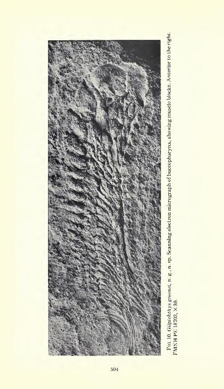
The buccopharyngeal structure itself comprises a series of about
20 segments which decrease in thickness posteriorly (fig. 10). Eachis made up of four block-like masses, presumably formed of mus-
cular tissue. There is no indication of hard structure, either bone,
cartilage, or keratin. The rigidity and tumescence of these units
suggests that they were dense and muscular, and were wrapped in
thick but elastic connective tissue. Their block-like, massive appear-ance is similar to that of the myomeres, and presumably their
shape was preserved because these animals were buried rapidly
under anaerobic conditions.
The dorsal muscular blocks, the largest and most massive in each
segment, cover the dorsal and dorsolateral surface of the buccopha-
ryngeal lumen. Short projections of tissue extend into the lumen
from the dorsolateral surfaces of the upper block, and support a
single, elongate, slender, fang-like tooth that projects medially and
posteriorly (fig. 11). These teeth, large anteriorly, diminish in size
posteriorly, and may be present only on the anterior two-thirds of
the buccopharyngeal chamber. They were presumably formed of a
c>>-
XI
oXI
"t^-'^K'* ^4
^x
4J< 00
O Oh
fa
504

a
Xc
=J3ao
o
aCO
3'2caoCO
-e
H tf
a.
505

506 FIELDIANA: GEOLOGY, VOLUME 33
keratin-like material, and are slightly striated longitudinally. Eachlateral wall segment of the buccopharynx is formed by an anteriorlyslanted muscle block. Near the ventral end of these lateral blocks
a single tooth, shorter than the upper tooth, points medially and
posteriorly. The ventral rim of the buccopharynx is completed byanother muscle block perhaps a quarter or a fifth the size of the dor-
sal block. At the anterior end of the mouth cavity there appears to
be a single mediodorsal block with a single large tooth which pro-
jects posteriorly.
In addition to the muscle blocks which surround the lumen of the
buccopharynx, other muscles involved in the function of this organare preserved. A series of elongated swellings and shallow valleysextends dorsoposteriorly from the dorsal muscle blocks. It is not
clear whether these attached dorsally to muscles of the body wall or
to cranial cartilages. Thinner bands run ventroposteriorly from the
ventral muscle blocks.
A series of semi-circular ridges and valleys, superimposed and
giving the appearance of the layers of an onion bulb in cross-section,
is connected to the back end of the buccopharynx, surrounding a
circular clear area which leads into a dorsoposteriorly directed tube-
like structure. Presumably all of these parts are components of the
digestive system.
The shape of Gilpichthys and its lack of fins suggests that it wasnot an active predator. Food material probably ranged from living
benthos (e.g., annelids, which are common in the fauna) to detrital
material that could be procured by simple wriggling movementsof the body coupled with ingestion through the specialized bucco-
pharynx. Insofar as intestinal content can be recognized in a few
specimens, it appears to include spore exines and crustacean frag-
ments. Presumably the buccopharynx functioned by peristaltic
movement. Thus, food could be moved along the buccopharyngealcanal by means of sequential circumferential contraction of the
muscular blocks surrounding the canal, coupled with vertical elong-
ation and compression of the canal effected by those muscle masses
which attach to the dorsal and ventral blocks. It is possible that the
anteromedial muscle block of the buccopharynx with its tooth,
and perhaps also the large teeth on the first entire set of muscle
blocks, could be everted to assist in picking up food materials. Sucheversion of lingual dentition occurs, for example, in hagfishes(Marinelli and Strenger, 1956) when the mouth is opened. It is not
clear why teeth should be present along most of the buccopharynx.

BARDACK & RICHARDSON: AGNATHOUS FISHES 507
Possibly they aided in restricting escape of ingested living material
or, in conjunction with muscular contractions, helped to comminuteand move food material. Finally, the bulbous structure at the pos-terior end of the buccopharynx probably assisted in forcing food
farther along the digestive tract.
Discussion:—To what group of animals may these new fossils
be assigned? If we list the principal characteristics of chordates
and of Vertebrata in particular (table 1), Pipiscius and Gilpichthys
require assignment to the vertebrates.
Table 1. Chordate and vertebrate characteristics of Pipiscius and Gilpichthys.
Characteristic
1. Notochord
2. Gill slits
3. Dorsal hollow nerve cord
4. Angular metamerism
5. Coelom
6. Fins
7. Post-anal caudal region
8. Otic structure
9. Otic capsule10. Digestive tract
1 1 . Pigmented blood
+ : character observed; — : character not observed.
Pipiscius Gilpichthys+++++
Characteristics 1 to 3 are considered definitive of the phylumChordata. Gilpichthys possesses all three. The seeming absence of
a notochord and dorsal hollow nerve cord in Pipiscius is probablydue to non-preservation rather than to absence in this animal. The
notochord, let alone the dorsal hollow nerve cord, is not to be ex-
pected in fossils, and appears in only three or four of the more than
100 specimens of Gilpichthys. We conclude that both Pipisciusand Gilpichthys are members of the phylum Chordata. Further-
more, we see no features that would suggest assignment to proto-
chordates sensu lato.
Among the vertebrates, the taxonomic affinity of Pipiscius and
Gilpichthys seems to lie among the fishes, and most likely with the
agnathans. This suggestion is based on body shape, lack of jaws,
apparently simple metameric pattern (like that of anaspids) and
presence of a fin (in Pipiscius). The lack of cartilaginous or ossified
structure even in individuals 10 cm. in length distinguishes these

508 FIELDIANA: GEOLOGY, VOLUME 33
fishes from other agnathans. Furthermore, no known agnathanexhibits the unique oral structures of Pipiscius and Gilpichthys.
The discovery of soft-bodied agnathans is not surprising in view
of the unusual, diverse, unskeletonized animals seen in the Essexfauna. This fauna has already yielded the first fossil lamprey (Bar-
dack and Zangerl, 1971). It is most likely that agnathans of the
Paleozoic included not only the typical hardbodied types but also
one or more groups that experimented with limited hard tissues or
developed such structures at a late ontogenetic stage. A survey of
osteostracans, anaspids, and heterostracans shows a variety of
mouth shapes and mouth positions. Yet we really know nothing of
the soft structures that surrounded these mouths and the
pharyngeal regions. The new genera represent additional evidence
of agnathan ventures with mechanisms for ingestion.
We are in a quandary as to whether these forms represent larval
or adult organisms. The absence of a mineralized skeletal structure
and the remnant of a yolk sac in Pipiscius may be evidence for a
larval condition. There are no unequivocally adult characteristics.
The incipient chondrification of the notochord in Gilpichthys,
strikingly shown in a specimen 9.5 cm. long (FMNH, PE 23464),
probably indicates that our specimens are juveniles. On that speci-
men, the buccal structure is not preserved, though the two eyesare present and are no larger than the eyes of the smallest speci-
mens (0.4 mm. in diameter, 0.6 per cent of the body length). Such
remarkably small eyes don't seem in keeping with a juvenile animal,
and would be small even for an adult. Perhaps, though no evidence
can be adduced, Gilpichthys was functionally blind and a denizen of
the infauna.
To what degree are Pipiscius and Gilpichthys related? Overall
similarities of these organisms are shown in the descriptions and in
Table 1. The principal differences lie in the short, circular mouth of
Pipiscius in contrast to the elongate, tubular mouth of Gilpichthys,and the presence of a fin in the former and not in the latter. While
belonging to the same class, they are not close kin.
Both of these animals are known only from the deltaic Francis
Creek Shale. Pipiscius occurs only in Pit Eleven in Will andKankakee counties, Illinois. Gilpichthys, the more common form,
has also been collected about 150 miles (about 250 km.) to the west
in Fulton County, in sediments of the same delta.

BARDACK & RICHARDSON: AGNATHOUS FISHES 509
ACKNOWLEDGEMENTS
This research was supported in part by National Science Founda-
tion grants GB-8266 to Eugene S. Richardson, Jr., and Ralph G.
Johnson, principal investigators, and BMS-72-02149 to Alan Solem,
principal investigator, both through the Field Museum of Natural
History. We wish to thank several fossil collectors who donated
specimens to the museum or loaned specimens for study, including:
Mr. and Mrs. Lincoln Douglass, Mr. and Mrs. James E. Konecny,Mr. and Mrs. Ted Piecko, Mr. and Mrs. Francis A. Wolff, Mr. and
Mrs. Anton Sobolik, Arthur Kimball, Roger Klocek, Frank Greene,
Lawrence Osterberger, Mrs. Phillip Rockwell, and the late JerryHerdina and Levi Sherman. Dr. Tibor Perenyi painstakingly and
accurately constructed a model of the mouth of Pipiscius that wasinvaluable in our study. The scanning electron micrographs were
taken and enhanced with uncommon expertise by the late Fred
Huysmans; the other pictures were printed by Rudy Chavez.
Finally, we thank Rainer Zangerl for his interest, encouragementand inspiration in the study of the unusual fishes of the Penn-
sylvanian.
REFERENCES
Bardack. D. and R. Zangerl
1971. Lampreys in the fossil record, pp. 67-84. In Hardisty, M. W. and I. C. Potter,
The biology of lampreys, 1.
Devillers. Ch.
1954. Structure et evolution de la colonne vertebrale, pp. 605-672. In Grasse, P. P.,
Traite de zoologie, torn. XII.
Johnson. R. G. and E. S. Richardson, Jr.
1966. A remarkable Pennsylvanian fauna from the Mazon Creek area, Illinois.
Jour. Geol., 74, pp. 626-631
1970. Fauna of the Francis Creek Shale in the Wilmington area, pp. 53-59. In
Depositional environments in parts of the Carbondale Formation, western and
northern Illinois. 111. State Geol. Surv., Guidebook Ser., no. 8.
Marinei.li, W. and A. Strenger
1956. Myxine glutinosa (L.). In Vergl. Anat. Morphol. Wirbeltiere, Lief. II.
Plavis. G. W.
1971. Embryology, pp. 361-400. In Hardisty, M. W. and I. C. Potter, The biology
of lampreys, vol. 1.
Richardson, E. S., Jr.
1966. The Tully Monster. Field Mus. Nat. Hist. Bull., 37, no. 7, pp. 4-6.

510 FIELDIANA: GEOLOGY, VOLUME 33
Richardson, E. S., Jr. and R. G. Johnson
1971. The Mazon Creek faunas. Proc. N. Amer. Paleontol. Conv. Chicago, 1969,
Part I, pp. 1,222-1,235.