Embed Size (px)
Citation preview


227
Sociedad Española de Medicina del Deporte
Junta de GobiernoPresidente: Pedro Manonelles MarquetaVicepresidente: Miguel E. Del Valle SotoSecretario General: Luis Franco BonafonteTesorero: Javier Pérez AnsónVocales: Carlos de Teresa GalvánJosé Fernando Jiménez Díaz Juan N. García-Nieto PortabellaTeresa Gaztañaga Aurrekoetxea José Naranjo Orellana
EditaSociedad Española de Medicina del DeporteIturrama, 43 bis. 31007 Pamplona. (España)Tel. 948 267 706 - Fax: 948 171 [email protected]
Correspondencia:Ap. de correos 120731080 Pamplona (España)
PublicidadESMON PUBLICIDADTel. 93 2159034
Publicación bimestralUn volumen por año
Depósito LegalPamplona. NA 123. 1984
ISSN0212-8799
Soporte válidoRef. SVR 389
Indexada en: EMBASE/Excerpta Medica, Índice Médico Español, Sport Information Resource Centre (SIRC), Índice Bibliográfi co Español de Ciencias de la Salud (IBECS), y Índice SJR (SCImago Journal Rank).
La Revista Archivos de Medicina del Deporte ha obtenido el Sello de Calidad en la V Convocatoria de evaluación de la calidad editorial y científi ca de las revistas científi -cas españolas, de la Fundación Española para la Ciencia y la Tecnología (FECYT).
La dirección de la revista no acepta responsabilida-des derivadas de las opiniones o juicios de valor de los trabajos publicados, la cual recaerá exclusivamen-te sobre sus autores.Esta publicación no puede ser reproducida total o parcialmente por ningún medio sin la autorización por escrito de los autores.Cualquier forma de reproducción, distribución, comunicación pública o transformación de esta obra sólo puede ser realizada con la autorización de sus titulares, salvo excepción prevista por la ley. Diríjase a CEDRO (Centro Español de Derechos Reprográfi cos, www.cedro.org) si necesita fotocopiar o escanear algún fragmento de esta obra.
Director
Pedro Manonelles Marqueta
Editor
Miguel E. Del Valle Soto
Administración
Mª Ángeles Artázcoz Bárcena
Comité EditorialNorbert Bachl. Centre for Sports Science and University Sports of the University of Vienna. Austria. Ramón Balius Matas. Consell Catalá de l'Esport. Generalitat de Catalunya. España. Araceli Boraita. Servicio de Car-diología. Centro de Medicina del Deporte. Consejo Superior de deportes. España. Josep Brugada Terradellas. Hospital Clinic. Universidad de Barcelona. España. Nicolas Christodoulou. President of the UEMS MJC on Sports Medicine. Chipre. Jesús Dapena. Indiana University. Estados Unidos. Franchek Drobnic Martínez. Servicios Médicos FC Barcelona. CAR Sant Cugat del Vallés. España. Tomás Fernández Jaén. Servicio Medi-cina y Traumatología del Deporte. Clínica Cemtro. España. Walter Frontera. Universidad de Vanderbilt. Past President FIMS. Estados Unidos. Pedro Guillén García. Servicio Traumatología del Deporte. Clínica Cemtro. España. Dusan Hamar. Research Institute of Sports. Eslovaquia. José A. Hernández Hermoso. Servicio COT. Hospital Universitario Germans Trias i Pujol. España. Pilar Hernández Sánchez. Universidad Católica San Antonio. Murcia. España. Markku Jarvinen. Institute of Medical Technology and Medical School. University of Tampere. Finlandia. Peter Jenoure. ARS Ortopedica, ARS Medica Clinic, Gravesano. Suiza. José A. López Calbet. Universidad de Las Palmas de Gran Canaria. España. Javier López Román. Universidad Católica San Antonio. Murcia. España. Alejandro Lucía Mulas. Universidad Europea de Madrid. España. Emilio Luengo Fernández. Servicio de Cardiología. Hospital General de la Defensa. España. Nicola Maffully. Universidad de Salerno. Salerno (Italia). Pablo Jorge Marcos Pardo. Universidad Católica San Antonio. Murcia. España. Alejandro Martínez Rodríguez. Universidad Católica San Antonio. Murcia. España. Estrella Núñez Delicado. Universidad Católica San Antonio. Murcia. España. Sakari Orava. Hospital Universitario. Universidad de Turku. Finlandia. Eduardo Ortega Rincón. Universidad de Extremadura. España. Nieves Palacios Gil-Antuñano. Centro de Medicina del Deporte. Consejo Superior de Deportes. España. Antonio Pelliccia. Institute of Sport Medicine and Science. Italia. José Peña Amaro. Facultad de Medicina y Enfermería. Universidad de Córdoba. España. Fabio Pigozzi. University of Rome Foro Italico, President FIMS. Italia. Per Renström. Stockholm Center for Sports Trauma Research, Karolinska Institutet. Suecia. Juan Ribas Serna. Universidad de Sevilla. España. Jordi Segura Noguera. Laboratorio Antidopaje IMIM. Presidente Asociación Mundial de Científi cos Antidopajes (WAADS). España. Giulio Sergio Roi. Education & Research Department Isokinetic Medical Group. Italia. Luis Serratosa Fernández. Servicios Médicos Sanitas Real Madrid CF. Madrid. España. Nicolás Terrados Cepeda. Unidad Regional de Medicina Deportiva del Principado de Asturias. Universidad de Oviedo. España. José Luis Terreros Blanco. Subdirector Adjunto del Gabinete del Consejo Superior de Deportes. España. Juan Ramón Valentí Nin. Universidad de Navarra. España. José Antonio Vega Álvarez. Facultad de Medicina. Universidad de Oviedo. España. José Antonio Villegas García. Académico de número de la Real Academia de Medicina de Murcia. España. Mario Zorzoli. International Cycling Union. Suiza.
Sociedad Española de Medicina del Deporte


Volumen 33(3) - Núm 174. July - August 2016 / Julio - Agosto 2016
Summary / Sumario
Editorial
Anterior Knee Pain: A Paradigm of Aversion Towards a Pathology Dolor anterior de rodilla: un ejemplo de aversión hacia una patología Vicente Sanchis-Alfonso .........................................................................................................................................................................................231
Original articles / Originales
Acute glycemic outcomes along the aerobic training in deep water in patients with type 2 diabetes Respuestas de la glucemia aguda a lo largo del entrenamiento aeróbico en aguas profundas en pacientes con diabetes tipo 2 Rodrigo Sudatti Delevatti, Nathalie de Souza Netto, Ana Carolina Kanitz, Cristine Lima Alberton, Carolina Dertzbocher Feil Pinho, Elisa Corrêa Marson, Luciana Peruchena Bregagnol, Salime Chedid Lisboa, Luiz Fernando Martins Kruel .................................................. 233
Effect of strength training on body composition, strength and aerobic capacity of Brazilians adolescents’ handball players related with peak growth rate Efecto del entrenamiento de la fuerza sobre la composición corporal, fuerza y capacidad aeróbica de los jugadores adolescentes de balonmano brasileños relacionados con el pico de crecimiento Luis Paulo Gomes Mascarenhas, William Cordeiro de Sousa, Josnei Franz, Valderi Lima de Abreu, Juliana Decimo, Marcio Cascante-Rusenhack, José Moncada-Jiménez ........................................................................................................................................ 239
Efficacy of motor activity in the quality of life in fibromyalgia: meta-analysis of clinical trials Eficacia del ejercicio físico sobre la calidad de vida en fibromialgia: meta-análisis de ensayos clínicos Jaiberth Cardona-Arias, Carmen Mantilla-Gutiérrez, Luis Felipe Higuita-Gutiérrez ............................................................................................244
Sedentary lifestyle level in nine cities of Colombia: cluster analysis El nivel de sedentarismo en nueve ciudades colombianas: análisis de clúster José A. Vidarte Claros, Consuelo Vélez Álvarez, José H. Parra Sánchez ............................................................................................................ 253
Reviews / Revisiones
Caffeine and its ergogenic effect in sport (second part) La cafeína y su efecto ergogénico en el deporte (segunda parte) Antonio García Moreno .......................................................................................................................................................................................... 259
The influence of fatigue in hamstrings: quadriceps ratio. A systematic review Efecto de la fatiga en el ratio isquiotibiales: cuádriceps. Revisión sistemática Juan P. Martín Martínez, Jorge Pérez Gómez, Jorge Carlos Vivas....................................................................................................................... 267
Books / Libros ............................................................................................................................................................................................................276
XVI Congreso Nacional de la Sociedad Española de Medicina del Deporte......................................................................................279
Agenda / Agenda ..................................................................................................................................................................................................... 286
Guidelines for authors / Normas de publicación ........................................................................................................................................ 292

Analizador Instantáneo de Lactato
c/ Lto. Gabriel Miro, 54, ptas. 7 y 946008 Valencia Tel: 963857395Móvil: 608848455 Fax: 963840104info@bermellelectromedicina.comwww.bermellelectromedicina.com
Sólo 0,3 μl de sangre
Determinación en 15 segundos
Más pequeño que su antecesor
Calibración automática
Memoria para 330 determinaciones
Conexión a PC
Rango de lectura: 0,5-25,0 mmol/litro
Conservación de tiras reactivas a temperatura ambiente y
Caducidad superior a un año
del
ciclismo
y fisiologíamedicina
Más informaciónFEMEDEwww.femede.es
FICHA TÉCNICAAutores: VV.AADirectores: José Fernando Jiménez Díaz, Nicolás Terrados Cepeda, J. Gerardo Villa VicenteCoordinador: Pedro Manonelles MarquetaDiseño: 17x24 cm - B/N2 VolúmenesVolumen 1: 780 páginasVolumen 2: en preparaciónEdita: FEMEDE / Nexus Médica EditoresAño 2009PVP (IVA incluido): 180 € + 15 € de gastos de envío.
VI Medicina del Deporte
Colegio de Médicos de BizkaiaBilbao, 27 y 28 de noviembre de 2015
Jornadas Nacionales de
VI Jornadas Nacionales de Medicina del Deporte

Anterior Knee Pain: A Paradigm of Aversion Towards a Pathology
231Arch Med Deporte 2016;33(4):231-232
Editorial
Anterior Knee Pain: A Paradigm of Aversion Towards a Pathology
Dolor anterior de rodilla: un ejemplo de aversión hacia una patología
Vicente Sanchis-AlfonsoMember of the International Patellofemoral Study Group. Member of the Editorial Board of the Patellofemoral Online Education, Patellofemoral Foundation, Farmington, CT, USA. Consultant Orthopaedic Surgeon, Hospital 9 de Octubre and Hospital Arnau de Vilanova, Valencia, Spain.
Correspondence: Vicente Sanchis-Alfonso E-mail: [email protected]
Anterior knee pain (AKP) is very common in the general population, causing chronic disability, lost time from work, limitations in partici-pating in sports, and a diminished quality of life. AKP represents one third or more of all complaints in a sports medicine clinic. In a study on participants from the U.S. Naval Academy, Boling et al.1 found an AKP prevalence of 15% in females and 12% in males. They also observed that the annual incidence of AKP was 33 of 1000 people in female patients and 15 of 1000 people in male patients. Nevertheless, in spite of its high incidence and prevalence, the etiology of AKP is obscure, which complicates treatment and hinders recovery. In a multicentre observational analysis study, Collins et al.2 showed that 40% of patients had an unfavourable recovery at 12 months after the initial diagnosis. Moreover, AKP is recurrent or chronic in 70% to 90% of individuals with the condition3. The etiology of AKP is multifactorial, with not only local (e.g., knee) factors but also proximal (e.g., hip and trunk) and distal (e.g., foot and ankle) ones. In fact, in many patients the primary cause of AKP does not lie within the patellofemoral joint, and there are several sub-groups within the AKP population. Therefore, the best treatment must be tailored to individual patients. Among all the subsets of patients with AKP, the most challenging type of AKP, from a therapeutic point of view, is neuropathic. Once conventional treatments have failed in these patients, alternatives such as radiofrequency neurotomy and the repetitive transcranial magnetic stimulation can be considered4. We are currently exploring these techniques in our work group in this subset of patients.
“Chondromalacia patellae” (soft cartilage on the knee cap) was previously used as a catch-all term for any pain in the anterior aspect of the knee, but the term has been replaced by “patellofemoral pain syndrome” in reference to patients with AKP. However, neither term ex-
presses a diagnosis but rather presents an admission of ignorance. Not all the patients with AKP have chondromalacia patellae, and many patients with chondromalacia patellae do not have from AKP. For example, van der Heijden et al.5 have not found any differences in the patellofemoral cartilage composition between AKP patients and healthy controls. Fur-ther, even patients with severe patellofemoral chondropathy may not have knee pain. Consequently, the International Patellofemoral Study Group advises against using these terms as a diagnosis and suggests that “anterior knee pain” might be better because it is descriptive, without implying anything more.
Yet, AKP is a pathology in which numerous clichés and false beliefs coexist. One of the clichés is that a patient with AKP has a peculiar psychological profile that might explain the pain. This belief is rein-forced by many patients having very disabling pain but insignificant radiological findings and unremarkable physical signs. The psychological explanation could not be further from the truth though. Domenech et al.6,7 have demonstrated that psychological factors modulate the pain, but they do not cause it. Rathleff et al.8 have shown that young female adults with long-standing AKP demonstrated impaired conditioned pain modulation. This is, AKP might have important central components that need to be studied in order to understand its extent and therapeutic implications. Another misconception is that AKP is a self-limiting and benign condition, which is why some physicians recommend “expec-tation” measures. That approach is a great mistake. Collins et al.2 have demonstrated that the success of the therapy depends on how recently the pain began. Rathleft et al.9 reported that AKP is not a self-limiting knee condition. Further, AKP in an adolescent has a high potential for becoming chronic. Conchie et al.10 brought into question the traditional belief that AKP in adolescence is a benign pathology, by showing that
“That those who know her, know her less, the nearer her they get”Emily Elizabeth Dickinson

Vicente Sanchis-Alfonso
232 Arch Med Deporte 2016;33(4):231-232
it is associated with patellofemoral osteoarthritis in adulthood. That is, AKP and patellofemoral osteoarthritis may form a continuum of disease. Therefore, we must take this pathology seriously.
In addition, to make the black legend of this pathology even bigger, we must point out that it is a source of iatrogenia11. We must be very cautious when recommending surgical treatment in AKP patients. This caution is particularly directed to those “well-meaning trigger-happy orthopaedic surgeons” educated in a purely structural/anatomical/biomechanical view of this pathology. These surgeons operate on what magnetic resonance imaging (MRI) shows. This approach is a blunder—the patient who began with just mild, intermittent symptoms beco-mes even worse. The same condemnation applies to inappropriately aggressive physical therapy. We must beware of structural anomalies. In fact, only a poor correlation exists between structural anomalies (chondropathy, patellar tilt and patellar subluxation) and AKP. We must avoid inappropriate or incorrect malalignment-oriented patellofemoral surgery. In agreement with Dye12, I believe that the loss of both osseous and soft tissue homeostasis is much more important in the genesis of AKP than structural alterations (Paradigm of Tissue/Joint Homeostasis).
Unluckily, the criteria for proper treatment of the AKP patient have largely been based on individual experience. The malalignment theory strongly supported by many orthopaedics surgeons, with an almost religious fervor, has enormously damaged many AKP patients and has given this pathology a bad reputation. We need to refine the indications for AKP management and surgery, and for this refinement to happen, more studies with a high level of evidence are needed. We should not be distracted by structural findings manifested on an MRI. In this way van der Heijden et al.13 have shown that structural abnormalities of the patellofemoral joint have on MRI are not associated with AKP.
We should instead treat symptoms and the patient as a whole. All pieces of the puzzle must fit. If the MRI says “small tear of the medial meniscus”, but the patient's pain is in the patellofemoral joint, then the structural finding on the MRI is likely not the cause of the pain, and it not should be used to justify an arthroscopy.
In most cases an AKP patient should be treated non-surgically. Phy-sical therapy must include the entire lower limb, with particular attention to the hip musculature4. Very often a patient's knee has suffered a loading event that has diminished the functional envelope of function12 in such a way that daily activities are beyond it. I must admit that in some cases it is hard to restrict activity below the new envelope of function. It is like asking a mechanic to complete an overhaul on the transmission of a car while driving it around town. But the worst part of unrestricted activity is that it obscures the underlying problem, that is the decrease of the functional envelope of function, and can lead to inappropriate surgery; that is, surgery that makes things worse.
The preceding text reveals why many orthopedic surgeons have an aversion towards treating the AKP patient. These patients are quickly sent (by other colleagues) to the orthopedic surgeon who excels at
treating this kind of pathology, although he or she is distant from the patient who experiences it. Surgeons appear to be put off by these patients even before studying their cases. However, in my eyes, AKP is one of the most intriguing pathologies from a clinical point of view because it obliges us to “think out of the box”, to look deeper into the anatomy, biomechanics, biology, anatomic pathology, physiopathology and psychology.
Many years ago a good friend of mine from the United States told me that to stand out in something I had to focus on a topic that was not well-known and that many did not like. Twenty years ago, AKP fulfilled both and continues to do so. It is currently not a well-known clinical entity and, moreover, orthopaedic surgeons do not usually like to treat it. Paraphrasing the great American poet Robert Frost14 in his poem “The Road Not Taken”, I took the least travel road 20 years ago; that is, I focused on the patella. As in his poem, it made all the difference. Without a doubt, I do not regret having chosen this road.
References 1. Boling M, Padua D, Marshall S, et al. Gender differences in the incidence and prevalence
of patellofemoral pain syndrome. Scand J Med Sci Sports. 2010;20:723-30.
2. Collins NJ, Bierma-Zeinstra SM, Crossley KM, van Linschoten RL, Vicenzino B, van Middelkoop M. Prognostic factors for patellofemoral pain: a multicentre observational analysis. Br J Sports Med. 2013;47(4):227-33.
3. Powers CM, Bolgla LA, Callaghan MJ, Collins N, Sheehan FT. Patellofemoral pain: proximal, distal, and local factors, 2nd International Research Retreat. J Orthop Sports Phys Ther. 2012;42(6):A1-54.
4. Sanchis-Alfonso V, McConnell J, Monllau JC, Fulkerson JP. Diagnosis and treatment of anterior knee pain. JISAKOS, 2016 doi.10.1136/jisakos-2015-000033.
5. van der Heijden RA, Oei EHG, Bron EE, et al. No difference on quantitative magnetic resonance imaging in patellofemoral cartilage composition between patients with patellofemoral pain and healthy controls. Am J Sports Med. 2015;44(5):1172-8.
6. Doménech J, Sanchis-Alfonso V, Espejo B. Changes in catastrophizing and kinesio-phobia are predictive of changes in disability and pain after treatment in patients with anterior knee pain. Knee Surg Sports Traumatol Arthrosc. 2014; 22(10):2295-300.
7. Domenech J, Sanchis-Alfonso V, Lopez L, Espejo B. Influence of kinesiophobia and catastrophizing on pain and disability in anterior knee pain patients. Knee Surg Sports Traumatol Arthrosc. 2013; 21(7):1562-8.
8. Rathleff MS, Petersen KK, Arendt-Nielsen L, Thorborg K, Graven-Nielsen T. Impaired Conditioned Pain Modulation in Young Female Adults with Long-Standing Patello-femoral Pain: A Single Blinded Cross-Sectional Study. Pain Med. 2016;17(5):980-8.
9. Rathleff MS, Rathleff CR, Olesen JL, Rasmussen S, Roos EM. Is Knee Pain During Ado-lescence a Self-limiting Condition? Prognosis of Patellofemoral Pain and Other Types of Knee Pain. Am J Sports Med. 2016;44(5):1165-71.
10. Conchie H, Clark D, Metcalfe A, Eldridge J, Whitehouse M. Adolescent knee pain and patellar dislocations are associated with patellofemoral osteoarthritis in adulthood: A case control study. Knee. 2016. doi: 10.1016/j.knee.2016.04.009.
11. Sanchis-Alfonso V, Merchant AC. Iatrogenic Medial Patellar Instability: An Avoidable Injury. Arthroscopy. 2015;31(8):1628-32.
12. Dye SF. The pathophysiology of patellofemoral pain: a tissue homeostasis perspective. Clin Orthop Relat Res. 2005;436:100-110.
13. van der Heijden RA, de Kanter JL, Bierma-Zeinstra SM, Verhaar JA, van Veldhoven PL, Krestin GP, Oei EH, van Middelkoop M. Structural Abnormalities on Magnetic Reso-nance Imaging in Patients With Patellofemoral Pain: A Cross-sectional Case-Control Study. Am J Sports Med. 2016. pii: 0363546516646107.
14. Frost R. The road not taken. http://www.bartleby.com/119/1.html

Acute glycemic outcomes along the aerobic training in deep water in patients with type 2 diabetes
233Arch Med Deporte 2016;33(4):233-238
Artículo original
Summary
Aims: The present study aimed to analyze the acute glucose responses in the first and last sessions of four mesocycles along an aquatic aerobic training periodization. Methods: Fourteen patients (6 men and 8 women; 54.3 ± 9.0 years; body mass index of 34.5 ± 3.9 kg/m2) with type 2 diabetes underwent a 12-week training program involving deep-water running. This exercise training was performed by an interval training method, with a frequency of 3 times a week, session duration of 35 minutes and intensity progressing from 85 to 90% to 95 to 100% of the anaerobic threshold heart rate (ATHR) along the periodization. Capillary glucose was assessed before and immediately after the first and last session of each mesocycle. A generalized estimated equation (time x session x mesocycle)was used to assess reductions in glucose levels in different sessions (first and last) along four mesocycles (α = 0.05). Results: All sessions resulted in a reduction in glucose levels (time effect: p <0.001), without differences between the first and last session of each mesocycle (session effect: p = 0.738). With regard to the mesocycles (mesocycle effect: p = 0.003), significant differences were found between mesocycles 2 and 3. In time*mesocycle interaction (p = 0.002), in most compa-risons, post-session values were lowest that pre-session values, regardless of mesocycle, except for the post-session value of mesocycle 3, which was similar to the pre-values of mesocycles 2 and 4. Conclusion: Aerobic training in deep water with crescent linear periodization over 12 weeks is able to reduce glucose levels in patients with type 2 diabetes.
Key words: Aquatic environment.
Exercise. Diabetes Mellitus.
Glycemia.
Resumen
Objetivo: Analizar las respuestas de glucemia aguda en las primeras y últimas sesiones de cuatro mesociclos a lo largo de una periodización de entrenamiento aeróbico acuático. Métodos: Catorce pacientes (6 hombres y 8 mujeres; 54,3±9,0 años; índice de masa corporal de 34,5±3,9 kg/m2) con diabe-tes tipo 2 fueron sometidos a un programa de entrenamiento de 12 semanas de carrera en aguas profundas. Se realizó un entrenamiento aeróbico de intervalos, realizado 3 veces por semana, con sesiones de 35 minutos y la intensidad progresando a lo largo de la periodización desde 85% - 90% a 95% - 100% de la frecuencia cardiaca del umbral anaeróbico (FCUA). La glucosa capilar fue evaluada antes e inmediatamente después de la primera y la última sesión de cada mesociclo. Se utilizó una ecuación generalizada estimada (tiempo x sesión x mesociclo) para evaluar las reducciones en los niveles de glucosa en las diferentes sesiones (primera y última) a lo largo de cuatro mesociclos (α = 0.05). Resultados: todas las sesiones resultaran en una reducción en los niveles de glucosa (efecto tiempo: p<0,001), sin diferencias entre la primera y la última sesión de cada mesociclo (efecto de sesión: p = 0,738). Con respecto a los mesociclos (efecto mesociclo: p=0,003) se encontraron diferencias significativas entre los mesociclos 2 y 3. En la interacción tiempo*mesociclo (p=0,002), en la mayoría de las comparaciones, los valores post-sesión fueron menores de los valores pre-sesión, indepen-dientemente de mesociclo, excepto para el valor después de la sesión del mesociclo 3, que fue similar a los valores antes de la sesión de los mesociclos 2 y 4. Conclusión: Doce semanas de entrenamiento aeróbico en aguas profundas con la periodización linear y creciente es capaz de reducir los niveles de glucosa en pacientes con diabetes tipo 2.
Palabras clave: Ambiente acuático.
Ejercicio. Diabetes Mellitus.
Glucemia.
Received: 15.07.2015 Accepted: 30.09.2015
Acute glycemic outcomes along the aerobic training in deep water in patients with type 2 diabetes
Rodrigo Sudatti Delevatti1,2, Nathalie de Souza Netto1, Ana Carolina Kanitz1,3, Cristine Lima Alberton4, Carolina Dertzbocher Feil Pinho1, Elisa Corrêa Marson1, Luciana Peruchena Bregagnol1, Salime Chedid Lisboa1, Luiz Fernando Martins Kruel1
1Exercise Research laboratory, Department of Physical Education, Universidade Federal do Rio Grande do Sul, Brazil. 2Faculdade Sogipa de Educação Física, Brazil. 3Department of Physical Education, Universidade Federal de Uberlândia, Brazil. 4Department of Physical Education, Universidade Federal de Pelotas, Rio Grande do Sul, Brazil.
Respuestas de la glucemia aguda a lo largo del entrenamiento aeróbico en aguas profundas en pacientes con diabetes tipo 2
Correspondence: Rodrigo Sudatti Delevatti E-mail: [email protected]

Rodrigo Sudatti Delevatti, et al.
234 Arch Med Deporte 2016;33(4):233-238
Introduction
Type 2 diabetes mellitus (T2DM) is a worldwide public health pro-blem predominantly resulting from obesity and a sedentary lifestyle. Interventions ensuring lifestyle changes have been effective both in the prevention and control of the disease, with exercise being one of the most effective nonpharmacological treatments for T2DM1,2, through its beneficial effects on outcomes such as blood pressure, lipid profile and glucose levels3.
According to the American Diabetes Association (ADA), patients with T2DM should do 150 minutes or more of aerobic exercise at moderate intensity per week, and when without complications must combine this with two or three sessions per week of resistance training4. These guide-lines demonstrate how important aerobic training is to this population, and it is generally recommended to all patients. However, despite a lot of evidence indicating the beneficial chronic effects, especially in glucose control in this population5-7, less is known about the acute glycemic effects of aerobic training during progressive training. Studies investi-gating this issue focused predominantly on the comparison between the effects of different exercise sessions in a specific training status8-10. To the best of the authors’ knowledge, no study has investigated these effects at different times during a periodization, in which the intensity and/or duration increases along the training while the patients improve their physical fitness and metabolic status.
With regard to the periodization of aerobic training, it is worth no-ting that patients with a type 2 diabetes clinical profile, usually obese or overweight, can complicate increases in exercise dosage, especially in intensity, because in greater intensity for glucose control, such as those near of anaerobic threshold11, patients are more susceptible to lower limb injury. An alternative to the necessary training progression is the training conducted in water, such as water aerobics, because the buoyancy provides attenuated impact forces, especially for the lower limbs12. Moreover, deep-water running is another interesting modality for the progression of exercise training because the practitioners perform aerobic exercise at high loads with reduced risk of injury, since a float vest is used to keep the body in an upright position, preventing contact between the feet and the bottom of the pool13.
Because of its characteristics, deep-water running is favorable to progress in training without increasing the impact on joints, thereby enabling patients to exercise at high intensity or for long duration, op-timizing glucose control. In addition, this modality has demonstrated a similar increase in strength to combined training in water14 and has glycemic metabolism benefits in glucose-intolerant women15. The fact that this modality allows progression in training intensity, including for patients with difficulties in supporting their own body mass16, indicates the need for knowledge about the acute glycemic effect in different stages of a linearly increasing periodization, because although it seems a great alternative, literature about “exercise and type 2 diabetes” is scar-ce on studies in water. Thus, the present study aimed to analyze acute glycemic responses in the first and last sessions in four mesocycles of a deep-water running program. Our hypothesis is that in mesocycles of three weeks, the acute glycemic effect not is attenuated, being similar in the first and last session of each mesocycle.
Materials and methods
Subjects
The sample consisted of 14 patients with T2DM (6 men and 8 wo-men) who had not undertaken any physical exercise in the previous three months and were receiving their usual medical treatment. Patients with the following conditions were excluded from the sample: uncontrolled hypertension, autonomic neuropathy, severe peripheral neuropathy, proliferative diabetic retinopathy, severe nonproliferative diabetic reti-nopathy, decompensated heart failure, limb amputations, chronic renal failure (MDRD-GFR < 30 ml/min)17 or any muscle or joint impairments that prevented individuals from engaging in physical exercise. The pre-sence of these conditions was confirmed by medical history as well as clinical and laboratory examinations. All patients had undergone effort electrocardiograms in the six months preceding the study.
Research design
Patients were identified from the records of the Endocrine Division of a tertiary hospital and were also recruited through advertisements in local newspapers between June and July 2012. All participants were fully informed of the procedures involved in the study, and provided written consent prior to participation. The study was approved by the Research Ethics Committees of the Universidade Federal do Rio Gran-de do Sul (protocol number 108.997) and of the Hospital de Clínicas de Porto Alegre (protocol number 54475). The research design, with intervention and evaluations performed are illustrated in the Figure 1.
Anthropometric measurements
Prior to the intervention, patients underwent anthropometric mea-surements. Body mass and height were assessed using a digital scale and a stadiometer (FILIZOLA; Sao Paulo, Brazil). These values were used to calculate patient body mass index (BMI) using the following formula: mass (kg)/height² (m). Waist circumference was measured at the mid-point between the iliac crest and the last rib. Additionally, skinfolds were measured at the following eight sites: tricipital, subscapular, suprailiac, abdominal, chest, midaxillary, thigh and leg. The equations proposed by Petroski18 were used to estimate the body density of men and women, while body fat percentages were estimated using the Siri formula19.
Blood analysis
Blood samples (4ml) were obtained from an antecubital vein after fasting for 12 to 14 h. The samples were collected in tubes with EDTA and kept frozen at -80 ºC as total blood (without centrifugation). After blood data collection, the levels of HbA1c were determined through high-performance liquid chromatography (HPLC) to characterize the glycemic control of the patients.
Capillary glycemia
Capillary glycemia was assessed before and immediately after the first and last sessions of each training mesocycle using a clinical

Acute glycemic outcomes along the aerobic training in deep water in patients with type 2 diabetes
235Arch Med Deporte 2016;33(4):233-238
glucometer (Accu-Check Performa, Roche, São Paulo, Brazil), which assesses glycemic levels in approximately 5 seconds, and a lancet device (Accu-ChekMulticlix, Sao Paulo, Brazil).
Intervention
Patients underwent 12-week training program involving deep-water running with a life vest. The interval-training program consisted of four mesocycles of three weeks each. Training was conducted three times per week (Monday, Wednesday and Friday), and each 45-minute session was divided into a warm-up period (5 min), followed by the main training program (35 min) and a cool-down section (5 min). The intensity of the physical exercise prescribed was adjusted according to each subject’s heart rate deflection point (HRDP), which was determined by a progres-sive maximal test conducted in the water environment. This method was chosen due to its ease of application and association with the second ventilatory threshold, a precise indicator of the relative stress caused by exercise20. Participants were asked to wear HR monitors (RSX 300, Polar) during the exercise sessions to control training intensity. Each individual was asked to read and report their heart rates to one of the three instruc-
tors who supervised the exercise sessions. Each instructor then used a table containing subjects’ training heart rate ranges to provide feedback on the recommended exercise intensity for each patient. The 12-week training program prescribed to each participant is described in Table 1.
Statistical analysis
Descriptive data of the subject characteristics are presented as mean and standard deviation for continuous variables and as n for ca-tegorical variables. Glycemic levels are presented as mean and standard error. A generalized estimation equation (GEE) was used to assess alte-rations in glucose levels (pre versus post) in different sessions (first and last) in each mesocycle of training, taking into consideration the three factors involved in the analysis (time, session and mesocycle). Given that exogenous insulin use could potentiate an exercise glucose-lowering effect, we performed the analyses for all patients, and excluding those patients using insulin. Multiple comparisons were performed with Bonferroni correction. The level of significance was set at α= 0.05. All analyses were performed using the Statistical Package for the Social Sciences (SPSS) software, version 19.0.
Table 1. 12-week aerobic training program.
Mesocycle Week Training sessions Duration (main part)
1 1 - 3 7x (3 min 85-90% ATHR with 2min <85% ATHR) 35 min
2 4 - 6 7x (4 min 85-90% ATHR with 1min <85% ATHR) 35 min
3 7 - 9 7x (4 min 90-95% ATHR with 1min <85% ATHR) 35 min
4 10 - 12 7x (4 min 95-100% ATHR with 1min <85% ATHR) 35 min
Note: ATHR: Anaerobic threshold heart rate.
Figure 1. Study design (evaluations and intervention).

Rodrigo Sudatti Delevatti, et al.
236 Arch Med Deporte 2016;33(4):233-238
Results
Sample data regarding disease duration, age, anthropometric measurements and medication use are shown in Table 2.
Table 2. Subject characteristics.
Age (years) 54.3 ± 2.4
Duration of DM2 (years) 5.4 ± 1.0
HbA1c (%) 7.9 ± 0.7
Body mass (kg) 93.1 ± 3.6
Body mass index (kg.m-2) 34.5 ± 1.0
Waist circumference (cm) 111.7 ± 2.9
WHR 0.68 ± 0.02
Fat mass (%) 37.5 ± 1.1
Sex (male/female) 6/8
Medical treatment
Metformin (n) 12
Sulphonylurea (n) 6
DPP-4-inhibitors (n) 1
Pioglitazone (n) 1
Diuretics (n) 4
Beta blockers (n) 4
ARAs II (n) 4
Acetyl-acetylsalicylic (n) 5
Statins (n) 7
Insulin (n) 3
DPP-4: dipeptidyl peptidase-4; ARAs: Angiotensin receptors antagonists; WHR: waist/height ratio; Values of age, duration of DM2 and anthropometric measures are expressed as the mean ± SE; Values of sex and medication are expressed by n.
All training sessions determined a reduction in glucose levels (time effect: p <0.001). In all mesocycles, the first session values did not differ from the values found in the last session (session 1 = session 9, session 10 = session 18, session 19 = session 27, session 28 = session 36; session effect: p = 0.738). Between the mesocycles (mesocycle effect: p = 0.003), significant differences were found only between the pre-session values of mesocycles 2 and 3 (Table 3). In time*mesocycle interaction (p = 0.002), significant differences were found between pre-session and post-session values in most comparisons, except for the post-session value of mesocycle 3, which was similar to the pre-values of mesocycles 2 and 4.
Given that exogenous insulin use could potentiate an exercise glucose-lowering effect, we performed an analysis excluding those patients using insulin. The results, however, were very similar: Exercise sessions determined reductions in glucose levels (time effect: p < 0.001), without differences between the first and last session of each meso-cycle (session effect: p = 0.889). Between the mesocycles (mesocycle effect: p = 0.018), significant differences were found only between the pre-session values of mesocycles 2 and 3 (Table 4). In time*mesocycle interaction (p = 0.012), significant differences were found between pre-session and post-session values in most comparisons, except for the post-session value of mesocycle 1, which was similar to the pre-value of mesocycle 2.
No hypoglycemic episodes or other adverse effects were reported over the course of the study.
Discussion
This study showed that acute exercise sessions performed in an aquatic environment are effective in reducing glycemia. The mesocycle composition of three weeks was enough to maintain the glucose mag-
Table 3. Glucose levels pre and post exercise sessions along the four mesocycles during a deep-water running periodization in all pa-tients (n=14).
First session Last session Pre session Post session Mean difference Pre session Post session Mean difference
Mesocycle 1 171.3 ± 20.5 138.7 ± 11.8* -32.6 174.0 ± 22.5 137.9 ± 17.2* -36.1
Mesocycle 2 163.2 ± 19.6a 131.2 ± 16.2* -32.0 164.7 ± 22.0a 144.6 ± 19.7* -20.1
Mesocycle 3 208.6 ± 28.1b 140.7 ± 18.5*a -67.9 192.4 ± 27.4b 150.7 ± 24.3*a -41.7
Mesocycle 4 168.8 ± 18.6a 130.2 ± 16.3* -38.6 169.8 ± 19.3a 141.0 ± 15.3* -28.8
Data are reported as mean and standard error; *indicates significant difference between pre vs. post session values; Different letters indicate significant difference between pre values of mesocycles 2 and 3; Same letters indicate that pre-session values of mesocycle 2 are similar to post-session values of mesocycle 3; Generalized estimated equation; Bonferroni correction.
Table 4. Glucose levels before and after the exercise sessions in the four mesocycles of deep-water running in patients excluding those using insulin (n=11).
First session Last session
Before session After session Mean difference Before session After session Mean difference
Mesocycle 1 146.8 ± 19.2 118.0 ± 5.7*a -28.8 135.2 ± 10.8 106.2 ± 7.4*a -29.0
Mesocycle 2 128.8 ± 8.5a 102.3 ± 5.2* -26.5 131.6 ± 12.1a 115.7 ± 7.5* -15.9
Mesocycle 3 165.0 ± 21.9b 109.10 ± 11.0* -55.9 145.0 ± 14.4b 112.8 ± 12.4* -32.2
Mesocycle 4 134.9 ± 7.5 100.1 ± 6.6* -34.8 135.5 ± 10.0 117.0 ± 8.2* -18.5
Data are reported as mean and standard error. *indicates significant difference between pre vs. post session values; Different letters indicate significant difference between pre values of mesocycles 2 and 3; Same letters indicate that post-session values of mesocycle 1 are similar to pre-session values of mesocycle 2; Generalized estimated equation; Bonferroni correction.

Acute glycemic outcomes along the aerobic training in deep water in patients with type 2 diabetes
237Arch Med Deporte 2016;33(4):233-238
nitude reduction during the mesocycles, without deteriorating effects as the patients adapted to the session model. The increasing intensity between the four different mesocycles was able to continue to impact glucose levels beneficially during the 12 weeks, underlining the similarity of post-exercise values in the third mesocycle to pre-exercise values in the fourth mesocycle, indicating a possible training adaptation.
Glucose reduction after training sessions has been the target of many investigations8-10. However, the aim of these studies is usually to compare the acute glycemic effects of different modalities. In this context, aerobic training has shown a greater glucose reduction than resistance training10 and a similar glucose reduction to combined training with a similar duration (40 min)9. Bachi et al.10 reported an area under the curve of glucose during 60 min of aerobic exercise lower than that observed during 60 min with no exercise. Figueira et al.9 found a glucose reduction of approximately 16% after sessions in both aerobic and combined training, a reduction that was sustained for only three hours after the end of the sessions. However, these investigations are restricted to land, which creates difficulties in performing exercise for some patients, because the disease is usually associated with obe-sity21,22, sarcopenia23, muscle weakness24 and chronic complications characteristic of the disease, such as peripheral neuropathy25 which end up limiting this population’s adherence to exercise programs. The-se patients’ clinical profile emphasizes the importance of our findings, because we found an expressive glucose reduction (average of the eight evaluated sessions: 34 ± 14 mg/dl; 19 ± 6%) without joint impact, something that is extremely important, especially because in addition to glycemic disorder, patients were obese and many had pain and a history of musculoskeletal injury.
One thing in common among studies analyzing glucose reduction with exercise is that they compared the effect of different training mo-dalities or different manipulations of the same modality in a given state of trainability (sedentary or trained). However, it is necessary to elucidate possible physiological adaptations caused by aerobic training, which can attenuate glucose reduction in a given dosage of exercise, needing an adequate progression in the variables of the training for continuity of this desirable effect. Among aerobic training adaptations, we find the improvement in muscle oxidative capacity, the increase in fatty acid oxidation26, and in the expression and activity of signaling enzymes and proteins, important for glucose metabolism, like glycogen synthase and GLUT 427. This leads to a greater use of lipid pathways at the same relative intensity and a greater storage of muscle glycogen, which sometimes is 50% smaller in patients with T2DM than in nondiabetic ones28. Thus, trained patients will be able to have a greater glucose supply derived from muscle storage during exercise, because skeletal muscle is the major site of available glucose in human29, with the same input of blood glucose not being necessary for a given activity compared to a sedentary situation, with low muscle glycogen storage. Because of these altera-tions, we believe that, in order for an expressive acute effect on glucose levels to continue to exist, it is necessary to have periodic increases in the dosage of the exercise (i.e. intensity). Although the adaptations to training referred to have been well demonstrated, the glucose effects of a same session model in different stages of a periodization have not been compared. With this proposal, we didn’t find differences between the first and the last sessions of each mesocycle, which demonstrated
that up to three weeks, the adopted prescription in the present study enables acute glucose reduction through all mesocycles.
When comparing the different mesocycles, the importance of pro-gression, either by changes in relationship stimulus: recovery (mesocycle 1 for mesocycle 2), or in stimulus of intensity (mesocycle 2 for mesocycle 3; mesocycle 3 for mesocycle 4) was also evidenced by the present findings. When analyzing the differences we found, it is noticeable that there was no attenuation in glycemic responses, something expected if the exercise dosage is not increased adequately. The fact that all ses-sions resulted in glucose reductions showed that the increases were efficient in terms of progression during the mesocycles. It is possibly for the fixed duration (35 min) and increases in intensity, which leads to increased volume and intensity, the main components of physical training. The association between training progression and reduced capillary glucose has not been sufficiently studied in patients with type 2 diabetes, which prevents comparisons between the present findings and those of other studies. Calculating an mean of the eight deltas in the present study, the reduction found (34 ± 14 mg/dl) was similar to the findings of Terada et al.8, in which a similar sample underwent a 12-week land-based aerobic training program, and it was found that participant glucose levels decreased by a mean of 34.2 ± 30.6 mg/dl. In the study in question, however, only exercise volume was increased, by increasing the duration over the course of the intervention, maintaining a mean intensity of 40% of oxygen uptake reserve over the mesocycles. Analyzing simultaneously this progression of training and the one used in the present study, we have two models that focus on different varia-bles, but with a similar magnitude of glycemic reduction. While Terada et al.8 adopted a progression focused on the duration of the training sessions, the present study adopted a progression based especially on intensity. Both strategies of periodization seem appropriate, and can be used as needed. The different progressions can be adapted to the profile of patients: While subjects with limitations for training at high intensity (i.e. land-based exercise, leading to higher joint impact) can progress in training duration, subjects with little available time for training may fix the duration of sessions and increase the intensity.
Another important issue to discuss is the effect of using or not exogenous insulin on glycemic responses and the possibility of exercise causing hypoglycemia. In a study30 analyzing the effect of a single session of aerobic exercise performed on a cycle ergometer, with moderate intensity (35-50% of maximal power), a similar glycemic reduction effect was shown between insulin users and nonusers, differing only with respect to glycemic variability and the prevalence of hypoglycemia, which was higher in the insulin users. In the present study, an analysis without exogenous insulin users showed minimal differences in relation to the overall analysis, these being the differences between the post-exercise values of the third mesocycle and the pre-exercise values of the fourth mesocycle, besides the similarity between the post-exercise values of the first mesocycle and the pre-exercise values of the second mesocycle, which was not demonstrated in the overall analysis. These small differences do not modify the posterior analysis, because the training periodization adopted allowed glucose reductions throughout its course (all patients or without insulin users).
In conclusion, deep-water aerobic training with an increasing linear periodization, especially progressive in intensity every three weeks, is

Rodrigo Sudatti Delevatti, et al.
238 Arch Med Deporte 2016;33(4):233-238
able to reduce glucose levels in patients with type 2 diabetes over 12 weeks – important information for structuring training aimed at con-trolling the glucose levels in this population. These findings suggest the need for further studies investigating glycemic behavior at the beginning and end of training cycles with different durations (>3 wee-ks), aimed at widening knowledge of the influence of different training adaptations on the effects of acute exercise on blood glucose levels.
References 1. Duclos M, Virally ML, Dejager S. Exercise in the management of type 2 diabetes mellitus:
what are the benefits and how does it work?. Phys and Sportsmed. 2011;39(2):98-106.
2. Asano RY, Sales MM, Browne RA, Moraes JF, Coelho Júnior HJ, Moraes MR, Simões HG. Acute effects of physical exercise in type 2 diabetes: A review. World J Diabetes. 2014;5(5):659-65.
3. Colberg SR, Sigal RJ, Fernhall B, Regensteiner JG, Blissmer BJ, Rubin RR, et al. American college of sports Medicine; American Diabetes Association. Exercise and type 2 diabe-tes: the American College of Sports Medicine and the American Diabetes Association: joint position statement. Diabetes Care. 2010;33:147-67.
4. American Diabetes Association. Standards of medical care in diabetes. Diabetes Care 2014;37 Suppl 1:S14-S80.
5. Balducci S, Zanuso S, Nicolucci A, Fernando F, Cavallo S, Cardelli P, et al. Anti-inflam-matory effect of exercise training in subjects with type 2 diabetes and the metabolic syndrome is dependent on exercise modalities and independent of weight loss. Nutr Metab Cardiovasc Dis. 2010;20(8):608-17.
6. Belli T, Ribeiro LFP, Ackermann MA, Baldissera V,Gobatto CA, Galdino DSR. Effects of 12-week overground walking training at ventilatory threshold velocity in type 2 diabetic women. Diabetes Res Clin Pract. 2011;93(3):337-43.
7. Li J, Zhang W, Guo Q, Liu X, Zhang Q, Dong R, et al. Duration of exercise as a key de-terminant of improvement in insulin sensitivity in type 2 diabetes patients. Tohoku J Exp Med. 2012;227(4):289-96.
8. Terada T, Friesen A, Chahal BS, Bell GJ, McCargar LJ, Boulé NG.Exploring the variability in acute glycemic responses to exercise in type 2 diabetes. J Diabetes Res. 2013;2013:591-674.
9. Figueira FR, Umpierre D, Casali KR, Tetelbom PS, Henn NT, Ribeiro JP, et al. Aerobic and Combined Exercise Sessions Reduce Glucose Variability in Type 2 Diabetes: Crossover Randomized Trial. PLoS ONE. 2013;8(3):e57733.
10. Bacchi E, Negri C, Trombetta M, Zanolin ME, Lanza M, Bonora E, et al. Differences in the Acute Effects of Aerobic and Resistance Exercise in Subjects with Type 2 Diabetes: Results from the RAED2 Randomized Trial. PLoS ONE. 2012;7(12): e49937.
11. Belli T, Ackermann MA, Ribeiro LF, Langeani R, Galdino da Silva R, Baldissera V. Lac-tate and ventilatory thresholds in type 2 diabetic women. Diabetes Res Clin Pract. 2007;76(1):18-23.
12. Alberton CL, Tartaruga MP, Pinto SS, Cadore EL, Antunes AH, Finatto P, et al. Vertical ground reaction force during water exercises performed at different intensities. Int J Sports Med. 2013;34(10):881-7.
13. Dowzer CN, Reilly T. Deep-water running. Sports Exerc Injury. 1998;4:56-61.
14. Kanitz, AC, Delevatti RS, Reichert T, Liedtke GV, Ferrari R, Almada BP, et al. Effects of two deep water training programs on cardiorespiratory and muscular strength responses in older adults. Exp Gerontol. 2015;64(2015):55-61.
15. Jones LM, Meredith-Jones K, Legge M. The Effect of water-based exercise on glu-cose and insulin response in overweight women: A Pilot Study. J Womens Health. 2009;18(10):1653-9.
16. Meredith-Jones K, Waters D, Legge M, Jones L. Upright water-based exercise to improve cardiovascular and metabolic health: A qualitative review. Complementary Therapies in Med. 2011;19(2):93-103.
17. Meara E, Chong K, Gardner R, Jardine A G, Neill JB, McDonagh TA. The Modification of Diet in Renal Disease (MDRD) equations provide valid estimations of glomerular filtration rates in patients with advanced heart failure. Eur J Heart Fail. 2006;8(1):63-7.
18. Petroski EL. Antropometria: ténicas e padronizações. Porto Alegre, Editorial Palloti; 2003.p.118.
19. Siri WE. Body composition from fluid spaces and density: Analysis of methods. Nutrition. 1993;9(5):480-91.
20. Meyer T, Lucia A, Earnest CP, Kindermann W. A conceptual framework for performance diagnosis and training prescription from submaximal gas exchange parameters-theory and application. Int J Sports Med. 2005;26(Suppl1):38-48.
21. Hills AP, Shultz SP, Soares MJ, Byrne NM, Hunter GR, King NA, et al.Resistance training for obese, type 2 diabetic adults: a review of the evidence. Obesity reviews 2010;11(10):740-9.
22. Whiting DR, Guariguata L, Weil C. Shaw, J. IDF Diabetes Atlas: Global estimates of the prevalence of diabetes for 2011 and 2030. Diabetes Res Clin Pract. 2011;94(3): 311-21.
23. Kim KS, Park KS, Kim MJ, Kim SK, Cho YW, Park SW. Type 2 diabetes is associated with low muscle mass in older adults. Geriatr Gerontol Int. 2014; 14(Suppl1):115-21.
24. Geirsdottir OG, Arnarson A, Briem K, Ramel A, Jonsson PV, Thorsdottir I. Effect of 12 week resistance exercise program on body composition, muscle strength, physical function, and glucose metabolism in healthy, insulin-resistant, and diabetic elderly Icelanders. J Gerontol A BiolSci Med Sci. 2012;67(11):1259-65.
25. Sacco IC, Amadio AC. Influence of the diabetic neuropathy on the behavior of electrom-yographic and sensorial responses in treadmill gait. Clin Biomech. 2003;18(5):426-34.
26. Schrauwen P, Van Aggel-Leijssen DP, Hul G, Wagenmakers AJ, Vidal H, Saris WH, et al. The Effect of a 3-Month Low-Intensity Endurance Training Program on Fat Oxidation and Acetyl-CoA carboxylase-2. Expression. Diabetes. 2002;51(7):2220-6.
27. Hughes VA, Fiatarone MA, Fielding RA, Kahn BB, Ferrara CM, Shepherd P, et al. Exercise increases muscle GLUT-4 levels and insulin action in subjects with impaired glucose tolerance. Am J Physiol. 1993;264(6):855-62.
28. De Fronzo RA, Gunnarsson R, Björkman O, Olsson M, Wahren J. Effects of insulin on peripheral and splanchnic glucose metabolism in noninsulin-dependent (type II) diabetes mellitus. J Clin Invest. 1985;76(1):149-55.
29. Burr JF, Rowan CP, Jamnik VK, Riddell MC. The role of physical activity in type 2 diabetes prevention: physiological and practical perspectives. Phys Sportsmed. 2010;38(1):72-82.
30. Van Djik JW, Manders RJF, Canfora EE, Van Mechelen W, Hartgens F, Stehouwer CDA, et al. Exercise and 24-h glycemic control: equal effects for all type 2 diabetes patients?. Med Sci Sports Exerc. 2013;45(4):628-35.
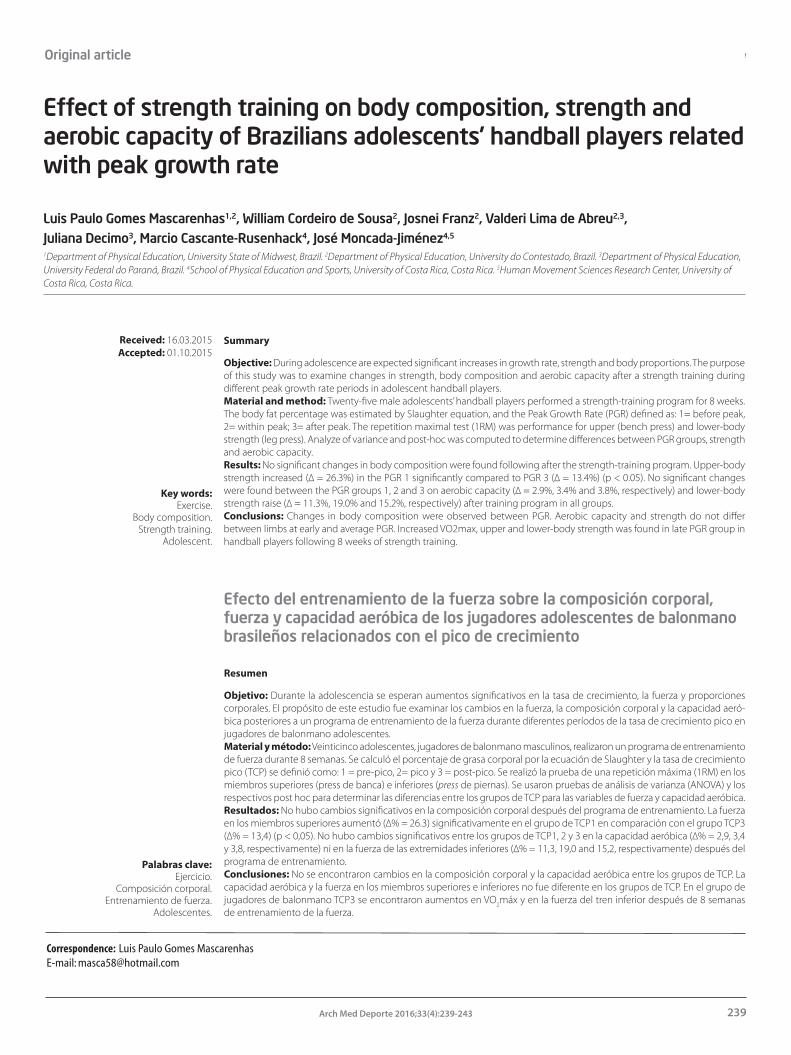
Effect of strength training on body composition, strength and aerobic capacity of Brazilians adolescents’ handball players related with peak growth rate
239Arch Med Deporte 2016;33(4):239-243
Original article
Summary
Objective: During adolescence are expected signi�cant increases in growth rate, strength and body proportions. The purpose of this study was to examine changes in strength, body composition and aerobic capacity after a strength training during di�erent peak growth rate periods in adolescent handball players. Material and method: Twenty-�ve male adolescents’ handball players performed a strength-training program for 8 weeks. The body fat percentage was estimated by Slaughter equation, and the Peak Growth Rate (PGR) de�ned as: 1= before peak, 2= within peak; 3= after peak. The repetition maximal test (1RM) was performance for upper (bench press) and lower-body strength (leg press). Analyze of variance and post-hoc was computed to determine di�erences between PGR groups, strength and aerobic capacity. Results: No signi�cant changes in body composition were found following after the strength-training program. Upper-body strength increased (∆ = 26.3%) in the PGR 1 signi�cantly compared to PGR 3 (∆ = 13.4%) (p < 0.05). No signi�cant changes were found between the PGR groups 1, 2 and 3 on aerobic capacity (∆ = 2.9%, 3.4% and 3.8%, respectively) and lower-body strength raise (∆ = 11.3%, 19.0% and 15.2%, respectively) after training program in all groups. Conclusions: Changes in body composition were observed between PGR. Aerobic capacity and strength do not di�er between limbs at early and average PGR. Increased VO2max, upper and lower-body strength was found in late PGR group in handball players following 8 weeks of strength training.
Key words: Exercise.
Body composition. Strength training.
Adolescent.
Resumen
Objetivo: Durante la adolescencia se esperan aumentos signi�cativos en la tasa de crecimiento, la fuerza y proporciones corporales. El propósito de este estudio fue examinar los cambios en la fuerza, la composición corporal y la capacidad aeró-bica posteriores a un programa de entrenamiento de la fuerza durante diferentes períodos de la tasa de crecimiento pico en jugadores de balonmano adolescentes. Material y método: Veinticinco adolescentes, jugadores de balonmano masculinos, realizaron un programa de entrenamiento de fuerza durante 8 semanas. Se calculó el porcentaje de grasa corporal por la ecuación de Slaughter y la tasa de crecimiento pico (TCP) se de�nió como: 1 = pre-pico, 2= pico y 3 = post-pico. Se realizó la prueba de una repetición máxima (1RM) en los miembros superiores (press de banca) e inferiores (press de piernas). Se usaron pruebas de análisis de varianza (ANOVA) y los respectivos post hoc para determinar las diferencias entre los grupos de TCP para las variables de fuerza y capacidad aeróbica. Resultados: No hubo cambios signi�cativos en la composición corporal después del programa de entrenamiento. La fuerza en los miembros superiores aumentó (∆% = 26.3) signi�cativamente en el grupo de TCP1 en comparación con el grupo TCP3 (∆% = 13,4) (p < 0,05). No hubo cambios signi�cativos entre los grupos de TCP1, 2 y 3 en la capacidad aeróbica (∆% = 2,9, 3,4 y 3,8, respectivamente) ni en la fuerza de las extremidades inferiores (∆% = 11,3, 19,0 and 15,2, respectivamente) después del programa de entrenamiento. Conclusiones: No se encontraron cambios en la composición corporal y la capacidad aeróbica entre los grupos de TCP. La capacidad aeróbica y la fuerza en los miembros superiores e inferiores no fue diferente en los grupos de TCP. En el grupo de jugadores de balonmano TCP3 se encontraron aumentos en VO2máx y en la fuerza del tren inferior después de 8 semanas de entrenamiento de la fuerza.
Palabras clave: Ejercicio.
Composición corporal. Entrenamiento de fuerza.
Adolescentes.
Received: 16.03.2015Accepted: 01.10.2015
Effect of strength training on body composition, strength and aerobic capacity of Brazilians adolescents’ handball players related with peak growth rate
Luis Paulo Gomes Mascarenhas1,2, William Cordeiro de Sousa2, Josnei Franz2, Valderi Lima de Abreu2,3, Juliana Decimo3, Marcio Cascante-Rusenhack4, José Moncada-Jiménez4,5
1Department of Physical Education, University State of Midwest, Brazil. 2Department of Physical Education, University do Contestado, Brazil. 3Department of Physical Education, University Federal do Paraná, Brazil. 4School of Physical Education and Sports, University of Costa Rica, Costa Rica. 5Human Movement Sciences Research Center, University of Costa Rica, Costa Rica.
Efecto del entrenamiento de la fuerza sobre la composición corporal, fuerza y capacidad aeróbica de los jugadores adolescentes de balonmano brasileños relacionados con el pico de crecimiento
Correspondence: Luis Paulo Gomes Mascarenhas E-mail: [email protected]

Luis Paulo Gomes Mascarenhas, et al.
240 Arch Med Deporte 2016;33(4):239-243
Introduction
Scienti�c evidence1,2 in children and adolescents have demonstra-ted the positive e�ects of physical activity as a stimulus for growth and development as well as in reducing health risk factors. In this period the maturational development expresses itself as a key process in the transition from childhood to adulthood and is characterized by rapid morpho-physiological changes3. During and after puberty signi�cant increases in physical performance are observed; these changes are explained, in part, by biomechanical factors and muscular, neural and hormonal development4-6.
The onset of resistance training during adolescence has been a topic of great interest and debate in the scienti�c community7-10. Several encourage the participation of adolescents in the resistance training program, provided they have proper planning and supervision of a competent professional7-10.
Research in the last two decades have provided valuable informa-tion on the responses of a young organism to such training11,12. Early research11 found that the children reported relatively similar strength gains than those for mature teens and young adults following resis-tance training at the onset of puberty. So strength training can induce adolescents neuromuscular adaptations resulting in signi�cant increase in muscle strength, but with little change in their anthropometric measurements13.
Resistance training is a key factor that stimulates growth, mus-cle hypertrophy, motor development, bone strength and increased strength14. In spite of this body of evidence, it has been suggested that resistance training should be done only after peak growth rate (PGR) to avoid impairing bone growth15,16. It is suggested that this type of training provides hormonal changes that a�ect the muscle strength already in prepubertal stages17. As a result, this type of training is being increasingly used by health professionals and adolescents.
Therefore, the purpose of this study was to examine changes in strength, body composition and aerobic power during di�erent perio-ds in adolescent handball players from Brazil undergo eight weeks of resistance training.
Materials and methods
Study model
This study has a quasi–experimental design with pre and post tests.
Participants
Volunteers were 25 adolescents’ male handball-players, with more than one years of expertise in handball and did not have any practice strength training at least six months prior to the program, all recruited from the community of São Bento do Sul, Brazil. They were divided into three groups according to the peak growth rate in late, average, and early.
Written informed consent was obtained from parents or legal guardians and from children participating in the study according to the Ethics Committee of the Brazil (Protocol 03682812.8.40.0117).
Adolescents were allowed to participate in the study if they met the following inclusion criteria: a) males, b) adolescents, c) handball players, and d) apparently-healthy showing no sign of physical injury in the past six months. Participants were excluded from the study if: a) presented any disease throughout the period of intervention that could interfere with testing measurements, b) did not show-up to the exercise training sessions, and c) did not complete the experimental protocol.
Procedures
Anthropometric assessment. Anthropometric measurements were obtained as described in the “Anthropometric Standardization Referen-ce Manual”18. Each measurement was taken three times and averaged for statistical analyses. Body height was measured to the nearest 0.1 cm using a stadiometer �xed to a wall. Individuals stood still with their heads in the Frankfort horizontal plane, barefoot, feet together, and the back surfaces of the calcaneus, pelvic, pectoral girdles and occipital regions in contact with the measuring equipment. Body mass was measured in kg on a digital platform balance, where individuals remain in light clothing, barefoot, feet positioned in the center of the platform, arms next to their bodies. The body mass index (BMI) in was calculated using the following formula: BMI = body weight in kg/body height in m2. A protocol by was used to estimate the body fat percentage (%BF)19. Tricipital and subscapular skinfold sites were measured to the nearest 0.1 mm with a clinical skinfold caliper (CESCORF). Finally, measures of waist and hip circumferences20 were also collected using a measuring tape. Then, the waist-to-hip ratio (WHR) was calculated.
Strength and aerobic power assessment. Muscle strength was asses-sed by the test of one-repetition maximum (1-RM) in the upper- (�at bench) and lower-limbs (leg press, 45°). The 1-RM consists in lifting the heaviest weight in a single maximum possible e�ort, with a full move-ment and without being able to repeat it again a second time21. The test starts with a brief warm-up with light weight below the maximum to prevent possible injuries. After a resting period of 3-min the 1-RM trial was performed. If the �rst attempt was successful then the following trials were preceded by a 3-min resting interval. Thus, the loads were increased until the individual failed to make a full-motion correctly. At that time was considered that the participant achieved the 1-RM.
Aerobic power was indirectly determined with a 20-m multistage run test and maximal oxygen consumption (ml · kg-1· min-1) was esti-mated according to a previously validated equation22.
Peak growth rate assessment. The PGR measurements included height trunk, leg length, height, weight and age. The calculation of PGR followed a pattern developed in Canada23 and validated in a Brazilian population14. The equation used was PGR = -9.236 + 0.0002708 (LL x TH) – 0.001663 (A x LL) + 0.007216 (A x TH) + 0.02292 (W/H), where CP: leg length, TH: trunk height-cephalic height, A: age, W: weight, and H: height. The PGR classi�cation is as follows: a) group 1 (more that -1 year = late), b) group 2 (between -1 and + 1 year = average), and c) group 3 (more that + 1 year = early).
Exercise training program. The resistance training program was per-formed in the mornings four days per week. This program was divided in two blocks, “A” and “B”. Following a light walk and jogging on a treadmill the participants performed the resistance training program at 75% of

Effect of strength training on body composition, strength and aerobic capacity of Brazilians adolescents’ handball players related with peak growth rate
241Arch Med Deporte 2016;33(4):239-243
their previously determined 1-RM with resting intervals of 1 and 3 min between 3 sets and exercises, respectively. The ‘A’ block was performed on Monday and Wednesday and comprised the following exercises: a) bench press in a �at and inclined bench, b) peck deck, c) front shoulder press, d) lateral raise, e) Triceps pulley, f ) leg press at 45°, g) “Smith” squats, h) leg extension, and i) rectus abdominis �oor exercise. The block ‘B’ was performed on Tuesday and Friday and included: a) open and closes pull-ups, b) dumbbell �y, c) barbell curl and barbell biceps curl on a “Scott” bench, d) abductor and adductor leg exercises on a machine, e) calf exercises, and f ) oblique abdominal exercises. All sessions always followed the same exercise order. All assessments and follow-up during the training sessions were performed by quali�ed trained sta� from the Physical Activity Unit of the Universidad do Costestado (UnC).
Statistical analysis
All analyses were computed using the MedCalc statistical software (Ostend, Belgium). Descriptive statistics mean (M), stan-dard deviation (±SD), frequencies and percentages were obtained. One-way analysis of variance (ANOVA) tests were used to determine differences between maturational stages and PGR periods. Tukey’s post hoc were computed following significant ANOVA’s F ratios. The variance equal Levene’s test was applied, and when your attended assumptions adopted the parametric statistics. Statistical significance was set a priori at α ≤ 0.05.
Results
Participant’s characteristics are presented in Table 1. Signi�cant between-group di�erences were found on mean age, weight, height, BMI, and WHR (Table 1).
ANOVA results showed that the mean VO2max was higher in the group 3 than in groups 1. Upper-body strength (in kg) was higher in groups 3 than in group 1 and 2 (p < 0.05) and upper-body strength increased in the PGR group 1 more than others (p < 0.05). Finally, mean lower-body strength was higher in the group 3 than in groups 1 and 2. (Table 2).
Discussion
The adolescence is a stage of life where major physical and matu-rational changes occur. In some individuals of the same chronological age but more mature than their respective counterparts, this stage may provide advantages in terms of sports performance due to grea-ter strength gains and increased muscle mass24. In this study, strength and aerobic capacity based on the PGR following a resistance training program in adolescent handball practitioners were evaluated.
Body composition (age, body weight, height, BMI, WHR), was di�erent between groups, with a gradual increase as the adolescents advance in their growth period; however, these changes are expected and natural once groups are in a period of growth, development and maturation25. In the present study, we did not observe changes in body fat percentage, which remained stable during periods of PGR. This �nding may be explained by the fact that teenagers were regular practitioners of handball, and regular physical activity stabilizes body fat in adolescents26.
The peak growth rate (PGR) considers the somatic age of adoles-cents, an indicator frequently used in studies for practical purposes. In this study, the PGR was found at about 14 years, similar to other reports19
and opposite to others27, where PGR was found close to 12 years of age.The PGR is related to other factors connected to physical �tness and
motor performance. In a longitudinal study of soccer players, the PGR was achieved at an age of 13.8 yr., with a concomitant development of VO2max and strength of upper- and lower-limbs compared to the present study28. However, others14, studied the association between PGR and motor performance and found a trend towards improvement in aerobic �tness and strength following the PGR, as corroborated in the present study. Peak force development occurs at about 1 to 1.5 years after the age of PGR of body height29, which was evidenced in the present study.
In this study there was signi�cant upper or lower-body strength change following a training program only for group of early develop-
Table 1. Descriptive statistics for participants based on peak growth rate.
Peak Growth Rate
Variable Group 1 Group 2 Group 3 P (n=7) (n=10) (n=8)
Age (yr.) 13.5 ± 0.3 13.9 ± 0.4 14.2 ± 0.7 0.055
Body weight (kg) 38.0 ± 7.7 48.1 ± 9.0a 60.3 ± 10.7a 0.005
Body height (cm) 148 ± 2.7b 157 ± 13.7a 170.0 ± 7.3a,b 0.002
BMI (kg/m2) 18.6 ± 1.7 22.1 ± 2.3a 23.8 ± 2.3a 0.001
Body fat (%) 18.6 ± 6.2 16.8 ± 4.6 18.1 ± 4.7 0.586
WHR 0.82 ± 0.03 0.77 ± 0.03a,c 0.81 ± 0.02 0.001
Note: Group 1: late PGR; Group 2: average; Group 3: early PGR; WHR: waist to-hip ratio. p < 0.05, a: di�erent from Group 1; b: di�erent from Group 2; c: di�erent from Group 3.
Table 2. Changes on aerobic power and strength variables after resistance training program by groups.
Variable PGR Pre Post Di�erence ∆% (Post – Pre)
VO2max (ml·kg-1·min-1) Group 1 45.6 46.9 1.3 2.9 Group 2 48.9 50.5 1.7 3.4 Group 3 52.1a 54.1a 2.0 3.8
Upper-body strength (kg) Group 1 19.0 24.0 5.0 26.3c
Group 2 27.4 33.8 6.5 23.6 Group 3 58.9a,b 66.7a,b 7.9 13.4
Lower-body strength (kg) Group 1 57.5 64.0 6.5 11.3 Group 2 110.4 131.4 21.0 19.0 Group 3 174.3a,b 200.7a,b 26.4 15.2
Note: p < 0.05, a: di�erent from Group 1; b: di�erent from Group 2; c: di�erent from Group 3.

Luis Paulo Gomes Mascarenhas, et al.
242 Arch Med Deporte 2016;33(4):239-243
ment (Table 2). Probably this changes can be because shortly after the PGR, there is a change in hormone pro�le, especially circulating testos-terone, which is known to a�ect muscle strength development30,31. In muscle testosterone stimulates protein synthesis and inhibits protein degradation, combined, these e�ects account for the promotion of muscle hypertrophy and subsequent increase in muscle strength in response to resistance training32. Hormonal changes that accompany puberty contribute to a signi�cant increase in strength depending on the increase in muscle mass33.
One of the �ndings of the present study was the 26% of ∆ variation at upper-body strength in late development group compared with early (Table 2). These �ndings reinforce the Lloyd et al (2009)34 highlights that muscle power and strength can be developed at the beginning of the PGR to adulthood. Strength training can elicit signi�cant gains in muscle strength above 10% when programs last from 4 to 19 weeks35,36. However, maturity has been found to be a signi�cant predictor of such changes36. The training program used in the present study (i.e., 8 weeks), did not elicit a su�cient stimulus to produce signi�cant changes in body composition and aerobic �tness in adolescents early or average, however the magnitude were di�erent.
A study in prepubescent children37, showed that resistance training during this stage is ine�cient and does not lead to strength gains. This assertion can be justi�ed with the pubertal growth, since it is in�uenced by the release of important hormones such as growth hormone (GH), insulin-like growth factor I (IGF-I), and sex steroids that induce increases in growth rate, muscle and bone maturation, functional ability and se-veral metabolic adaptations38. These alterations can and will in�uence the physical development, capacity and performance during childhood and adolescence6.
A limitation of this study was the small number of individuals eva-luated; however, various studies reported in a meta-analysis36 included smaller samples than in this study. Nevertheless, further research is needed to better understand the in�uence of PGR on strength training in adolescents.
Conclusion
Adolescents at di�erent times of the PGR showed di�erent body weight, height, BMI and WHR. Following 8 weeks of a resistance training program, no signi�cant changes in VO2max, upper-body strength and lower-body strength were observed in late and average PGR. In contrast players at after PGR show a signi�cant change after the program for VO2max, upper and lower strength gain. The early PGR show a signi�-cant magnitude variance in response to training sessions than late PGR.
References1. Janssen I, Leblanc AG. Systematic review of the health bene�ts of physical activity and
�tness in school-aged children and youth. Int J Behav Nutr Phys Act. 2010;7:40-56.
2. tabelini-Neto A, Sasaki JE, Mascarenhas LP, Boguszewski MC, Bozza R, Ulbrich AZ, et al. Physical activity, cardiorespiratory �tness, and metabolic syndrome in adolescents: a cross-sectional study. BMC Public Health. 2011;11:674-81.
3. Bond L, Clements J, Bertalli N, Evans-Whipp T, McMorris BJ, Patton GC. A comparison of self-reported puberty using the pubertal development scale and the sexual maturation scale in a school-based epidemiologic survey. J Adolesc. 2006;29:709-20.
4. Beunen G, Malina R, Van’t Hof M, Simons J, Ostyn M, Renson R. Adolescent growth and motor performance : a longitudinal study of Belgian boys. Champaign, IL.: Human Kinetics; 1988:153–5.
5. Seger JY, Thorstensson A. Muscle strength and electromyogram in boys and girls followed through puberty. Eur J Appl Physiol. 2000;81:54-61.
6. Tsolakis C, Bogdanis GC. In�uence of resistance training on anabolic hormones in pre-pubertal and pubertal males. Journal of Exerc Sci & Phys. 2007;3:1-11.
7. American College of Sports Medicine. ACSM’s guidelines for exercise testing and pres-cription. 6th ed. Baltimore: Lippincot, Willians & Wilkins, 2006:123-34.
8. Cahill BR (ed):American Orthopaedic Society for Sports Medicine. Prodeedings of the conference on strength training and the prepubesent. American Orthopaedic Society for Sports Medicine. 1988:1-14.
9. Bernhardt DT, Gomez J, Johnson MD, Martin TJ, Rowland TW, Small E, et al.. American Academy of Pediatrics. Strength training by children and adolescents. Pediatrics. 2001; 107:1470-2.
10. Faigenbaum A, Kraemer W, Cahill B, Chandler J, Dziados J, Elfrink L, et al. Youth Resistance training: position statement paper and literature review. Strength and Conditioning Journal. 1996;18(6):62-75.
11. Blimkie CJ. Resistance training during pre- and early puberty: e�cacy, trainability, mechanisms, and persistence. Can J Sport Sci. 1992;17:264-79.
12. Falk B, Tenenbaum G. The e�ectiveness of resistance training in children. A meta-analysis. Sports Med. 1996;22:176-86.
13. Oliveira AR, Lopes AG, Risso S. Preparation of Strength Training Programs for Children. Seminar: Biological and Health Sciences. 2003;24:85-96.
14. Robergs RA, Roberts SO. Fundamental principles of exercise physiology: For �tness, performance and health. Dubuque, IA: McGraw-Hill; 2000:350-9.
15. Machado DRL, Bon�m MR, Costa LT. Peak growth rate as an alternative for maturational classi�cation associated with motor performance. Braz J Kinanth Hum Perf. 2009;11:14-21.
16. Mortatti AL, Honorato RC, Moreira A, Arruda Md. The use of somatic maturation in the morphofunctional identi�cation in young soccer players. Rev And Med Dep. 2013;6:108- 14.
17. Fleck SJ, Kraemer W J. Muscular Strength Training Basics. Artmed. 2ª Ed. 1997:185-6.
18. Lohman TG, Roche AF, Martorell R. Anthropometric standardization reference manual. Champaign, IL: Human Kinetics Books. 1988:98
19. Slaughter MH, Lohman TG, Boileau RA, Horswill CA, Stillman RJ, Van Loan MD Skinfold equations for estimation of body fatness in children and youth. Hum Biol. 1988;60:709-23.
20. Fernandes Filho J. The practice of physical assessment. Rio de Janeiro, Brazil: Shape. 2003:23-8.
21. Carnaval PE. Measurement and evaluation in sport sciences. 6th ed. Rio de Janeiro, Brazil: Sprint. 2004:34-9.
22. Lèger LA, Mercier D, Gadoury C, Lambert J. The multistage 20 metre shuttle run test for aerobic �tness. J Sports Sci. 1988;6:93-101.
23. Mirwald RL, Baxter-Jones AD, Bailey DA, Beunen GP. An assessment of maturity from anthropometric measurements. Med Sci Sports Exerc. 2002;34:689-94.
24. Fleck SJ, Kraemer WJ. The basics of muscular strength training. Porto Alegre, Brazil: Artes Médicas. 1999:115-25.
25. Malina RM, Bouchard C. Physical activity of the young athlete: from growth to maturation. São Paulo, Brazil: Roca. 2002:85-8.
26. Mascarenhas LPG, Stabelini Neto A, Bozza R, Cezar CJ, Campos W. Comportment of maximal oxygen consumption and body composition during the maturational stages in male adolescents participants of soccer training. Rev Bras de Ciênc Mov. 2006;14:41-8.
27. Bergmann GG, de Araújo Bergmann ML, Del Corona Lorenzi T, dos Santos Pinheiro E, Garlipp DC, Baptista Moreira R, et al. Peak growth velocity of height, body mass and subcutaneous fat in 10 to 14-year-old boys and girls. Braz J Kinanth Hum Perf. 2007;9:333-8.
28. Philippaerts RM, Vaeyens R, Janssens M, Van Renterghem B, Matthys D, Craen R. The relationship between peak height velocity and physical performance in youth soccer players. J Sports Sci. 2006;24:221-30.
29. Colantonio E, Fernandes da Costa R, Colombo E, Silveira Böhme MT, Dal Molin Kiss MAP. Evaluation of growth and physical performance of children and adolescents. Rev Bras Ativ Fís & Saúd. 1999;4:17-29.
30. Lee PA. Physiology of puberty. Becker KL, editor. Philadelphia, PA.: Lipincott; 1995:885- 889.
31. Sale DG. Strength training in children. In: Gisol� CV. & Lamb DR., editor. Perspectives in Exercise Science and Sports Medicine. 2. Indianapolis, IN: Benchmark Press; 1989.

Effect of strength training on body composition, strength and aerobic capacity of Brazilians adolescents’ handball players related with peak growth rate
243Arch Med Deporte 2016;33(4):239-243
32. Vingren JL, Kraemer WJ, Ratamess NA, Anderson JM, Volek JS, Maresch CM. Testosterone physiologi in resistance exercise and trainning: the up - stream regulatory elements. Sports Med. 2010;40(12):1037-53.
33. Wilmore JH, Costill DL, Kenney WL. Physiology of sport and exercise. Champaign, IL: Human Kinetics. 2008:516-21.
34. Lloyd RS, Oliver JL. The youth physical development model: a new approach to long-term athletic development. Strength and Conditioning Journal. 2012;34(3):61-72.
35. Meinhardt U, Witassek F, Petrò R, Fritz C, Eiholzer U. Strength training and physical activity in boys: a randomized trial. Pediatrics. 2013;132:1105-11.
36. Behringer M, vom Heede A, Yue Z, Mester J. E�ects of resistance training in children and adolescents: a meta-analysis. Pediatrics. 2010;126:e1199-e1210.
37. Chia M. Suitability of resistance training and strength trainability in young people. Teaching and Learning. 2000;20:71-7.
38. Rogol AD. Growth at puberty: interaction of androgens and growth hormone. Med Sci Sports Exerc. 1994;26:767-70.
Fallecimiento del Dr. D. Ramon Balius i Juli
La Sociedad Española de Medicina del Deporte lamenta co-municar el fallecimiento de uno de sus miembros más queridos, el Dr. D. Ramón Balius i Juli. El Dr. Balius era Miembro de Honor de la Sociedad y hasta recientes fechas, y a pesar de su avanzada edad, gozó de una lucidez e inteligencia excepcionales para poder seguir obsequiando a los lectores de la Revista Archivos de Medicina del Deporte con su sección "Arte en el Estadio" que era un elemento caracterizador y exclusivo de la revista.
Fue uno de los padres de la Medicina del Deporte y desarrolló una excelente carrera profesional.
Lamentamos muy sentidamente su irreparable ausencia.
D.E.P.

Original article
244 Arch Med Deporte 2016;33(4):244-252
Summary
Introduction: Physical activity is effective in reducing fatigue, anxiety and depression in patients with fibromyalgia, and to improve the quality of life.Objective: To evaluate the efficacy of physical activity in improving the quality of life of people with fibromyalgia, 2004-2014.Methods: Meta-analysis of controlled clinical trials evaluating the efficacy of physical activity on the FIQ (Fibromyalgia Impact Questionnaire) and MOSSF-36 (Medical Outcome Study Short Form) scores, with 18 search strategies in four multidisciplinary databases. A protocol that containing criteria for inclusion, exclusion, assessment of methodological quality and extraction of information, were applied by two researchers to ensure reproducibility. Random effects meta-analysis, sensitivity analysis, DerSimonian-Lairds heterogeneity and publication bias with Begg test was performed.Results: 10 studies were included, the most in Spain and Brazil; 203 patients with intervention and 238 in the control group. The meta-analysis showed homogeneity in the FIQ and MOSSF-36 scores between groups, prior to the implementation of the intervention; after the application of intervention were identified difference of 14.9 points (95% CI 10.3; 19.5) in the FIQ and 2.0 on the MOSSF-36, was best in the group that received exercise therapy.Conclusion: The major efficacy of regular physical exercise is evident, compared with conventional treatment, to improve the quality of life of patients with fibromyalgia. Measuring the quality of life as a primary outcome in clinical trials should be performed with the FIQ.
Key words: Fibromyalgia. Quality of fife.
Health status. Motor activity. Controlled clinical trials as
topic. Meta-analyses.
Resumen
Introducción: La actividad física es eficaz para disminuir fatiga, ansiedad y depresión en pacientes con fibromialgia, esto redunda en un mejoramiento de su calidad de vida.Objetivo: Evaluar la eficacia de la actividad física en el mejoramiento de la calidad de vida de personas con fibromialgia, 2004-2014.Métodos: Meta-análisis de ensayos clínicos que evaluaron la eficacia de la actividad física sobre los puntajes del FIQ (Fi-bromyalgia Impact Questionaire) y MOSSF-36 (Medical Outcome Study Short Form), con 18 estrategias de búsqueda en cinco bases de datos multidisciplinarias. Se aplicó un protocolo que, a priori, contenía criterios de inclusión, exclusión, evaluación de la calidad metodológica y extracción de la información, aplicado por dos investigadores para garantizar reproducibilidad. Se realizó meta-análisis de efectos aleatorios, análisis de sensibilidad, heterogeneidad con DerSimonian-Lairds y sesgo de publicación con estadístico de Begg.Resultados: Se incluyeron 10 estudios, la mayoría desarrollados en España y Brasil; se aplicó la intervención a 203 pacientes y el control a 238. El meta-análisis demostró homogeneidad en los puntajes del FIQ y MOSSF-36 entre los grupos de estudio previo a la aplicación de la intervención; posterior a ella la diferencia en el FIQ fue de 14,9 puntos (IC 95% 10,3; 19,5) a favor del grupo que recibió la terapia con ejercicio físico; mientras que en los componentes del MOSSF-36 fue de 2,0.Conclusión: se evidencia la mayor eficacia del ejercicio físico regular, en comparación con el tratamiento convencional, para mejorar la calidad de vida de pacientes con fibromialgia. La medición de la calidad de vida como desenlace primario en estudios clínicos debe realizarse con el FIQ.
Palabras clave: Fibromialgia. Calidad de vida.
Estado de salud. Actividad física. Ensayos clínicos como asunto.
Meta-análisis.
Received: 20.07.2015 Accepted: 13.11.2015
Efficacy of motor activity in the quality of life in fibromyalgia patients: meta-analysis of clinical trials
Jaiberth Cardona-Arias1,2, Carmen Mantilla-Gutiérrez2, Luis Felipe Higuita-Gutiérrez2
1Universidad Cooperativa de Colombia. Medellín, Colombia. 2Universidad de Antioquia. Medellín, Colombia.
Eficacia del ejercicio físico sobre la calidad de vida en fibromialgia: meta-análisis de ensayos clínicos
Correspondence: Jaiberth Antonio Cardona Arias E-mail: [email protected]

Efficacy of motor activity in the quality of life in fibromyalgia patients: meta-analysis of clinical trials
245Arch Med Deporte 2016;33(4):244-252
Introduction
Fibromyalgia is a chronic illness of unknown aetiology, cha-racterised by the presence of generalised pain that may become incapacitating for the patient; it affects the patient’s biological, psychological and social spheres, generating difficulties at work, sleep pattern alterations, and a reduction in the performance of everyday activities1. Furthermore, it is associated with fatigue, tiredness, de-pression, anxiety, headaches and paraesthesia in the extremities2,3. Its diagnosis is carried out following the criteria of the American College of Rheumatology, which include widespread pain lasting over three months, muscular-skeletal pain and pain upon palpating at least 11 of 18 painful points2,4,5. This illness represents a worldwide health problem due to its high prevalence, morbidity rate and high consumption of health resources1. Added to this, it greatly affects the patient’s quality of life, including development in the workplace, home life and social sphere5.
To improve the pain and quality of life of these patients, phar-macological and non-pharmacological treatments are undertaken; the non-pharmacological treatments include exercise programmes, relaxation, physiotherapy and psychological or psychiatric support5. With reference to physical activity, previous studies have revealed their efficiency in reducing the impact of the illness by improving symptoms, including low levels of fatigue, anxiety, depression, and in general an improvement to the health-related quality of life (HRQL) and the overall state of health6-7.
The assessment of health-related quality of life in this illness has been performed using specific and generic instruments, including in particular the Fibromyalgia Impact Questionnaire (FIQ) and the Medical Outcome Study Short Form (MOSSF-36), which both enable clinical monitoring, help guide decision making for health providers, obtain clinical outcome measures and perform cost-effectiveness or cost-usefulness analyses1,8.
In view of the aforementioned, various controlled clinical trials about the effectiveness of physical activity in improving health-rela-ted quality of life and the state of health of people with fibromyalgia have been carried out. Despite this, there are no clear conclusions, with powerful and solid statistics, which is why a systematic review must be performed to meta-analyse the evidence from previous articles and to give greater external validation to the outcomes and conclusions. With this objective, in 2008 a meta-analysis was publis-hed with controlled clinical trials that collected research carried out up to 20059; however, this information requires updating, as syste-matic reviews on this issue have revealed that the most research has been concentrated over the past two three-year periods, meaning that it is possible that the effects of physical activity on the quality of life of fibromyalgia patients have changed over these past 6 years with the diversification of exercise programmes8. Added to this, this meta-analysis concentrated the results on a specific scale (FIQ), lea-
ving aside the generic MOSSF-36 scale that may be more sensitive to clinical changes10.
In accordance with the aforementioned, this study was desig-ned with the aim of assessing the effectiveness of physical activity on improving the health-related quality of life and overall state of health of fibromyalgia sufferers, based on articles published between 2004 and 2014.
Material and methods
Type of study
Meta-analysis of clinical trials.
PICO: Population Intervention Comparison Outcome
Population. Patients diagnosed with Fibromyalgia according to the criteria established by the American College of Rheumatology11.
Intervention. Physical activity for at least 3 weeks, by which physical activity is understood to mean aerobic exercise in which the heart rate is maintained at over 50% of the maximum frequency, flexibility exercises, stretching, strengthening or a mixture of these according to the American College of Sports Medicine guidelines for exercise testing and prescription12.
Comparison. Patients with fibromyalgia that do not perform physical activity or that do so with less intensity than that established in the intervention. This group includes conventional education or guidance interventions about lifestyle provided by the rheumatologist; pharmacological treatment prescribed by the doctor and light physical activity such as stretching or routines that do not exceed 30 minutes per session or three days per week.
Outcome. Quality of life score obtained using the FIQ or MOS-SF36 scales, or both, for the study group as well as the control group, before and after the intervention. Note that this review was not restricted to studies that simultaneously applied both scales, given that the index generated by the FIQ is independent from the physical and mental components of the MOSSF-36, meaning that this restriction would reduce the exhaustive nature of this study.
Measuring health-related quality of life
The FIQ is a specific instrument to measure the impact of fibromyal-gia in functional capacity and the quality of life of the people affected. It is composed of 10 dimensions that evaluate physical capacity, habitual work, impact on paid labour activity, pain, fatigue, feeling of tiredness, stiffness, feeling of well-being, anxiety and depression; the domains generate a score between 0 and 100, with 0 being the best result in HRQL, or the least impact caused by the fibromyalgia13.
The MOSSF-36 is a generic HRQL questionnaire composed of 36 items that generate an 8-dimension profile: body pain, physical per-formance, physical function and overall health that is summarised in a

Jaiberth Cardona-Arias, et al.
246 Arch Med Deporte 2016;33(4):244-252
physical component, emotional performance, social function, mental health and vitality that conform to the mental component. The instru-ment generates a score that can vary from 0 to 100 points; the higher the score, the better the HRQL, and the reference value established in a healthy population is 50±10 points14.
Research Protocol according to PRISMA phases (Preferred Reporting Items for Systematic reviews and Meta-Analyses)15
Article identification or search. A sensitivity search was carried out in the literature related to quality of life in fibromyalgia from the Pubmed database, Science Direct, Lilacs and Scielo; using the following search strategies: “Calidad de vida & Fibromialgia”, “Calidad de vida relacionada con la salud & Fibromialgia”, “Calidad de vida & FIQ”, “Calidad de vida & SF-36”, “Calidad de vida relacionada con la salud & FIQ”, “Calidad de vida relacionada con la salud & SF-36” and their counterparts in English and Portuguese. As well as this, a search was performed of clinical trials in the Cochrane Library with the terms “physical activity in the quality of life in fibromyalgia” and “motor activity in the quality of life in fibromyalgia”.
Screening or applying the inclusion criteria − Contain search terms in the title or abstract, − research studies published between 2004 and September 2014, − original articles and − being a controlled clinical trial.
Some syntax used were: − (quality of life [Title/Abstract]) AND fibromyalgia [Title/Abstract]; − (health related quality of life[Title/Abstract]) AND fibromyalgia impact
questionnaire [Title/Abstract]; − TITLE-ABSTR-KEY (health related quality of life) and TITLE-ABSTR-KEY
(fibromyalgia impact questionnaire); − pub-date > 2003 y iv) TITLE-ABSTR-KEY (quality of life) and TITLE-ABSTR-
KEY (fibromyalgia).
Selecting or applying the exclusion criteria
− Publications with interventions different to physical activity or multi-disciplinary interventions (educational, pharmacological, physiotherapeutic, psychological);
− articles with internal validation problems due to the failure to control bias or confusing variables;
− articles that do not explicitly mention the quality of life scores before and after the intervention in each of the compared groups.To assess the methodological quality of the studies, the following
criteria were applied: randomisation, concealment, calculation of the sam-ple size, analysis by treatment intentions, safety report and homogeneity analysis. A form was made categorising the study with 1 if it contained the criteria, and with 0 for those that did not apply. With these a total was reached, taking scores equal or greater than 4 as good quality. Given that in the studies included the majority had a similar character (between 2 and 4 points), a meta-regression was not carried out for this variable.
Gathering the information
Two researchers applied the research protocol independently to ensure the ability to reproduce the review; discrepancies were resol-ved by consensus or through reference to a third party. The articles obtained were exported to an Endnote Web programme to eliminate duplicates. Once this stage was finished, the classification process of the studies began by carefully reading the abstracts and by creating a data extraction format stored in an Excel-designed database. Extracting the information was performed independently by two researchers with the aim of ensuring the inter-observer reproducible nature, in which the kappa coefficient of 1.00 for the year and place of study variables was obtained; and Intraclass Correlation Coefficient (CCI) of 1.0 for quality of life scores.
Analysing the information
The studies were characterised according to the place and year of performance. The standard deviation of the overall scores of each of the MOSSF-36 and FIQ dimensions was established based on the variation coefficient calculated in the individual studies.
To ensure the homogeneity of the groups, a meta-analysis was performed for average differences with the quality of life scores at the start of the study. The aforementioned was carried out given that in the studies this analysis was not performed, nor was a health-related quality of life score defined as inclusion criteria.
The efficacy of physical activity on the quality of life of fibromyalgia patients was assessed using a meta-analysis for average differences of scores after the intervention in both the treated and control group. With the aim of equalling out the direction of the FIQ and MOSSF-36 scales, the following formula was applied: “101- FIQ score”.
The heterogeneity was assessed with the Q statistic of the DerSimo-nian and Lairds test, the publication bias using the Funnel Plot, and the Begg’s test statistic and a sensitivity analysis was carried out to explore the influence of each study on the size of the overall effect.
For the analyses, the Excel and Programme for Epidemiological Analysis of Tabulated Data from the Pan American Health Organisation (EPIDAT) version 3.0 was used.
Results
In the initial search, 31 articles were identified that complied with the inclusion criteria, of which 21 were eliminated for not assessing the effect of the physical activity in fibromyalgia symptoms (n=15), for using multiple treatment simultaneously, such as sociotherapy, physiotherapy, psychotherapy and art-therapy (n=4) and for not explicitly mentioning the changes in the FIQ or MOSSF-36 scores before and after the inter-vention (Figure 1). Based on the Cochrane search with the “motor activity quality of life fibromyalgia, in Title, Abstract, Keywords in Trials” strategy, six results of 893,010 articles were found that contained one or more of the search terms, and all focused on pain as the central link; with the

Efficacy of motor activity in the quality of life in fibromyalgia patients: meta-analysis of clinical trials
247Arch Med Deporte 2016;33(4):244-252
“physical activity quality of life fibromyalgia, in Title, Abstract, Keywords in Trials” strategy, 28 results were obtained, from which only two complied with the protocol and were duplicated in the findings on Pubmed.
In the studies included in the quantitative synthesis (n = 10), the majority were published between 2012 and 2014, mainly in Spain and Brazil and all included women with fibromyalgia, apart from the study by Elijk-Hustings, which included one male in the control group (n = 48). The physical intervention was performed with aerobic exercises, hydrothera-py, bio-dance and strengthening. The frequency was between 2 and 3 times a week and the total duration of the intervention was between 3 and 24 weeks. With regards to the characteristics of the people included, there was an intra-study homogeneity for the variables of age and years with the illness; however, there was inter-study variability regarding the same variable in that the average age fluctuated between 42 and 59 years. The same occurred with the average duration of the symptoms of the illness, which oscillated between 4 and 26 years (Table 1). It should be specified that the majority of the studies did not reveal other variables in the analyses of homogeneity and in the inclusion criteria the majority circumscribe to the diagnostic criteria of the illness and the type of institutions where the patients were found; this prevents further research into potential variables for a meta-regression.
All the test groups applied interventions in which they reached a minimum of 50% of their maximum heart rate, though in some studies this intensity was higher with increases between 55-64%16, 60-85%18, 50-69%23 or 60-70%25 according to ACSM recommendations.
Author Year Country Group n Age Years with the Treatment illness
Eijk-Hustings16 2013 Netherlands EG 19 43.9±7.6 6.2±7 Aerobic exercises CG 48 42.9±11 7.1 ± 6.4 Conventional treatmentFernandes de Melo17 2006 Brazil EG 24 48.9±9.2 --- Hydrotherapy CG 23 46.6±8.4 --- Conventional physiotherapyGarcía-Martínez18 2012 Spain EG 12 59.3±4.8 9.9±3.8 Physical exercise CG 13 58.6±7.8 10.6 ±4.1 Conventional treatmentLatorre19 2013 Spain EG 42 50.9±7.7 9.1 ± 3.8 Physical exercise CG 30 52.4±8 9.0±4.8 Conventional treatmentLópez-Rodríguez20 2012 Spain EG 19 55.5±7.7 12.5±7.4 Aquatic bio-dance CG 20 55.3±7.5 14.3±8.3 StretchingMartín-Nogueras21 2012 Spain EG 15 52.6±8.4 4.6±3.2 Physiotherapy and aerobics CG 13 48.1±6.9 6.4±6 Conventional treatmentOliveira22 2014 Brazil EG 35 44.3±7.9 --- Strengthening CG 31 48.6±7.6 --- FLEXSañudo23 2007 Spain EG 14 57.9±6.2 --- Physical exercise and vibration training CG 12 60.1±9.4 --- Physical exerciseTomas-Carus24 EG 17 51±10 24±9 Aquatic exercise CG 17 51±9 27±4 Conventional treatmentVinícius letieri25 2013 Brazil EG 33 58.2±10.6 --- Hydro-kinesiotherapy (PSE) CG 31 59.6±9.4 --- Conventional treatment
Figure 1. Algorithm of the article selection.
Table 1. Description of the articles included in the quantitative synthesis.
Group EG: Experimental group; CG: Control group.
Identi
ficati
on
Ele
cti
on
Scre
enin
gIn
clu
sio
n
Articles with search
terms in title and/or
abstract n= 253
Duplicates n= 50
They are not originals n= 27
They do not fall within the time
frame n= 42
Validation studies n= 22
Analytical studies n= 38
Descriptive studies n=43
Screened articles
n=134
Articles complete text
analysed for election
n=31
Does not assess physical activity
n= 15
Multiple intervention
n=4
Does not explicitly mention scores
n=2
Studies included
in qualitative synthesis
n=10
Studies included in
qualitative synthesis
(Meta-analysis)
n=10
Articles identified
in the search
n= 23303

Jaiberth Cardona-Arias, et al.
248 Arch Med Deporte 2016;33(4):244-252
All the studies applied the intervention with a duration of one hour per session, with the exception of two studies which lasted 45 minutes22,25, each session included between 5-10 minutes of warming up, 30-40 minutes of physical aerobic exercise and/or strengthening, with 5-10 minutes of stretching and relaxing. The intervention time varied with six23, twelve16-18,20,21, fifteen25, 1622 or 24 weeks19; in half of the studies 2 sessions per week were applied16,21,22,23,25 and in the rest, three.
Regarding the instruments to assess the effect of physical activity on the quality of life, 4 used both the FIQ and the MOSSF-36, 4 studies only applied the FIQ and 2 only applied the MOSSF-36. Before starting the intervention, the FIQ scores in the test group and the control group fluctuated between 22.5 ± 11.1 and 52.1 ± 12.1 points; the same procedure was performed after the intervention and in this case the
test group scores were between 44 ± 16 and 57.2 ± 12.3 and for the control group between 22.0 ± 10 and 51.2 ± 14.9. With regards to the HRQL according to the MOSSF-36 in the physical component prior to treatment, scores were obtained between 9.7 ± 26.4 and 44.6 ±12.7; following intervention the test group displayed scores between 33.6 ± 35.8 and 75.6 ± 14.2. Mental health was also assessed before treatment in both groups with results between 38.4 ± 17.0 and 58.0 ± 16.5 points and after the intervention the scores in the treated group were between 50.1 ± 25.0 and 69.3 ± 20.9 (Table 2).
Upon performing the meta-analysis of the FIQ scores before and after the intervention, both the treated and control groups revealed heterogeneity in the studies included (Statistic Q Vp 0.000), lack of pu-blication bias (Begg Test 0.915) and good robustness of the combined
Table 2. Description of the FIQ scores and the SF-36 pre and post treatment in the experimental and control groups.
Author Group FIQ score 36 Score Physical 36 Score Mental X±DS X±DS X±DS
Pre* Post** Pre* Post** Pre* Post**
García-Martínez EG 28.9±13.1 50.8±12.9 33.9±18.5 50.6±27.4 46.6±18.2 61.7±18.5
CG 32±12.5 36.2±16.7 39.6±17.5 35.4±17.2 48.0±21.3 41.5±20.3
Latorre EG 35.5±13.5 47.7±16.3 38.7±16.2 41.5±21.3 43.1±13.4 52.7±17.4
CG 36.2±11.6 35.3±11.8 34±17.0 34.3±16.6 48.0±18.1 39.7±17.4
Oliveira EG 33.2±15.4 49.9±18.4 12.1±24.5 33.6±35.8 39.0±21.5 50.1±25.0
CG 34.2±17.2 49.9±18.4 9.7±26.4 28.2±38.6 45.9±24.5 61.0±24.5
Tomas-Carus EG 38.0±20 44.0±16 36.0±23 48.0±21 48.0±20 62.0±27
CG 42.0±16 41.0±17 33.0±19 37.0±17 51.0±24 50.0±20
Fernandes de Melo EG --- --- 35.5±20.1 75.6±14.2 42.4±19.4 69.3±20.9
CG --- --- 35.5±15.0 67.7±23.5 38.4±17.0 66.8±23.9
Martín-Nogueras EG --- --- 39.7±13.3 55.3±12.9 52.5±15.3 62.9±20.7
CG --- --- 44.6±12.7 40.0±14.9 58.0±16.5 54.0±23.8
Eijk-Hustings EG 41.0±2.1 49.0±3.2 --- --- --- ---
CG 45.6±2.3 44.8±2.9 --- --- --- ---
López-Rodríguez EG 33.9±10.5 48.8±16.2 --- --- --- ---
CG 31.5±13.0 31.8±12.9 --- --- --- ---
Sañudo EG 52.1±12.1 57.2±12.3 --- --- --- ---
CG 44.1±11.6 51.2±14.9 --- --- --- ---
Vinicius Letieri EG 22.5±11.1 48.5±15.6 --- --- --- ---
CG 25.3±10.3 22.0±10 --- --- --- ---
Group EG: Experimental group; CG: Control group; *Pre: Pre-treatment; **Post: Post-treatment.

Efficacy of motor activity in the quality of life in fibromyalgia patients: meta-analysis of clinical trials
249Arch Med Deporte 2016;33(4):244-252
measures, given that in the sensitivity analysis the exclusion of each of the studies did not affect the overall result. In the Funnel Plot of random effects, no significant differences were found in the FIQ scores before starting the intervention 0.90 (CI 95% -0.39; 0.89) between the treated and control group; on the other hand, after intervention the difference in the average FIQ score was 14.9 points (CI 95% 10.3; 19.5) in favour of the group that received the therapy with physical exercise, proving the effectiveness of the intervention (Figure 2).
In the domain of physical health of the MOSSF-36, heterogeneity in the studies was observed (Statistic Q Vp 0.0002), lack of publication bias (Begg Test Vp 0.1329), equality in the test and control group sco-res prior to the intervention 0.20 (CI 95% -0.36; 0.76) and difference in favour of the group with physical activity of 2.06 points (CI 95% 1.18; 2.95) following intervention.
In the domain of mental health, the heterogeneity is maintained (Statistic Q Vp 0.000), the lack of publication bias (Begg Test Vo 0.7071), equality in the test and control groups before initiating treatment 0.07 (CI 95% -0.56; 0.71) and the difference in favour of the group that received the physical therapy, in this case of 1.86 points (CI 95% 0.30; 3.40) (Figure 3).
Discussion
Fibromyalgia strongly affects the quality of life of sufferers, especially in the physical and mental domain. The physical alterations are revealed as chronic pain, disability to perform everyday activities and the on-going use of medication. The psychological and mental limitations are characterised by stress, depression and anxiety. Previous studies have
described the impact of the illness on the physical and mental domain on the quality of life of patients, using instruments such as the FIQ and the MOSSF-36. The latter reveals scores below 50 which is the benchmark for healthy populations10, which is in keeping with this research in that the pretreatment scores were lower than 50 points.
Among the therapeutic options to treat the illness and reduce the impact on the patient’s quality of life, performing regular physical exercise has been established as one of the main non-pharmacological strategies to control the symptoms. Despite this, there are still discrepan-cies regarding the muscular and physical capacity of these patients to take on an exercise programme, as well as the physiological mechanism by which these types of therapy generate a reduction in symptoms26.
Discrepancies regarding the capacity of the patients to perform exercise arise because they can perceive a greater effort during physical activity; however, previous studies have revealed that muscular stren-gth and resistance in patients with fibromyalgia are similar to those in healthy control subjects. Furthermore, in patients that undergo physical activity therapy, the beneficial effects are evident, such as reduced pain, increased strength, improved sleep quality, less fatigue and an improved psychological state27.
In keeping with the above, in this study it was clear that the patients with physical activity experienced improvements to their HRQL by the end of the treatment, proven by the increase in the FIQ and MOSSF-36 scores. The FIQ scores of the treated group increased by 14.9 points whilst in the physical and mental domain of MOSSF-36 the increase was 2.06 and 1.86 respectively. The difference in the score of both treatments could be attributed to the nature of the scales, as the FIQ is a specific scale for fibromyalgia patients, making it more sensitive to detecting
Figure 2. Forest Plot for the difference in averages of the FIQ scores in the test and control groups.

Jaiberth Cardona-Arias, et al.
250 Arch Med Deporte 2016;33(4):244-252
changes associated to the illness in aspects such as stiffness, morning tiredness, pain, depression and anxiety; on the other hand, MOSSF-36 is a generic measurement, in that its sensitivity is greater when it comes to detecting unforeseen effects of the illness via the domains of physical function, the physical role, body pain, general health, vitality, social role, emotional role and mental health28.
In allusion to the mechanism that explains the beneficial effects of exercise in fibromyalgia patients, some studies have described a clear reduction in the levels of the systemic inflammatory markers such as the IL-8, and of stress such as the noradrenaline and extra-cellular heat shock proteins 72 kDa (eHsp72), which are usually increased in patients with the illness and its regulation would in part explain the reduction
Figure 3. Forest Plot for the difference in averages of the Mental and Physical scores of the MOSSF-36 in the test and control groups.
Study (Year)
Fernandes de Melo Vitorino (2006)
Tomas Carus (2007)
Martín Nogueras (2012)
García Martínez (2012)
Latorre PA (2013)
De Oliveira Gavi (2014)
OVERALL (Fixed ef.)
OVERALL (Random ef.)
Study (Year)
Fernandes de Melo Vitorino (2006)
Tomas Carus (2007)
Martín Nogueras (2012)
García Martínez (2012)
Latorre PA (2013)
De Oliveira Gavi (2014)
OVERALL (Fixed ef.)
OVERALL (Random ef.)
n -
47 -
34 -
28 -
25 -
72 -
66 -
272 -
272 -
n -
47 -
34 -
28 -
25 -
72 -
66 -
272 -
272 -
CI measurement averages (95.0%)
CI measurement averages (95.0%)
A. Pre-treatment
C. Pre-treatment
Mental Component of the MOSSF-36
B. Post-treatment
D. Post-treatment
-1.5 0-0.5-1 0.5 1 1.5 -3.6 -1.2 0 1.2 2.4 64.83.6-2.4-4.8
Physical Component of the MOSSF-36
-0.8 -0.4 0-1.2-1.6 0.4 1.2 2 43210-1-2-3-41.60.8

Efficacy of motor activity in the quality of life in fibromyalgia patients: meta-analysis of clinical trials
251Arch Med Deporte 2016;33(4):244-252
of the symptoms and the increase in the HRQL. Despite this, there is no consensus on this matter; furthermore physical activity should be regulated because acute exercise, with no physiological adaptation or training, could exacerbate, rather than improve, the systemic state of stress or inflammation in women with fibromyalgia29.
Research studies into the effectiveness of physical activity in impro-ving the symptoms of fibromyalgia and its impact on FIQ and MOSSF-36 scores, are summarised in this meta-analysis, and in this respect it pro-vides useful information for orientating later studies and consolidating causal hypotheses, as it includes an exhaustive literary search, groups the results of various research studies into one sole measurement, gives a more precise estimation of the effect, and improves the statistical power by increasing the sample size30.
It is important to highlight that the methodological quality of the articles included in the quantitative synthesis of this research were conditioned by aspects such as the calculation of the sample size, blinding, the analysis of the results with the treatment intentions and the analysis of the safety of the intervention. The analysis with treatment intentions implies including all the patients in the analysis, regardless of whether or not they received the whole treatment or if the originally established protocol was removed. Omitting this analysis influences the results, as the motives of the lack of adherence may be related to the prognostic or effectiveness of the treatment31. Blinding refers to the fact that the participants and researchers did not know which group received the intervention, reducing the bias in the physiological or physical response motivated by the type of intervention received. This is particularly important in studies in which the result measurements are far from objective, such as pain or quality of life. However, the majority of the patients involved in this meta-analysis could not be blinded regarding the treatment, as in general terms one group received phy-sical therapy whilst the other did not, impeding blinding for obvious reasons32. Safety refers to the protection of patients faced with adverse and secondary effects of the intervention, in the studies included in this analysis, no long-term analysis was performed on the safety of physical activity; however, it is clearly better than the effects of pharmacological treatments with anti-depressives and anticonvulsants33.
Among the limitations of this study, which at the same time constitute suggestions for later research that aims to assess the effect of physical activity on the HRQL of patients with fibromyalgia, is the diversity of physical interventions included, which highlight the need for experts in the field to unify criteria such as intensity, duration, frequency, and even the design of the exercise regime prescribed, particularly when dealing with an intervention to assess in a clinical trial. Furthermore, it should be noted that the Begg statistic provides good statistical power for meta-analyses with 75 or more studies, and moderate power for analyses with 25, meaning that in meta-analyses with few research studies the publication bias should be interpreted with caution. However, the number of studies included should not be taken categorically, as there are other parameters that affect the power of this statistic, even more importantly than the number of studies, such
as the research selection criteria, the variations in the size of the effect and global effect. In addition, it should not be forgotten that this statistic is exploratory, and is performed as a formal procedure to complement the Funnel_plot34.
Conclusion
This exercise highlights that practising regular physical activity is statistically better than conventional treatment in improving the quality of life of fibromyalgia patients, though the quality of the recommenda-tion is low due to the limitations of the studies included, the variability in defining the test group and the low number of analysed research studies. However, the hypothesis is consolidated regarding the benefits of physical activity on this illness, as a base for the design of posterior random controlled clinical trials, and it is recommended that the measu-rement of quality of life as a primary outcome in clinical studies should be performed with the FIQ.
References1. Collado A, Alijotas J, Benito P, Alegre C, Romera M, Sañudo I, et al. Documento de
consenso sobre el diagnóstico y tratamiento de la fibromialgia en Cataluña. Med Clin. Barcelona 2002;118(19):745-9.
2. Rivera J, Alegre C, Ballina F, Carbonell J, Carmona L, Castel B, et al. Documento deconsenso de la Sociedad Española de Reumatología sobre la fibromialgia. Reumatol Clin. 2006;2(Supl 1):55-66.
3. Häuser W, Hayo S, Biewer W, Gesmann M, Kühn-Becker H, Petzke F, et al. Diagnosis of fibromyalgia syndrome-a comparison of Association of the Medical Scientific Socie-ties in Germany, survey, and American College of Rheumatology criteria. Clin. J. Pain. 2010;26(6):505-11.
4. Santos A, Assumpção A, Matsutani L, Pereira C, Lage L, Marques A. Depressão e qua-lidade de vida em pacientes com fibromialgia. Rev. Bras. Fisioter. 2006;10(3):317-24.
5. Secretaría de Salud de México. Guía de Práctica Clínica para diagnóstico y tratamien-to de fibromialgia en el adulto. 2009 [sitio de Internet; consultado: 01 Nov 2014].Disponible en: http://www.cenetec.salud.gob.mx/descargas/gpc/CatalogoMaes-tro/075_GPC_FirbomialgiaAdulto/FIBROMIALGIA_EVR_CENETEC.pdf
6. Carbonell-Baeza A, Aparicio VA, Chillón P, Femia P, Delgado-Fernandez M, Ruiz JR.Effectiveness of multidisciplinary therapy on symptomatology and quality of life in women with fibromyalgia. Clin. Exp. Rheumatol. 2011;29(6):97-103.
7. Guymer EK, Maruff P, Littlejohn GO. Clinical characteristics of 150 consecutive fi-bromyalgia patients attending an Australian public hospital clinic. Int J Rheum Dis. 2012;15(4):348-57.
8. Hernández-Petro AM, Cardona-Arias JA. Sistematización de las investigaciones sobre calidad de vida relacionada con la salud en fibromialgia, 2004-2014. Archivos de Me-dicina. 2014;10(1):1-12.
9. Busch A, Schachter C, Overend T, Peloso P, Barber K. Exercise for Fibromyalgia: ASystematic Review. J. Rheumatol. 2008;35(6):1130-44.
10. Hernández-Petro AM, Cardona-Arias JA. Efecto de la fibromialgia sobre el estado de salud y la calidad de vida relacionada con la salud, 2004-2014. Rev. Colomb. Reumatol. 2015;22(2):110-8.
11. Wolfe F, Smythe HA, Yunus MB, Bennett RM, Bombardier C, Goldenberg DL, et al. The American College of Rheumatology 1990 Criteria for the Classification of Fibromyalgia. Report of the Multicenter Criteria Committee. Arthritis Rheum. 1990;33:160-72.
12. American College of Sports Medicine: ACSM’s guidelines for exercise testing andprescription. 9th Edition. Baltimore: Lippincott, Williams & Wilkins, 2010.
13. Monterde S, Salvat I, Montull S, Fernández J. Validación de la versión española delFibromyalgia Impact Questionnaire. Rev Esp Reumatol. 2004;31(9):507-13.
14. Campolina AG, Ciconelli RM. SF-36 and the development of new assessment tools for quality of life. Acta Reumatol Port. 2008;33(2):127-33.
15. Moher D, Liberati A, Tetzlaff J, Altman D, The PRISMA Group. Preferred Reporting Items for Systematic Reviews and Meta-Analyses: The PRISMA Statement. PLoS Med. 2009 6(7): e1000097.doi:10.1371/journal.pmed.1000097.

Jaiberth Cardona-Arias, et al.
252 Arch Med Deporte 2016;33(4):244-252
16. Eijk-Hustings Y, Kroese M, Tan F, Boonen A, Bessems-Beks M, Landewé R. Challenges in demonstrating the effectiveness of multidisciplinary treatment on quality of life, participation and health care utilization in patients with fibromyalgia: a randomised controlled trial. Clin Rheumatol 2013;32:199-209. DOI 10.1007/s10067-012-2100-7
17. Fernandes de Melo D, Bizari L, Fernandes do Prado G. Hydrotherapy and conventional physiotherapy improve total sleep time and quality of life of fibromyalgia patients: Randomized clinical trial. Sleep Med. 2006;7(3):293-6.
18. García-Martínez A, De Paz J, Márquez S. Effects of an exercise programme on self-esteem, self-concept and quality of life in women with fibromyalgia: a randomized controlled trial. Rheumatol. Int. 2012;32:1869-76. DOI 10.1007/s00296-011-1892-0
19. Latorre PA, Santos MA, Heredia-Jiménez JM, Delgado-Fernández M, Soto VM, Mañas A, et al. Effect of a 24-week physical training programme (in water and on land) on pain, functional capacity, body composition and quality of life in women with fibromyalgia. Clin. Exp. Rheumatol. 2013;31(Suppl. 79):S72-S80.
20. López-Rodríguez M, Castro-Sánchez AM, Fernández-Martínez M, Matarán-Peñarrocha G, Rodríguez-Ferrer ME. Comparación entre biodanza en medio acuático y stretching en la mejora de la calidad de vida y dolor en los pacientes con fibromialgia. Aten Primaria. 2012;44(11):641-50.
21. Martín-Nogueras AM, Calvo-Arenillas JI. Eficacia del tratamiento de fisioterapia en la mejora del dolor y la calidad de vida en pacientes con fibromialgia. Rehabilitación (Madr). 2012;46(3):199-206.
22. Oliveira Gavi MB, Vassalo DV, Tadeu Amaral F, Felício Macedo DC, Gava PL, Miranda Dantas M, et al. Strengthening exercises improve symptoms and quality of life but do not change autonomic modulation in fibromyalgia: a randomized clinical trial. PLoS One. 2014; 9(3): e90767. doi:10.1371/journal.pone.0090767.
23. Sañudo B, de Hoyo M, Carrasco L, McVeigh JG, Corral J, Cabeza R, et al. The effect of a 6-week exercise programme and whole body vibration on strength and quality of life in women with fibromyalgia: a randomized study. Clin. Exp. Rheumatol. 2010;28(6 Suppl 63):S40-5.
24. Tomas-Carus P, Hakkinen A, Gusi N, Leal A, Hakkinen K, Ortega-Alonso A. Aquatic Training and Detraining on Fitness and Quality of Life in Fibromyalgia. Med Sci Sports Exerc. 2007;39(7):1044-50.
25. Vinícius Letieri R, Furtado GE, Letieri M, Góes SM, Borba Pinheiro CJ, Veronez SO, et al. Pain, quality of life, self-perception of health, and depression in patients with fibromyal-gia treated with hydrokinesiotherapy. Rev Bras Reumatol. 2013;53(6):494-500.
26. Mannerkorpi K. Exercise in fibromialgia. Curr Opin Rheumatol. 2005;17:190-4
27. Álvarez A. Ejercicio físico en la fibromialgia. Rehabilitación (Madr). 2003;37(6):363-74.
28. Chen T, Li L, Kochen M. A systematic review: How to choose appropriate health-related quality of life (HRQOL) measures in routine general practice? J Zhejiang Univ Sci B. 2005;6(9):936-40.
29. Ortega E. Mecanismos de efectividad del ejercicio físico en el tratamiento del síndro-me de la fibromialgia: respuestas anti-inflamatorias y anti-estrés. Arch Med Deporte. 2013;30(2):108-13.
30. Marín Martínez F, Sánchez Meca J, López López JA. El meta-análisis en el ámbito de las Ciencias de la Salud: una metodología imprescindible para la eficiente acumulación del conocimiento. Fisioterapia. 2009;31(3):107-14.
31. Gómez-Restrepo C, Okuda Benavides M. Análisis por intención de tratamiento: su importancia en psiquiatría. Rev. Colomb. Psiquiatr. 2006;35(4):583-9.
32. Lazcano-Ponce E, Salazar-Martínez M, Gutiérrez-Castrellón P, Angeles-llerenas A, Hernández-Garduño A, Viramontes JL. Ensayos clínicos aleatorizados: variantes, mé-todos de aleatorización, análisis, consideraciones éticas y regulación. Salud Públ. Méx. 2004;46(6):559-84.
33. Häuser W, Walitt, Fitzcharles MA, Sommer C. Review of pharmacological therapies in fibromyalgia syndrome. Arthritis Res & Ther. 2014;16:201 doi:10.1186/ar4441.
34. Begg C, Mazumdar M. Operating characteristics of a rank correlation test for publication bias. Biometrics. 1994;50(4):1088-101.

Sedentary lifestyle level in nine cities of Colombia: cluster analysis
253Arch Med Deporte 2016;33(4):253-258
Original article
Summary
Objective: To set the level of sedentary lifestyle in the population between 18 and 60 years of age in nine cities of Colombia. Materials and methods: Descriptive correlational study with multivariate analysis. The sample was collected with 4,383 people between 18 and 60 years of age in the cities of Manizales, Pereira, Monteria, Armenia, Sincelejo, Medellin, Bogotá, Neiva and Tunja. Sample selection technique: Probabilistic and K systematic. The sedentary lifestyle test by Perez-Rojas was applied. Variables analyzed: genre, age, marital status, schooling, BMI and physical activity. Results: From the total sample, 53.4% were men with an average age of 35.5 ± 12.9, 57.2% with High School and/or university studies and their average BMI was 24.5 Kg/m2 ± 3.9 Kg/m2;. The dendrogram showed two groups: active and sedentary. From the active group, 73% lived in Popayan, Bogotá, Medellin, Sincelejo and Armenia, they were between 18 and 24 years of age, men were predominant with normal BMI who exercised between 30 and 60 minutes three times per week. The sedentary groups was 27% of the population from the cities of Pereira, Neiva and Manizales, 35 years and older, predominantly women, single who did not practice any physical activity and were overweight. Conclusions: A sedentary lifestyle is a widespread problem in the nine cities studied. Two groups were established and in the sedentary group female over 35, overweight and not being physically active stand.
Key words: Motion. Adult.
Body mass index. Sedentary lifestyle.
Resumen
Objetivo: Establecer el nivel de Sedentarismo en la población, de 18 y 60 años de edad, de nueve ciudades de Colombia. Materiales y métodos: Estudio descriptivo – correlacional con análisis multivariado. Muestra 4.383 personas de 18 a 60 años de las ciudades de Manizales, Pereira, Montería, Armenia, Sincelejo, Medellín, Bogotá, Neiva y Tunja. Técnica de Selección de la Muestra: Probabilística, K sistemática. Se aplicó el test de sedentarismo de Perez- Rojas. Variables analizadas: Género, edad, estado civil, escolaridad, IMC y Actividad Física. Resultados: El 53,4% fueron hombres, con una media de edad de 35,5 ± 12,9 años, estudios secundarios y/o universitarios en un 57,2% y un IMC cuya media fue de 24,5 Kg/m2 ± 3,9 Kg/m2. El dendograma mostró 2 grupos, activos y sedentarios; El grupo de activos residían en un 73% en Popayán, Bogotá, Medellín, Sincelejo y Armenia., entre los 18 y 24 años de edad, predominan los hombres, con IMC normal, realizaban ejercicio, con tiempo de práctica entre 30 y 60 minutos y tres veces a la semana. Los sedentarios eran el 27%, de las ciudades de Pereira, Neiva y Manizales. mayores de 35 años, predominantemente mujeres, solteras, no practican ningún nivel de actividad física, y presentan sobrepeso. Conclusiones: El sedentarismo es un problema generalizado en las nueve ciudades estudiadas, se establecieron dos clases, en la clase sedentaria se destaca ser mujer, mayor de 35 años, tener sobrepeso y no realizar actividad física.
Palabras clave: Movimiento. Adulto.
Índice de masa corporal. Estilo de vida sedentario.
Received: 11.06.2015 Accepted: 16.11.2015
Sedentary lifestyle level in nine cities of Colombia: cluster analysis
José A. Vidarte Claros1, Consuelo Vélez Álvarez1, José H. Parra Sánchez2
1Universidad Autónoma de Manizales, Colombia. 2Universidad Nacional de Colombia sede Manizales.
El nivel de sedentarismo en nueve ciudades colombianas: análisis de clúster
Correspondence: Consuelo Vélez Álvarez E-mail: [email protected]

José A. Vidarte Claros, et al.
254 Arch Med Deporte 2016;33(4):253-258
Introduction
Physical inactivity is considered to be the time spent sitting down, which today also constitutes a worldwide concern and public health issue, with physical inactivity being the fourth highest mortality risk factor in the world1,2. Physical inactivity implies a minimum energy expenditure3, and some of the activities considered to have low energy expenditure are sitting, lying, and leaning back watching television4. Different authors recommend limiting the consecutive time spent doing these activities, and establish that after 30 minutes of sitting, we should stand and perform some movements that may include stretching or taking a few paces, even if this is just walking around, with the aim of interrupting the amount of time spend being sedentary as much as possible4-6.
In Latin America almost three quarters of the population has a se-dentary lifestyle, a large part of the population of all ages is inactive, and women display a greater tendency to be sedentary, as do low-income demographics7. The proportion of the population whose health is at risk because of an inactive lifestyle is approximately 60%8.
The information obtained regarding physical inactivity in Colombia is similar to international data. There are numerous studies claiming that the prevalence of physical inactivity is 52%9. These figures have been determined using universal instruments such as the International Phy-sical Activity Questionnaire (IPAQ). In the search for even more precise and objective data, the level of physical inactivity has been measured in different cities in the country, applying the physical inactivity test10. The aims of this work were:
− To analyse the Body Mass Index (BMI) depending on the age, gender, civil status and physical activity.
− To determine the dendrogram and the groups to be analysed in this study.
− To analyse different variables depending on the previously obtai-ned groups in the dendrogram (active vs. sedentary participants).
Materials and methods
Participants
A total of 4383 people from 9 cities in Colombia (Manizales, Neiva, Tunja, Pereira, Medellín, Popayán, Bogotá, Sincelejo and Armenia) par-ticipated in this study. The bioethics committee from the Autonomous University of Manizales approved this work.
Design
A correlating descriptive study with a multi-varied analysis, per-formed between 2012 and 2014, the levels of physical inactivity of the participants in the research were assessed. To gather the information an instrument designed by the researchers was used that was assessed by experts. The aim was to establish the socio-demographic variables of perception on the practice of physical exercise. This instrument was authorised by the people selected. Next the height and body weight were measured to calculate the BMI.
To define the level of physical inactivity, the physical inactivity test was applied10 and once finished, the person was informed of their classification level according to the established test.
Material
The level of physical inactivity was assessed based on the physical inactivity classification test; its objectivity, reliability and validity were proven in the general population10.
− The par-q questionnaire11: was applied to establish the cardio-vascular risk, adding to the blood pressure reading using a blood pressure gauge and a LORD brand stethoscope, aspects defined as participant inclusion criteria.
− Heart rate: once the participant was established as apt to participa-te in the physical inactivity test, this was taken using the Fingertip Pulse Oximeter version 3.0.
− Test rhythm: an MA-30 KORG brand digital metronome was used. − Execution timekeeping: measured with a SEIKO brand stopwatch. − Physical Inactivity Classification Test: consisted in going up and
down a 25 cm-high step at the rhythm of: 1st load 17 steps per mi-nute for 3 minutes. After the 3 minute load was finished, the pulse rate was taken in the first 15 seconds of 1 minute of recovery. If the pulse rate remained below 120 beats/minute after one minute of recovery, the participant moved on to the second load. If the figure was higher, the test finished and a classification was given, in this case, sedentary. The second load followed the same procedure (26 steps per minute and the third load 34 steps per minute, always applying a 3-minute load and one minute of recovery)10. With this procedure, and in accordance with the participant’s response, he/she was classified as severely sedentary, moderately sedentary, active or very active.
− BMI: To measure the weight, a Tanita digital scale was used and a height rod to measure the height.For the statistical analysis, data about the heart rate was used,
which enabled the final classification of the physical inactivity levels of the participants and the socio-demographic variables, BMI and physical activity that allowed for the final configuration of the clusters.
Statistical analysis
For the statistical analysis of the data, the SPSS 20.0 was used. The Cluster or Conglomerate analysis was taken with the SPAD version 4.1 statistics package, from which the dendrogram was constructed, which consisted in a graphical representation in the shape of a tree that summarised the individual grouping process; those with similar characteristics are connected with links whose position on the diagram is determined by the degree of similarity or dissimilarity between the individuals.
The normality test for quantitative values was established via the Shapiro-Wilk’ test and the homoscedasticity via the Levene test. The agglomerative hierarchical method was used, meaning that each indi-vidual constitutes a small group, does not change group in the entire grouping process, until there is only one group which contains all the observations. The analysis metric was the Euclidean distance of the

Sedentary lifestyle level in nine cities of Colombia: cluster analysis
255Arch Med Deporte 2016;33(4):253-258
squared standardised variables table, and the conglomeration criteria used was the Ward system or the minimum variance criteria, whose objective is to minimise the variance between each cluster. The level of statistical significance was established at p < 0.05.
Results
Regarding age, this oscillated between 18-60 years with an average of 35.5 ± 12.9 years, the BMI with a range of 14.2-48.4 Kg/m2 with an average of 24.5 ± 3.9 Kg/m2. Table 1 displays the general description of the participants in terms of socio-demographic variables such as gender, civil status, level of education and physical activity undertaken.
The first approach for detecting the number of groupings or clusters is to perform a classification dendrogram. Once this has been performed, there are 2 clearly defined groups (cluster 1 and cluster 2), which are both homogeneous between individuals and heterogeneous between themselves (Figure 1).
Once the number of groups has been chosen the partition is obtai-ned as well as the indicators of the homogeneity of the classes obtained. The partition of the classification tree in 2 classes has provided groups composed of 73% (n=3,194), and 27% (n=1,189) individuals respectively. Table 2 displays the inertia of each of the clusters and their distances to the centre of gravity of the sample. The most homogeneous and the smallest is class 2 (inertia 0.2741), class 1 is the most heterogeneous (1.1498) and includes the most individuals.
Figure 2 and Table 3 display the coordinates of the classes on the factorial axes and their test-values. The classes may be interpreted as well as the position of the individuals on the first factorial plane.
Class 1 is formed of 73% (n=3,194) of the people studied, 1,577 males and 1,617 females, aged between 18 and 24 years, in the BMI classification they are considered within the normal group. There is a prevalence of practising exercise, the time spent doing so is between
Table 1. General characteristics of the participating sample in this study.
Variables Category Frequency Percentage
Gender Male 2,043 46.6 Female 2,340 53.4
Civil status Single 1,032 23.5 Married 1,058 24.1 Separated 346 7.9 Divorced 437 10.0 Widowed 1,104 25.2 Free partnership 406 9.3
Level of education No studies 54 1.2 Primary 1,180 26.9 Secondary 1,408 32.1 University 1,117 25.5 Others 624 14.2
Physical activity None 1,191 27.2 Physical activity 1,059 24.2 Sport 603 13.8 Exercise 1,530 34.9
Figure 1. Dendrogram of the cluster analysis.
The dendrogram shows the heterogeneity of all the individuals in the sample. Cluster 1 shows the majority of individuals (72.9%) and it is the class with the greatest inertia (1,232). Cluster 2 is the most homogeneous group and has less individuals, its inertia is just 0.2301.
Cluster 1 Cluster 2
Figure 2. Factorial plan of the classes.
The plan shows all the individuals divided in two large groups. Class 1 can be called the class or group of the active participants (non sedentary), whilst class 2 is the sedentary group.
Facteur 2 - 4.23%
Facteur 1 - 6.52%
Table 2. Breakdown of the inertia.
Inertia Inertia Strength Distances
Inter-classes 0.2780 Intra-classes
Class 1 1.1498 3,194 0.1035
Class 2 0.2741 1,189 0.7467
Table 3. Coordinates and test-values about the factorial axes.
Classes Test values Coordinates Axis 1 Axis 2 Axis 1 Axis 2
Clase 1 54.3 -30.7 0.29 -0.13
Clase 2 -54.3 30.7 -0.77 0.35
The level of statistical significance was established at p < 0.05

José A. Vidarte Claros, et al.
256 Arch Med Deporte 2016;33(4):253-258
30 and 60 minutes, with a frequency of three times a week. They were classified in the physical inactivity level as active participants. Cluster name: Active participants (Table 4).
Class 2 is displayed in Table 5, and was made up of 27% (n=1,189), 725 women and 464 men, aged over 35 years, predominantly single women that do not practise any kind of physical activity, are overweight and are sedentary. Cluster name: Sedentary participants.
Discussion
The general aim was to establish the level of physical inactivity in the population aged between 18 and 60 years, in nine Colombian cities. The results reveal a greater level of physical activity among men aged between 18 and 24, and the reduction of physical activity with age. Different studies suggest that these inequalities in sex in physical activity levels may be due to the different attitude that men and women have towards physical exercise12-14. Specifically, men consider sporting activities as congruent to the masculine role and acquire prestige through competition, whilst women are less likely to relate sporting activity with the process of being a woman, which may mean they avoid participating in any activities that may be perceived as threatening to their femininity15.
An aspect that may be affected by cultural factors, and as presen-ted by Inchley and Currie16, is that the existing norms for practising physical activity have been less restrictive for men. Despite this analysis not focusing on the state of health, there is a positive link between a high level of physical activity during youth and other important health indicators, such as the perception of health, emotional well-being and life satisfaction17-21.
Likewise both sedentary conduct and physical activity are perfectly compatible because they occur at different times of the day21.
In terms of the BMI, the results of this study are consistent with the research that establish positive connections between this and levels of physical inactivity. (24,5 ± 3,9 Kg/m2, vs 24,18 ± 3,61 Kg/m2; 24, 4 ± 3,45 kg/m2)13,22. Some research studies on physical inactivity describe subjects with high levels of physical inactivity as sedentary; over 60% of adults over 17 years suffer from excess weight or obesity, the majority of which results from not just inactivity, but also from poor eating habits22-25.
In accordance with that established in the results of this study, the participants are associated to two groups: active and sedentary parti-cipants. The group of active participants corresponds with the criteria established for the practice of physical activity, it mainly consists of men aged between 18 and 24 years, with a normal BMI, mainly performing exercise, spending 30 and 60 minutes doing so and performing exercise three or more times a week.
This information is similar to that suggested by González et al26, who state that the factors that increase the possibility of complying with recommendations to perform physical activity in leisure time were age (18-29 years), a secondary or higher level of education, being a student, looking for work, working in home jobs or retired and not being disabled.
The participants in group 1 revealed a tendency to uphold the universally established criteria for practising physical activity, changes can be seen in the lifestyle habits of the population, including using a bicycle, walking and practising sports, among others. This conduct is displayed to a greater extent in participating males, data that is coherent
Table 4. Description of class 1: active participants
The level of statistical significance was established at p < 0.05
Table 5. Description of class 2: sedentary.
The level of statistical significance was established at p < 0.05
Variables Categories Percentage Probability
Participant’s city Popayán 11.34 0.000
Participant’s city Bogotá 10.20 0.000
Participant’s city Medellín 12.07 0.000
Participant’s city Sincelejo 10.43 0.000
Participant’s city Armenia 10.15 0.001
BMI classification Normal 52.91 0.004
Age by ranges 15-24 years 27.95 0.000
Civil status Widowed 25.19 0.000
Weekly frequency Once 26.26 0.000
Weekly frequency Three times 15.20 0.000
Weekly frequency Twice 12.75 0.000
Weekly frequency Four times 9.83 0.000
Weekly frequency Five times 8.76 0.000
Gender Male 46.61 0.000
Level of physical activity Exercise 34.91 0.000
Level of physical activity Physical activity 24.16 0.000
Level of physical activity Sport 13.76 0.000
Level of education Others 14.24 0.000
Level of education Primary studies 26.92 0.000
Level of physical inactivity Active 31.30 0.000
Level of physical inactivity Very active 13.39 0.000
Codified physical inactivity Active 43.85 0.000
Time practising Between 30 and 60 mins 29.02 0.000
Time practising Over 60 mins 22.18 0.000
Time practising Less than 30 mins 21.63 0.000
Variables Categories Percentage Probability
Participant’s city Pereira 10.50 0.000 Neiva 10.52 0.000 Manizales 14.40 0.000
BMI classification Overweight 34.57 0.007
Age by ranges 35-39 years 9.63 0.000 50 plus 19.32 0.009
Civil status Single 23.55 0.000 Married 24.14 0.000
Gender Female 53.39 0.000
Level of physical activity None 27.17 0.000
Level of education Students 25.48 0.000 University Secondary 32.12 0.006 students
Level of physical inactivity Moderately 36.37 0.000 sedentary Severely 18.94 0.000 sedentary
Codified physical inactivity Sedentary 56.15 0.000

Sedentary lifestyle level in nine cities of Colombia: cluster analysis
257Arch Med Deporte 2016;33(4):253-258
with that found by Muros et al27, who highlight that 79.66% of male sub-jects prove to be active compared to 40.43% of women who are active.
Cluster 2 was formed by 27% of people, residing in the cities of Pereira, Neiva and Manizales. They are over 35 years, predominantly female, single, do not practise any kind of sport, are overweight and are sedentary. This data is coherent with that found in other studies that reveal that a large percentage of women lack physical activity and present sedentary conduct28-30. Likewise, it is important to highlight that in Latin America almost three quarters of the population has a sedentary lifestyle, a large part of the population of all ages is inactive, and women display a greater tendency to be sedentary, as do low-income demographics31.
Conclusions
In the Colombian context the results of this study enable the de-termination of sedentary behaviour in the population studied based on the study of physiological variables such as the heart rate, which objectively highlights a widespread problem, suggesting that measures should be taken in public policy in the search for behavioural changes.
There are variables that are associated with levels of physical inactivity in the population studied in each of the conglomerates; in particular being female, over 35 years, overweight and not practising physical activity.
References 1. OMS. Centro de Prensa. Actividad física. Nota descriptiva No.384. 2014. Recomendacio-
nes mundiales sobre actividad física para la salud. OMS. 2010. (Consultado 2911/2014). Disponible en: http://whqlibdoc.who.int/publications/2010/9789243599977_spa.pdf
2. Lee I-M, Shiroma EJ, Lobelo F, Puska P, Blair SN, Katzmarzyk PT. Impact of Physical Inactivity on the World’s Major Non-Communicable Diseases. Lancet. 2012; 380 (9838):219-29.
3. Ainsworth BE, Haskell WL, Herrmann SD, Meckes N, Bassett DR Jr, Tudor-Locke C. Compendium of physical activities: a second update of codes and MET values. Med Sci Sports Exerc. 2011;43:1575-81.
4. Healy G, Owen N. Conducta sedentaria y biomarcadores del riesgo cardiometabólico en adolescentes: un problema científico y de salud pública emergente. Rev Esp Cardiol. 2010;63(3):261-4.
5. Cristi-Montero C, Rodríguez FR. Paradoja: “activo físicamente pero sedentario, se-dentario pero activo físicamente”. Nuevos antecedentes, implicaciones en la salud y recomendaciones. Rev Med Chile. 2014;142:72-8.
6. Rutten GM, Savelberg HH, Biddle SJH, Kremers SPJ. Interrupting long periods of sitting: good STUFF. Int J Behav Nutr Phys Act. 2013;10:1. (Consultado 2501/2015). Disponible en: http://www.ijbnpa.org/content/10/1/1
7. Jacoby E, Bull F, Neiman A. Cambios acelerados del estilo de vida obligan a fomentar la actividad física como prioridad en la región de las Américas. Rev Panam Salud Pública. 2003;14:223-5.
8. World Health Organization. 2002 world health report: reducing risks, promoting healthy life. Geneva: WHO; 2002.
9. Gómez LF, Duperly J, Lucumi DI, Gámez R, Venegas AS. Nivel de actividad física global en la población adulta de Bogotá (Colombia). Prevalencia y factores asociados. Gac Sanit. 2005;19(3):206-13.
10. Pérez Fuentes A, Suárez Surí R, García Castillo G, Espinosa Brito A, Linares Girela D. Propuesta de variante del test de clasificación de sedentarismo y su validación es-tadística. Cienfuegos, Cuba. Foro de Cardiología Transdisciplinaria [Internet]. Buenos Aires: Federación Argentina de Cardiología (Consultado 2303/2015). Disponible en: http://www.fac.org.ar/fec/foros/ cardtran/colab/SedentarismoCuba.htm.
11. Borraccino A, Lemma P, Iannotti R, Zambon A, Dalmasso P, Lazzeri G, et al. Socioecono-mic effects on meeting physical activity guidelines: Comparisons among 32 countries. Med Sci Sports Exerc. 2009;41:749-56.
12. Physical Activity and Public Health: Updated Recommendation for Adults From American College of Sports Medicine and the American Heart Association. Circulation 2007;116:1081-93.
13. Vidarte JA, Vélez C, Parra JH. Niveles de sedentarismo en población de 18 a 60 años. Manizales, Colombia. Rev. Salud pública. 2012;14(3):417-28.
14. Olds T, Wake M, Patton G, Ridley K, Waters E, Williams J, et al. How do school-day activity patterns differ with age and gender across adolescence? J Adolesc Health. 2009;44:64-72.
15. Coakley J, White A. Making decisions: gender and sport participation among British adolescents. Sociol Sport J. 1992;9:20-35.
16. Inchley J. y Currie C. Summary of key findings from the 2003/04 pupil survey. Findings from the Physical Activity in Scottish Schoolchildren (PASS) Survey, 2004. (Consultado 0205/2015). Disponible en: http;//www.education.ed.ac.uk/cahru/publications/reports downloads/PASS_SummaryReport0304.pdf
17. Aarnio M., Winter T, KujalaU, Kaprio J. Associations of health related behaviour, social relatioships, and health status with persistent physical activity and inactivity: A study of finnish adolescent twings. Br J Sports Med. 2002;36:360-4.
18. Castillo I, Tomás I, García-Merita M, Balaguer I. Participación en deporte y salud percibida en la adolescencia. Rev Psicol Gen Apl. 2003;56:77-88.
19. Iannotti R, Kogan M, Janssen I, Boyce W. Patterns of adolescent physical activity, screen-based media use, and positive and negative health indicators in the U.S. and Canada. J Adolesc Health. 2009;44:493-9.
20. Shoup JA, Gattshall M, Dandamudi P, Estabrooks P. Physical activity, quality of life, and weight status in overweight children. Qual Life Res. 2008;17:407-12.
21. Ussher M, Owen C, Cook D, Whincup P. The relationship between physical activity, sedentary behaviour and psychological wellbeing among adolescents. Soc Psychiatry Psychiatr Epidemiol. 2007;42(10):851-6.
22. Vélez C, Vidarte JA, Parra JH. Niveles de sedentarismo en población entre 18 y 60 años en Manizales, Pereira y Armenia. Análisis multivariado. Aquichán. 2014;14(3):303-19.
23. Marshall S, Biddle SJ, Sallis JF, Mckenzie TL, Conway TL. Clustering of sedentary behaviours and physical activity among youth a cross-national study. Ped Exerc Sci. 2002;14:401-17.
24. Pate RR, O’Neill JR, Lobelo F. The evolving definition of «sedentary». Exerc Sport Sci Rev. 2008;36:173-8.
25. Rodríguez-Hernández A, De la Cruz-Sánchez E, Feu S, Martínez-Santos R. Sedentarismo, obesidad y salud mental en la población española de 4 a 15 años de edad. Rev Esp Salud Pública. 2011;85:373-82.
26. González S, Sarmiento O, Ramírez A, Grijalba C. Niveles de actividad física de la población colombiana: desigualdades por sexo y condición económica. Biomédica. 2014;34:447-59.
27. Muros Molina JJ, Som Castillo A, López García de la Serrana H, Zabala Díaz M. Asociacio-nes entre el IMC, la realización de actividad física y la calidad de vida en adolescentes. Cultura, Ciencia y Deporte 2009; 4:159-165. (Consultado 2507/2015). Disponible en: http://www.redalyc.org/articulo.oa?id=163013073004.
28. Lavielle Sotomayor P, Pineda V, Jauregui O, Castillo M. Actividad física y sedentarismo: Determinantes sociodemográficos, familiares y su impacto en la salud del adolescente. Rev. Salud Pública. 2014;16 (2):161-72.
29. Tribess S, Virtuoso-Junior JS, Petroski EL. Factores asociados a inatividade física em mulheres idosas em comunidades de baixa renda. Rev. Salud Pública. 2009;11(1):39-49.
30. Vidarte CJ, Vélez AC, Sandoval CC, Alfonso MM. Actividad física: estrategia de promoción de la salud. Hacia Promoc. Salud. 2011;16(1):202-18.
31. Organización Panamericana de la Salud (OPS). La inactividad física: Un factor de riesgo para la salud en las Américas. Programa de Alimentación y nutrición/División de promoción y protección de la salud. 2002. (Consultado 1203/2015). Disponible en: www.ops-oms.org/Spanish/HPP/HPN/whd2002-factsheet3.pdf

José A. Vidarte Claros, et al.
258 Arch Med Deporte 2016;33(4):253-258
· Gestión y Plani�cación de Servicios Sanitarios
· Gestión Integral del Riesgo Cardiovascular
· Ingeniería Biomédica
· Investigación en Ciencias Sociosanitarias
· Investigación en Educación Física y Salud
· Neuro-Rehabilitación
· Nutrición Clínica
· Nutrición y Seguridad Alimentaria
· Nutrición en la Actividad Física y Deporte
· Osteopatía y Terapia Manual
· Patología Molecular Humana
· Psicología General Sanitaria
· Actividad Física Terapéutica
· Alto Rendimiento Deportivo:
Fuerza y Acondicionamiento Físico
· Performance Sport:
Strength and Conditioning
· Audiología
· Balneoterapia e Hidroterapia
· Desarrollos Avanzados
de Oncología Personalizada Multidisciplinar
· Enfermería de Salud Laboral
· Enfermería de Urgencias,
Emergencias y Cuidados Especiales
· Fisioterapia en el Deporte
· Geriatría y Gerontología:
Atención a la dependencia
POSTGRADOS OFICIALES: SALUD Y DEPORTE
UniversitarioUniversitario
(1) Presencial (2) Semipresencial
(2)
(2)
(2)
(2)
(2)
(2)
(2)
(2)
(2)
(2)
(2)
(2)
(1)
(1)
(1)
(1)
(1)
(1)
(1)
(1)
(1)
(1)
Miguel Ángel LópezCampeón del Mundo en 20 km. marcha (Pekín, 2015)Estudiante y deportista de la UCAM