Embed Size (px)
Citation preview
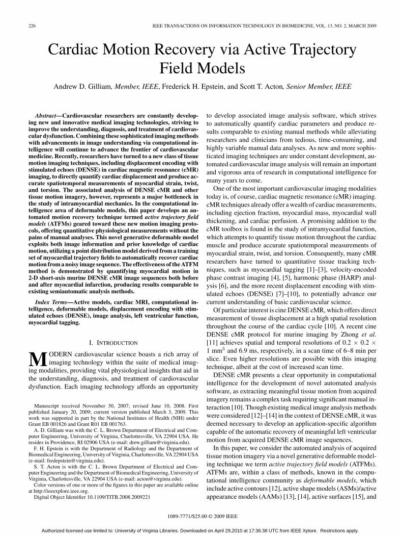
226 IEEE TRANSACTIONS ON INFORMATION TECHNOLOGY IN BIOMEDICINE, VOL. 13, NO. 2, MARCH 2009
Cardiac Motion Recovery via Active TrajectoryField Models
Andrew D. Gilliam, Member, IEEE, Frederick H. Epstein, and Scott T. Acton, Senior Member, IEEE
Abstract—Cardiovascular researchers are constantly develop-ing new and innovative medical imaging technologies, striving toimprove the understanding, diagnosis, and treatment of cardiovas-cular dysfunction. Combining these sophisticated imaging methodswith advancements in image understanding via computational in-telligence will continue to advance the frontier of cardiovascularmedicine. Recently, researchers have turned to a new class of tissuemotion imaging techniques, including displacement encoding withstimulated echoes (DENSE) in cardiac magnetic resonance (cMR)imaging, to directly quantify cardiac displacement and produce ac-curate spatiotemporal measurements of myocardial strain, twist,and torsion. The associated analysis of DENSE cMR and othertissue motion imagery, however, represents a major bottleneck inthe study of intramyocardial mechanics. In the computational in-telligence area of deformable models, this paper develops an au-tomated motion recovery technique termed active trajectory fieldmodels (ATFMs) geared toward these new motion imaging proto-cols, offering quantitative physiological measurements without thepains of manual analyses. This novel generative deformable modelexploits both image information and prior knowledge of cardiacmotion, utilizing a point distribution model derived from a trainingset of myocardial trajectory fields to automatically recover cardiacmotion from a noisy image sequence. The effectiveness of the ATFMmethod is demonstrated by quantifying myocardial motion in2-D short-axis murine DENSE cMR image sequences both beforeand after myocardial infarction, producing results comparable toexisting semiautomatic analysis methods.
Index Terms—Active models, cardiac MRI, computational in-telligence, deformable models, displacement encoding with stim-ulated echoes (DENSE), image analysis, left ventricular function,myocardial tagging.
I. INTRODUCTION
MODERN cardiovascular science boasts a rich array ofimaging technology within the suite of medical imag-
ing modalities, providing vital physiological insights that aid inthe understanding, diagnosis, and treatment of cardiovasculardysfunction. Each imaging technology affords an opportunity
Manuscript received November 30, 2007; revised June 10, 2008. Firstpublished January 20, 2009; current version published March 3, 2009. Thiswork was supported in part by the National Institutes of Health (NIH) underGrant EB 001826 and Grant R01 EB 001763.
A. D. Gilliam was with the C. L. Brown Department of Electrical and Com-puter Engineering, University of Virginia, Charlottesville, VA 22904 USA. Heresides in Providence, RI 02906 USA (e-mail: [email protected]).
F. H. Epstein is with the Department of Radiology and the Department ofBiomedical Engineering, University of Virginia, Charlottesville, VA 22904 USA(e-mail: [email protected]).
S. T. Acton is with the C. L. Brown Department of Electrical and Com-puter Engineering and the Department of Biomedical Engineering, University ofVirginia, Charlottesville, VA 22904 USA (e-mail: [email protected]).
Color versions of one or more of the figures in this paper are available onlineat http://ieeexplore.ieee.org.
Digital Object Identifier 10.1109/TITB.2008.2009221
to develop associated image analysis software, which strivesto automatically quantify cardiac parameters and produce re-sults comparable to existing manual methods while alleviatingresearchers and clinicians from tedious, time-consuming, andhighly variable manual data analyses. As new and more sophis-ticated imaging techniques are under constant development, au-tomated cardiovascular image analysis will remain an importantand vigorous area of research in computational intelligence formany years to come.
One of the most important cardiovascular imaging modalitiestoday is, of course, cardiac magnetic resonance (cMR) imaging.cMR techniques already offer a wealth of cardiac measurements,including ejection fraction, myocardial mass, myocardial wallthickening, and cardiac perfusion. A promising addition to thecMR toolbox is found in the study of intramyocardial function,which attempts to quantify tissue motion throughout the cardiacmuscle and produce accurate spatiotemporal measurements ofmyocardial strain, twist, and torsion. Consequently, many cMRresearchers have turned to quantitative tissue tracking tech-niques, such as myocardial tagging [1]–[3], velocity-encodedphase contrast imaging [4], [5], harmonic phase (HARP) anal-ysis [6], and the more recent displacement encoding with stim-ulated echoes (DENSE) [7]–[10], to potentially advance ourcurrent understanding of basic cardiovascular science.
Of particular interest is cine DENSE cMR, which offers directmeasurement of tissue displacement at a high spatial resolutionthroughout the course of the cardiac cycle [10]. A recent cineDENSE cMR protocol for murine imaging by Zhong et al.[11] achieves spatial and temporal resolutions of 0.2 × 0.2 ×1 mm3 and 6.9 ms, respectively, in a scan time of 6–8 min perslice. Even higher resolutions are possible with this imagingtechnique, albeit at the cost of increased scan time.
DENSE cMR presents a clear opportunity in computationalintelligence for the development of novel automated analysissoftware, as extracting meaningful tissue motion from acquiredimagery remains a complex task requiring significant manual in-teraction [10]. Though existing medical image analysis methodswere considered [12]–[14] in the context of DENSE cMR, it wasdeemed necessary to develop an application-specific algorithmcapable of the automatic recovery of meaningful left ventricularmotion from acquired DENSE cMR image sequences.
In this paper, we consider the automated analysis of acquiredtissue motion imagery via a novel generative deformable model-ing technique we term active trajectory field models (ATFMs).ATFMs are, within a class of methods, known in the compu-tational intelligence community as deformable models, whichinclude active contours [12], active shape models (ASMs)/activeappearance models (AAMs) [13], [14], active surfaces [15], and
1089-7771/$25.00 © 2009 IEEE
Authorized licensed use limited to: University of Virginia Libraries. Downloaded on April 29,2010 at 17:36:38 UTC from IEEE Xplore. Restrictions apply.

GILLIAM et al.: CARDIAC MOTION RECOVERY VIA ACTIVE TRAJECTORY FIELD MODELS 227
Fig. 1. Single-frame murine DENSE cMR analysis. (a) Short-axis anatomical diagram. (b) Magnitude image. (c) Horizontal encoded phase image. (d) Phase-unwrapped image. (e) Displacement field.
splines [16]. The ATFM method introduced here exploits bothimage information and prior knowledge of the cardiac motionwe wish to recover, using a training set of myocardial trajectoryfields to automatically recover tissue motion from a noisy imagesequence. This technique is able to eliminate our dependence onfinite-element approximations of complex biomechanical mod-els by instead deriving a myocardial motion model directly fromreal-world training data.
As with ASMs/AAMs, the ATFM technique relies on ourability to represent a motion trajectory field as a discrete set ofspatiotemporal points. After choosing points in the same man-ner from every trajectory field within a training set of fields,we can examine the statistics of these labeled point positionsand develop a point distribution model describing how the spa-tiotemporal locations vary. We are then able to move about inthis model space by varying a small number of parameters as-sociated with the modes of variation within the training data.To automatically recover motion from a noisy image sequence,we must choose parameter values that best fit model to noisyimagery.
The development of the ATFM motion recovery techniqueis wrought with challenges, including the preliminary semi-automatic analysis of a training set of complex spatiotempo-ral trajectory fields, the characterization of variability withinthe training set, and a solution to a combinatorial optimizationproblem of searching for the trajectory field of best fit to a noisyimage sequence. As DENSE cMR resolutions are well suitedfor studies that use imaging of transgenic mice to elucidate theroles of individual genes in contractile function, we focus ourefforts on left ventricular motion recovery within 2-D short-axismurine cine DENSE cMR imagery, discussed in detail in thenext section. Though we concentrate on the analysis of this sin-gle imaging technique, the ATFM method can be adapted to 2-Dhuman DENSE cMR imagery (differing primarily in scale andresolution), other displacement data such as myocardial tagging(differing primarily in resolution), as well as 3-D DENSE cMRmotion recovery (requiring an extension to 3-D space and 3-Ddisplacements).
In the next section, we present necessary background infor-mation including an in-depth discussion of a state-of-the-artDENSE cMR analysis, as well as an introduction to deformablemodels and their application to cardiac segmentation problems.We then develop the proposed ATFM method in detail, including
a novel preliminary semiautomatic training set analysis, modeldefinition, and the final automated motion field recovery. Wedemonstrate the effectiveness of the ATFM method by quanti-fying myocardial motion in 2-D short-axis murine DENSE cMRimage sequences both before and after myocardial infarction.
II. BACKGROUND
This section presents two topics essential to the proposedresearch: a discussion on the typical cine DENSE cMR imageanalysis method and a short review of deformable models forcardiac segmentation.
A. Typical Cine DENSE cMR Analysis
A typical MR acquisition consists of a complex valueddataset, and conventionally researchers and clinicians discard allphase information and make use of the magnitude informationonly. DENSE cMR, however, exploits this phase space by di-rectly encoding tissue displacement into the phase-reconstructedimages [7]. DENSE cMR first uses a spatial magnetic fieldgradient to impart a location-dependent phase shift to the MRsignal at the initial cardiac configuration. A similar gradient isapplied at subsequent cardiac configurations such that, if no dis-placement occurred, the initial imparted phase shift would beremoved. Any residual phase shift remaining after applicationof the second gradient pulse directly reflects tissue displacementthat occurred during the time between the two gradient pulses.A more detailed discussion of the cine DENSE cMR acquisitionmethod, beyond the scope of this paper, can be found in [8].
Fig. 1 illustrates a typical single-frame DENSE cMR acqui-sition and analysis. We acquire complex DENSE cMR imageryconsisting of magnitude [Fig. 1(b)] and phase [Fig. 1(c)] infor-mation, where phase is directly proportional to displacement ina single direction. In this example, phase is encoded in the hor-izontal direction. A similar procedure produces phase imageryencoded in the vertical direction (not shown).
Let us now briefly consider the typical transformation of a2-D+time DENSE cMR image acquisition into meaningfultissue motion, as described by Spottiswoode et al. [10]. AsMR phase is inherently bounded between −π and π, large dis-placements produce wrapping artifacts, visible at the 3 o’clockposition in Fig. 1(c). This wrapping effect is corrected by a2-D phase unwrapping technique described in [17], known as
Authorized licensed use limited to: University of Virginia Libraries. Downloaded on April 29,2010 at 17:36:38 UTC from IEEE Xplore. Restrictions apply.

228 IEEE TRANSACTIONS ON INFORMATION TECHNOLOGY IN BIOMEDICINE, VOL. 13, NO. 2, MARCH 2009
Fig. 2. Typical murine DENSE cMR trajectory field.
quality-guided path following. This technique unwraps thephase image one pixel at a time along a 2-D path guided bythe phase quality image, proportional to the variance of the par-tial derivatives of the locally unwrapped phase within a smallneighborhood around each pixel. Fig. 1(d) illustrates unwrappedphase values within the myocardial region of interest, wheredark pixels represent movement to the left and bright pixelsrepresent movement to the right.
After unwrapping the vertically encoded phase image (notshown) via a similar procedure, the associated horizontal andvertical displacements are combined to obtain the displacementfield illustrated in Fig. 1(e). This field consists of noisy vectorswith heads located at pixel centers and tails located at the pixelpoints of origin. A series of these single-frame displacementfields measured over the course of the cardiac cycle is con-catenated to recover a meaningful myocardial trajectory field,as illustrated in Fig. 2. This generally involves spatial filter-ing, linear interpolation to determine frame-to-frame motion,measurement of coarse trajectories using these frame-to-framevectors, and temporal smoothing of the coarse trajectories viaFourier basis functions.
To gain more insight into myocardial function, strain can bedirectly calculated from the recovered trajectory fields accordingto the method described in [18]. Consider a single trajectory[x0(t), y0(t)] and its nearest neighbor [x1(t), y1(t)] defined bythe Euclidean distance between spatial origins. We define thedistance between these points at an arbitrary time t as
dx(t) =[
x1(t) − x0(t)y1(t) − y0(t)
]. (1)
A 2-D deformation gradient tensor F (t), of size [2 × 2], mapsvectors from the original configuration dx(0) to the currentconfiguration dx(t) and is defined as
dx(t) = F (t)dx(0). (2)
Given a trajectory with at least two neighbors, F (t) canbe determined via a least-squares technique. The associatedLagrangian strain tensor S(t) of size [2 × 2] is given as
S(t) =12
[F (t)T F (t) − I
](3)
where I is the identity matrix. This 2-D Cartesian strain tensoris typically decomposed into its radial and circumferential com-ponents, pointing toward the myocardial center and along thecircumference of the myocardium, respectively. Fig. 3 illustrates
Fig. 3. End-systolic (left) radial and (right) circumferential strain.
typical end-systolic (full cardiac contraction) strain fields. Notethat the average end-systolic radial strain of approximately 0.35and average end-systolic circumferential strain of approximately−0.15 are in good agreement with previous measurements ofstrain in the mouse heart [9], [19].
Though this state-of-the-art analysis method provides reveal-ing myocardial trajectory fields, it does suffer from several dis-tinct disadvantages. It first requires a significant amount of userinput, as the left ventricle must be manually delineated on at leastone frame, and the phase unwrapping algorithm performance isimproved when the left ventricle is delineated on all frames. Inour laboratory, manual myocardial segmentation within a typ-ical murine DENSE cMR acquisition of 30 frames may take5–10 min. Performing this segmentation on multiple datasetsquickly becomes monotonous and time-consuming. Manualsegmentation is especially problematic early in the cardiac cycle,as blood encoded with the DENSE cMR pulse sequence remainsin the blood pool, virtually eliminating myocardial contrast. Ad-ditionally, little effort is directed toward noise compensationamong the displacement vectors beyond some spatial filteringand an individual temporal fit to each trajectory. There is aclear opportunity for a more automated solution via advances incomputational intelligence.
B. Deformable Models for Cardiac Segmentation
One of the most successful classes of medical image segmen-tation methods, termed deformable models, attempts to exploitboth image information and prior knowledge of the anatomicalstructure to be delineated. Here, we will consider some of themost relevant prior research on deformable models for myocar-dial segmentation.
The popularization of deformable models for image segmen-tation is commonly attributed to the introduction of active con-tours by Kass et al. [12]. An active contour captures a desiredimage feature by minimizing a corresponding energy functional,typically the weighted sum of an image-based external energy,attracting the contour to features of interest, and a contour-based internal energy, ensuring a smooth segmentation solution.While active contours have been utilized for cardiac segmenta-tion by a number of researchers [20]–[22], this method does notallow for the inclusion of any known size, shape, or appearanceinformation.
Cootes et al. [13] introduced a technique termed ASMs thatintegrates an expected feature shape into the deformable modelframework. An ASM measures the variability of a set of trainingshapes via principal component analysis (PCA) and constrains
Authorized licensed use limited to: University of Virginia Libraries. Downloaded on April 29,2010 at 17:36:38 UTC from IEEE Xplore. Restrictions apply.

GILLIAM et al.: CARDIAC MOTION RECOVERY VIA ACTIVE TRAJECTORY FIELD MODELS 229
the segmentation solution to this shape space. Given the propertraining set, the corresponding model is able to encapsulateall shape variability within a small subset of principal compo-nent weighting parameters. The ASM technique has been usedsuccessfully to delineate a number of different anatomicalshapes including the myocardium [13], [23], but the locationof anatomical features with ASM is driven by often spuriouslow-level image features.
To improve their segmentation results, Cootes et al. [14]introduced an extension to the ASM technique termed AAMs.This technique considers not only the expected object shape,but also the expected pattern of intensity or color in and aroundthe object termed the object appearance. AAMs belong to asubclass of deformable models termed generative deformablemodels, as we can generate a synthetic image corresponding toany shape and appearance within the model space. Segmentationvia this method proceeds via analysis-by-synthesis, finding theset of model parameters that minimizes the difference betweennoisy imagery and model-derived synthetic imagery. AAM tech-niques have shown significant promise in the segmentations ofa wide range of cardiac imagery [24]–[27].
Unfortunately, the aforementioned algorithms are ill-suitedto the analysis of cardiac displacement imagery such asDENSE cMR. Myocardial edge contrast is nonexistent earlyin the cardiac cycle, causing purely edge-based segmentationmethods such as active contours and ASMs to fail or requiresignificant correction. AAMs show more promise as they couldconsider the entire myocardial muscle appearance rather thanjust the myocardial borders; however, we would still requirethe complex displacement analysis methods discussed in theprevious section to recover meaningful myocardial motionfrom the segmentation solution.
We therefore endeavor to create a novel motion recoverymethod geared specifically to tissue motion imagery. Ofparticular interest is the concept of a generative deformablemodel, as it is a relatively simple matter to generate idealDENSE cMR phase imagery from a given trajectory field. Wehypothesize that it is possible to characterize the variabilityof a set of training trajectory fields and then recover cardiacmotion via an analysis-by-synthesis technique that minimizesthe difference between ideal phase imagery and a noisy DENSEcMR sequence. The ATFM approach described in this paper ac-complishes this goal, developing a novel generative deformablemodel to recover cardiac motion in tissue motion imagery thatexploits known intramyocardial spatiotemporal variability.
III. METHODS
This section presents an in-depth description of the proposedATFM technique toward the goal of automated motion recoveryfrom tissue displacement imagery. The ATFM method definesa point distribution model characterizing the variability ofspatiotemporal myocardial landmarks within a training setof discrete trajectory fields and then automatically recoversmotion from a noisy image sequence by finding the best fit ofmodel to imagery.
The development of this method is not a trivial task, as wemust consider the preliminary semiautomatic analysis of atraining set of DENSE cMR image sequences, a characteriza-tion of variation within recovered trajectory field training set,and a solution to the combinatorial optimization problem ofsearching a noisy image sequence for the trajectory field ofbest fit. As mentioned previously, though we focus our effortson murine DENSE cMR imagery, the ATFM method can beadapted to human data, other tissue motion imaging techniques,as well as 3-D motion recovery.
A. Training Set Analysis
The current state-of-the-art DENSE cMR motion recoverymethod discussed in Section II-A does not lend itself to the con-struction of our cardiac point distribution model, as each heartin the training set is sampled at a unique set of spatiotemporallocations. To address this concern, as well as improve noisecompensation within the acquired datasets, we introduce anovel semiautomatic DENSE cMR analysis method. Thismethod defines a smooth and continuous myocardial trajectoryfield via two spatiotemporal splines, given phase-unwrappedimage sequences and expertly delineated myocardial contours.Note that this novel semiautomatic training set analysis is onlya stepping stone toward the fully automated ATFM solutiondiscussed in subsequent sections.
Consider deformation in a single direction. Let zn , n ∈[1, . . . , N ] represent the finite set of N irregularly spacedspatiotemporal deformations at the Cartesian coordinates(xn , yn ) ∈ Ω and temporal locations tn ∈ [0, . . . , 1] (varyingfrom the start to the end of the cardiac cycle, respectively).We define the function f(x, y, t) as the unique solution thatminimizes
Espline(f) = λ1
∫ 1
0
[∫ ∫Ω(f 2
xx + 2f 2xy + f 2
yy )dx dy
]dt
+ λ2
∫ ∫Ω
[∫ 1
0f 2
ttdt
]dx dy (4)
+ (1 − λ1 − λ2)N∑
n=1
||zn − f(xn , yn , tn )||
where (λ1 , λ2) ∈ [0, . . . , 1] define the relative weight betweenenergy terms and fdd is the second derivative of f(x, y, t) inthe direction d. We apply an additional boundary constraintto our solution that forces trajectories to begin and end at theirrespective spatial origins by requiring zero-valued deformationsat these temporal locations.
The first energy term in (4) is derived from a 2-D thin-platespline [16], [28], the 2-D analog of the 1-D cubic spline. Thisterm quantifies the spatial bending energy at each temporallocation in the function f(x, y, t), ensuring a spatially smoothdeformation field. The second energy term is derived froma 1-D cubic spline [16]. This term quantifies the temporalcurvature of our solution, i.e., the sum of squared accelerationacross time within the spatial region of interest, ensuring atemporally smooth deformation field when minimized. Our
Authorized licensed use limited to: University of Virginia Libraries. Downloaded on April 29,2010 at 17:36:38 UTC from IEEE Xplore. Restrictions apply.

230 IEEE TRANSACTIONS ON INFORMATION TECHNOLOGY IN BIOMEDICINE, VOL. 13, NO. 2, MARCH 2009
Fig. 4. Typical spatiotemporal spline fitting. (Top row) Original displacementfields. (Bottom row) Corresponding spline fields.
Fig. 5. End-systolic (left) radial and (right) circumferential strain using thespatiotemporal spline method.
energy functional also includes an L2-norm data constraint,forcing the solution toward the least-squares approximation ofthe input deformations. Minimizing the weighted sum of thesethree energy terms produces a spatially and temporally smoothsolution that closely matches the input deformation data. Wenote that although (4) could be formulated in the L1-norm sense,the range and variability of noise present in the displacementdata has lent itself to an L2-norm minimization scheme.
To ensure we do not oversmooth and lose local tissue motionfeatures, our choice of λ1 and λ2 is governed by the cross-validation technique described in [29]. We divide our entiretraining set of displacement fields into estimation data and vali-dation data, choosing D random displacements from each fieldas the validation data. We then search the parameter space forthe spatiotemporal splines derived from the estimation data thatbest fit the validation data.
Fig. 4 illustrates a typical spatiotemporal spline fit. The toprow shows the original displacement fields and the bottom rowshows the corresponding spline deformation fields. We can ad-ditionally calculate strain fields by discretizing this continuoustrajectory field and using the method described in Section II-A,as illustrated in Fig. 5.
B. Model Definition
With a training set of continuous spatiotemporal trajectoryfields in hand, we may now consider the construction of a tra-jectory field model for subsequent motion recovery [30]. This
Fig. 6. Alignment of training data to reference coordinate system. (a) Expertsegmentation at rest. (b) Corresponding trajectory field. (c) Aligned restingsegmentation. (d) Aligned trajectory field.
is a three-step process, wherein we align the training data to asingle reference coordinate system, discretize each continuoustrajectory field at the same set of myocardial landmarks and car-diac phases, and define a point distribution model that measuresthe variability among these discrete trajectory fields via PCA.
Let us first consider training data alignment. The center ofeach resting heart within the training set lies at a different spatiallocation within its respective image acquisition space. Addition-ally, each resting heart has a different orientation with respect tothis center. Finally, two hearts that differ in size may still exhibitthe same motion characteristics. To more accurately character-ize motion variability within the training set, we eliminate thesedifferences in cardiac position, orientation, and scale by align-ing each resting heart to a single reference coordinate system.We refer to the resulting aligned trajectory field as normalized.
As it is difficult to manually delineate the myocardium inDENSE cMR imagery early in the cardiac cycle, we define theresting heart by projecting expert drawn contours and features[right ventricle insertion points; see Fig. 1(a)] from the frame ofgreatest myocardial contrast back to their resting positions viathe spatiotemporal spline definition. Alignment of this restingconfiguration is achieved via a Procrustes analysis [13], [31],with respect to a reference myocardium centered at the spatialorigin with right ventricle insertion points aligned to the verti-cal axis and unity epicardial radius. Fig. 6 illustrates a typicalalignment, transforming a resting configuration and motion field[Fig. 6(a) and (b)] to the reference coordinate system [Fig. 6(c)and (d)]. To aid visualization, the right ventricle insertion pointsare marked as squares.
Authorized licensed use limited to: University of Virginia Libraries. Downloaded on April 29,2010 at 17:36:38 UTC from IEEE Xplore. Restrictions apply.

GILLIAM et al.: CARDIAC MOTION RECOVERY VIA ACTIVE TRAJECTORY FIELD MODELS 231
Fig. 7. Trajectory field discretization. (a) Sampling template. (b) Normalizedmyocardial borders. (c) Discretized myocardial definition. (d) Correspondingtrajectory field.
After alignment, the next challenge in model construction isthe spatial and temporal discretization of each trajectory fieldwithin the training set using an identical set of myocardiallandmarks. To elaborate, if trajectory #10 in the first trainingfield corresponds to the anterior right ventricle insertionpoint, trajectory #10 in all training fields must correspond tothe same anterior right ventricle insertion point. We achievethis trajectory matching by mapping a sampling templateto the aligned resting myocardial definition and recoveringthe discrete trajectories associated with each of these spatialorigins. Fig. 7 illustrates this procedure, using a sampling tem-plate [Fig. 7(a)] to sample the normalized resting myocardialdefinition [Fig. 7(b)], and produce a discrete set of restingspatial locations [Fig. 7(c)] and a corresponding trajectoryfield [Fig. 7(d)]. Temporal discretization is achieved bysampling each training trajectory field at a known set of cardiacphases.
Given the set of normalized discrete training trajectoryfields, we are able to characterize the data variability viaPCA. Let Xijk = [xijk , yijk ] represent the x–y position of theith trajectory at the jth cardiac phase of the kth normalizedtrajectory field, where i ∈ [1, . . . , Ni], j ∈ [1, . . . , Nj], andk ∈ [1, . . . , Nk]. We vectorize the kth trajectory field as
Φk = [
phase 1︷ ︸︸ ︷X1,1,k ,X2,1,k , . . . , XN i,1,k , . . .
. . .
phase j︷ ︸︸ ︷X1,j,k , . . . , XN i,j,k , . . .
phase N j︷ ︸︸ ︷,X1,N j,k , . . . , XN i,N j,k ]T
(5)
of size [(2NiNj) × 1]. The average normalized trajectoryfield is defined as
Φ =1
Nk
N k∑k=1
Φk (6)
and the sample distribution covariance matrix is defined by
Cov =1
Nk
N k∑k=1
(Φk − Φ)(Φk − Φ)T . (7)
The eigenvectors P of the covariance matrix define themodes of variation within the training set and the correspondingeigenvalues describe the relative significance of each mode. Thepercentage of variation accounted for by each eigenvector isdefined as the corresponding eigenvalue divided by the sum of alleigenvalues.
We can recover any normalized trajectory field within thetraining set by a linear combination of the average normalizedtrajectory field with the modes of variation. Moreover, we canapproximate any normalized trajectory field within the trainingdata by a linear combination of the average normalized trajectoryfield with the most significant eigenvectors, termed principalcomponents, as in
Mk ≈ M + P bk (8)
where P is a subset of the modes of variation P and bk is a setof weighting coefficients corresponding to the kth trajectoryfield. In practice, we define an amount of variation we wishto approximate (e.g., 95% variation) and use the principalcomponents that account for this level of variation.
These principal components of variation, combined witha rotation, translation, and scale to transform the normalizedtrajectory fields back to the image space of a new DENSE cMRsequence, define our trajectory field model. We are able toachieve any trajectory field within the search space by varyinga small number of parameters, i.e., the principal componentweights, orientation, translation, and scale.
C. ATFM Motion Recovery
Given the aforementioned model space definition, we areable to attempt the automatic recovery of cardiac motion from anewly acquired DENSE cMR sequence. In this section, we willfirst consider the search criterion that defines the best matchbetween trajectory field and noisy imagery. We then discussour method of attack to solve the combinatorial optimizationproblem of locating the best match within the model searchspace.
We measure the correspondence between a given trajectoryfield and a noisy image sequence via an analysis-by-synthesistechnique. Given any myocardial trajectory field, one cansynthesize a set of “ideal” DENSE cMR values that representthe noise-free DENSE cMR data that would have produced thetrajectory field in question. This is accomplished by measuringdisplacement from the spatial origin for each trajectory ateach phase of the cardiac cycle, and scaling according to theDENSE cMR encoding parameter. Let Ψ = [xn (t), yn (t)],
Authorized licensed use limited to: University of Virginia Libraries. Downloaded on April 29,2010 at 17:36:38 UTC from IEEE Xplore. Restrictions apply.

232 IEEE TRANSACTIONS ON INFORMATION TECHNOLOGY IN BIOMEDICINE, VOL. 13, NO. 2, MARCH 2009
n ∈ [1, . . . , N ] represent a set of N temporally continuoustrajectories. The ideal DENSE cMR value corresponding toeach trajectory at some time t are defined as
Ixn (t) = ke [xn (t) − xn (0)]
Iyn (t) = ke [yn (t) − yn (0)]
(9)
where Ixn (t) and Iy
n (t) are the x-direction and y-directionideal values, respectively, and ke is the DENSE cMR encodingparameter.
Let Ix(x, y, t) and Iy (x, y, t) represent the discrete noisyDENSE cMR image sequence encoded in the x-direction andy-direction, respectively, where (x, y) ∈ Ω and t takes on k ∈[1, . . . , Nk] unique values within the range [0, . . . , 1]. We definethe distance D(·) ∈ [0,∞]of a given trajectory field Ψ to a noisyDENSE cMR sequence in the L2-norm sense as
D(Ψ) =N k∑k=1
N∑n=1
(Ix [xn (tk ), yn (tk ), tk ] − Ixn [tk ])2
+N k∑k=1
N∑n=1
(Iy [xn (tk ), yn (tk ), tk ] − Iyn [tk ])2.
(10)
To determine the trajectory field of highest similarity to agiven noisy DENSE cMR image sequence, we must traverse thecombinatorial optimization space of the trajectory field modelto locate the field of minimum distance defined by (10). Adirect search of the model space for the globally minimumdistance is computationally prohibitive. We therefore search themodel space via simulated annealing [32], analogous to themetallurgical annealing process wherein hot metal is slowlycooled to form a perfect crystalline structure with minimumfree energy. In simulated annealing, we slowly “cool” our modelsearch to locate the point within the model space of minimumenergy, where “coolness” reflects the decreasing probability ofmoving to an inferior solution in terms of an energy measurethat quantifies solution quality.
Let Ψ1 represent the current trajectory field state of ourmodel search, with some distance to the noisy DENSE cMRimagery D(Ψ1). We perturb the model parameters associatedwith this trajectory field, obtaining a new trajectory field Ψ2with corresponding distance D(Ψ2), and consider changingthe state of our system to this new trajectory field. If weaccepted only good moves, i.e., when D(Ψ2) < D(Ψ1), wewould quickly find a local minimum within our search space,but would become trapped in this local minimum and unableto discover the global minimum. In simulated annealing, weutilize a more sophisticated acceptance condition, i.e., changesin the model state are accepted if
11 + exp[(D(Ψ2) − D(Ψ1))/T ]
< U(0, 1) (11)
where T is the current system temperature and U(0, 1) is auniformly distributed random variable between 0 and 1. At hightemperatures, all changes have an equal probability of beingaccepted. As the temperature is lowered, moves that increasethe system energy have a lower probability of acceptance, until
the algorithm reduces to the greedy method at T = 0, acceptingonly those moves that reduce the system energy.
We begin the annealing process at some initial temperatureT0 (in which every possible change to the system state is equallyprobable) and follow a geometric annealing schedule of reduc-ing the temperature by some constant τ (Tk+1 = τTk ) at eachtemperature until a final temperature Tfinal is reached. At eachtemperature, we test NC candidate points within the modelspace. When Tfinal is achieved, the algorithm is essentially agreedy one, for which achieving a local minimum is guaranteed.To provide a small reduction in the search space dimensionality,we define the orientation, position, and scaling of the normal-ized trajectory field model to the DENSE cMR image spacevia a small number of user-defined myocardial landmarks. Thecomputational complexity of this annealing algorithm scaleslinearly according to the number of principal components used,the number of candidate point tested at each temperature, andthe number of temperatures evaluated.
IV. RESULTS
To demonstrate the effectiveness of the ATFM techniquefor tissue motion recovery within displacement imagery, weattempted to quantify myocardial motion in 2-D short-axismurine DENSE cMR imagery. We considered a datasetcontaining two distinct murine conditions: healthy mice andmice seven days after induction of an experimental heart attack.The former condition consisted of 13 healthy mice imaged witha standard 2-D cine DENSE cMR protocol, obtaining between20 and 27 short-axis midplane images of the cardiac cycle permouse. The latter condition consisted of six unhealthy micewith an induced myocardial infarction in the anterolateral wall,again imaged with a standard 2-D cine DENSE cMR protocol,obtaining between 17 and 21 short-axis midplane images of thecardiac cycle per mouse. For each image sequence, a trainedtechnician delineated the endocardial and epicardial borders onevery frame and labeled the right ventricle insertion points onthe last sequence frame.
In this section, we present our findings. We first illustratethe failings of traditional segmentation methods toward thegoal of automated cardiac motion recovery. We then present aphysiological comparison of DENSE cMR analysis techniques,evaluating agreement between the traditional semiautomaticDENSE cMR analysis described in Section II-A and the novelATFM technique presented within this paper.
A. Motion Analysis via Traditional Automated Segmentation
Semiautomatic DENSE cMR analysis can be broken intotwo distinct steps: 1) the manual delineation of myocardialborders within the magnitude imagery throughout the cardiaccycle by a trained technician and 2) tissue motion recoveryvia phase analysis as described in Section II-A or III-A. Thisnatural division implies a plausible alternative for automatedtissue motion recovery, i.e., the delineation of endocardial andepicardial borders via a traditional automated segmentationtechnique followed by DENSE cMR phase analysis. Manyresearchers have had significant success with deformable
Authorized licensed use limited to: University of Virginia Libraries. Downloaded on April 29,2010 at 17:36:38 UTC from IEEE Xplore. Restrictions apply.

GILLIAM et al.: CARDIAC MOTION RECOVERY VIA ACTIVE TRAJECTORY FIELD MODELS 233
Fig. 8. Example active contour segmentation. (a) Early in the cardiac cycle.(b) Late in the cardiac cycle.
models for cMR segmentation [20], [22]–[25], and thus, wechose to examine such an alternative.
To evaluate the effectiveness of such a system, we consideredmyocardial segmentation via the classical active contoursegmentation technique described in [12]. The active contourwas driven by a negative gradient magnitude external force,derived from DENSE cMR magnitude imagery. Endocardialand epicardial borders were separately initialized on each frameof a given image sequence as circles particularly close to thecorrect myocardial locations. After segmentation, we appliedthe traditional DENSE cMR phase analysis method described inSection II-A to recover tissue motion.
This traditional segmentation technique was unable toaccurately define myocardial borders on every image frame ofany healthy DENSE cMR image sequence, which rendered thesubsequent trajectory fields meaningless. Across the dataset,endocardial and epicardial active contour segmentation hadroot mean square errors of 0.87 and 0.38 mm, respectively,exceedingly large as compared to the average myocardial wallthickness of ∼2 mm.
Fig. 8 illustrates two typical segmentation problems. Fig. 8(a)shows an attempted segmentation early in the cardiac cycle,which fails due to a lack of myocardial contrast. Fig. 8(b) showsan attempted segmentation at the end of the cardiac cycle, whichfails as the epicardial border is drawn to the higher edge strengthof the endocardial border. These failures lead us to concludethat a more comprehensive tissue motion analysis solution isrequired.
B. ATFM Motion Recovery Analysis
Let us now consider the evaluation of the novel automatedATFM motion recovery technique. As we desire to automat-ically reproduce physiologically meaningful measurements ofcardiac motion similar to traditional semiautomatic techniques,we compare strain values measured by both the automatedATFM technique and a traditional semiautomatic DENSE cMR
Fig. 9. Typical six-segment strain analysis. (a) Anatomical diagram. (b) Radialstrain. (c) Circumferential segmental strain.
analysis. We will quantify agreement between the two methodsvia correlation and a Bland–Altman analysis [33].
The preliminary analysis of the ATFM training datautilized the same manually delineated myocardial borders andmyocardial features of interest as the traditional DENSE cMRanalysis. ATFM analysis was performed via a leave-one-outcross-validation study, attempting motion recovery on eachnoisy DENSE cMR image sequence within the dataset usingthe remaining trajectory fields as training data. Parameters usedfor a given motion recovery technique remained consistentacross the entire dataset.
Spatiotemporal strain was calculated at many points withinthe myocardium, as described in Section II-A. For comparisonacross motion recovery methods, we divide the left ventricle intosix segments, as standardized in [34] and illustrated in Fig. 9(a).We quantify and plot the average radial and circumferentialstrain in each segment throughout the cardiac cycle, as illustratedin Fig. 9(b) and (c), respectively.
As a quantitative comparison, we sample ten evenly spacedcardiac phases throughout the cardiac cycle for each leftventricular segment, resulting in 60 data points per heart. Thetwo methods show good correlation, as illustrated in Fig. 10(a)and (b). The average correlation value of radial strain is 0.83(p < 0.001) and the average correlation value of circumferentialstrain is 0.86 (p < 0.001). A Bland–Altman analysis of the data,shown in Fig. 10(c) and (d), reveals further similarity betweenthe two methods. We measure an average difference of −0.02in radial strain (95% confidence interval of −0.16 to 0.11) andan average difference of <0.01 in circumferential strain (95%confidence interval of −0.05 to 0.07) between the two methods.
Authorized licensed use limited to: University of Virginia Libraries. Downloaded on April 29,2010 at 17:36:38 UTC from IEEE Xplore. Restrictions apply.

234 IEEE TRANSACTIONS ON INFORMATION TECHNOLOGY IN BIOMEDICINE, VOL. 13, NO. 2, MARCH 2009
Fig. 10. Strain correlation of traditional and ATFM methods. Radial/circum-ferential [(a) and (b)] correlation and [(c) and (d)] Bland–Altman analyses.
V. CONCLUSION
The medical image analysis field continues to benefit fromthe advent of new and exciting medical imaging techniques.One class of techniques, tissue motion imaging such as DENSEcMR, boasts the ability to quantify tissue motion throughoutthe cardiac muscle and produce accurate spatiotemporalmeasurements of myocardial strain, twist, and torsion. Thoughcurrent manual and semiautomatic analysis methods cangenerate remarkable trajectory fields, researchers and clinicianswould benefit from a fully automated analysis softwarepackage.
As an advance in computational intelligence in biomedicine,this paper has presented a novel generative deformable modelingtechnique, termed ATFM, for the automated analysis of acquiredtissue motion imagery. The development of this technique wasa complex task, requiring a novel preliminary semiautomaticDENSE cMR analysis technique, the alignment and subsequentvariability characterization of a complex set of spatiotemporaltrajectory fields, and the solution of a combinatorial optimiza-tion problem to find the trajectory field of best fit to a givennoisy image sequence.
We validated the ATFM method by quantifying myocardialmotion in 2-D short-axis murine DENSE cMR image sequencesboth before and after myocardial infarction, producing resultscomparable to existing semiautomatic analysis methods.Though we focused our efforts on left ventricular motionrecovery within 2-D short-axis murine cine DENSE cMRimagery, the ATFM method can be adapted to human data,other tissue motion imaging techniques, as well as 3-D motionrecovery.
ACKNOWLEDGMENT
The authors would like to thank several researchers at theUniversity of Virginia for their continued support: Dr. B. A.French of the Departments of Biomedical Engineering,Radiology, and Medicine; Dr. J. A. Hossack of the Departmentof Biomedical Engineering; and Dr. C. M. Kramer of theDepartments of Radiology and Medicine.
REFERENCES
[1] L. Axel and L. Dougherty, “MR imaging of motion with spatial modulationof magnetization,” Radiology, vol. 171, pp. 841–845, 1989.
[2] L. Axel and L. Dougherty, “Heart wall motion: Improved method of spatialmodulation of magnetization for MR imaging,” Radiology, vol. 172,pp. 349–350, 1989.
[3] E. A. Zerhouni, D. M. Parish, W. J. Rogers, A. Yang, and E. P. Shapiro,“Human heart: Tagging with MR imaging—A method for noninvasiveassessment of myocardial motion,” Radiology, vol. 169, pp. 59–63, 1988.
[4] P. V. Dijk, “Direct cardiac NMR imaging of heart wall and blood flowvelocity,” J. Comput. Assisted Tomogr., vol. 8, pp. 429–436, 1984.
[5] D. Bryant, J. Payne, D. Firmin, and D. Longmore, “Measurement of flowwith NMR imaging using gradient pulse and phase difference technique,”J. Comput. Assisted Tomogr., vol. 8, pp. 588–593, 1984.
[6] N. F. Osman, E. R. McVeigh, and J. L. Prince, “Imaging heart motionusing harmonic phase MRI,” IEEE Trans. Med. Imag., vol. 19, no. 3,pp. 186–202, Mar. 2000.
[7] A. H. Aletras, S. Ding, R. S. Balaban, and H. Wen, “DENSE: Displacementencoding with stimulated echoes in cardiac functional MRI,” J. Magn.Reson., vol. 137, pp. 247–252, 1999.
[8] D. Kim, W. D. Gilson, C. M. Kramer, and F. H. Epstein, “Myocardial tissuetracking with two-dimensional cine displacement-encoded MR imaging:Development and initial evaluation,” Radiology, vol. 230, pp. 862–871,2004.
[9] W. D. Gilson, Z. Yang, B. A. French, and F. H. Epstein, “Measurement ofmyocardial mechanics in mice before and after infarction using multislicedisplacement-encoded MRI with 3D motion encoding,” Amer. J. Physiol.Heart Circ. Physiol., vol. 288, pp. H1491–H1497, 2005.
[10] B. S. Spottiswoode, X. Zhong, A. T. Hess, C. M. Kramer, E. M. Meintjes,B. M. Mayosi, and F. H. Epstein, “Tracking myocardial motion from cineDENSE images using spatiotemporal phase unwrapping and temporalfitting,” IEEE Trans. Med. Imag., vol. 26, no. 1, pp. 15–30, Jan. 2007.
[11] X. Zhong, R. Janiczek, B. French, R. Roy, C. Kramer, C. Meyer, andF. Epstein, “Spiral cine DENSE MRI at 7T for quantification of regionalfunction in the mouse heart,” presented at the 16th Sci. Meeting Int. Soc.Magn. Reson. Med., Toronto, ON, Canada, 2008, Program 580.
[12] M. Kass, A. Witkin, and D. Terzopoulos, “Snakes: Active contour models,”Int. J. Comput. Vis., vol. 1, pp. 321–331, 1988.
[13] T. F. Cootes, C. J. Taylor, D. H. Cooper, and J. Graham, “Active shapemodels—Their training and application,” Comput. Vis. Image Under-standing, vol. 61, pp. 38–59, 1995.
[14] T. F. Cootes, G. J. Edwards, and C. J. Taylor, “Active appearance models,”IEEE Trans. Pattern Anal. Mach. Intell., vol. 23, no. 6, pp. 681–685, Jun.2001.
[15] D. Terzopoulos, A. Witkin, and M. Kass, “Constraints on deformablemodels—Recovering 3D shape and nonrigid motion,” Artif. Intell.,vol. 36, pp. 91–123, 1988.
[16] G. Wahba, Spline Models for Observational Data. Philadelphia, PA:SIAM, 1990.
[17] D. C. Ghiglia and M. D. Pritt, Two-Dimensional Phase Unwrapping:Theory, Algorithms and Software. New York: Wiley/Interscience, 1998.
[18] C. Truesdell and W. Noll, The Non-Linear Field Theories of Mechanics.New York: Springer-Verlag, 2004.
[19] R. Zhou, S. Pickup, J. D. Glickson, C. H. Scott, and V. A. Ferrari, “As-sessment of global and regional myocardial function in the mouse usingcine and tagged MRI,” Magn. Reson. Med., vol. 49, pp. 760–764, 2003.
[20] C. Pluempitiwiriyawej, J. M. F. Moura, W. Y.-J. Lin, and H. Chien,“STACS: New active contour scheme for cardiac MR image segmen-tation,” IEEE Trans. Med. Imag., vol. 24, no. 5, pp. 593–603, May 2005.
[21] T. McInerney and D. Terzopoulos, “A dynamic finite element surfacemodel for segmentation and tracking in multidimensional medical imageswith application to cardiac 4D image analysis,” Comput. Med. Imag.Graph., vol. 19, pp. 69–83, 1995.
Authorized licensed use limited to: University of Virginia Libraries. Downloaded on April 29,2010 at 17:36:38 UTC from IEEE Xplore. Restrictions apply.

GILLIAM et al.: CARDIAC MOTION RECOVERY VIA ACTIVE TRAJECTORY FIELD MODELS 235
[22] V. Chalana, D. T. Linker, D. R. Haynor, and K. Yongmin, “A multipleactive contour model for cardiac boundary detection on echocardiographicsequences,” IEEE Trans. Med. Imag., vol. 15, no. 3, pp. 290–298, Jun.1996.
[23] T. F. Cootes, A. Hill, C. J. Taylor, and J. Haslam, “Use of active shapemodels for locating structures in medical images,” Image Vis. Comput.,vol. 12, pp. 355–366, 1994.
[24] S. C. Mitchell, B. P. F. Lelieveldt, R. J. Van Der Geest, H. G. Bosch, J.H. C. Reiver, and M. Sonka, “Multistage hybrid active appearance modelmatching: Segmentation of left and right ventricles in cardiac MR images,”IEEE Trans. Med. Imag., vol. 20, no. 5, pp. 415–423, May 2001.
[25] S. C. Mitchell, J. G. Bosch, B. P. F. Lelieveldt, R. J. Van Der Geest, J. H.C. Reiber, and M. Sonka, “3-D active appearance models: Segmentationof cardiac MR and ultrasound images,” IEEE Trans. Med. Imag., vol. 21,no. 9, pp. 1167–1178, Sep. 2002.
[26] J. G. Bosch, S. C. Mitchell, B. P. F. Lelieveldt, F. Nijland, O. Kamp,M. Sonka, and J. H. C. Reiber, “Automatic segmentation of echocardio-graphic sequences by active appearance motion models,” IEEE Trans.Med. Imag., vol. 21, no. 11, pp. 1374–1383, Nov. 2002.
[27] R. Beichel, H. Bischof, F. Leberl, and M. Sonka, “Robust active appear-ance models and their application to medical image analysis,” IEEE Trans.Med. Imag., vol. 24, no. 9, pp. 1151–1169, Sep. 2005.
[28] F. L. Bookstein, “Principal warps: Thin-plate splines and the decomposi-tion of deformations,” IEEE Trans. Pattern Anal. Mach. Intell., vol. 11,no. 6, pp. 567–585, Jun. 1989.
[29] S. T. Acton and A. C. Bovik, “Piecewise and local image models forregularized image restoration using cross-validation,” IEEE Trans. ImageProcess., vol. 8, no. 5, pp. 652–665, May 1999.
[30] A. D. Gilliam and S. T. Acton, “Murine spatiotemporal cardiac segmen-tation,” in Proc. Asilomar Conf. Signals, Syst. Comput. (ACSSC), 2007,pp. 737–740.
[31] J. C. Gower and G. B. Dijksterhuis, Procrustes Problems. New York:Oxford Univ. Press, 2004.
[32] E. Aarts and J. Korst, Simulated Annealing and Boltzmann Machines: AStochastic Approach to Combinatorial Optimization and Neural Comput-ing. New York: Wiley, 1989.
[33] J. Bland and D. Altman, “Statistical methods for assessing agreementbetween two methods of clinical measurement,” Lancet, vol. i, pp. 307–310, 1986.
[34] M. D. Cerqueira, N. J. Weissman, V. Dilsizian, A. K. Jacobs, S. Kaul,W. K. Laskey, D. J. Pennell, J. A. Rumberger, T. Ryan, and M. S.Verani, “Standardized myocardial segmentation and nomenclature for to-mographic imaging of the heart: A statement for healthcare professionalsfrom the cardiac imaging committee of the council on clinical cardiologyof the American Heart Association,” Circulation, vol. 105, pp. 539–542,2002.
Andrew D. Gilliam (M’05) received the B.S. degreein electrical engineering and the Ph.D. degree fromthe University of Virginia, Charlottesville, in 2002and 2008, respectively, and the M.S. degree in elec-trical engineering from the University of Illinois atUrbana-Champaign, Urbana, in 2004.
He is currently an independent Image AnalysisConsultant and resides in Providence, RI. His currentresearch interests include cardiac MRI and echocar-diographic image analysis, and image analysis soft-ware development.
Frederick H. Epstein received the B.A. degree inmathematics and the B.S. degree in physics from theUniversity of Rochester, Rochester, NY, in 1988, theM.S. degree in engineering physics and the Ph.D.degree in biomedical engineering from the Univer-sity of Virginia, Charlottesville, in 1990 and 1993,respectively.
From 1994 to 1999, he was a Senior Engineer anda Research Scientist at GE Medical Systems. From1999 to 2000, he was a Staff Scientist at the NationalHeart, Lung, and Blood Institute. In 2000, he returned
to the University of Virginia as an Associate Professor of radiology and biomed-ical engineering. His current research interests include MRI of cardiac functionand perfusion, as well as molecular and cellular MRI in heart disease.
Dr. Epstein was named an Established Investigator of the American HeartAssociation in 2005. In 2007, he was inducted into the University of Virginia’sAcademy of Distinguished Educators. He is currently the Chair of the ScienceCommittee of the Society for Cardiovascular Magnetic Resonance and the Chairof the Cardiac MR Study Group of the International Society for Magnetic Res-onance in Medicine.
Scott T. Acton (S’89–M’93–SM’99) received theGraduate degree from Oakton High School, Vienna,VA, in 1984, the B.S. degree in electrical engineer-ing from Virginia Tech, Blacksburg, in 1988, and theM.S. and Ph.D. degrees in electrical and computerengineering from the University of Texas at Austin,Austin, in 1990 and 1993, respectively.
He was in industry with AT&T, Oakton, VA; theMITRE Corporation, McLean, VA; and Motorola,Inc., Phoenix, AZ. He was also with Oklahoma StateUniversity, Stillwater. He is currently a Professor of
electrical and computer engineering, and biomedical engineering at the Univer-sity of Virginia (UVa), Charlottesville. During 2007–2008, he was on sabbaticalin Santa Fe, NM. His current research interests include anisotropic diffusion,basketball, active models, biomedical segmentation problems, and biomedicaltracking problems.
Prof. Acton is an Associate Editor for the IEEE TRANSACTIONS ON IMAGE
PROCESSING. He was also an Associate Editor of the IEEE SIGNAL PROCESSING
LETTERS. He was the 2004 Technical Program Chair and the 2006 General Chairfor the Asilomar Conference on Signals, Systems and Computers. At UVa, hewas named a Virginia Scholar, the Outstanding New Teacher in 2002, a FacultyFellow in 2003, and the Walter N. Munster Chair for Intelligence Enhancementin 2003.
Authorized licensed use limited to: University of Virginia Libraries. Downloaded on April 29,2010 at 17:36:38 UTC from IEEE Xplore. Restrictions apply.