Embed Size (px)
Citation preview

Please do not adjust margins
a. Chemistry, University of Southampton, Southampton, SO17 1BJ, UK. [email protected]
Electronic Supplementary Information (ESI) available: Syntheses details, 1H NMR and 13C NMR spectra, X-ray crystallographic data, vesicle transport assay details, Hill plots, hydrolysis data, in vitro assays details, and other supporting figures. In line with Research Councils UK policy on data sharing, the raw data for the above experiments is available from the University of Southampton data repository upon request.
Please do not adjust margins
Journal Name
ARTICLE
Received 00th January 20xx,Accepted 00th January 20xx
DOI: 10.1039/x0xx00000x
www.rsc.org/
Perenosins: a new class of anion transporter with anti-cancer activityWim Van Rossom,a Daniel J. Asby,a Ali Tavassoli a and Philip A. Galea,*
A new class of anion transporter named ‘perenosins’ consisting of a pyrrole linked through an imine to either an indole, benzimidazole or indazole is reported. The indole containing members of the perenosin family function as effective transmembrane Cl-/NO3
- antiporters and HCl cotransporters in a manner similar to the prodigiosenes. The compounds reduce the viability of MDA-MB-231 and MCF-7 .
IntroductionProdigiosin is naturally occurring tripyrrolic compound,
1
produced by a group of microorganisms including Serratia marcescens (Figure 1).
2 Although the compound was first
isolated in pure form in 1929,3
it was not until that 1977 Fullan and co-workers demonstrated that prodigiosin has anti-tumour activity. Since that time the anti-cancer properties of many different natural and synthetic prodigiosenes have been explored.
4,5 Prodigiosin has been shown to passively transport
HCl across lipid bilayer membranes and it is proposed that the anti-cancer properties of this class of compounds may be linked to this process.
6,7 Many prodigiosenes have also shown
potent antimicrobial,8
antimalarial9
and immunosuppressive activity.
10 Unfortunately, the high toxicity of prodigiosin and its
analogues prevents their use in the clinic.11
Closely related compounds including the tambjamines
12,13 and obatoclax,
14
having similar structures, have been shown to exhibit similar biological properties (Figure 1). Obatoclax mesylate GX15-070, an indole-based prodigiosin analogue, is currently in clinical trials being evaluated in solid tumors and hematological neoplasms.
Figure 1. Prodigiosin, tambjamine C and obatoclax
Pyrrole and indole groups are found in many synthetic anion
receptor systems. Examples that have been employed in lipid bilayer anion transport include amidopyrrole functionalized with a basic methylimidazole group that was shown to co-transport HCl,
15 calixpyrrole-based transporters
16 including
strapped systems that trigger apoptosis in cells due to influx of NaCl,
17 and indole functionalized thioureas.
18 This latter class
of compound have also been used as carboxylate transporters.
19
In this paper we report the synthesis of a new class of anion transporter with structures inspired by prodigiosin. Known as ‘perenosins’
20 these compounds contain a pyrrole
hydrogen bond donor linked through an imine to an indole, benzimidazole or indazole. Compounds with a range of lipophilicities have been prepared and their anion complexation and transport properties studied.
Results and discussionSynthesis and characterization
This journal is © The Royal Society of Chemistry 20xx J. Name., 2013, 00, 1-3 | 1
Please do not adjust margins
Please do not adjust margins
ARTICLE Journal Name
Perenosins 1a–e, 2 and 3 (Figure 2) were prepared using a condensation reaction (EtOH, MgSO4, room temperature, 24h) of 3,5-dimethylpyrrole-2-carboxaldehyde with a reduced 7-nitroindole, 7-nitrobenzimidazole or 7-nitroindazole (H2, Pd/C 10%, EtOH, room temperature, 5h). The non-commercial 7-nitroindoles (except for R = MeO) were readily obtained from the respective 2-nitroaniline starting material through an iodination – Sonagashira reaction – base assisted cyclization pathway. For 5-methoxy-7-nitroindole an alternative Fisher indole synthesis – decarboxylation route was followed. Further details are provided in the ESI.
Figure 2. The structures of the perenosins reported in this paper
Insert Table 1 here – see below.
Compounds 1a–e, 2 and 3 all obey ‘Lipinski’s rule of 5’ (except the non-protonated form of compound 1d which has a clog P slightly over 5).23 The clog P of 1a–e, 2, 3 were calculated with VCCLabs (Table 1).22 Although initially hypothesised by J.T. Davis et al., no direct correlation between the basicity of prodigiosenes and their anti-cancer properties was found.24
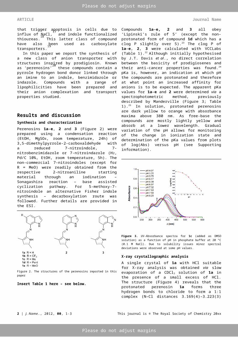
pKa is, however, an indication at which pH the compounds are protonated and therefore at what point an increased affinity for anions is to be expected. The apparent pKa values for 1a–e and 2 were determined via a spectrophotometric method, previously described by Manderville (Figure 3; Table 1).25 In solution, protonated perenosins are dark yellow to orange with absorbance maxima above 380 nm. As free-base the compounds are mostly lightly yellow and absorb at a lower wavelength. Gradual variation of the pH allows for monitoring of the change in ionization state and determination of the pKa values from plots of log(Abs) versus pH (see Supporting information).
Figure 3. UV-Absorbance spectra for 1c (added as DMSO solution) as a function of pH in phosphate buffer at 20 °C (0.1 M NaCl). Due to solubility issues minor spectral deviations were observed at some pH values.
X-ray crystallographic analysis
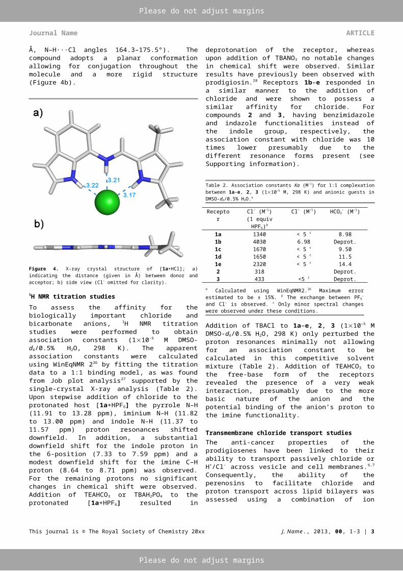
A single crystal of 1a with HCl suitable for X-ray analysis was obtained via slow evaporation of a CDCl3 solution of 1a in the presence of a small excess of HCl. The structure (Figure 4) reveals that the protonated perenosin 1a forms three hydrogen bonds to chloride to form a 1:1 complex (N–Cl distances 3.169(4)–3.223(3) Å, N–H···Cl angles 164.3–175.5°). The compound adopts a planar conformation allowing for conjugation throughout the molecule and a more rigid structure (Figure 4b).
Figure 4. X-ray crystal structure of [1a+HCl]; a) indicating the distance (given in Å) between donor and acceptor; b) side view (Cl– omitted for clarity).
1H NMR titration studies
To assess the affinity for the biologically important chloride and bicarbonate anions, 1H NMR titration studies were
2 | J. Name., 2012, 00, 1-3 This journal is © The Royal Society of Chemistry 20xx
Please do not adjust margins
Please do not adjust margins
Journal Name ARTICLE
performed to obtain association constants (110–5 M DMSO-d6/0.5% H2O, 298 K). The apparent association constants were calculated using WinEqNMR 226 by fitting the titration data to a 1:1 binding model, as was found from Job plot analysis27
supported by the single-crystal X-ray analysis (Table 2). Upon stepwise addition of chloride to the protonated host [1a+HPF6] the pyrrole N–H (11.91 to 13.28 ppm), iminium N–H (11.82 to 13.00 ppm) and indole N–H (11.37 to 11.57 ppm) proton resonances shifted downfield. In addition, a substantial downfield shift for the indole proton in the 6-position (7.33 to 7.59 ppm) and a modest downfield shift for the imine C–H proton (8.64 to 8.71 ppm) was observed. For the remaining protons no significant changes in chemical shift were observed. Addition of TEAHCO3 or TBAH2PO4 to the protonated [1a+HPF6] resulted in deprotonation of the receptor, whereas upon addition of TBANO3 no notable changes in chemical shift were observed. Similar results have previously been observed with prodigiosin.28 Receptors 1b–e responded in a similar manner to the addition of chloride and were shown to possess a similar affinity for chloride. For compounds 2 and 3, having benzimidazole and indazole functionalities instead of the indole group, respectively, the association constant with chloride was 10 times lower presumably due to the different resonance forms present (see Supporting information).
Table 2. Association constants Ka (M–1) for 1:1 complexation between 1a–e, 2, 3 (110–
5 M, 298 K) and anionic guests in DMSO-d6/0.5% H2O.a
Receptor Cl– (M–1)(1 equiv HPF6)b
Cl– (M–1) HCO3– (M–1)
1a 1340 < 5 c 8.981b 4030 6.98 Deprot.1c 1670 < 5 c 9.501d 1650 < 5 c 11.51e 2320 < 5 c 14.42 318 Deprot.3 433 <5 c Deprot.
a Calculated using WinEqNMR2.26 Maximum error estimated to be ± 15%. b
The exchange between PF6– and Cl– is observed. c Only minor spectral
changes were observed under these conditions.
Addition of TBACl to 1a–e, 2, 3 (110–5 M DMSO-d6/0.5% H2O, 298 K) only perturbed the proton resonances minimally not allowing for an association constant to be calculated in this competitive solvent mixture (Table 2). Addition of TEAHCO3 to the free-base form of the receptors revealed the presence of a very weak interaction, presumably due to the more basic nature of the anion and the potential binding of the anion’s proton to the imine functionality.
Transmembrane chloride transport studiesThe anti-cancer properties of the prodigiosenes have been linked to their ability to transport passively chloride or H+/Cl–
across vesicle and cell membranes.6,7 Consequently, the ability of the perenosins to facilitate chloride and proton transport across lipid bilayers was assessed using a combination of ion selective electrode (ISE) and fluorescence assays. To quantify
the chloride efflux rate Hill plots were determined for 200 nm POPC:cholesterol liposomes (Table 1; see Supporting Information). Typically, unilamellar vesicles were prepared from 1-palmitoyl-2-oleoyl-sn-glycero-3-phosphocholine (POPC) and cholesterol (7:3 ratio), containing an intravesicular sodium chloride solution (489 mM with 5 mM phosphate buffer at pH 7.2), were suspended in an isotonic sodium nitrate solution (489 mM with 5 mM phosphate buffer at pH 7.2). Perenosins 1a–e, 2, 3 were added as a DMSO solutions and the resulting chloride efflux was monitored by a chloride selective electrode. At the end of the experiment detergent (octaethylene glycol monododecyl ether) was added to lyse the liposomes and calibrate the electrode to 100% chloride release.This assay and others described below are evidence that the perenosins are mediating chloride/nitrate antiport in this case. Through the addition of transporter 1a in various concentrations a Hill plot29 was derived giving EC50,270 sec of 0.0773 mol% (carrier to lipid). The more electron-deficient analogue 1b (R = CF3), having a higher affinity for chloride but a lower pKa, proved to be a less efficient chloride transporter with EC50,270 sec of 1.1922 mol% (Figure 5). The introduction of a methyl or pentyl substituent in the 5-position of the indole moiety provided a decrease of the EC50,270 sec value (EC50,270 sec
0.0301 and 0.0299 mol%, respectively). Presumably the increase in clog P with respect to 1a resulted in improved chloride efflux. A higher pKa via the use of methoxy-derivative 1e which should be protonated more easily and is therefore expected to transport more efficiently, resulted in a less effective transporter than 1a–d (EC50,270 sec 0.1859 mol%) most probably attributed to the receptor’s lower clog P value (Table 1). The benzimidazole and indazole derivatives were shown to be poor lipid bilayer chloride transporters. Perenosin 1d with the lowest EC50,270 sec was found to be two orders of magnitude slower than prodigiosin (EC50,270 sec 0.0299 and 0.0002, respectively).
Figure 5. Chloride efflux promoted by a DMSO solution of compound 1a–e, 2, 3 (1 mol% carrier to lipid) from unilamlellar POPC:cholesterol vesicles loaded with 489 mM NaCl buffered to pH 7.2 with 5 mM sodium phosphate salts. The vesicles were dispersed in 489 mM NaNO3 buffered to pH 7.2 with 5 mM sodium phosphate salts. At the end of the experiments, detergent was added to lyse the vesicles and calibrate the ISE to 100% chloride efflux. Each point represents the average of three trials. DMSO was used as a control.
This journal is © The Royal Society of Chemistry 20xx J. Name., 2013, 00, 1-3 | 3

Please do not adjust margins
Please do not adjust margins
ARTICLE Journal Name
To investigate the effect of the pH on the transport activity of perenosins 1a–e and 2, chloride/nitrate antiport was followed at different pH values (pH 4.0, 6.2, 7.2 and 8.2; Figure 6). Upon decreasing the pH from 7.2 to 6.2, an increase in transport is observed corresponding to the increased amount of receptor molecules being protonated. At pH 8.2 there is a significant drop in transport activity as presumably a significant proportion of the transporters are not protonated. This is evidence that only the protonated form of this perenosin is capable of transporting anions.
Figure 6. Anion exchange assay promoted by a DMSO solution of compound 1d (1 mol% carrier to lipid) from unilamlellar POPC:cholesterol vesicles loaded with 489 mM NaCl buffered to a given pH with 5 mM sodium phosphate salts (pH 7.2 and 8.2), piperazine (pH 6.2) or citric acid (pH 4.0). The vesicles were dispersed in 489 mM NaNO3 buffered to a given pH with 5 mM sodium phosphate salts (pH 7.2 and 8.2), piperazine (pH 6.2) or citric acid (pH 4.0). At the end of the experiments, detergent was added to lyse the vesicles and calibrate the ISE to 100% chloride efflux. Each point represents the average of three trials. DMSO was used as a control.
To further explore the transport mechanism operating in this system, a variety of ISE and fluorescence vesicle assays were performed altering the bilayer and the intra- or extravesicular solution composition. To probe whether metal ion-anion symport occurs POPC vesicles were loaded with different group 1 metal (Na+, K+, Cs+) chloride salts (see Supporting Information). The metal was found to have no effect on the rate of chloride efflux from the vesicles upon addition of compound 1a, evidence in support of a transport mechanism not involving metal cations.The sulfate ion is highly hydrophilic and is more challenging to transport across the lipid bilayer than nitrate.30 Upon addition of perenosin to vesicles loaded with sodium chloride suspended in a sodium sulfate solution, no chloride efflux was observed (see Supporting Information). Upon addition of bicarbonate to the extravesicular solution, a chloride/bicarbonate antiport mechanism may be initiated. After the bicarbonate pulse at t = 120 sec, a modest increase in extravesicular chloride concentration was noted (Figure 7). The compounds proved to be quite poor bicarbonate transporters presumably due to deprotonation of the protonated perenosin. This is evidence in support of the protonated form
of the receptor being the species that is capable of transporting anions across the bilayer.
Figure 7. Change of extravesicular chloride concentration over time for 1a–e, 2 (1 mol% carrier to lipid) of unilamlellar POPC:cholesterol vesicles loaded with 489 mM NaCl buffered to pH 7.2 with 20 mM sodium phosphate salts. The vesicles were dispersed in 167 mM Na2SO4 buffered to pH 7.2 with 20 mM sodium phosphate salts. At t = 120 sec, a solution of NaHCO3 spike was introduced such that the external NaHCO3
concentration was 40 mM. At the end of the experiments, detergent was added to lyse the vesicles and calibrate the ISE to 100% chloride efflux. Each point represents the average of three trials.
In the sulfate assays no anion transport was observed, however due to the very small intravesicular volume, HCl co-transport along a pH gradient using ion-selective electrode assays is very hard to quantify. The possible presence of HCl co-transport was studied by fluorescence using a pH gradient assay. Vesicles containing sodium chloride (489 mM) and 1 mM 8-hydroxy-1,3,6-pyrenetrisulfonate (HPTS), a pH sensitive fluorescent dye were prepared.31 The vesicles were suspended in a solution of sodium sulfate (167 mM) and the HPTS fluorescence measured upon addition of a DMSO solution of compounds 1ae, 2 (Figure 8). An increase in pH was observed, corresponding to the deacidification of the vesicles via a H+/Cl
co-transport mechanism (with Cl-/OH- antiport being ruled out due to the decrease in transport observed at higher pH and with basic anions such as bicarbonate).
4 | J. Name., 2012, 00, 1-3 This journal is © The Royal Society of Chemistry 20xx

Please do not adjust margins
Please do not adjust margins
Journal Name ARTICLE
Figure 8. Fluorescence assay (ex = 403 and 460nm, em = 510 nm) for 1a–e, 2 (1 mol% carrier to lipid) using unilamellar POPC:cholesterol vesicles loaded with 489 mM NaCl buffered to pH 7.2 with 20 mM sodium phosphate salts and 1 mM HPTS. The vesicles were dispersed in 167 mM Na2SO4 buffered to pH 7.2 with 20 mM sodium phosphate salts. Each point represents the average of three trials.
Prodigiosenes have been shown to transport HCl across the lipid bilayer via a mobile carrier mechanism.1 The Hill coefficients found for all indole-based perenosins and prodigiosin have a value of approximately 1 evidence in support of the hypothesis that the transport of a chloride ion can be performed by a single carrier molecule.32 The non-indole perenosins 2 and 3 have a Hill coefficient of 1.93 and 2.21, respectively, evidence in support of cooperative mechanism involving two carrier molecules transporting one chloride ion. Evidence for a carrier mechanism was derived from U-tube experiments.33 Transporters 1a, 1c-e, 2 as a solution in chloroform (1 mM) were kept between two aqueous phases as a membrane model mimicking a vesicle assay (see Supporting Information). The source aqueous phase was loaded with sodium chloride (489 mM buffered to pH 7.2 with 5 mM sodium phosphate salts) and the receiving aqueous phase was loaded with sodium nitrate (489 mM buffered to pH 7.2 with 5 mM sodium phosphate salts). The large separation between the two aqueous phases rules out the possibility of transport via channel formation. Chloride transport was monitored using an ISE and showed that all the tested perenosins yielded an increase in chloride concentration in the receiving phase over time (5 days). These results support the hypothesis of a mobile carrier mechanism being the most likely mode of transport in this case.Transport studies with the prodigiosenes show that the pKa of the transporter correlates well with the EC50,270sec,24 however no clear correlation could be found between the pKa of perenosins and their EC50,270sec. However, compounds with a pKa value higher than 6.65 and a clog P between than 2.85 and 5.16, (supported by the log P range stipulated by Quesada et al.,13b) appear to exhibit the best chloride transport properties. Taking all the transport studies together the results show that the indole perenosins behave similarly to prodigiosin namely functioning as both a HCl cotransporter and a Cl-/NO3
-
antiporter6 and forming a 1:1 complex with the anion.
Hydrolysis studiesThe rate of hydrolysis is an important factor within the set of pharmokinetic properties, the collective of bioavailability and processes of absorption, distribution, metabolism and elimination. As a model for all perenosins, the hydrolysis rate of 1a in phosphate buffer (0.1 M NaCl) was determined following the perturbation of the UV/Vis spectroscopic data over time (see Supporting Information). At pH 7.2 the half-life time of 1a was calculated, following first-order kinetics, to be 10.8 h.34 Lowering the pH to 4.0 shortened the half-life time of 1a to 6.6 h, consistent with the presence of the imine linker. Entrapment of 1a inside a vesicular lipid bilayer at pH 7.2 extended the life span of perenosin 1a six fold (half-life 60.2 h).
Vesicles that fuse with the cancer cell membrane, potentially decorated with cancer cell selective receptors and fluorophores, therefore offer a potential route to administer future anti-cancer agents based upon the perenosin scaffold. An additional benefit would be the reduced toxicity for highly active compounds when administered whilst embedded inside liposomes.35
Cell-based analysisWe performed preliminary studies to assess the effect of perenosins on the viability of cancerous and non-cancerous model cell lines. Compounds 1a–e, 2 were assessed for their effect on the viability of breast carcinoma MDA-MB-231 (invasive) and MCF-7 (non-invasive) model cell lines, as well as MCF-10A normal mammary model cells (Table 3). The cell lines were treated with increasing doses of perenosins for 24 h and the effect on the degree of cell viability was determined by MTT assays giving a dose-response curve (see Supporting Information Figures S85-S90) that was used to determine the IC50 values for each compound in each cell line.
Table 3. IC50 (M) values for 1a–e, 2 on MDA-MB-231, MCF-7 and MCF-10A cells.
Compound MDA-MB-231(M)
MCF-7(M)
MCF-10A(M)
1a 9.07 ± 1.30 6.02 ± 1.27 15.43 ± 5.751b 3.67 ± 0.05 4.13 ± 0.22 12.93 ± 3.231c 5.10 ± 1.08 4.38 ± 0.30 11.92 ± 3.371d 4.39 ± 0.80 3.84 ± 0.12 22.37 ± 7.741e 9.92 ± 0.80 5.61 ± 1.48 18.28 ± 6.692 28.78 ± 2.33 20.22 ± 7.35 51.20 ± 15.31
All indole-based perenosins 1a–e were cytotoxic to the two malignant cell lines at low µM, with 1b, 1c and 1d being most potent. All the molecules tested here were less potent in the normal MCF-10A cells. Interestingly, 1d showed the largest selectivity (~5.5 fold) for the cancerous cell lines tested here. The benzimidazole derivative 2 was found to be substantially less active (IC50 24.32 M) than the other molecules. The most active compounds in cells (1b and 1d) were also the most lipophilic in the series (clog P 3.84 and 5.06, respectively). The reduced cytotoxicity observed in the normal MCF-10A cells suggests a potential mechanism for selective targeting of cancer cells with more potent derivatives of these molecules.
ConclusionsThe perenosins represent a new class of highly effective anion transporters based upon structure of prodigiosin. It has been demonstrated that indole-based perenosins are highly efficient chloride transporters and function as a mobile-carrier by an antiport and H+/Cl– symport mechanism of anion transport. The most lipophilic derivatives affect the viability of two breast cancer cell lines with ~5.5-fold selectivity over normal breast cells. These compounds therefore represent excellent lead structures for further exploration of the potentially selective anti-cancer activities of this new class of molecules.
This journal is © The Royal Society of Chemistry 20xx J. Name., 2013, 00, 1-3 | 5

Please do not adjust margins
Please do not adjust margins
ARTICLE Journal Name
AcknowledgementsW.VR. and P.A.G. thank the European Union for a Marie Curie Career Integration Grant. P.A.G. thanks the Royal Society and the Wolfson Foundation for a Royal Society Wolfson Research Merit Award. D.J.A. and A.T. thank the Engineering and Physical Sciences Research Council (for EP/H04986X/1). The authors thank Dr Ethan Howe for assistance with the preparation of this manuscript.
Notes and references.1 J.T. Davis, Topics in Heterocyclic Chemistry, 2010, 24, 145-
176.2 B. Bizio, Biblioteca Italiana o sia Giornale di Letteratura,
Scienze e Arti Tomo, 1823, 30, 275-295.3 F. Wrede and O. Hettche, Ber. Dtsch. Chem Ges. B 1929, 62B,
2678-2687.4 N.P. Fullan, D.L. Lynch and D.H. Ostrow, Microbiol. Lett.
1977, 5, 157-161.5 a) R.A. Manderville, Curr. Med. Chem.: Anti-Cancer Agents
2001, 1, 195-218; b) R. Perez-Tomas, Curr. Med. Chem. 2006, 13, 1859-1876; c) R. Perez-Tomas, B. Montaner, E. Llagostera and V. Soto-Cerrato, Biochem. Pharmacol. 2003, 66, 1447-1452; d) J. Regourd, A. A.-S.Ali and Thompson, J. Med. Chem. 2007, 50, 1528-1536. e) R.I. Sáez Díaz, S.M. Bennett and A. Thompson, A. ChemMedChem 2009, 4, 742-745.
6 J.L. Seganish and J.T. Davis,. Chem. Commun. 2005, 5781-5783.
7 a) T. Sato, H. Konno, Y. Tanaka, T. Kataoka, K. Nagai, H.H. Wasserman and S. Ohkuma, J. Biol. Chem. 1998, 273, 21455-21462; b) S. Ohkuma, T. Sato, M. Okamoto, H. Matsuya, K. Arai, T. Kataoka, K.Nagai and H.H. Wasserman, Biochem. J. 1998, 334, 731-741; c) D. Yamamoto, Y. Kiyozuka, Y. Uemura, C. Yamamoto, H. Takemoto, H. Hirata, K. Tanaka, K. Hioki, A. Tsubura, J. Cancer Res. Clin. Oncol. 2000, 126, 191-197.
8 R.F. Tsuji, J. Magae, M. Yamashita, K. Nagai, M. Yamasaki, J. Antibiot. 1992, 45, 1295-1302; b) E. Marchal, Md. I. Uddin, D.A. Smithen, C.L.A. Hawco, M. Lanteigne, D.P. Overy, R.G. Kerr,and A. Thompson, RSC Adv. 2013, 3, 22967-22971.
9 a) A.J. Castro, Nature 1967, 213, 903-904; b) E. Marchal, D.A. Smitchen, Md. I. Uddin, A.W. Robertson, D.L. Jakeman, V. Mollard, C.D. Goodman, K.S. MacDougall, S.A. McFarland, G.I. McFadden and A. Thompson, Org. Biomol. Chem. 2014, 12, 4132-4142.
10 a) N.R. Williamson, P.C. Fineran, T. Gristwood, S.R. Chawrai, F.J. Leeper, D.P.C. Salmond, Future Microbiol. 2007, 2, 605-618. b) P.S. Kim, C. Jochems, I. Grenga, R.N. Donahue, K.Y. Tsang, J.L. Gulley, J. Scholm and D. Fursaci, J. Immunol. 2014, 192, 2622-2633.
11 R.H. Wier, R.O. Egeberg, A.R. Lack and G. Leiby, Am. J. Med. Sci. 1952, 224, 70-76.
12 P.I. Hernandez, D. Moreno, A.A. Javier, T. Torroba, R. Perez-Tomas and R. Quesada, Chem. Commun. 2012, 48, 1556-1558.
13 a) N.J. Knight, E. Hernando, C.J.E. Haynes, N. Busschaert, H.J. Clarke, K. Takimoto, M. García-Valverde, J.G. Frey, R. Quesada and P.A. Gale, Chem. Sci. 2016, DOI: 10.1039/c5sc03932k. b) V. Saggiomo, S. Otto, I. Marques, V. Félix, T. Torroba and R. Quesada, Chem. Commun. 2012, 48, 5274-5276.
14 B.D. de Grenu, P.I. Hernandez, M. Espona, D. Quinonero, M.E. Light, T. Torroba, R. Perez-Tomas and R. Quesada, Chem. Eur. J. 2011, 17, 14074-14083.
15 P.A. Gale, M.E. Light, B. McNally, K. Navakhun, K.E. Sliwinski, and B.D. Smith, Chem. Commun. 2005, 3773-3775.
16 a) C.C. Tong, R. Quesada, J.L. Sessler and P.A. Gale, P.A. Chem. Commun. 2008, 6321-6323. b) M.G. Fisher, P.A. Gale, J.R. Hiscock, M.B. Hursthouse, M.E. Light, F.P. Schmidtchen, C.C. Tong, Chem. Commun. 2009, 3017-3019. c) P.A. Gale, C.C. Tong, C.J.E. Haynes, O. Adeosun, D.E. Gross, D.E.; E. Karnas, E. Sedenberg, R. Quesada and J.L. Sessler, J. Am. Chem. Soc. 2010, 132, 3240-3241. d) M. Yano, C.C. Tong, M.E. Light, F.P. Schmidtchen and P.A. Gale, Org. Biomol. Chem. 2010, 8, 4356-4363.
17 S.K. Ko, S.K. Kim, A. Share, V.M. Lynch, J. Park, W. Namkung, W. Van Rossom, N. Busschaert, P.A. Gale, J.L. Sessler and I. Shin, Nature Chem., 2014, 6, 885-892.
18 N.J. Andrews, C.J.E. Haynes, M.E. Light, S.J. Moore, C.C. Tong, J.T. Davis, W.A. Harrell Jr. and P.A. Gale, Chem. Sci., 2011, 2, 256-260. b) Moore, S.J.; Wenzel, M.; Light, M.E.; Morley, R.; Bradberry, S.J.; Gómez-Iglesias, P.; Soto-Cerrato, V.; Pérez-Tomás, R.; Gale, P.A. Chem. Sci., 2012, 3, 2501-2508.
19 C.J.E. Haynes, S.N. Berry, J. Garric, J. Herniman, J.R. Hiscock, I.L. Kirby, M.E. Light, G. Perkes and P.A. Gale, Chem. Commun. 2013, 49, 246-248.
20 The name ‘perenosin’ was derived from a fusion of the name prodigiosin and the Russian word Переносчик [perenoschik], which translates as ‘carrier’.
21 V. Rizzo, A. Morelli, V. Pinciroli, D. Sciangula, R. D’Alessio, J. Pharm. Sci. 1999, 88, 73-78.
22 VCCLAB Virtual Computational Chemistry Laboratory. http://www.vcclab.org.
23 C.A. Lipinski, F. Lombardo, B.W. Dominy and P.J. Feeney, Adv. Drug. Deliver. Rev. 2001, 46, 3-26.
24 E. Marchal, S. Rastogi, A. Thompson and J.T. Davis, Org. Biomol. Chem. 2014, 12, 7515-7522.
25 M.S. Melvin, J.T. Tomlinson, G. Park, C.S. Day, G.R. Saluta, G.L. Kucera and R.A. Manderville, Chem. Res. Toxicol. 2002, 15, 734-741.
26 M.J. Hynes, J. Chem. Soc., Dalton Trans. 1993, 311-312.27 P. Job, Ann. Chim. 1928, 9, 113-203.28 J.T. Davis, P.A. Gale, O.A. Okunola, P. Prados, J.C. Iglesias-
Sanchez, T. Torriba and R. Quesada, Chem. Nat. 2009, 1, 138–144.
29 A.V. Hill, Biochem. J. 1913, 7, 471-480.30 Y. Marcus, J. Chem. Soc., Faraday Trans. 1991, 87, 2995-
2999.31 N.R. Clement and J.M. Gould, Biochemistry, 1981, 20, 1534-
1539.32 A.V. Jentzsch, D. Emery, J. Mareda, S.K. Nayak, P.
Metrangolo, G. Resnati, N. Sakai and S. Matile, Nat. Commun. 2012, 3, 905.
33 a) N. Busschaert, M. Wenzel, M.E. Light, P. Iglesias-Hernandez, R. Perez-Tomas and P.A. Gale, J. Am. Chem. Soc. 2011, 133, 14136-14148. b) S.N. Berry, N. Busschaert, C.L. Frankling, D. Salter and P.A. Gale, Org. Biomol. Chem. 2015, 13, 3136-3143.
34 P.L. Toutain and A. Bousquet-Mélou, J. Vet. Pharmacol. Therap. 2004, 27, 427-439.
35 C.R. Dass, Methods Mol. Biol. 2008, 437, 177-182.
6 | J. Name., 2012, 00, 1-3 This journal is © The Royal Society of Chemistry 20xx

Please do not adjust margins
Please do not adjust margins
Journal Name ARTICLE
Table 1. Experimentally determined EC50,270sec, Hill coefficient (n), and pKa and calculated log P (clog P) (free-base and protonated form) for perenosins 1a–e, 2 and 3.
Transporter pKa EC50,270sec (mol%)b n clog P (error)c clog P protonated (error)c
1a 6.84 0.0773 1.11 2.98 (±0.60) 2.05 (±1.20)1b 5.38 1.1922 1.52 3.84 (±0.61) 3.37 (±0.59)1c 6.81 0.0301 1.00 3.36 (±0.66) 2.41 (±1.17)1d 6.65 0.0299 1.00 5.16 (±0.98) 4.02 (±1.46)1e 7.11 0.1859 1.33 2.85 (±0.74) 2.00 (±1.16)2 7.18 6.4084 1.93 2.32 (±0.44) 1.58 (±1.38)3 n.d.d 5.4305 2.21 2.44 (±0.50) 1.48 (±1.26)
prodigiosin 7.16a 0.0002 1.00 4.12 (±0.78) 3.28 (±1.15)
a Literature value.21 b Cl–/NO3– assay using POPC/chol (7:3) at pH 7.2. c Values calculated with VCCLabs.22 d not determined.
This journal is © The Royal Society of Chemistry 20xx J. Name., 2013, 00, 1-3 | 7


![Crystallographic relations in the Fe[bond]Zn system · Crystallographic Relations in the Fe-Zn System The crystallographic relations between the various Fe-Zn compounds have been](https://img.dokumen.tips/doc/110x75/5f0570af7e708231d412f970/crystallographic-relations-in-the-febondzn-system-crystallographic-relations-in.jpg)