Embed Size (px)
Citation preview

7/26/2019 20306 Lecture 14 Primary Immunodeficiency Disorder
http://slidepdf.com/reader/full/20306-lecture-14-primary-immunodeficiency-disorder 1/18
1
LECTURE: 14
Tit le: PRIMARY IMMUNODEFICIENCY DISORDERS
LEARNING OBJECTIVES:
The student should be able to:
Discuss the importance of knowing the normal concentration values ofcomplement proteins, providing some important diagnostic complementproteins.
Discuss the importance of knowing the normal concentrations values of thedifferent immunoglobulins isotypes and their subclasses.
Explain the benefit of comparing patient's clinical manifestations withlaboratory f inding for the various soluble humoral immunological elements.
Explain the valuable values of identifying the predominant site for eachimmunoglobulin isotype.
Explain the significance of detecting the immunoglobul in isotype IgM in thecord blood of a pregnant lady, and provide one pathological case which isimportant to identify the type of the immunoglobulin isotype in the cordblood.
Explain the indication of having pyogenic infections, in relation of
immunoglobulin concentrations.
Explain the indication of having extraordinary infections with Neisseriaspecies.
Explain the indication of having hereditary angioedema in relation withcomplement protein concentration.
Enumerate some common serological techniques used in evaluatingantibodies and complement concentrations.
LECTURE REFRENCE:
1. TEXTBOOK: ROITT, BROSTOFF, MALE. IMMUNOLOGY. 6th edition. Chapter4. pp. 65, chapter 3. pp 47. Chapter 19. pp 303-311.2. TEXTBOOK: ABUL K. ABBAS. ANDREW H. LICHTMAN. CELLULAR ANDMOLECULAR IMMUNOLOGY. 5TH EDITION. Chapter 20. pg 453.3. HANDOUT.

7/26/2019 20306 Lecture 14 Primary Immunodeficiency Disorder
http://slidepdf.com/reader/full/20306-lecture-14-primary-immunodeficiency-disorder 2/18
2
Primary Immunodeficiency
• Defective antibody responses result in increased susceptibility to pyogenic infections
and are due to failure of B-cell function, such as occurs in X-linkedagammaglobulinaemia, or from failure of proper T-cell signals to B cells such as
occurs in hyper-IgM syndrome, common variable immunodeficiency (CVID) and
transient hypogammaglobulinaemia of infancy.• Defective cell-mediated immunity results in increased susceptibility to opportunistic
infections and is due to failure of T-cell function such as occurs in severe combined
immunodeficiency (SCID), MHC class II deficiency, ataxia-telangiectasia, the
Wiskott-Aldrich syndrome and the DiGeorge anomaly.
• Hereditary complement component defects are found in a number of clinical
syndromes, the most common of which is that of the C1 inhibitor, which results in
hereditary angioedema.
• Hereditary complement deficiencies of the terminal complement components (C5,
C6, C7 and C8) and the alternative pathway proteins (Factor H, Factor I and
properdin) lead to extraordinary susceptibility to infections wit the two Neisseria species, N. gonorrhoeae and N. meningitides.
• Defects in the oxygen reduction pathway of phagocytes, so that the phagocytescannot assemble NADPH oxidase and produce the hydrogen peroxide and oxygen
radicals that kill bacteria, are the basis of chronic granulomatous disease. The resulting
persistence of bacterial products in phagocytes leads to abscesses or granulomas
depending on the pathogen.
• Leucocyte adhesion deficiency is associated with a persistent leukocytosis because
phagocytic cells with defective integrin molecules cannot migrate through the vascular
endothelium from the blood stream into the tissues.
Immunodeficiency disease results from the absence, or failure of normal function, of one or
more elements of the immune system. Specific immunodeficiency diseases involve
abnormalities of T or B cells, the cells of the adaptive immune system. Non-specific
immunodeficiency diseases involve abnormalities of elements such as complement or
phagocytes, which act non-specifically in immunity. Primary immunodeficiency diseases are
due to intrinsic defects in cells of the immune system and are for the most part genetically
determined.
Immunodeficiency diseases cause increased susceptibility to infection in patients. The
infections encountered in immunodeficient patients fall, broadly speaking, into two
categories. Patients with defects in immunoglobulins complement proteins or phagocytes are
very susceptible to recurrent infections with encapsulated bacteria such as Haemophilus
influenzae, Streptococcus pneumoniae and Staphylococcus aureus. These are called pyogenic infections, because the bacteria give rise to pus formation. On the other hand,
patients with defects in cell-mediated immunity, i.e. in T cells, re susceptible to
overwhelming, even lethal, infections with microorganisms that are ubiquitous in the
environment and to which normal people rapidly develop resistance. For this reason, theseare called opportunistic infections; opportunistic microorganisms include yeast and common
viruses such as chickenpox.
7/26/2019 20306 Lecture 14 Primary Immunodeficiency Disorder
http://slidepdf.com/reader/full/20306-lecture-14-primary-immunodeficiency-disorder 3/18
3
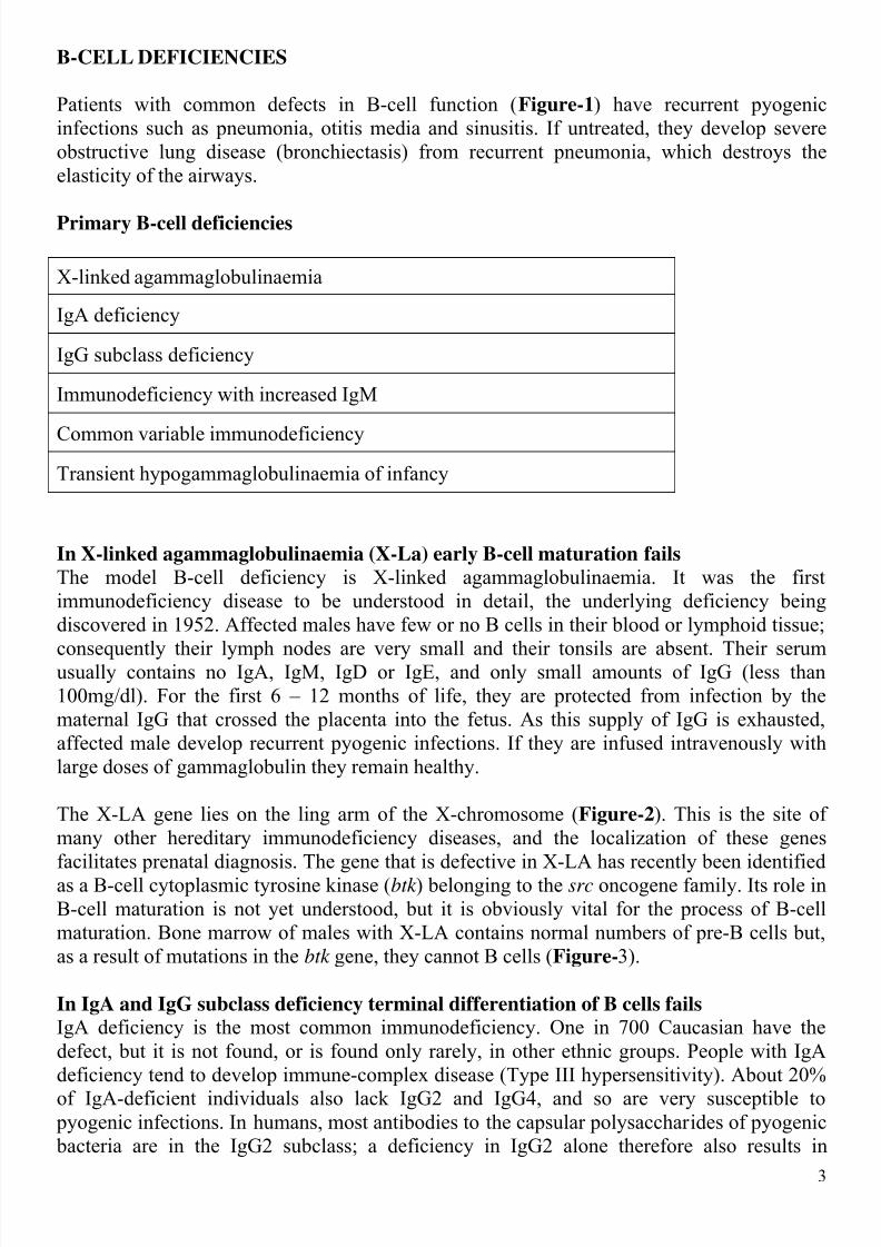
B-CELL DEFICIENCIES
Patients with common defects in B-cell function (Figure-1) have recurrent pyogenic
infections such as pneumonia, otitis media and sinusitis. If untreated, they develop severe
obstructive lung disease (bronchiectasis) from recurrent pneumonia, which destroys the
elasticity of the airways.
Primary B-cell deficiencies
X-linked agammaglobulinaemia
IgA deficiency
IgG subclass deficiency
Immunodeficiency with increased IgM
Common variable immunodeficiency
Transient hypogammaglobulinaemia of infancy
In X-linked agammaglobulinaemia (X-La) early B-cell maturation fails
The model B-cell deficiency is X-linked agammaglobulinaemia. It was the first
immunodeficiency disease to be understood in detail, the underlying deficiency being
discovered in 1952. Affected males have few or no B cells in their blood or lymphoid tissue;
consequently their lymph nodes are very small and their tonsils are absent. Their serumusually contains no IgA, IgM, IgD or IgE, and only small amounts of IgG (less than
100mg/dl). For the first 6 – 12 months of life, they are protected from infection by the
maternal IgG that crossed the placenta into the fetus. As this supply of IgG is exhausted,
affected male develop recurrent pyogenic infections. If they are infused intravenously with
large doses of gammaglobulin they remain healthy.
The X-LA gene lies on the ling arm of the X-chromosome (Figure-2). This is the site of
many other hereditary immunodeficiency diseases, and the localization of these genes
facilitates prenatal diagnosis. The gene that is defective in X-LA has recently been identified
as a B-cell cytoplasmic tyrosine kinase (btk ) belonging to the src oncogene family. Its role in
B-cell maturation is not yet understood, but it is obviously vital for the process of B-cell
maturation. Bone marrow of males with X-LA contains normal numbers of pre-B cells but,
as a result of mutations in the btk gene, they cannot B cells (Figure-3).
In IgA and IgG subclass deficiency terminal differentiation of B cells fails
IgA deficiency is the most common immunodeficiency. One in 700 Caucasian have the
defect, but it is not found, or is found only rarely, in other ethnic groups. People with IgA
deficiency tend to develop immune-complex disease (Type III hypersensitivity). About 20%
of IgA-deficient individuals also lack IgG2 and IgG4, and so are very susceptible to pyogenic infections. In humans, most antibodies to the capsular polysaccharides of pyogenic
bacteria are in the IgG2 subclass; a deficiency in IgG2 alone therefore also results in

7/26/2019 20306 Lecture 14 Primary Immunodeficiency Disorder
http://slidepdf.com/reader/full/20306-lecture-14-primary-immunodeficiency-disorder 4/18
4
recurrent pyogenic infections. Individuals with deficiency of only IgG2 are also susceptible
to recurrent infections. These class and subclass deficiencies result from failure in terminal
differentiation of B cells (Figure-3).
In immunodeficiency with increased IgM (HIgM) isotype switching does not occur
A particular immunodeficiency results in patients who are IgG and IgA-deficient but
synthesizes large amounts (more than 200mg/dl) of polyclonal IgM. They are susceptible to
pyogenic infections and should be treated with intravenous gammaglobulin. They trend to
form IgM autoantibodies to neutrophils, platelets and other elements of the blood, as well asto tissue antigens, thereby adding the complexities of autoimmune disease to the
immunodeficiency. The tissues, particularly of the gastrointestinal tract, become infiltrated
with IgM-producing cells (Figure-4). In HIgM the B cells cannot make the switch from IgM
to IgG, IgA and IgE synthesis that normally occurs in B-cell maturation. For example, in
normal B cells, this switch to IgE is induced by two factors: IL-4 must bind to the B-cell
receptor for IL-4, and the CD40 molecule on the B-cell surface must bind to the CD40 ligand
on activated T cells. In 70% of cases HIgM is inherited as an X-linked recessive that results
from mutations in the CD40 ligand, whose gene maps to precisely the same location on the
long arm of the X-chromosome as HIgM.
In common variable immunodeficiencies (CVID) there are defects in T-cell signaling to
B cells
Individuals with CVID have acquired agammaglobulinaemia in the second or third decade oflife, or later. Both males and females are equally affected and the cause is generally not
known, but may follow infection with viruses such as Epstein – Barr virus (EBV). Patients
with CVID, like males with X-LA, are very susceptible to pyogenic organisms and to the
intestinal protozoan, Giardia lamblia (Figure-5), which cause serve diarrhoea. Most patients
(80%) with CVID have B cells that do not function properly and are immature. The B cellsare not defective; instead, they fail to receive proper signals from the T cells. However, the
T-cell defects have not been defined well in CVID. Patients with CVID should be treatedwith intravenous gammaglobulin as it provides protection against recurrent pyogenic
infections. Many patients develop autoimmune diseases, most prominently pernicious
anaemia, and the reason for this is not known. CVID is not hereditary, but is commonly
associated with the MHC haplotypes HLAB8 and HLA-DR3.
IgG production is delayed in transient hypogammaglobulinaemia of infancy
As mentioned above, infants are protected initially by their mother's IgG. The maternal IgG
is catabolized, with a half-life of approximately 30 days. By 3 months of age, normal infants begin to synthesize their own IgG, although formation of antibody to bacterial capsular
polysaccharides does not commence in earnest until the second year of life. In some infants,
the onset of normal IgG synthesis can be delayed for as long as 36 months and, until then,
such infants are susceptible to pyogenic infections. The B cells of these infants are normal
but they appear to lack help from CD4+
T cells in synthesizing antibodies.
T-CELL DEFICIENCIES
The major T-cell deficiencies are shown in Figure-6. Patients with no T cells, or poor T-cellfunction, are susceptible to opportunistic infections. Since B-cell function in humans is
largely T-cell dependent, T-cell deficiency also results in humoral immunodeficiency; in

7/26/2019 20306 Lecture 14 Primary Immunodeficiency Disorder
http://slidepdf.com/reader/full/20306-lecture-14-primary-immunodeficiency-disorder 5/18
5
other words, T-cell deficiency leads to a combined deficiency of both humoral and cell-
mediated immunity.
In severe combined immunodeficiency (SCID) there is lymphocyte deficiency and the
thymus does not develop
The most profound hereditary deficiency of cell-mediated immunity occurs in infants with
SCID who develop recurrent infections early in life (in contrast to X-LA). They have
prolonged diarrhoea due to rotavirus or bacterial infection of the gastrointestinal tract and
develop pneumonia, usually due to the protozoan, Pneumocystis carinii. The common yeastorganism Candida albicans grows luxuriantly in their mouth or on their skin (Figure-7). If
they are vaccinated with live organisms, such as poliovirus or bacille Calmette-Guerin
(BCG) (used for immunization against tuberculosis), they die of progressive infection from
these ordinarily benign organisms, SCID is incompatible with life and affected infants
usually die within the first 2 years unless they are rescued with transplants of bone marrow.
In this case they become lymphocyte chimeras and can survive and live normally.
Infants with SCID have very few lymphocytes in their blood (fewer than 3000/ml). Their
lymphoid tissue also contains few or no lymphocytes. The thymus has a fetal appearance(Figure-8), containing the endodermal stromal cells derived embryonically from the third
and fourth pharyngeal pouch. Lymphoid stem cells, which normally populate the thymus by
6 weeks of human gestation (see Chapter 2), fail to appear and the thymus does not become a
lymphoid organ.
SCID is more common in male than female infants (3:1) because over 50% of SCID cases
are caused by a gene defect on the X-chromosome. The defective gene encodes the γ chain
of the IL-2 receptor. This γ chain also forms part of the receptors for IL-4, 7, 11 and 15. Of
these, the binding of interleukin-7 to the IL-7 receptor is most important for T-cellmaturation. Thus, the lymphoid stem cells are incapable of receiving a number of signals for
growth and maturation. The remaining cases of SCID are due to recessive genes on other
chromosomes. Of these, half have a genetic deficiency of adenosine deaminase (ADA) or purine nucleoside phosphorylase (PNP). Deficiency of these purine degradation enzymes
results in the accumulation of metabolites that are toxic to lymphoid stem cells, namely
dATP and dGTP (Figure-9). These metabolites inhibit the enzyme ribonucleotide reductase,which is required for DNA synthesis and, therefore, for cell replication. Since ADA and PNP
are found in all mammalian cells, why should these defects only affect lymphocytes? The
explanation appears to lie in the relative deficiency of 5' nucleotidase in lymphoid cells; in
other cells, this enzyme compensates for defective ADA or PNP by preventing dAMP anddGMP accumulation.
The two recombinase activation genes, Rag-1 and Rag-2, are absolutely required for cleaving
double-stranded DNA prior to recombination of DNA to form the immunoglobulin genes
and the genes encoding the T-cell receptor. If these gene rearrangements do not occur, B and
T lymphocytes do not develop; an autosomal recessive form of SCID results from mutation
in either of the genes encoding Rag-1 or Rag-2.
The optimal treatment for SCID is a bone-marrow transplant from a completelyhistocompatible donor, usually a normal sibling. About 70% of patients do not have a
histocompatible sibling, in which case parental marrow, which would have one haplotype

7/26/2019 20306 Lecture 14 Primary Immunodeficiency Disorder
http://slidepdf.com/reader/full/20306-lecture-14-primary-immunodeficiency-disorder 6/18
6
identical, has been transplanted successfully. Recently a retroviral vector, into which the
ADA gene had been inserted, has been used to transfect the lymphocytes of children who are
ADA deficient. This was the firs example of successful 'gene therapy'.
In MHC class II deficiency TH-cell deficiency results
The failure to express class II MHC molecules on antigen-presenting cells (macrophages and
B cells) is inherited as an autosomal recessive characteristics, which is not linked to the
MHC locus on the short arm of chromosome 6. Affected infants have recurrent infections,
particularly of the gastrointestinal tract. Because the development of CD4+ TH cells (T-helpercells) depends on positive selection by MHC class II molecules in the thymus, MHC class II
deficient infants have a deficiency of CD4+ T cells. This lack of TH cells leads to a
deficiency in antibodies as well. The MHC class II deficiency results from defects in
promoter proteins that bind to the 5' untranslated region of the class II genes.
The destruction of intracellular microorganisms that flourish in macrophages depends on the
activation of microbicidal activity in macrophages by interferon-γ. When microorganisms
are taken up by macrophages these cells secrete interleukin-12 (Il-12). IL-12 binds to the IL-
12 receptor on T cells and this provokes T cells to secrete interferon-γ receptor sustainrecurrent infection with non-pathogenic mycobacteria and, to a lesser extent, with
salmonella. These various defects are inherited as autosomal recessive traits. The defects can
be fatal unless treatment with interferon-γ is undertaken.
The DiGeorge anomaly arises from a defect in thymus embryogenesis
As previously mentioned, the thymic epithelium is derived from the third and fourth
pharyngeal by the sixth week of human gestation. Subsequently the endodermal anlage is
invaded by lymphoid stem cells that undergo development into T cells. The parathyroid
glands are also derived from the same embryonic origin. A congenital defect in the organsderived from the third and fourth pharyngeal pouches results in the DiGeorge anomaly. The
T-cell deficiency is variable, depending on how badly the thymus is affected. Affected
infants have distinctive facial features (Figure-10) in that their eyes are widely separated(hypertelorism), the ears are low set, and the philtrum of the upper lip is shortened. They also
have congenital malformations of the heart or aortic arch and neonatal tetany from the
hypoplasia or aplasia of the parathyroid glands.
X-linked proliferateive syndrome (LLP)
This results from a failure to control the normal proliferation of cytotoxic T cells following
an infection with Epstein-Barr virus (EBV), which causes infectious mononucleosis.Affected males appear normal until they encounter EBV, when they develop either fatal
infectious mononucleosis, or have complete destruction of their B cells so that
agammaglobulinaemia ensures, or develop a fatal lymphoid malignancy or aplastic anaemia.
The defective gene on the X-chromosome encodes an adapter protein of T and B cells called
SAP or the SLAM associated protein. SLAM is expressed on the surface or T and B cells. Its
intracellular tail interacts with the adapter protein, SAP. By a mechanism that is not
understood, SAP controls the limitless proliferation of cytotoxic T cells. A genetic defect in
SAP results in the destruction of lymphoid and other haematopoietic tissue by uncontrolled
proliferation of cytotoxic T cells and maternal killer cells.
7/26/2019 20306 Lecture 14 Primary Immunodeficiency Disorder
http://slidepdf.com/reader/full/20306-lecture-14-primary-immunodeficiency-disorder 7/18
7
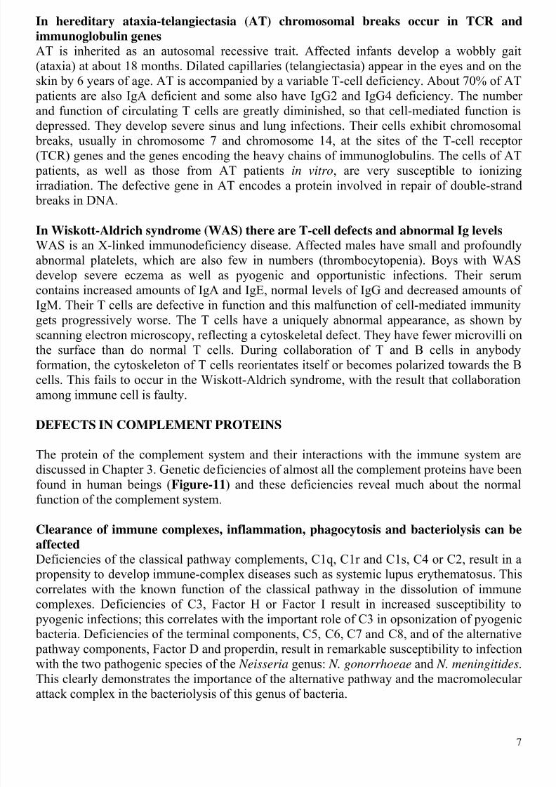
In hereditary ataxia-telangiectasia (AT) chromosomal breaks occur in TCR and
immunoglobulin genes
AT is inherited as an autosomal recessive trait. Affected infants develop a wobbly gait
(ataxia) at about 18 months. Dilated capillaries (telangiectasia) appear in the eyes and on the
skin by 6 years of age. AT is accompanied by a variable T-cell deficiency. About 70% of AT
patients are also IgA deficient and some also have IgG2 and IgG4 deficiency. The number
and function of circulating T cells are greatly diminished, so that cell-mediated function is
depressed. They develop severe sinus and lung infections. Their cells exhibit chromosomal
breaks, usually in chromosome 7 and chromosome 14, at the sites of the T-cell receptor(TCR) genes and the genes encoding the heavy chains of immunoglobulins. The cells of AT
patients, as well as those from AT patients in vitro, are very susceptible to ionizing
irradiation. The defective gene in AT encodes a protein involved in repair of double-strand
breaks in DNA.
In Wiskott-Aldrich syndrome (WAS) there are T-cell defects and abnormal Ig levels
WAS is an X-linked immunodeficiency disease. Affected males have small and profoundly
abnormal platelets, which are also few in numbers (thrombocytopenia). Boys with WAS
develop severe eczema as well as pyogenic and opportunistic infections. Their serumcontains increased amounts of IgA and IgE, normal levels of IgG and decreased amounts of
IgM. Their T cells are defective in function and this malfunction of cell-mediated immunity
gets progressively worse. The T cells have a uniquely abnormal appearance, as shown by
scanning electron microscopy, reflecting a cytoskeletal defect. They have fewer microvilli onthe surface than do normal T cells. During collaboration of T and B cells in anybody
formation, the cytoskeleton of T cells reorientates itself or becomes polarized towards the B
cells. This fails to occur in the Wiskott-Aldrich syndrome, with the result that collaboration
among immune cell is faulty.
DEFECTS IN COMPLEMENT PROTEINS
The protein of the complement system and their interactions with the immune system are
discussed in Chapter 3. Genetic deficiencies of almost all the complement proteins have been
found in human beings (Figure-11) and these deficiencies reveal much about the normal
function of the complement system.
Clearance of immune complexes, inflammation, phagocytosis and bacteriolysis can be
affected
Deficiencies of the classical pathway complements, C1q, C1r and C1s, C4 or C2, result in a propensity to develop immune-complex diseases such as systemic lupus erythematosus. This
correlates with the known function of the classical pathway in the dissolution of immune
complexes. Deficiencies of C3, Factor H or Factor I result in increased susceptibility to
pyogenic infections; this correlates with the important role of C3 in opsonization of pyogenic
bacteria. Deficiencies of the terminal components, C5, C6, C7 and C8, and of the alternative
pathway components, Factor D and properdin, result in remarkable susceptibility to infection
with the two pathogenic species of the Neisseria genus: N. gonorrhoeae and N. meningitides.
This clearly demonstrates the importance of the alternative pathway and the macromolecular
attack complex in the bacteriolysis of this genus of bacteria.

7/26/2019 20306 Lecture 14 Primary Immunodeficiency Disorder
http://slidepdf.com/reader/full/20306-lecture-14-primary-immunodeficiency-disorder 8/18
8
All these genetic complement component deficiencies are inherited as autosomal recessive
traits, except for properdin deficiency, which is inherited as an X-linked recessive, and C1
inhibitor deficiency, which is inherited as an autosomal dominant.
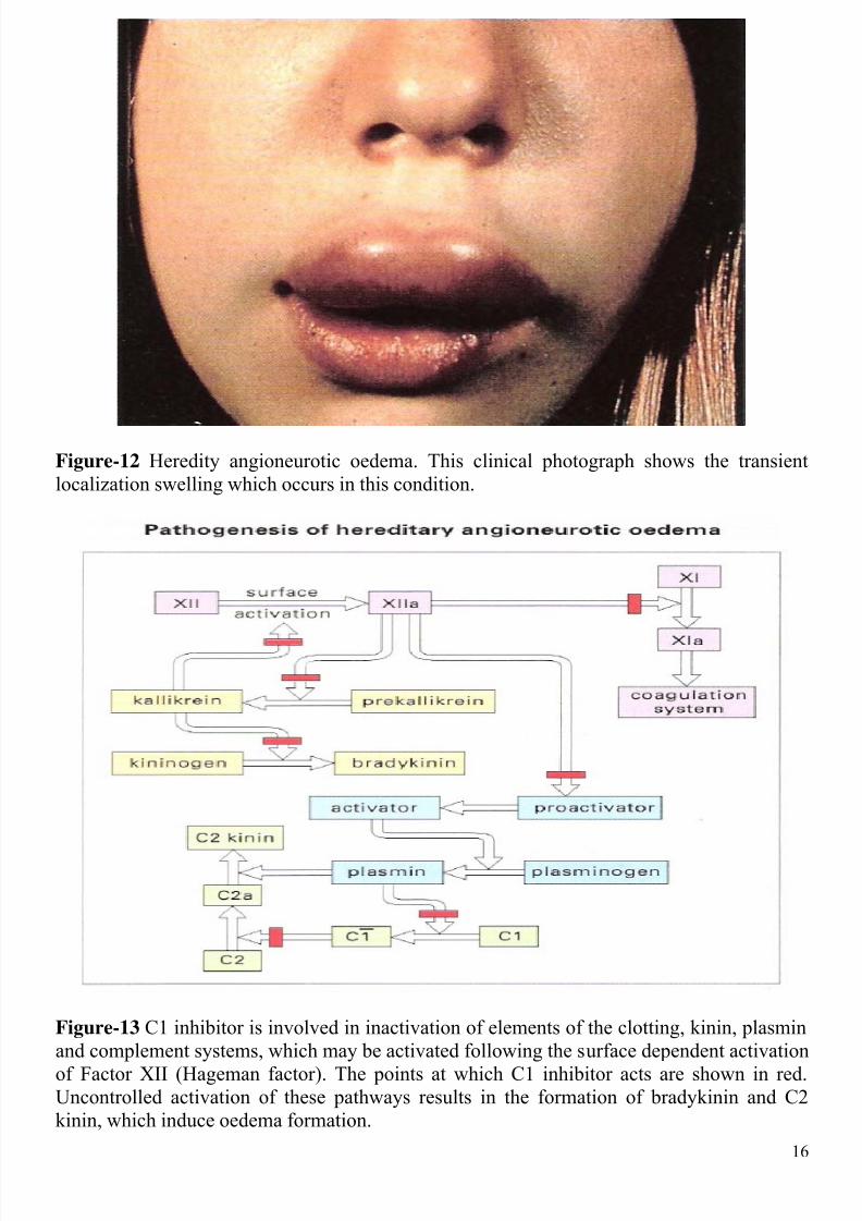
Hereditary angioneurotic oedema (HAE) is due to C! Inhibitor deficiency
Clinically, the most important deficiency of the complement system is that of the C1
inhibitor. This molecule is responsible for dissociation of activated C1, by binding to C1r 2
C1s2. The deficiency results in the well-known disease, hereditary angioneurotic oedema(HAE) (Figure-12). This disease is inherited as an autosomal dominant trait. Patients with
HAE have recurrent episodes of circumscribed swelling of various parts of the body
(angioedema). When the oedema involves the intestine, excruciating abdominal pains ad
cramps results, with severe vomiting. When the oedema involves the upper airway, the
patients may choke to death from respiratory obstruction. Angioedema of the upper airway
therefore presents a medical emergency, which requires rapid action to restore normal
breathing.
C1 inhibitor nor only inhibits the classical pathway of complement but also joint elements ofthe kinin, plasmin and clotting systems. The oedema is mediated by two peptides generated
by uninhibited activation of the complement and contact systems: a peptide derived from the
activation of C2, called C2 kinin, and bradykinin derived from the activation of the contact
system (Figure-13). The effect of these peptides is on the postcapillary venule, where theycause endothelial cell to retract, forming gaps that allow leakage of plasma.
There are two genetically determined forms of HAE. In Type I, the C1 inhibitor gene is
defective and no transcripts are formed. In Type II, there are point mutations in the C1
inhibitor gene with the consequence that defective molecules are synthesized. Thisdistinction is important because the diagnosis of Type II disease cannot be made by
quantitative measurement of serum C1 inhibitor alone. Simultaneous measurements of serum
C4 must also be done. C4 is always decreased in the serum of HAE patients, because of itsdestruction by uninhibited, activated C1.
C1 inhibitor deficiency may be acquired later in life. In some cases an autoantibody to C1inhibitor is found. In others, there is a monoclonal B-cell proliferation such as occurs in
chronic lymphocytic leukaemia, multiple myeloma or B-cell lymphoma. Such patients make
an anti-idiotype to their over-produced immunoglobulin; the idiotype – anti-idiotype
interaction, for unknown reasons, causes consumption of C1, C4 and C2 and of C1 inhibitorwithout formation of an effective C3 convertase (which would cause C3 deposition and
removal of the complement complex).
DEFECTS IN PHAGOCYTES
Phagocytic cells – polymorphonuclear leucocytes and cells of the monocyte /macrophage
lineage – are important in host defence against pyogenic bacteria and other intracellular
microorganisms. A severe deficiency of polymorphonuclear leucocytes (neutropenia) can
result in overwhelming bacterial infection. Two genetic defects of phagocytes are clinicallyimportant in that they result in susceptibility to severe infections and are often fatal: chronic
granulomatous disease and the leucocyte adhesion deficiency.
7/26/2019 20306 Lecture 14 Primary Immunodeficiency Disorder
http://slidepdf.com/reader/full/20306-lecture-14-primary-immunodeficiency-disorder 9/18
9
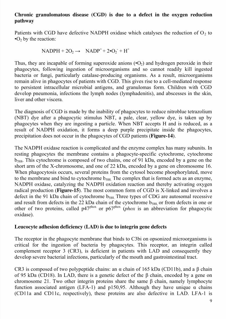
Chronic granulomatous disease (CGD) is due to a defect in the oxygen reduction
pathway
Patients with CGD have defective NADPH oxidase which catalyses the reduction of O2 to
•O2 by the reaction:
NADPH + 2O2 → NADP+
+ 2•O2- + H
+
Thus, they are incapable of forming superoxide anions (•O2) and hydrogen peroxide in their
phagocytes, following ingestion of microorganisms and so cannot readily kill ingested
bacteria or fungi, particularly catalase-producing organisms. As a result, microorganisms
remain alive in phagocytes of patients with CGD. This gives rise to a cell-mediated response
to persistent intracellular microbial antigens, and granulomas form. Children with CGD
develop pneumonia, infections the lymph nodes (lymphadenitis), and abscesses in the skin,
liver and other viscera.
The diagnosis of CGD is made by the inability of phagocytes to reduce nitroblue tetrazolium(NBT) dye after a phagocytic stimulus NBT, a pale, clear, yellow dye, is taken up by
phagocytes when they are ingesting a particle. When NBT accepts H and is reduced, as a
result of NADPH oxidation, it forms a deep purple precipitate inside the phagocytes,
precipitation does not occur in the phagocytes of CGD patients (Figure-14).
The NADPH oxidase reaction is complicated and the enzyme complex has many subunits. In
resting phagocytes the membrane contains a phagocyte-specific cytochrome, cytochrome
b588. This cytochrome is composed of two chains, one of 91 kDa, encoded by a gene on the
short arm of the X-chromosome, and one of 22 kDa, encoded by a gene on chromosome 16.When phagocytosis occurs, several proteins from the cytosol become phosphorylated, move
to the membrane and bind to cytochrome b588. The complex that is formed acts as an enzyme, NADPH oxidase, catalyzing the NADPH oxidation reaction and thereby activating oxygen
radical production (Figure-15). The most common form of CGD is X-linked and involves a
defect in the 91 kDa chain of cytochrome b588. Three types of CDG are autosomal recessive
and result from defects in the 22 kDa chain of the cytochrome b588, or from defects in one orother of two proteins, called p47 phox
or p67 phox
( phox is an abbreviation for phagocytic
oxidase).
Leucocyte adhesion deficiency (LAD) is due to integrin gene defects
The receptor in the phagocyte membrane that binds to C3bi on opsonized microorganisms is
critical for the ingestion of bacteria by phagocytes. This receptor, an integrin called
complement receptor 3 (CR3), is deficient in patients with LAD and consequently they
develop severe bacterial infections, particularly of the mouth and gastrointestinal tract.
CR3 is composed of two polypeptide chains: an α chain of 165 kDa (CD11b), and a β chain
of 95 kDa (CD18). In LAD, there is a genetic defect of the β chain, encoded by a gene on
chromosome 21. Two other integrin proteins share the same β chain, namely lymphocytefunction associated antigen (LFA-1) and p150,95. Although they have unique α chains
(CD11a and CD11c, respectively), these proteins are also defective in LAD. LFA-1 is
7/26/2019 20306 Lecture 14 Primary Immunodeficiency Disorder
http://slidepdf.com/reader/full/20306-lecture-14-primary-immunodeficiency-disorder 10/18
10
important in cell adhesion and interacts with intercellular adhesion molecule-1 (ICAM-1) on
endothelial cell surfaces and other cell membranes. Because of the defect in LFA-1,
phagocytes from patients with LAD cannot adhere to vascular endothelium and thus cannot
migrate out of blood vessels into areas of infection. Thus patients with LAD cannot form pus
efficiently; this allows the rapid spread of bacterial invaders.
When leucocytes in the circulation enter an area of inflammation their speed of movement is
greatly retarded by the interaction of selectins that are expressed on the surface of the
leucocytes with ligands that are expressed on the surface of the endothelium in areas ofinflammation. The leucocytes start to roll on the endothelial surface prior to the interaction
of the leucocyte integrins with adhesion molecules such as intracellular adhesion molecule-1
(ICAM-1). The ligands with which the selectins interact are glycoproteins that contain
fucosylated sugars such as the blood group sialyl Lewisx.
A genetic defect in the conversion
of mannose to fucose results in the failure of normal synthesis in these selectin ligands.
Consequently the leucocytes of such patients cannot roll on the endothelium. This causes a
second form of LAD, called LAD type 2.
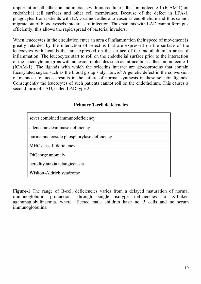
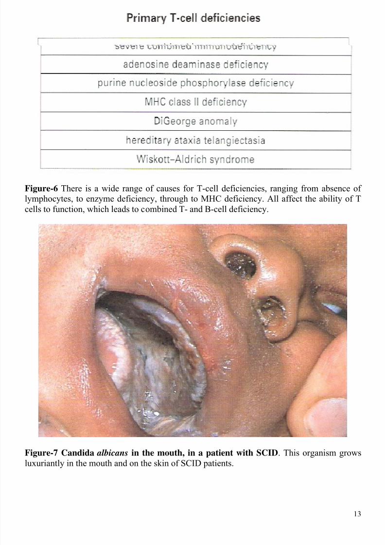
Primary T-cell deficiencies
sever combined immunodeficiency
adenosine deaminase deficiency
purine nucleoside phosphorylase deficiency
MHC class II deficiency
DiGeorge anomaly
heredity ataxia telangiectasia
Wiskott-Aldrich syndrome
Figure-1 The range of B-cell deficiencies varies from a delayed maturation of normal
immunoglobulin production, through single isotype deficiencies to X-linked
agammaglobulinaemia, where affected male children have no B cells and no serum
immunoglobulins.
7/26/2019 20306 Lecture 14 Primary Immunodeficiency Disorder
http://slidepdf.com/reader/full/20306-lecture-14-primary-immunodeficiency-disorder 11/18
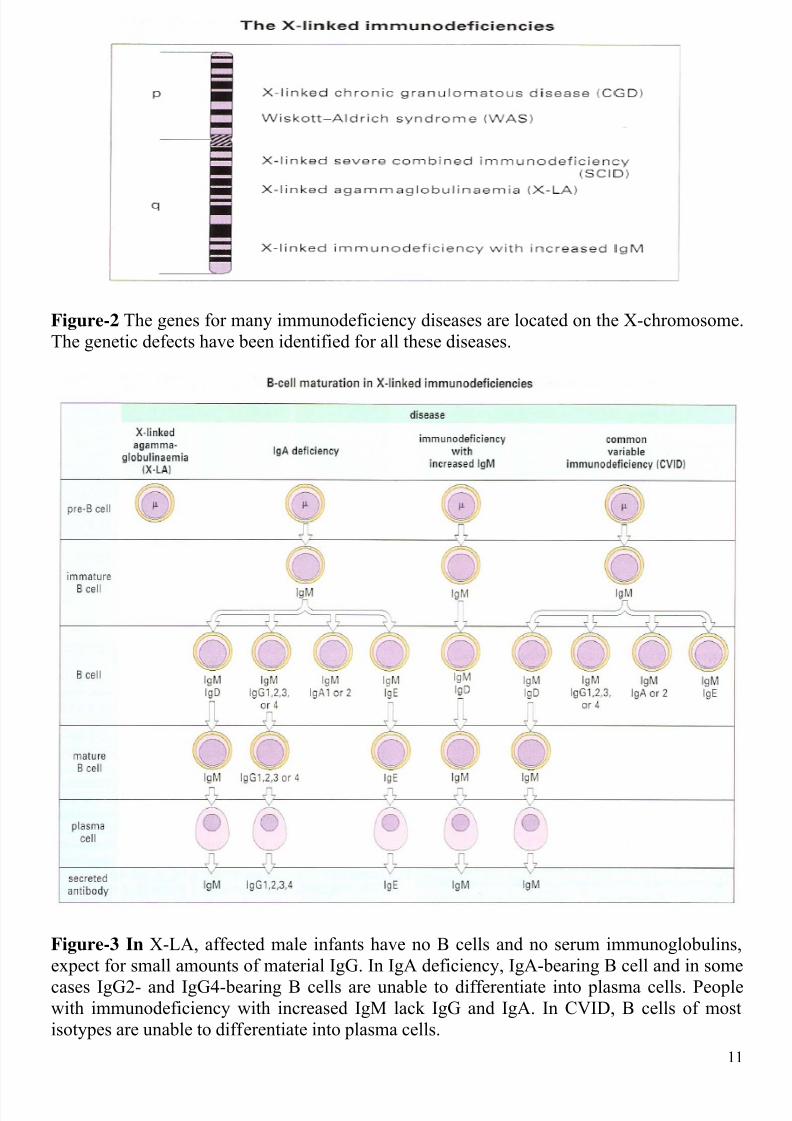
Figure-2 The genes for many immunodeficiency diseases are located on the X-chromosome.
The genetic defects have been identified for all these diseases.
Figure-3 In X-LA, affected male infants have no B cells and no serum immunoglobulins,
expect for small amounts of material IgG. In IgA deficiency, IgA-bearing B cell and in some
cases IgG2- and IgG4-bearing B cells are unable to differentiate into plasma cells. Peoplewith immunodeficiency with increased IgM lack IgG and IgA. In CVID, B cells of most
isotypes are unable to differentiate into plasma cells.
11

7/26/2019 20306 Lecture 14 Primary Immunodeficiency Disorder
http://slidepdf.com/reader/full/20306-lecture-14-primary-immunodeficiency-disorder 12/18
Figure-4 Gall bladder from a patient with immunodeficiency with increased IgM. Thesubmucosa is filled with cells with pink-staining cytoplasm and eccentric nuclei. The cells
are synthesizing and secreting IgM.
Figure-5 Giardia lamblia Innumerable Giardia parasites can be seen swarming over themucosa of the jejunum of a patient with CVID.
12
7/26/2019 20306 Lecture 14 Primary Immunodeficiency Disorder
http://slidepdf.com/reader/full/20306-lecture-14-primary-immunodeficiency-disorder 13/18
Figure-6 There is a wide range of causes for T-cell deficiencies, ranging from absence oflymphocytes, to enzyme deficiency, through to MHC deficiency. All affect the ability of T
cells to function, which leads to combined T- and B-cell deficiency.
Figure-7 Candida albicans in the mouth, in a patient with SCID. This organism grows
luxuriantly in the mouth and on the skin of SCID patients.
13

7/26/2019 20306 Lecture 14 Primary Immunodeficiency Disorder
http://slidepdf.com/reader/full/20306-lecture-14-primary-immunodeficiency-disorder 14/18
Figure-8 Thymus of SCID: Note that the thymic stroma has not been invaded by lymphoid
cells and no Hassall's corpuscles are seen. The gland has a fetal appearance.
Figure-9 It is thought that deficiencies of ADA and PNP lead to accumulations of dATP and
dGTP respectively. Both of these metabolites are powerful inhibitors of ribonucleotidereductase, an essential enzyme for DNA synthesis.
14

7/26/2019 20306 Lecture 14 Primary Immunodeficiency Disorder
http://slidepdf.com/reader/full/20306-lecture-14-primary-immunodeficiency-disorder 15/18
Figure-10 DiGeorge anomaly. Note the wide-set eyes, low-set ears and shortened philtrum
of upper lip. Congenital malformations of the cardiovascular system may also occur.
Figure-11 Genetic deficiencies of human complement: (AR=phenotypically autosomalrecessive; AD=autosomal dominant; XL=X-linked recessive).
15
7/26/2019 20306 Lecture 14 Primary Immunodeficiency Disorder
http://slidepdf.com/reader/full/20306-lecture-14-primary-immunodeficiency-disorder 16/18
Figure-12 Heredity angioneurotic oedema. This clinical photograph shows the transient
localization swelling which occurs in this condition.
Figure-13 C1 inhibitor is involved in inactivation of elements of the clotting, kinin, plasmin
and complement systems, which may be activated following the surface dependent activation
of Factor XII (Hageman factor). The points at which C1 inhibitor acts are shown in red.Uncontrolled activation of these pathways results in the formation of bradykinin and C2
kinin, which induce oedema formation.
16

7/26/2019 20306 Lecture 14 Primary Immunodeficiency Disorder
http://slidepdf.com/reader/full/20306-lecture-14-primary-immunodeficiency-disorder 17/18
Figure-14 Nitroblue tetrazolium (NBT) test. In normal polymorphs and monocytes, reactive
oxygen intermediates (ROIs) are activated by phagocytosis, and yellow NBT is converted to
purple-blue formazan (1). Patients with CGD cannot form ROIs and so the dye stays yellow(2).
Figure-15 Prevailing knowledge of the NADPH oxidase suggest that, in its dormant state,some of its component parts are in the membrane (cytochrome b558 and possibly rap-1) while
others are in the cytosol (p47 phox,
p67 phox,
the NADPH-binding component, N, and a putative
fourth component, α). After the stimulus provided phagocytosis, the cytosolic componentsassociate and move to the membrane, an event possibly mediated by phosphorylation (P) of
p47 phox.
Once the cytosol components are associated with the membrane components, the
oxidase becomes catalytically active and p47 phox
is phosphorylated further. In the differentforms of CGD, there are defects in the genes for different components of the oxidase.
Dr. MUSTAFA HASSAN LINJAWI
17

7/26/2019 20306 Lecture 14 Primary Immunodeficiency Disorder
http://slidepdf.com/reader/full/20306-lecture-14-primary-immunodeficiency-disorder 18/18
18


![Lung diseases in children with primary immunodeficiency children with primary immunodeficiency, susceptible to a range of diseases, interstitial lung tissue disorder can develop [3]](https://img.dokumen.tips/doc/110x75/5edc03d8ad6a402d66667fa3/lung-diseases-in-children-with-primary-immunodeficiency-children-with-primary-immunodeficiency.jpg)