Embed Size (px)
Citation preview

Treatment
TREATMENT
revised 08/22/19 10.010 - Page 1
10.010Abdominal Pain
Treatment
Abdominal Pain
HX PE DDX
Pain: nature, duration, location, radiation, intensity
Associated symptoms: fever, nausea and vomiting, diarrhea, melena, painful urination
Last menstrual period
Prior abdominal surgery
Distension
Tenderness
Guarding
Rigidity
Rebound
Masses
Diffuse: Perforation, intraabdominal bleeding (trauma, ectopic, AAA), gastroenteritis
RUQ: cholecystitis, hepatitis, pancreatitis
Epigastric: peptic ulcer, pancreatitis, gastritis
LUQ: spleen, pancreatitis, stomach (PUD)
Flank: kidney stone, pyelonephritis
RLQ: Appendicitis , kidney stone, PID, ovarian cyst, cystitis
LLQ: diverticulitis, kidney stone, PID, ovarian cyst, cystitis
Specific Precautions: A. Abdominal pain may be the first warning of catastrophic internal bleeding (ruptured aneurysm, liver, spleen, ectopic pregnancy, perforated viscus, etc.). B. Since the bleeding is not apparent, you must think of volume depletion and monitor patient closely to recognize shock. C. For transgender or non-binary patients, ask if any prior surgery.
Treatment: A. Start O
2, follow Airway Management procedure.
B. If shock syndrome is present and MAP > 65 mmHg (SBP > 90 mmHg), follow Shock protocol, and consider IV/IO, NS, large bore, TKO or as needed. If traumatic event, enter into trauma system. Rapid transport is of primary importance. C. Place patient in comfortable position. D. Do not allow patient to eat or drink. E. Obtain vital signs frequently.
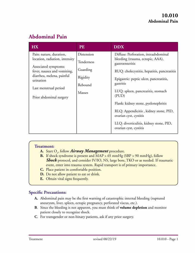
Treatment: A. Determine level of consciousness. B. Start O
2, follow Airway Management procedure. Unless intubated, transport on
left side, if possible, to protect airway. C. Monitor vital signs and respiratory status during transport. D. Start IV/IO as needed. E. Monitor cardiac rhythm and follow Cardiac Dysrhythmia protocol.
Consider underlying causes: Altered mental status has many causes, and may require the use of multiple protocols.
Treatment
Altered Mental Status and Coma
HX PE DDX
Onset / changes LOC
Recent history: headache, nausea and vomiting, trauma
Diabetes
CVA
Hypertension
Seizure
Medications
Pregnancy
LOC
Evidence of traumatic injury
Vital signs
Pupils
Breath odor
Nuchal rigidity
Neuro deficits (weakness)
Confusion
Hypoglycemia (diabetes)
Hypoxia/hypercarbia/CO
Shock (MI, hypovolemia)
Drug/toxin
Trauma
Cerebrovascular (CVA, intracranial hemorrhage, infection, tumor)
Metabolic (e.g., electrolyte imbalance, hypothermia, hyperthermia)
Seizure (postictal)
Infection (meningitis/encephalitis)
revised 08/31/17 10.020 - Page 1
10.020Altered Mental Status and Coma
Treatment
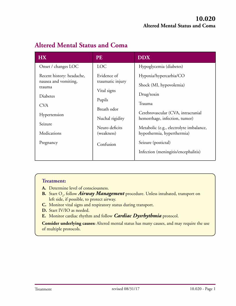
Hypoglycemia:Determine capillary blood glucose level using blood glucose meter. If the blood glucose reading is less than 60 mg% or glucose less than 80 mg% in a known diabetic: A. Administer glucose: 1. Do not administer oral glucose to patients without a gag reflex or with a rapidly diminishing level of consciousness. 2. If patient is unable to take sugar orally, administer dextrose 50%, 25-50 mL (in large vein) or dextrose 10%, 125-250 mL.
B. Repeat capillary blood glucose level after 10 minutes and treat if it remains low. C. If unable to administer oral glucose or establish IV/IO, give glucagon 1 mg IM.
Overdose: A. Follow Poisoning and Overdose protocol, if indicated. B. If opioid intoxication is suspected:
1. If no IV/IO has been established, administer naloxone 2 mg IM/IN.2. If IV/IO already established, administer naloxone 0.5 mg IV/IO and observe for improved respiration, IV/IO dose may be repeated every 2 minutes up to 2 mg.3. In most instances, a total dose of 2 mg IM/IN or IV/IO will be sufficient to reverse opioid intoxication. In some cases (methadone or designer drugs), larger doses of naloxone may be necessary. In these cases, additional doses of naloxone (2 mg IM/IN or IV/IO every 3-5 minutes) up to a MAX of 8 mg of naloxone may be administered to reverse opioid intoxication.
Psychiatric Disorders: A. Almost never cause disorientation or alteration in level of consciousness. If the patient is disoriented, assume a medical cause. B. Follow Psychiatric and Behavioral Disorders protocol. C. If a non-organic cause of coma in adults (over age 16) is suspected, ammonia inhalants or other noxious stimuli may be considered. 1. Response to noxious stimuli does not rule out medical or traumatic causes of initial coma. 2. Never place inhalants in nostrils or inside O
2 mask.
Seizure:Follow Seizure protocol.
Stroke:
Follow Stroke/CVA protocol.
Toxemia: Follow OB/GYN Emergencies protocol, if indicated.
revised 10/30/17 10.020 - Page 2
10.020Altered Mental Status and Coma

Treatment
Trauma: A. Maintain spinal precautions. B. If GCS score is < 13, enter patient into the Trauma System. C. Perform all treatment possible en route. D. Maintain ventilation as per End-tidal CO
2 protocol.
1. Secure protected airway if GCS score is < 8.
Pediatric Considerations: 1. Consider etiology and appropriate protocols: shock, toxic exposure, head trauma (consider intentional injury), seizure. 2. Vascular access. 3. Rapid blood glucose determination. If glucose determination is less than 60 mg% (less than 40 mg% for neonates), administer oral glucose to conscious patient, OR,
a. If no IV/IO established and airway protective reflexes are intact, administer D50
, or other glucose containing substance, orally. b. If IV/IO established, administer Dextrose 10% (5 mL/kg) for neonates, infants, and children, may repeat once. c. If no IV/IO established and airway protective reflexes are not intact, administer glucagon 0.02 mg/kg IM to a MAX of 1 mg. d. Repeat blood glucose determination and treat if it remains low. 4. If mental status and respiratory effort are depressed, administer naloxone 0.1 mg/kg, MAX 2 mg IV/IO/IM. a. Do Not Administer naloxone to neonates. b. May repeat every 5 minutes with strong suspicion of opiate overdose, or if partial response is noted.
revised 10/15/14 10.020 - Page 3
10.020Altered Mental Status and Coma

Treatment
Anaphylaxis and Allergic Reactions
HX PE DDX
Difficulty breathing / speaking (hoarseness)
Chest tightness
Subjective airway impairment or swelling
Itching
Exposure: Meds, insects or stings, food / toxic substance
Known allergies
Prior allergic reactions
LOC
Edema (face, tongue, extremities)
Respiratory (wheezing, hoarseness, stridor etc.)
Rash, flushing, hives
Hypotension/shock
Anaphylaxis
Upper airway infections
Angioedema (medication)
Asthma (bronchospasm)
Urticaria
Foreign body
revised 08/01/18 10.030 - Page 1
10.030Anaphylaxis and Allergic Reactions
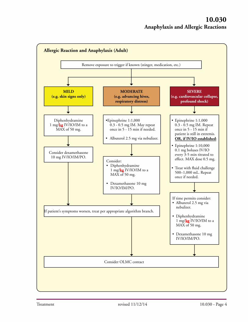
Treatment: A. Protect airway; suction as needed. 1. Follow Airway Management procedure. 2. Cricothyrotomy may be required if unable to secure protected airway or ventilate by BVM after epinephrine has been administered. B. Start IV/IO as needed. If shock syndrome is present and MAP < 65 mmHg (SBP < 90 mmHg), follow Shock protocol. C. Monitor cardiac rhythm and if dysrhythmia is present, follow Cardiac Dysrhythmia protocol.
D. Assess severity of allergic reaction. 1. Mild (Skin signs only) a. Diphenhydramine 1 mg/kg IV/IO/IM to a MAX of 50 mg. b. Consider dexamethasone 10 mg IV/IO/IM/PO. 2. Moderate (Advancing hives, respiratory distress, etc.) a. Epinephrine 1:1,000 0.3 - 0.5 mg IM. May repeat once in 5-15 minutes if needed. b. Albuterol 2.5 mg via nebulizer. c. Consider: i. Diphenhydramine 1 mg/kg IV/IO/IM to a MAX of 50 mg. ii. Dexamethasone 10 mg IV/IO/IM/PO.

Treatment
3. Severe (cardiovascular collapse, profound shock). a. Epinephrine 1:1,000 0.3 - 0.5 mg IM. Repeat once in 5-15 minutes if patient is still in shock Or, if IV/IO established, b. Epinephrine 1:10,000 0.1 mg boluses IV/IO every 3-5 min titrated to effect. MAX dose 0.5 mg. c. If hypotensive, fluid challenge 500–1,000 mL. Repeat once if needed. d. If time permits consider: i. Albuterol 2.5 mg via nebulizer. ii. Diphenhydramine 1 mg/kg IV/IO/IM to a MAX of 50 mg. iii. Dexamethasone 10 mg IV/IO/IM/PO.
E. SPECIAL NOTE: 1. If 1:10,000 not available, you may dilute 1 mL of 1:1,000 epinephrine with 9 mL of NS (1 mg/10 mL) and administer 1 mL IV or IO.
revised 11/12/14 10.030 - Page 2
10.030Anaphylaxis and Allergic Reactions
Treatment
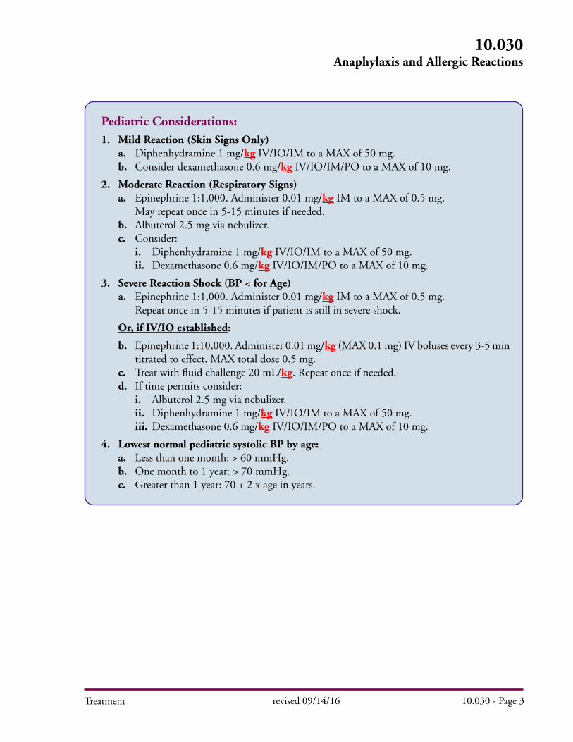
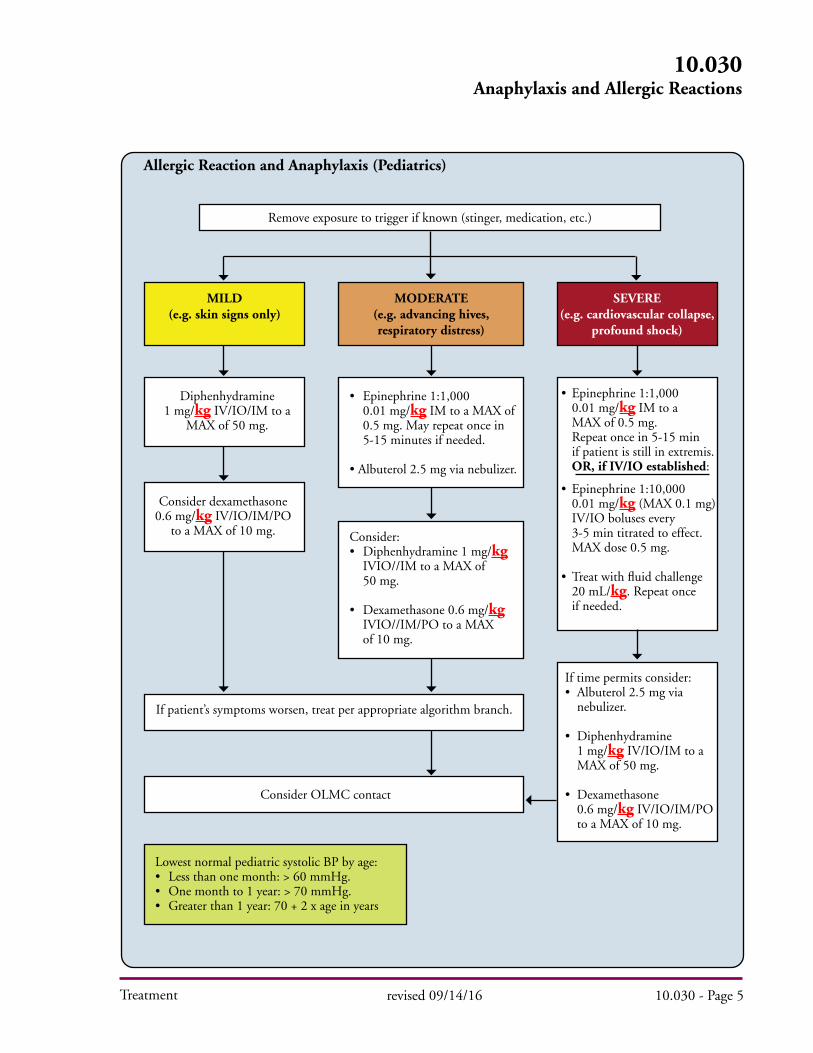
Pediatric Considerations:1. Mild Reaction (Skin Signs Only) a. Diphenhydramine 1 mg/kg IV/IO/IM to a MAX of 50 mg. b. Consider dexamethasone 0.6 mg/kg IV/IO/IM/PO to a MAX of 10 mg.
2. Moderate Reaction (Respiratory Signs) a. Epinephrine 1:1,000. Administer 0.01 mg/kg IM to a MAX of 0.5 mg. May repeat once in 5-15 minutes if needed. b. Albuterol 2.5 mg via nebulizer. c. Consider: i. Diphenhydramine 1 mg/kg IV/IO/IM to a MAX of 50 mg. ii. Dexamethasone 0.6 mg/kg IV/IO/IM/PO to a MAX of 10 mg.
3. Severe Reaction Shock (BP < for Age) a. Epinephrine 1:1,000. Administer 0.01 mg/kg IM to a MAX of 0.5 mg. Repeat once in 5-15 minutes if patient is still in severe shock.
Or, if IV/IO established:
b. Epinephrine 1:10,000. Administer 0.01 mg/kg (MAX 0.1 mg) IV boluses every 3-5 min titrated to effect. MAX total dose 0.5 mg. c. Treat with fluid challenge 20 mL/kg. Repeat once if needed. d. If time permits consider: i. Albuterol 2.5 mg via nebulizer. ii. Diphenhydramine 1 mg/kg IV/IO/IM to a MAX of 50 mg. iii. Dexamethasone 0.6 mg/kg IV/IO/IM/PO to a MAX of 10 mg.
4. Lowest normal pediatric systolic BP by age: a. Less than one month: > 60 mmHg. b. One month to 1 year: > 70 mmHg. c. Greater than 1 year: 70 + 2 x age in years.
revised 09/14/16 10.030 - Page 3
10.030Anaphylaxis and Allergic Reactions
Treatment revised 11/12/14 10.030 - Page 4
10.030Anaphylaxis and Allergic Reactions
Allergic Reaction and Anaphylaxis (Adult)
MILD(e.g. skin signs only)
MODERATE(e.g. advancing hives, respiratory distress)
SEVERE(e.g. cardiovascular collapse,
profound shock)
Remove exposure to trigger if known (stinger, medication, etc.)
Diphenhydramine 1 mg/kg IV/IO/IM to a
MAX of 50 mg.
•Epinephrine 1:1,000 0.3 - 0.5 mg IM. May repeat once in 5 - 15 min if needed.
• Albuterol 2.5 mg via nebulizer.
Consider:• Diphenhydramine 1 mg/kg IV/IO/IM to a MAX of 50 mg.
• Dexamethasone 10 mg IV/IO/IM/PO.
Consider dexamethasone 10 mg IV/IO/IM/PO.
If patient’s symptoms worsen, treat per appropriate algorithm branch.
Consider OLMC contact
If time permits consider:• Albuterol 2.5 mg via nebulizer.
• Diphenhydramine 1 mg/kg IV/IO/IM to a MAX of 50 mg.
• Dexamethasone 10 mg IV/IO/IM/PO.
• Epinephrine 1:1,000 0.3 - 0.5 mg IM. Repeat once in 5 - 15 min if patient is still in extremis. OR, if IV/IO established:
• Epinephrine 1:10,000 0.1 mg boluses IV/IO every 3-5 min titrated to effect. MAX dose 0.5 mg.
• Treat with fluid challenge 500–1,000 mL. Repeat once if needed.
Treatment revised 09/14/16 10.030 - Page 5
10.030Anaphylaxis and Allergic Reactions
Allergic Reaction and Anaphylaxis (Pediatrics)
Remove exposure to trigger if known (stinger, medication, etc.)
Diphenhydramine 1 mg/kg IV/IO/IM to a
MAX of 50 mg.
• Epinephrine 1:1,000 0.01 mg/kg IM to a MAX of 0.5 mg. May repeat once in 5-15 minutes if needed.
• Albuterol 2.5 mg via nebulizer.
• Epinephrine 1:1,000 0.01 mg/kg IM to a MAX of 0.5 mg. Repeat once in 5-15 min if patient is still in extremis. OR, if IV/IO established:
• Epinephrine 1:10,000 0.01 mg/kg (MAX 0.1 mg) IV/IO boluses every 3-5 min titrated to effect. MAX dose 0.5 mg.
• Treat with fluid challenge 20 mL/kg. Repeat once if needed.
If time permits consider:• Albuterol 2.5 mg via nebulizer.
• Diphenhydramine 1 mg/kg IV/IO/IM to a MAX of 50 mg.
• Dexamethasone 0.6 mg/kg IV/IO/IM/PO to a MAX of 10 mg.
Consider:• Diphenhydramine 1 mg/kg IVIO//IM to a MAX of 50 mg.
• Dexamethasone 0.6 mg/kg IVIO//IM/PO to a MAX of 10 mg.
Lowest normal pediatric systolic BP by age:• Less than one month: > 60 mmHg.• One month to 1 year: > 70 mmHg.• Greater than 1 year: 70 + 2 x age in years
Consider dexamethasone 0.6 mg/kg IV/IO/IM/PO
to a MAX of 10 mg.
If patient’s symptoms worsen, treat per appropriate algorithm branch.
Consider OLMC contact
MILD(e.g. skin signs only)
MODERATE(e.g. advancing hives, respiratory distress)
SEVERE(e.g. cardiovascular collapse,
profound shock)
Treatment revised 11/12/14 10.030 - Page 6
10.030Anaphylaxis and Allergic Reactions
Specific Precautions:A. Epinephrine should not be given unless signs of cardiovascular or respiratory distress are present.B. Preferred location for IM administration, if feasible, is the mid-anterolateral aspect of thigh.C. Common side effects include anxiety, tremor, palpations, tachycardia, and headache.D. Acute coronary syndromes (angina, myocardial infarction, arrhythmias) may occur in both treated and untreated patients of all age groups.E. Patients presenting with anaphylaxis who are on beta-blockers and ACE inhibitors may have a more severe anaphylaxis presentation and more adverse effects with epinephrine treatment. 1. Epinephrine administered to a patient taking beta-blockers may produce unopposed alpha-adrenergic and reflex vagotonic effects, possibly leading to severe hypertension and the risk of cerebral hemorrhage. 2. Anaphylaxis in a patient on beta-blockers may present with severe bronchospasm, profound hypotension and bradycardia. 3. If epinephrine is ineffective in treating anaphylaxis in patients taking beta-blockers, both glucagon administration (1-5 mg in adults, 20-30 microgram/kg (MAX 1 mg) in children (OLMC required) and isotonic volume expansion (in some circumstances, up to several liters of crystalloid) may be necessary.
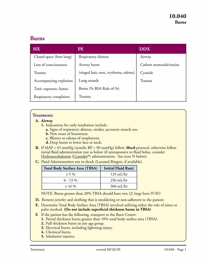
Treatment: A. Airway 1. Indications for early intubation include: a. Signs of respiratory distress, stridor, accessory muscle use. b. New onset of hoarseness. c. Blisters or edema of oropharynx. d. Deep burns to lower face or neck. B. If MAP < 65 mmHg (systolic BP < 90 mmHg) follow Shock protocol, otherwise follow initial fluid administration rate as below (if unresponsive to fluid bolus, consider Hydroxocobalamin (Cyanokit®) administration - See item N below). C. Fluid Administration not in shock (Lactated Ringers, if available).
NOTE: Burns greater than 20% TBSA should have two (2) large bore IV/IO
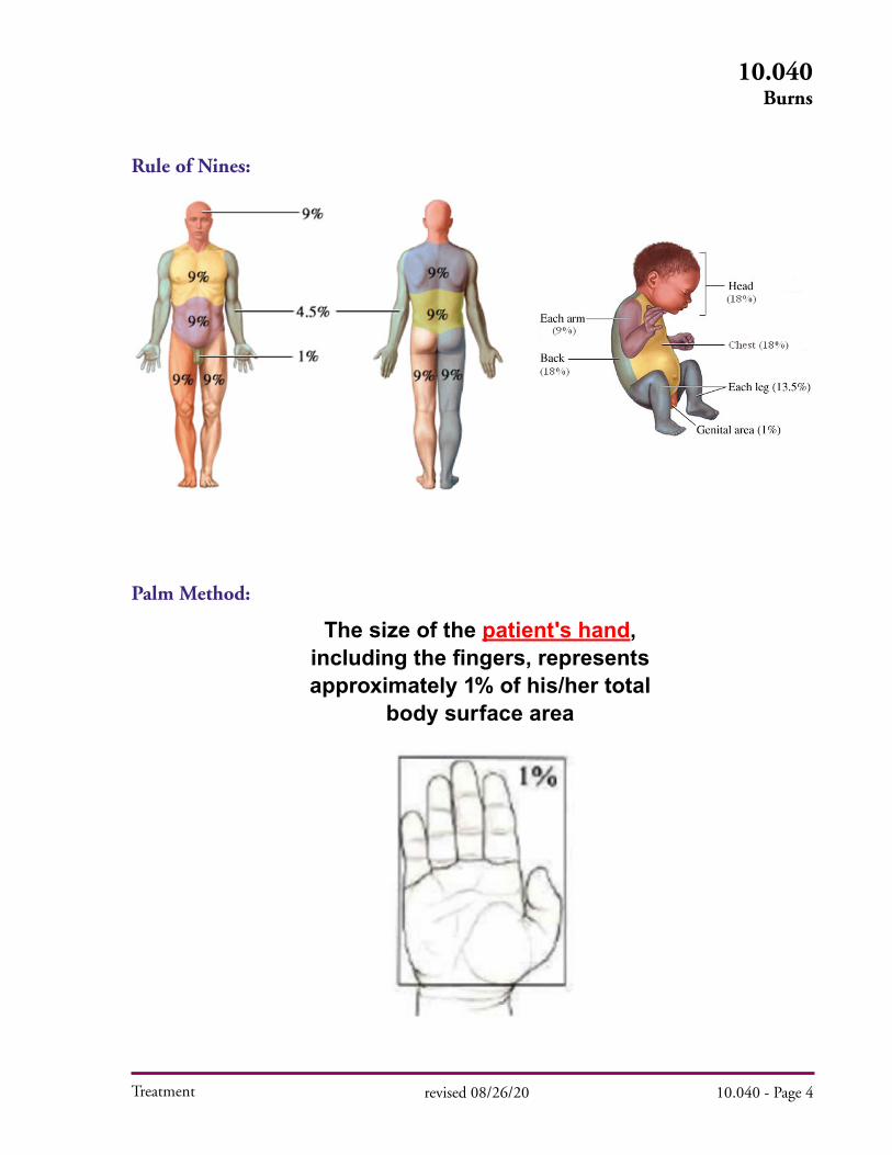
D. Remove jewelry and clothing that is smoldering or non-adherent to the patient. E. Determine Total Body Surface Area (TBSA) involved utilizing either the rule of nines or palm method. (Do not include superficial thickness burns in TBSA) F. If the patient has the following, transport to the Burn Center: 1. Partial thickness burns greater than 10% total body surface area (TBSA). 2. Full thickness burns in any age group. 3. Electrical burns, including lightning injury. 4. Chemical burns. 5. Inhalation injuries.
Total Body Surface Area (TBSA)≤ 5 %
6 - 13 %≥ 14 %
Initial Fluid Rate)125 mL/hr250 mL/hr500 mL/hr
Treatment
Burns
HX PE DDX
Closed space (how long)
Loss of consciousness
Trauma
Accompanying explosion
Toxic exposure, fumes
Respiratory complaints
Respiratory distress
Airway burns
(singed hair, soot, erythema, edema)
Lung sounds
Burns (% BSA Rule of 9s)
Trauma
Airway
Carbon monoxide/toxins
Cyanide
Trauma
revised 08/26/20 10.040 - Page 1
10.040Burns
6. Burns to face, hands, feet, genitalia, perineum, major joints, or circumferential burns. 7. Burns in high-risk patients (pediatrics, elderly, significant underlying cardiac or respiratory problems). 8. Trauma system patients with burns meeting the above criteria should be transported to Emanuel Trauma Center, if possible. G. Cool burned areas (no more than 5 minutes) then cover with clean, warm, and dry sheet or blanket or clean saran wrap (if available). Discontinue cooling if patient begins to shiver. Attempt to leave unbroken blisters intact.
H. Wound care 1. Transport using clean, dry sheets or blankets, or saran wrap. I. Prevent hypothermia J. Treat pain per Pain Management protocol. K. Apply carbon monoxide (e.g. Rad-57) monitor if available and measure carbon monoxide level. If symptoms are present (e.g. headache, dizziness, weakness, nausea) and carbon monoxide level (COHb) is greater than 15%, administer 100% oxygen. L. If chemical burn: 1. Consider HazMat response. 2. Protect yourself from contamination. (See Hazardous Materials - 50.060 protocol) 3. Flush contaminated areas with copious amounts of water. 4. If chemical is dry, carefully brush off prior to flushing. 5. Do not use a neutralizer. M. If electrical burn: 1. Apply sterile dressings to entry and exit wounds. As with other injuries, keep clean, warm, and dry. 2. Treat any dysrhythmias per appropriate Cardiac Dysrhythmia protocol. 3. Specify arc flash or contact and voltage if known. N. If cyanide toxicity is suspected based on findings (soot in mouth, nose, or oropharynx) and patient is comatose, in cardiac or respiratory arrest, or has persistent hypotension despite fluid resuscitation: 1. Administer Hydroxocobalamin (Cyanokit®) 5 g IV/IO as an infusion over 15 minutes and monitor for clinical response. Contact OLMC for advice regarding a second dose. 2. If Hydroxocobalamin (Cyanokit®) is not available, then administer Sodium Thiosulfate 50 ml of 25% solution over 10-20 minutes. 3. Hydroxocobalamin (Cyanokit®) and Sodium Thiosulfate may be administered to the same patient but NOT at the same time. 4. Treat other presenting symptoms per appropriate protocol. 5. Initiate emergent transport to appropriate facility. 6. Notify receiving facility if either Hydroxocobalamin or Sodium Thiosulfate are administered.
Treatment revised 08/26/20 10.040 - Page 2
10.040Burns
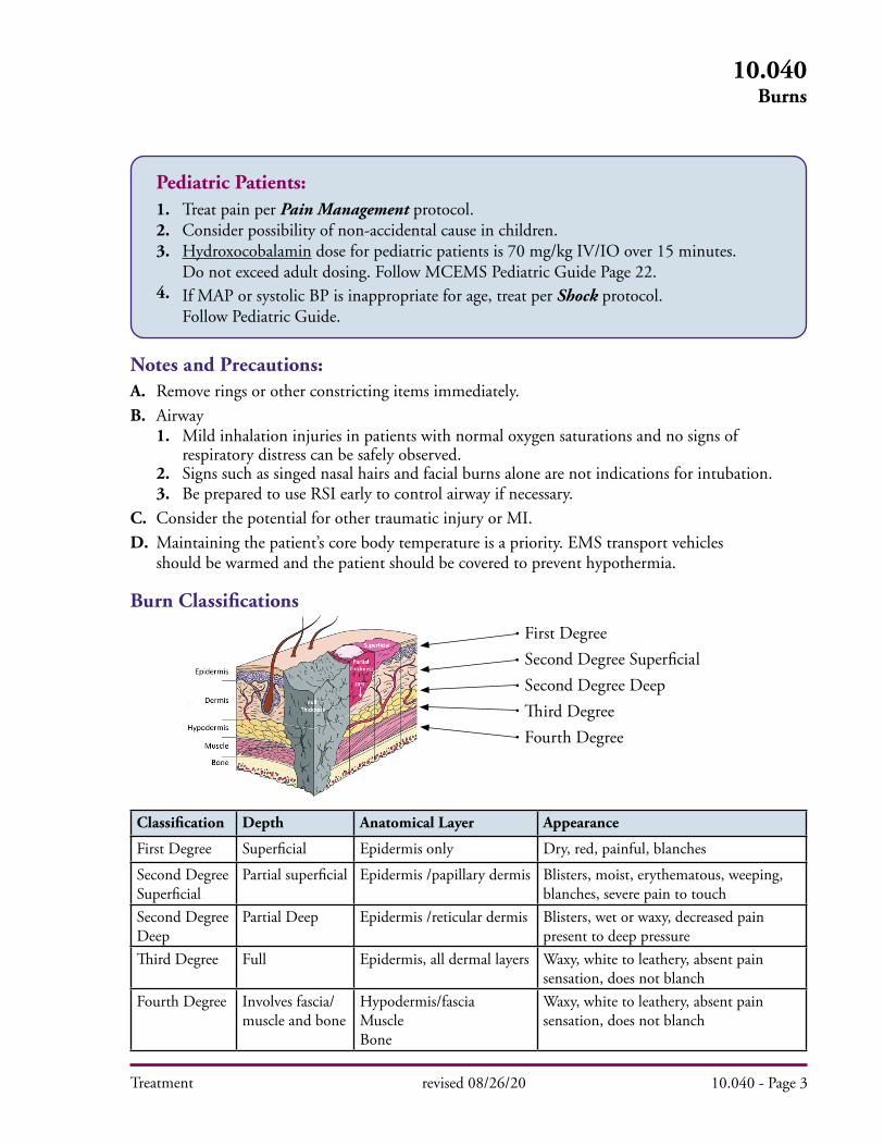
Classification Depth Anatomical Layer Appearance
First Degree Superficial Epidermis only Dry, red, painful, blanches
Second Degree Superficial
Partial superficial Epidermis /papillary dermis Blisters, moist, erythematous, weeping, blanches, severe pain to touch
Second Degree Deep
Partial Deep Epidermis /reticular dermis Blisters, wet or waxy, decreased pain present to deep pressure
Third Degree Full Epidermis, all dermal layers Waxy, white to leathery, absent pain sensation, does not blanch
Fourth Degree Involves fascia/muscle and bone
Hypodermis/fasciaMuscleBone
Waxy, white to leathery, absent pain sensation, does not blanch
First DegreeSecond Degree SuperficialSecond Degree DeepThird DegreeFourth Degree
Treatment revised 08/26/20 10.040 - Page 3
10.040Burns
Pediatric Patients: 1. Treat pain per Pain Management protocol. 2. Consider possibility of non-accidental cause in children. 3. Hydroxocobalamin dose for pediatric patients is 70 mg/kg IV/IO over 15 minutes. Do not exceed adult dosing. Follow MCEMS Pediatric Guide Page 22. 4. If MAP or systolic BP is inappropriate for age, treat per Shock protocol. Follow Pediatric Guide.
Notes and Precautions:A. Remove rings or other constricting items immediately.
B. Airway 1. Mild inhalation injuries in patients with normal oxygen saturations and no signs of respiratory distress can be safely observed. 2. Signs such as singed nasal hairs and facial burns alone are not indications for intubation. 3. Be prepared to use RSI early to control airway if necessary.
C. Consider the potential for other traumatic injury or MI.
D. Maintaining the patient’s core body temperature is a priority. EMS transport vehicles should be warmed and the patient should be covered to prevent hypothermia.
Burn Classifications
Treatment revised 08/26/20 10.040 - Page 4
10.040Burns
Rule of Nines:
Palm Method:
The size of the patient's hand,including the fingers, representsapproximately 1% of his/her total
body surface area
Treatment
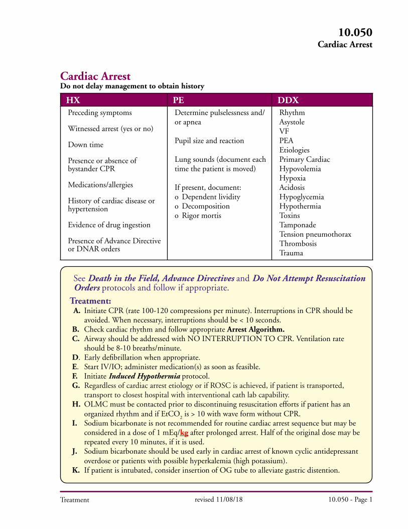
HX PE DDXPreceding symptoms
Witnessed arrest (yes or no)
Down time
Presence or absence of bystander CPR
Medications/allergies
History of cardiac disease or hypertension
Evidence of drug ingestion
Presence of Advance Directive or DNAR orders
Determine pulselessness and/or apnea
Pupil size and reaction
Lung sounds (document each time the patient is moved)
If present, document:o Dependent lividityo Decompositiono Rigor mortis
RhythmAsystoleVFPEAEtiologiesPrimary Cardiac HypovolemiaHypoxiaAcidosisHypoglycemiaHypothermiaToxinsTamponadeTension pneumothoraxThrombosisTrauma
Do not delay management to obtain history
revised 11/08/18 10.050 - Page 1
10.050Cardiac Arrest
See Death in the Field, Advance Directives and Do Not Attempt Resuscitation Orders protocols and follow if appropriate.
Treatment: A. Initiate CPR (rate 100-120 compressions per minute). Interruptions in CPR should be avoided. When necessary, interruptions should be < 10 seconds.
B. Check cardiac rhythm and follow appropriate Arrest Algorithm. C. Airway should be addressed with NO INTERRUPTION TO CPR. Ventilation rate should be 8-10 breaths/minute. D. Early defibrillation when appropriate. E. Start IV/IO; administer medication(s) as soon as feasible. F. Initiate Induced Hypothermia protocol. G. Regardless of cardiac arrest etiology or if ROSC is achieved, if patient is transported, transport to closest hospital with interventional cath lab capability. H. OLMC must be contacted prior to discontinuing resuscitation efforts if patient has an organized rhythm and if EtCO
2 is > 10 with wave form without CPR.
I. Sodium bicarbonate is not recommended for routine cardiac arrest sequence but may be considered in a dose of 1 mEq/kg after prolonged arrest. Half of the original dose may be repeated every 10 minutes, if it is used. J. Sodium bicarbonate should be used early in cardiac arrest of known cyclic antidepressant overdose or patients with possible hyperkalemia (high potassium). K. If patient is intubated, consider insertion of OG tube to alleviate gastric distention.
Cardiac Arrest
Treatment
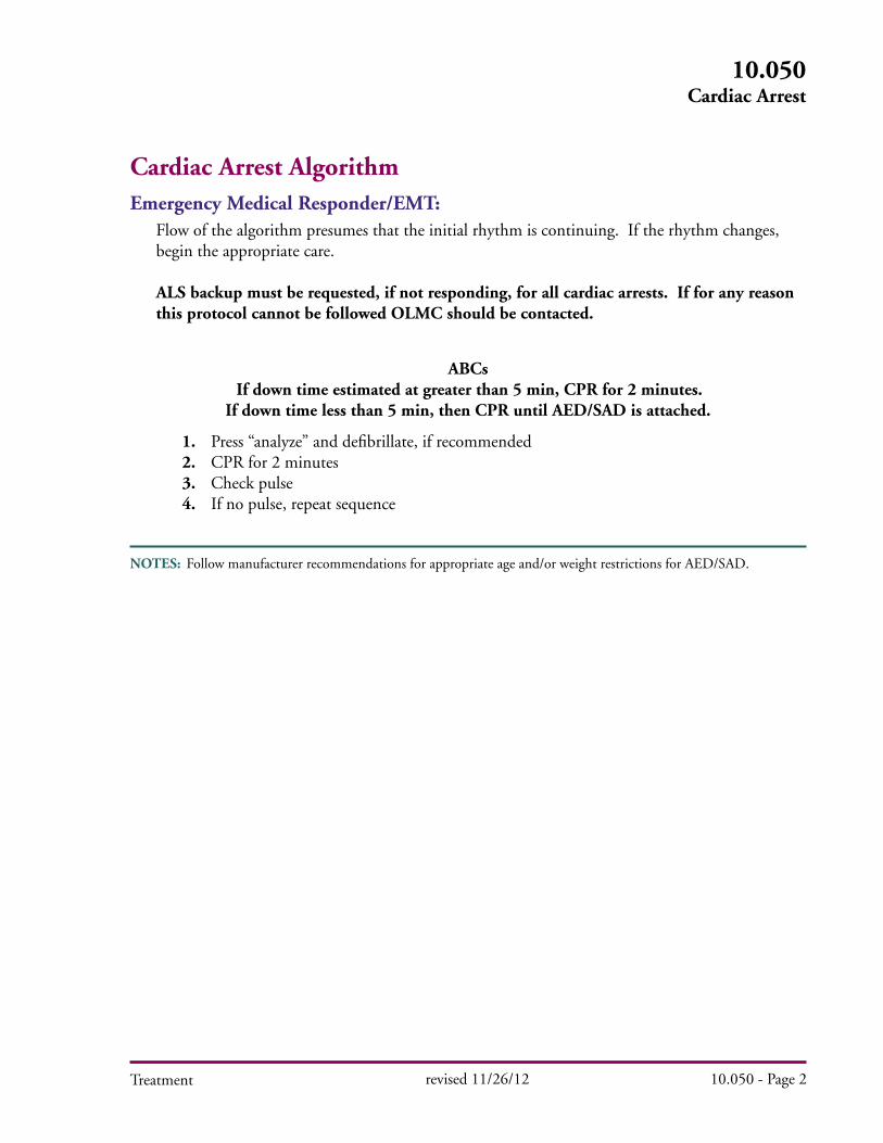
Cardiac Arrest AlgorithmEmergency Medical Responder/EMT:
Flow of the algorithm presumes that the initial rhythm is continuing. If the rhythm changes, begin the appropriate care.
ALS backup must be requested, if not responding, for all cardiac arrests. If for any reason this protocol cannot be followed OLMC should be contacted.
ABCs If down time estimated at greater than 5 min, CPR for 2 minutes.
If down time less than 5 min, then CPR until AED/SAD is attached.
1. Press “analyze” and defibrillate, if recommended 2. CPR for 2 minutes 3. Check pulse 4. If no pulse, repeat sequence
NOTES: Follow manufacturer recommendations for appropriate age and/or weight restrictions for AED/SAD.
revised 11/26/12 10.050 - Page 2
10.050Cardiac Arrest
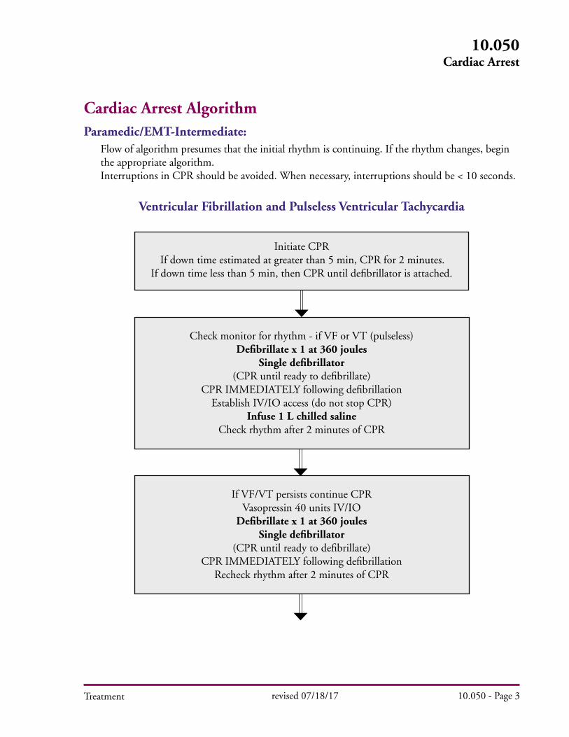
Cardiac Arrest AlgorithmParamedic/EMT-Intermediate:
Flow of algorithm presumes that the initial rhythm is continuing. If the rhythm changes, begin the appropriate algorithm. Interruptions in CPR should be avoided. When necessary, interruptions should be < 10 seconds.
Ventricular Fibrillation and Pulseless Ventricular Tachycardia
revised 07/18/17 10.050 - Page 3
10.050Cardiac Arrest
Initiate CPR If down time estimated at greater than 5 min, CPR for 2 minutes.
If down time less than 5 min, then CPR until defibrillator is attached.
Check monitor for rhythm - if VF or VT (pulseless)Defibrillate x 1 at 360 joules
Single defibrillator(CPR until ready to defibrillate)
CPR IMMEDIATELY following defibrillationEstablish IV/IO access (do not stop CPR)
Infuse 1 L chilled salineCheck rhythm after 2 minutes of CPR
If VF/VT persists continue CPRVasopressin 40 units IV/IO
Defibrillate x 1 at 360 joulesSingle defibrillator
(CPR until ready to defibrillate)CPR IMMEDIATELY following defibrillation
Recheck rhythm after 2 minutes of CPR
Treatment
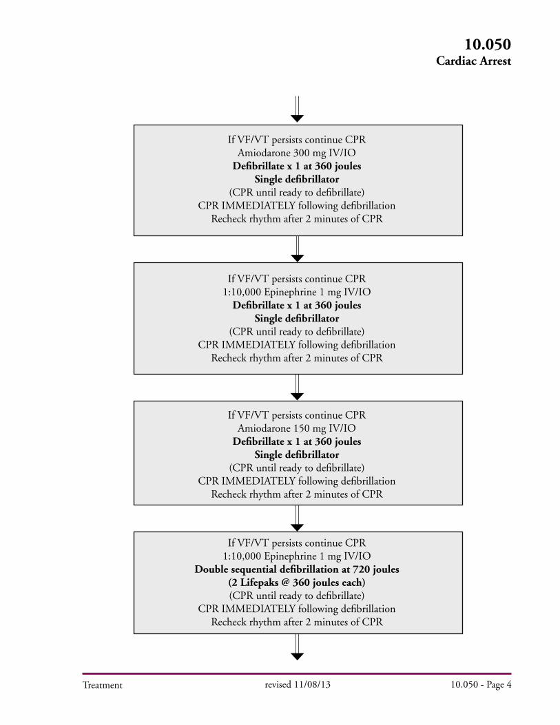
If VF/VT persists continue CPRAmiodarone 300 mg IV/IO
Defibrillate x 1 at 360 joulesSingle defibrillator
(CPR until ready to defibrillate)CPR IMMEDIATELY following defibrillation
Recheck rhythm after 2 minutes of CPR
If VF/VT persists continue CPR1:10,000 Epinephrine 1 mg IV/IO
Defibrillate x 1 at 360 joulesSingle defibrillator
(CPR until ready to defibrillate)CPR IMMEDIATELY following defibrillation
Recheck rhythm after 2 minutes of CPR
If VF/VT persists continue CPRAmiodarone 150 mg IV/IO
Defibrillate x 1 at 360 joulesSingle defibrillator
(CPR until ready to defibrillate)CPR IMMEDIATELY following defibrillation
Recheck rhythm after 2 minutes of CPR
If VF/VT persists continue CPR1:10,000 Epinephrine 1 mg IV/IO
Double sequential defibrillation at 720 joules (2 Lifepaks @ 360 joules each)(CPR until ready to defibrillate)
CPR IMMEDIATELY following defibrillationRecheck rhythm after 2 minutes of CPR
revised 11/08/13 10.050 - Page 4
10.050Cardiac Arrest
Treatment
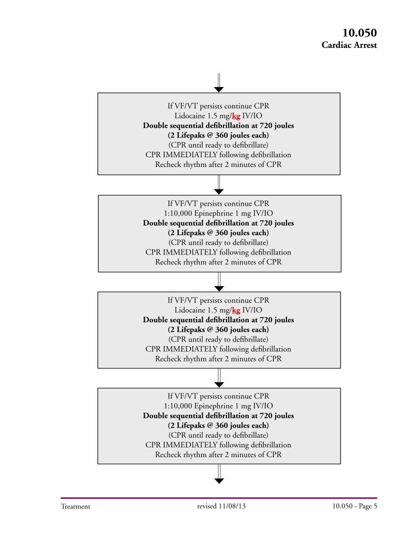
If VF/VT persists continue CPRLidocaine 1.5 mg/kg IV/IO
Double sequential defibrillation at 720 joules (2 Lifepaks @ 360 joules each)(CPR until ready to defibrillate)
CPR IMMEDIATELY following defibrillationRecheck rhythm after 2 minutes of CPR
If VF/VT persists continue CPR1:10,000 Epinephrine 1 mg IV/IO
Double sequential defibrillation at 720 joules (2 Lifepaks @ 360 joules each)(CPR until ready to defibrillate)
CPR IMMEDIATELY following defibrillationRecheck rhythm after 2 minutes of CPR
revised 11/08/13 10.050 - Page 5
10.050Cardiac Arrest
Treatment
revised 09/29/20 10.050 - Page 6
10.050Cardiac Arrest
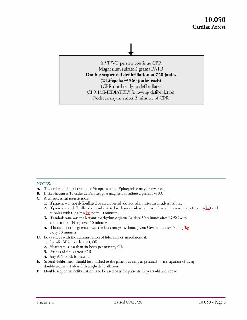
If VF/VT persists continue CPRMagnesium sulfate 2 grams IV/IO
Double sequential defibrillation at 720 joules (2 Lifepaks @ 360 joules each)(CPR until ready to defibrillate)
CPR IMMEDIATELY following defibrillationRecheck rhythm after 2 minutes of CPR
Treatment
NOTES:
A. The order of administration of Vasopressin and Epinephrine may be reversed. B. If the rhythm is Torsades de Pointes, give magnesium sulfate 2 grams IV/IO. C. After successful resuscitation: 1. If patient was not defibrillated or cardioverted, do not administer an antidysrhythmic. 2. If patient was defibrillated or cardioverted with no antidysrhythmic: Give a lidocaine bolus (1.5 mg/kg) and re-bolus with 0.75 mg/kg every 10 minutes. 3. If amiodarone was the last antidysrhythmic given: Re-dose 30 minutes after ROSC with amiodarone 150 mg over 10 minutes. 4. If lidocaine or magnesium was the last antidysrhythmic given: Give lidocaine 0.75 mg/kg every 10 minutes. D. Be cautious with the administration of lidocaine or amiodarone if: 1. Systolic BP is less than 90, OR 2. Heart rate is less than 50 beats per minute, OR 3. Periods of sinus arrest, OR 4. Any A-V block is present. E. Second defibrillator should be attached to the patient as early as practical in anticipation of using double sequential after fifth single defibrillation. F. Double sequential defibrillation is to be used only for patients 12 years old and above.

Treatment
Cardiac Arrest AlgorithmParamedic/EMT-Intermediate:
Asystole(Confirm in two leads, increase gain to rule out fine VF; if rhythm is unclear and possibly
Ventricular Fibrillation, defibrillate as for VF)
ABCs
Initiate CPR If down time estimated at greater than 5 min, CPR for 2 minutes.
If down time less than 5 min, then CPR until defibrillator is attached.Establish IV/IO access
Infuse 1 L chilled saline
Vasopressin 40 units IV/IO x 1Continuous CPR for 2 minutes
1:10,000 Epinephrine 1 mg IV/IO every 3 to 5 minutes
revised 08/13/20 10.050 - Page 7
10.050Cardiac Arrest
NOTES:
The order of administration of Vasopressin and Epinephrine may be reversed. Consider and treat other possible causes: Acidosis — consider sodium bicarbonate 1 mEq/kg IV/IO Cardiac Tamponade - immediate transport Cyclic antidepressants - consider sodium bicarbonate 1 mEq/kg IV/IO Hyperkalemia- consider calcium gluconate or sodium bicarbonate 1 mEq/kg IV/IO Hypothermia- see Hypothermia protocol Hypovolemia- fluid challenge Hypoxia- oxygenate and ventilate Pulmonary Embolism - immediate transport Tension Pneumothorax - needle decompression.
If unresponsive to at least epinephrine 3 mg consider termination of efforts if asystole is confirmed in all six limb leads (with full gain).Administration of lidocaine or amiodarone is not indicated unless VF/PVT was present during resuscitation.

Treatment
Pulseless Electrical Activity (PEA)
1. Electromechanical dissociation 4. Pulseless bradycardic rhythm 2. Idioventricular rhythm 5. Post defibrillation idioventricular 3. Ventricular escape rhythm rhythm
ABCs
Initiate CPRIf down time estimated at greater than 5 min, CPR for 2 minutes.
If down time less than 5 min, then CPR until defibrillator is attached.Establish IV/IO access
Infuse 1 L chilled saline
Vasopressin 40 units IV/IO x 1Continuous CPR for 2 minutes
1:10,000 Epinephrine 1 mg IV/IO every 3 to 5 minutes
NOTES:
The order of administration of Vasopressin and Epinephrine may be reversed.
If EtCO2 > 20, with organized rhythm, initiate fluids per Shock protocol and consider norepinephrine (4-12 micrograms/min). Continue CPR until palpable pulse.
Consider and treat other possible causes: Acidosis — consider sodium bicarbonate 1 mEq/kg IV/IO Cardiac Tamponade - immediate transport Cyclic antidepressants - consider sodium bicarbonate 1 mEq/kg IV/IO Hyperkalemia - consider calcium gluconate or sodium bicarbonate 1 mEq/kg IV Hypothermia - see Hypothermia protocol Hypovolemia - fluid challenge Hypoxia - oxygenate and ventilate Pulmonary Embolism - immediate transport Tension Pneumothorax - needle decompression
Administration of lidocaine or amiodarone is not indicated unless VF/PVT was present during resuscitation.
revised 08/13/20 10.050 - Page 8
10.050Cardiac Arrest
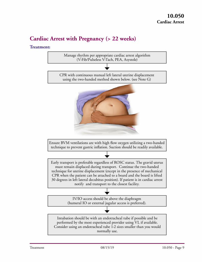
Cardiac Arrest with Pregnancy (> 22 weeks)Treatment:
Manage rhythm per appropriate cardiac arrest algorithm (V-Fib/Pulseless V-Tach, PEA, Asystole)
CPR with continuous manual left lateral uterine displacement using the two-handed method shown below. (see Note G)
Ensure BVM ventilations are with high flow oxygen utilizing a two-handed technique to prevent gastric inflation. Suction should be readily available.
IV/IO access should be above the diaphragm (humeral IO or external jugular access is preferred).
Intubation should be with an endotracheal tube if possible and be performed by the most experienced provider using VL if available.
Consider using an endotracheal tube 1-2 sizes smaller than you would normally use.
Early transport is preferable regardless of ROSC status. The gravid uterus must remain displaced during transport. Continue the two-handed
technique for uterine displacement (except in the presence of mechanical CPR when the patient can be attached to a board and the board is lifted 30 degrees in left lateral decubitus position). If patient is in cardiac arrest
notify and transport to the closest facility.
Treatment 08/15/19 10.050 - Page 9
10.050Cardiac Arrest
Treatment 08/22/19 10.050 - Page 10
10.050Cardiac Arrest
Notes and Precautions (Pregnancy):A. Early transport prior to achieving ROSC, especially if a mechanical CPR device is available.
B. Alert the receiving facility early in order to have an OB team present upon arrival in the emergency department. If you have not achieved ROSC go to the closest facility regardless of OB capabilities.
C. If ROSC has been achieved prior to, or during transport, bypass to an OB and NICU capable facility
D. Lidocaine is preferable (Class B in Pregnancy) to amiodarone (Class C in Pregnancy) in the setting of ventricular fibrillation or pulseless ventricular tachycardia.
E. In the setting of ventricular fibrillation or pulseless ventricular tachycardia, no adjustments need to be made to defibrillation energy settings. Immediately following defibrillation, resume the left lateral uterine displacement.
F. If mechanical CPR is in place, continue the left lateral uterine displacement by tilting the backboard 30° to the left or by continuing manual displacement.
G. If ROSC is achieved continue left lateral uterine displacement by placing the patient in the left lateral decubitus position or by manually displacing the gravid uterus.
H. High flow oxygen needs to be maintained in all peri-arrest patients.
I. Consider OG placement when possible.
Treatment
Cardiac Arrest Algorithm
Paramedic/EMT-Intermediate:
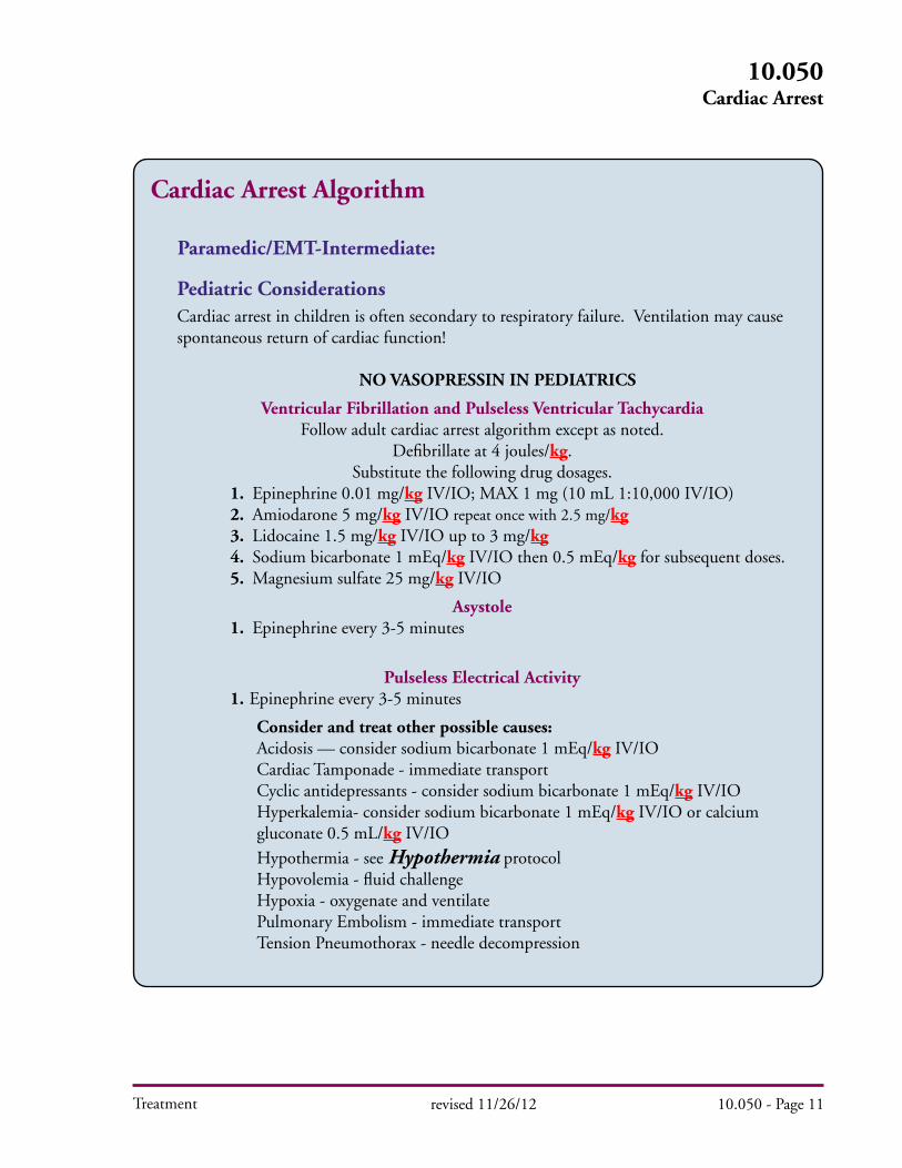
Pediatric ConsiderationsCardiac arrest in children is often secondary to respiratory failure. Ventilation may cause spontaneous return of cardiac function!
NO VASOPRESSIN IN PEDIATRICS
Ventricular Fibrillation and Pulseless Ventricular TachycardiaFollow adult cardiac arrest algorithm except as noted.
Defibrillate at 4 joules/kg. Substitute the following drug dosages.
1. Epinephrine 0.01 mg/kg IV/IO; MAX 1 mg (10 mL 1:10,000 IV/IO)2. Amiodarone 5 mg/kg IV/IO repeat once with 2.5 mg/kg3. Lidocaine 1.5 mg/kg IV/IO up to 3 mg/kg4. Sodium bicarbonate 1 mEq/kg IV/IO then 0.5 mEq/kg for subsequent doses.5. Magnesium sulfate 25 mg/kg IV/IO
Asystole 1. Epinephrine every 3-5 minutes
Pulseless Electrical Activity1. Epinephrine every 3-5 minutes
Consider and treat other possible causes: Acidosis — consider sodium bicarbonate 1 mEq/kg IV/IO Cardiac Tamponade - immediate transport Cyclic antidepressants - consider sodium bicarbonate 1 mEq/kg IV/IO Hyperkalemia- consider sodium bicarbonate 1 mEq/kg IV/IO or calcium gluconate 0.5 mL/kg IV/IO
Hypothermia - see Hypothermia protocol Hypovolemia - fluid challenge Hypoxia - oxygenate and ventilate Pulmonary Embolism - immediate transport Tension Pneumothorax - needle decompression
revised 11/26/12 10.050 - Page 11
10.050Cardiac Arrest
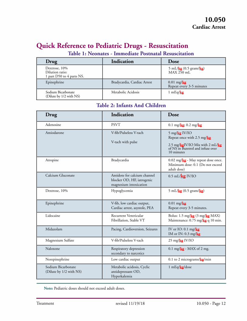
Drug Indication DoseDextrose, 10%Dilution ratio: 1 part D50 to 4 parts NS.
5 mL/kg (0.5 gram/kg)MAX 250 mL
Epinephrine Bradycardia, Cardiac Arrest 0.01 mg/kgRepeat every 3-5 minutes
Sodium Bicarbonate (Dilute by 1/2 with NS)
Metabolic Acidosis 1 mEq/kg
Table 2: Infants And Children
Drug Indication Dose
Adenosine PSVT 0.1 mg/kg; 0.2 mg/kg
Amiodarone V-fib/Pulseless V-tach
V-tach with pulse
5 mg/kg IV/IORepeat once with 2.5 mg/kg
2.5 mg/kgIV/IO Mix with 2 mL/kg of NS in Buretrol and infuse over 10 minutes
Atropine Bradycardia 0.02 mg/kg - May repeat dose once. Minimum dose: 0.1 (Do not exceed adult dose)
Calcium Gluconate Antidote for calcium channel blocker OD, HF, iatrogenic magnesium intoxication
0.5 mL/kg IV/IO
Dextrose, 10% Hypoglycemia 5 mL/kg (0.5 gram/kg)
Epinephrine V-fib, low cardiac output, Cardiac arrest, asystole, PEA
0.01 mg/kg Repeat every 3-5 minutes.
Lidocaine Recurrent Ventricular Fibrillation, Stable VT
Bolus: 1.5 mg/kg (3 mg/kg MAX)Maintenance: 0.75 mg/kg q 10 min.
Midazolam Pacing, Cardioversion, Seizures IV or IO: 0.1 mg/kgIM or IN: 0.3 mg/kg
Magnesium Sulfate V-fib/Pulseless V-tach 25 mg/kg IV/IO
Naloxone Respiratory depression secondary to narcotics
0.1 mg/kg - MAX of 2 mg.
Norepinephrine Low cardiac output 0.1 to 2 micrograms/kg/min
Sodium Bicarbonate(Dilute by 1/2 with NS)
Metabolic acidosis, Cyclic antidepressant OD, Hyperkalemia
1 mEq/kg/dose
Note: Pediatric doses should not exceed adult doses.
Treatment
Quick Reference to Pediatric Drugs - ResuscitationTable 1: Neonates - Immediate Postnatal Resuscitation
10.050Cardiac Arrest
revised 11/19/18 10.050 - Page 12

Treatment
Cardiac Dysrhythmias
HX PE DDX
Past medical history
Medications• Beta blockers• Calcium Channel blockers• Clonidine• Digitalis
Pacemaker
AMS
Respiratory distress
Hypotension / shock
Chest pain
CHF
Syncope
Seizures
Sinus bradycardia
AV blocks
Acute MI
Hypoxia
Hypothermia
Head injury (increased ICP)
Spinal cord lesion
Sick sinus
Overdose
Treatment: A. Start O
2, follow Airway Management procedure, and apply pulse oximeter.
B. Start IV/IO, NS and follow Shock protocol if indicated. C. Monitor cardiac rhythm, see the following cardiac dysrhythmias on the next pages:
revised 12/08 10.060 - Page 1
10.060Cardiac Dysrhythmias
NOTES:
If the patient is asymptomatic, dysrhythmias may not require treatment in the field.

Treatment
Cardiac Dysrhythmias - Adult Tachycardia (Stable)
revised 12/11/13 10.060 - Page 2
10.060Cardiac Dysrhythmias
Narrow QRS (≤ 0.12 sec) Wide QRS (> 0.12 sec)
Attempt vagal maneuvers
Consider Adenosine 6 mg
rapid IV/IO if possible aberrancy
Amiodarone150 mg IV/IO
over 10 min
If no conversion, repeat amiodarone 150 mg IV/IO over
10 min
If possible Torsades
Magnesium Sulfate 2 grams
IV/IO over 1-2 minutes
Adenosine 6 mgrapid IV/IO
Adenosine 12 mgrapid IV/IO
Adenosine 12 mgrapid IV/IO
Consider wide complex
irregular rhythms including
WPW, AFib with aberrancy
Start oxygen per Airway Management procedure.Monitor vital signs, ECG and oxygen saturation. Establish IV/IO access.
Patient does not have signs or symptoms of poor perfusion caused by the dysrhythmia. (Altered mental status, chest pain, hypotension or other signs of shock.)
Rate-related symptoms uncommon if HR < 150 bpm. Consider other causes.
Obtain 12-lead ECG
n Obtain post treatment 12-lead ECGn Contact OLMC for advicen Consider contributing factors and other treatments
Irregular IrregularRegular Regular
Monitor Patient
Consider:n Atrial fibn Atrial fluttern Multifocal atrial tachycardia
Treatment
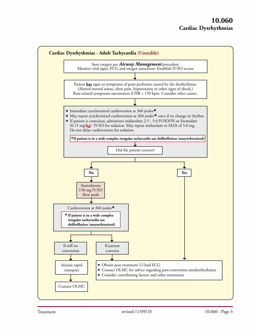
Cardiac Dysrhythmias - Adult Tachycardia (Unstable)
10.060Cardiac Dysrhythmias
n Immediate synchronized cardioversion at 360 joules.n May repeat synchronized cardioversion at 360 joules once if no change in rhythm.n If patient is conscious, administer midazolam 2.5 - 5.0 IV/IO/IN or Etomidate (0.15 mg/kg) IV/IO for sedation. May repeat midazolam to MAX of 5.0 mg. Do not delay cardioversion for sedation.
Start oxygen per Airway Management procedure.Monitor vital signs, ECG and oxygen saturation. Establish IV/IO access.
Patient has signs or symptoms of poor perfusion caused by the dysrhythmia. (Altered mental status, chest pain, hypotension or other signs of shock.)
Rate-related symptoms uncommon if HR < 150 bpm. Consider other causes.
n Obtain post treatment 12-lead ECGn Contact OLMC for advice regarding post-conversion antidysrhythmicsn Consider contributing factors and other treatments
Did the patient convert?
Yes
Cardioversion at 360 joules
If still no conversion
If patient converts
Initiate rapid transport
Contact OLMC
revised 11/09/18 10.060 - Page 3
If patient is in a wide complex irregular tachycardia use defibrillation (unsynchronized)
If patient is in a wide complex irregular tachycardia use defibrillation (unsynchronized)
No
Amiodarone150 mg IV/IO
Slow push

Treatment revised 06/23/16 10.060 - Page 4
10.060Cardiac Dysrhythmias
Notes and Precautions: (Tachycardia):A. In stable wide complex tachycardia which is monomorphic, consider adenosine if SVT with aberrancy is suspected.B. If the patient is asymptomatic, tachycardia may not require treatment in the field. Continue to monitor the patient for changes during transport. The acceptable upper limit for heart rate for sinus tachycardia is 220 minus the patient’s age.C. Other possible causes of tachycardia include: 1. Acidosis 2. Hypovolemia 3. Hyperthermia/fever 4. Hypoxia 5. Hypo/Hyperkalemia 6. Hypoglycemia 7. Infection 8. Pulmonary embolus 9. Tamponade 10. Toxic exposure 11. Tension pneumothoraxD. If pulseless arrest develops, follow Cardiac Arrest protocol.E. All doses of adenosine should be reduced to one-half (50%) in the following clinical settings: 1. History of cardiac transplantation. 2. Patients who are on carbamazepine (Tegretol) and dipyridamole (Persantine, Aggrenox). 3. Administration through any central line (Porta Cath, Broviac, Hickman, etc).F. Adenosine may initiate atrial fibrillation with rapid ventricular response in patients with Wolff-Parkinson-White syndrome.G. Adenosine should be used with caution in patients with asthma as it may cause a reactive airways response in some cases.

Treatment
Cardiac Dysrhythmias - Pediatric Tachycardia (Stable)
revised 10/24/14 10.060 - Page 5
10.060Cardiac Dysrhythmias
Start oxygen per Airway Management procedure.Monitor vital signs, ECG and oxygen saturation. Establish IV/IO access.
Are signs or symptoms of poor perfusion caused by the dysrhythmia present?
No - Pt stable. Obtain 12-lead ECG
Narrow complex QRS (≤ 0.09 sec) Wide complex QRS (> 0.09 sec)
Attempt vagal maneuvers
If rhythm is regular and QRS is monomorphic, consider adenosine 0.1 mg/kg rapid IV/IO
Probable Sinus Tachycardian P waves presentn Variable RR; Consistant PRn HR < 220 infants n HR <180 children
Possible Ventricular Tachycardia
Probable SVTn Compatible historyn P waves absent/abnormaln HR not variablen HR ≥ 220 infants n HR ≥180 children
Adenosine 0.1 mg/kg
rapid IV/IO
If no conversion may repeat adenosine x2 at 0.2 mg/kg rapid IV/IO
Monitor patient
n Obtain post treatment 12-lead ECGn Contact OLMC for advicen Consider contributing factors and other treatments
Treatment
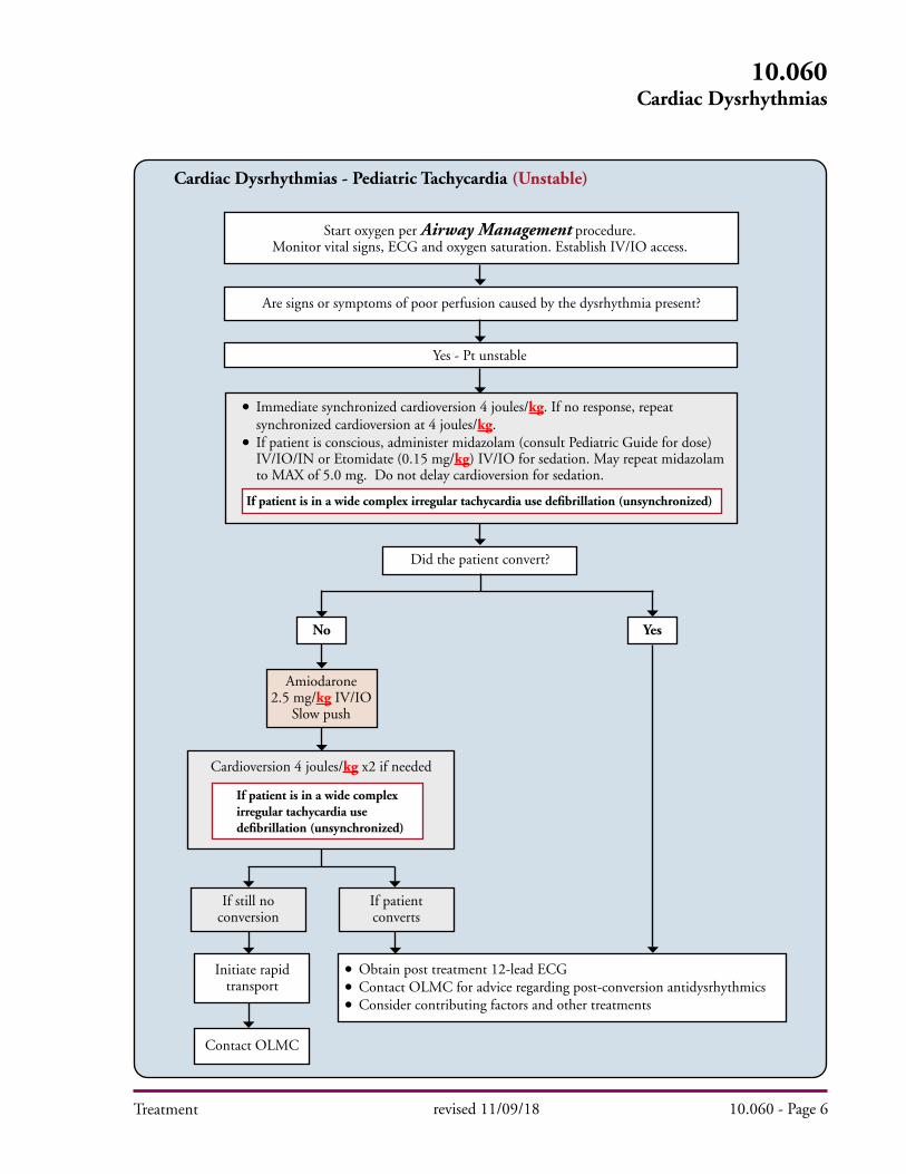
Cardiac Dysrhythmias - Pediatric Tachycardia (Unstable)
Start oxygen per Airway Management procedure.Monitor vital signs, ECG and oxygen saturation. Establish IV/IO access.
Are signs or symptoms of poor perfusion caused by the dysrhythmia present?
10.060Cardiac Dysrhythmias
n Immediate synchronized cardioversion 4 joules/kg. If no response, repeat synchronized cardioversion at 4 joules/kg.n If patient is conscious, administer midazolam (consult Pediatric Guide for dose) IV/IO/IN or Etomidate (0.15 mg/kg) IV/IO for sedation. May repeat midazolam to MAX of 5.0 mg. Do not delay cardioversion for sedation.
Yes - Pt unstable
revised 11/09/18 10.060 - Page 6
n Obtain post treatment 12-lead ECGn Contact OLMC for advice regarding post-conversion antidysrhythmicsn Consider contributing factors and other treatments
Did the patient convert?
YesNo
Amiodarone2.5 mg/kg IV/IO
Slow push
Cardioversion 4 joules/kg x2 if needed
If still no conversion
If patient converts
Initiate rapid transport
Contact OLMC
If patient is in a wide complex irregular tachycardia use defibrillation (unsynchronized)
If patient is in a wide complex irregular tachycardia use defibrillation (unsynchronized)
Treatment
NOTES:
A. Use pediatric pads for cardioversion for children less than 15 kg. B. Place on anterior chest in sternal-apical location. C. If pediatric pads are not available, use adult pads placed anterior-posterior on the chest wall with firm contact. D. If available defibrillator will not “dial down” to appropriate energy level, use lowest possible energy level available.
revised 11/10/11 10.060 - Page 7
10.060Cardiac Dysrhythmias

Treatment
Cardiac Dysrhythmias - Adult Bradycardia
revised 06/23/16 10.060 - Page 8
10.060Cardiac Dysrhythmias
Observe and monitor patient. Consider 12-lead ECG if
patient is stable.
Start oxygen per Airway Management procedure.Monitor vital signs, ECG and oxygen saturation. Establish IV/IO access.
Are signs or symptoms of poor perfusion caused by the bradycardia present? (Altered mental status, chest pain, hypotension or other signs of shock)
HEART RATE < 50
n If capture is achieved and patient is uncomfortable, consider Midazolam 2.5 - 5.0 mg IV/IO or 5 mg IM/IN. May repeat IV/IO/IN dose once to a MAX of 5 mg.n If capture is not achieved, try repositioning pads.n Goal of therapy is to improve perfusion and maintain a BP of > 90 mmHg systolic.
No - Pt stable Yes - Pt unstable
2nd degree Type II, or 3rd degree heart block, or
Cardiac transplant?
Atropine 0.5 mg IV/IO. May repeat every 3-5 minutes
to a MAX of 3 mg.
Monitor patient
Monitor patient
• If no response to pacing or atropine: Consider epinephrine infusion 2-10 micrograms/min
titrated to effect or norepinephrine 4 micrograms/min (increase every 5 min in 4 micrograms/min increments to MAX 12 micrograms/min).
• Consider OLMC contact.
Capture?
Transcutaneous Pacingper protocol
Atropine 0.5 mg IV/IO. May repeat every 3-5 minutes
to a MAX of 3 mg.
If no response to atropine, Transcutaneous Pacing per protocol
Capture?
YesNo
YesNo
Yes No
Treatment
Notes and Precautions: (Bradycardia):A. Hypoxia is a common cause of bradycardia.B. Bradycardia may be protective in the setting of cardiac ischemia and should only be treated if associated with serious signs and symptoms of hypoperfusion. Increasing heart rate may worsen ischemia or increase infarct size.C. Hyperkalemia may cause bradycardia. If the patient has a wide complex bradycardia with a history of renal failure, muscular dystrophy, paraplegia, crush injury or serious burn > 48 hours prior, consider treatment per Hyperkalemia protocol.D. Immediate Transcutaneous Pacing can be considered in unstable patients when vascular access is not available.E. Transcutaneous Pacing is at best a temporizing measure and is not useful in asystole.F. If Transcutaneous Pacing capture is not achieved, try repositioning pads.G. If capture is achieved with Transcutaneous Pacing and patient is experiencing discomfort administer midazolam 2.5 - 5.0 mg IV/IO or 5 mg IM. May repeat IV/IO dose once to a MAX of 5 mg.H. Atropine will likely be ineffective in heart transplant recipients because they lack vagal innervation.I. 3rd degree heart blocks with a wide complex QRS (>0.12 sec) are less likely to respond to atropine than those with a narrow complex.
revised 06/23/16 10.060 - Page 9
10.060Cardiac Dysrhythmias

Treatment
Cardiac Dysrhythmias - Pediatric Bradycardia
revised 12/17/13 10.060 - Page 10
10.060Cardiac Dysrhythmias
n Start CPR if despite oxygenation and ventilation patient’s heart rate is < 60 bpm with poor perfusion.n Reassess after 2 minutes of CPR.
n Continue to support ABCs as needed.n Monitor patient.n Consider OLMC contact.
Start oxygen per Airway Management procedure. Monitor vital signs, ECG and oxygen saturation. Establish IV/IO access.
Support ABCs
Bradycardia causing cardiorespiratory compromise or poor perfusion?
BRADYCARDIA WITH A PULSE
n Give 1:10,000 epinephrine 0.01 mg/kg IV/IO. Repeat epinephrine every 3-5 minutes.n If increased vagal tone or AV block, consider Atropine 0.02 mg/kg IV/IO. Minimum single dose 0.1 mg; MAX single dose 0.5 mg. MAX total dose 1 mg.n Consider pacing per Transcutaneous Pacing procedure. n If capture is achieved and patient is uncomfortable, consider Midazolam 0.1mg/kg IV/IO to a MAX of 2.5 mg.n If capture is not achieved, try repositioning pads.n Goal of therapy is to improve perfusion.
Persistent symptomatic bradycardia?No
Yes
Specific Precautions: A. Most pediatric bradycardia is due to hypoxia. Oxygenate and ventilate aggressively.
No - Pt stable Yes - Pt unstable
Treatment
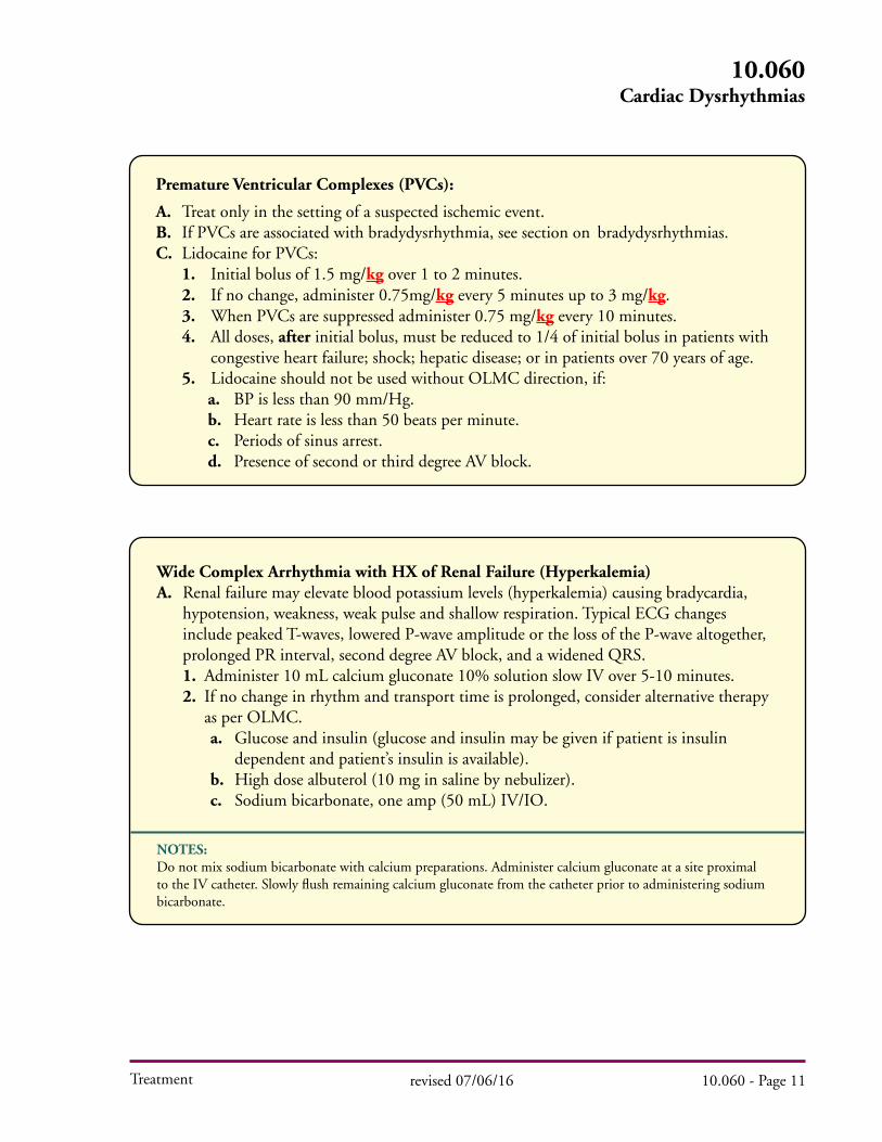
Premature Ventricular Complexes (PVCs):
A. Treat only in the setting of a suspected ischemic event. B. If PVCs are associated with bradydysrhythmia, see section on bradydysrhythmias. C. Lidocaine for PVCs: 1. Initial bolus of 1.5 mg/kg over 1 to 2 minutes. 2. If no change, administer 0.75mg/kg every 5 minutes up to 3 mg/kg. 3. When PVCs are suppressed administer 0.75 mg/kg every 10 minutes. 4. All doses, after initial bolus, must be reduced to 1/4 of initial bolus in patients with congestive heart failure; shock; hepatic disease; or in patients over 70 years of age. 5. Lidocaine should not be used without OLMC direction, if:
a. BP is less than 90 mm/Hg.b. Heart rate is less than 50 beats per minute.c. Periods of sinus arrest.d. Presence of second or third degree AV block.
Wide Complex Arrhythmia with HX of Renal Failure (Hyperkalemia) A. Renal failure may elevate blood potassium levels (hyperkalemia) causing bradycardia, hypotension, weakness, weak pulse and shallow respiration. Typical ECG changes include peaked T-waves, lowered P-wave amplitude or the loss of the P-wave altogether, prolonged PR interval, second degree AV block, and a widened QRS.
1. Administer 10 mL calcium gluconate 10% solution slow IV over 5-10 minutes.2. If no change in rhythm and transport time is prolonged, consider alternative therapy as per OLMC.
a. Glucose and insulin (glucose and insulin may be given if patient is insulin dependent and patient’s insulin is available). b. High dose albuterol (10 mg in saline by nebulizer). c. Sodium bicarbonate, one amp (50 mL) IV/IO.
NOTES:
Do not mix sodium bicarbonate with calcium preparations. Administer calcium gluconate at a site proximal to the IV catheter. Slowly flush remaining calcium gluconate from the catheter prior to administering sodium bicarbonate.
revised 07/06/16 10.060 - Page 11
10.060Cardiac Dysrhythmias
Treatment
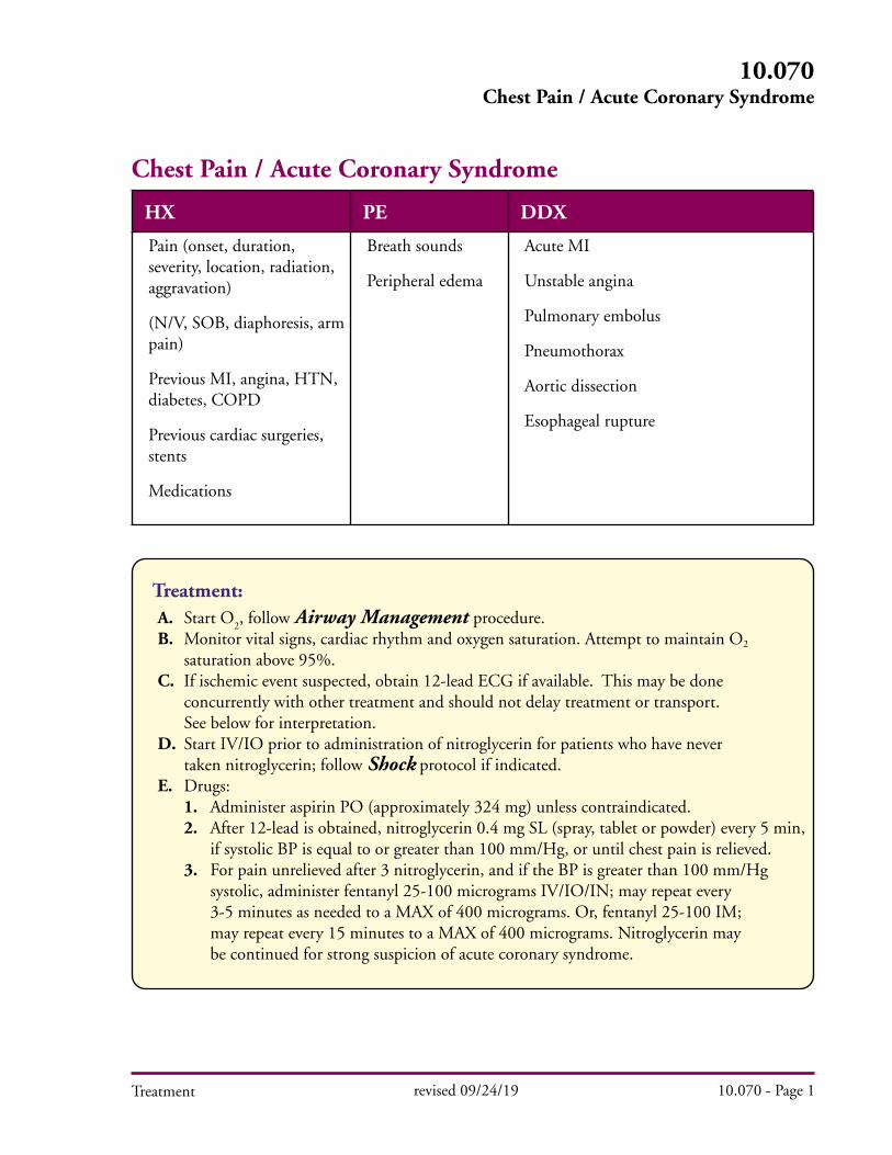
Chest Pain / Acute Coronary Syndrome
HX PE DDX
Pain (onset, duration, severity, location, radiation, aggravation)
(N/V, SOB, diaphoresis, arm pain)
Previous MI, angina, HTN, diabetes, COPD
Previous cardiac surgeries, stents
Medications
Breath sounds
Peripheral edema
Acute MI
Unstable angina
Pulmonary embolus
Pneumothorax
Aortic dissection
Esophageal rupture
Treatment: A. Start O
2, follow Airway Management procedure.
B. Monitor vital signs, cardiac rhythm and oxygen saturation. Attempt to maintain O2
saturation above 95%.
C. If ischemic event suspected, obtain 12-lead ECG if available. This may be done concurrently with other treatment and should not delay treatment or transport. See below for interpretation. D. Start IV/IO prior to administration of nitroglycerin for patients who have never taken nitroglycerin; follow Shock protocol if indicated.
E. Drugs: 1. Administer aspirin PO (approximately 324 mg) unless contraindicated.
2. After 12-lead is obtained, nitroglycerin 0.4 mg SL (spray, tablet or powder) every 5 min, if systolic BP is equal to or greater than 100 mm/Hg, or until chest pain is relieved. 3. For pain unrelieved after 3 nitroglycerin, and if the BP is greater than 100 mm/Hg systolic, administer fentanyl 25-100 micrograms IV/IO/IN; may repeat every 3-5 minutes as needed to a MAX of 400 micrograms. Or, fentanyl 25-100 IM; may repeat every 15 minutes to a MAX of 400 micrograms. Nitroglycerin may be continued for strong suspicion of acute coronary syndrome.
revised 09/24/19 10.070 - Page 1
10.070 Chest Pain / Acute Coronary Syndrome
Treatment
Specific Precautions A. DO NOT DELAY ADMINISTRATION OF ASPIRIN TO OBTAIN 12 LEAD B. NTG administration to patients with an acute inferior wall myocardial infarction should be performed with close monitoring of vital signs and rhythm. NTG in these patients may result in symptomatic hypotension and/or shock which should be treated with usual measures (fluids, changes in position, medications if necessary). C. Do not administer nitroglycerin without OLMC if patient has taken Viagra or other similar drugs in the last 24 hours or Cialis (tadalafil) within last 48 hours. D. Contraindications to administration of aspirin: 1. Allergy to aspirin or aspirin induced asthma. 2. History of active bleeding disorder, (i.e., hemophilia). 3. Current ulcer or GI bleeding. 4. Suspected aortic dissection. E. If 12-Lead ECG is obtained, use biological sex as assigned at birth. If patient does not disclose, use default setting (male).
Field Identified ST-Elevation MI (STEMI)
Indication 12-lead ECG with: • Paramedic interpretation of probable STEMI: 1 mm elevation in 2 contiguous limb leads or 2 mm elevation in 2 contiguous chest leads
Action A. As soon as STEMI is recognized, prompt and early notification of the receiving facility of “STEMI patient” or “STEMI alert” should be performed. B. Rapid transport to destination hospital ED with interventional capability. C. Non-diagnostic ECGs with potential “imitators” of ACS or ECGs that are clinically concerning should be described to the receiving hospital or OLMC. These may include: • LBBB or RBBB • LVH • SVT with aberrancy • Paced rhythms • Pericarditis • Benign early repolarization • Digitalis effect
revised 10/25/19 10.070 - Page 2
10.070 Chest Pain / Acute Coronary Syndrome

Treatment
10.075Crush Injury
Crush Injury
revised 06/30/16 10.075 - page 1
HX PE DDX
Body part entrapped
Mechanism of entrapment
Length of time entrapped
Function of body part (e.g. able to feel, move extremity)
LOC
Airway
Extremity pulses, neurologic function
Respiratory distress
Dehydration
Pain
Hypo and/or hyperthermia
Treatment: A. Control/stop hemorrhage. B. Airway Management procedure if indicated. C. Spinal immobilization if indicated. D. Monitor cardiac rhythm for signs of hyperkalemia. (Widening QRS > .08ms, peaked T-waves, sine wave, etc). E. Start IV/IO if feasible. F. Consider pain management. G. Wound care. 1. Remove all restrictive dressings (clothing, jewelry, etc). 2. Continually monitor distal pulse, motor and sensation in involved extremity. 3. Bandage all open wounds. (Irrigate if needed.) 4. Stabilize all protruding foreign bodies (impaled objects). 5. Splint/immobilize injured areas. 6. For suspected pelvic crushing injuries, follow the Pelvic Wrap procedure if indicated. H. If severe crushing injury/compartment syndrome: 1. If cardiac monitor is available, treat for hyperkalemia if present. 2. If feasible, administer 1000 – 2000 mL (pediatric: 10 to 20 mL/kg) of isotonic fluid prior to extrication and maintain IV/IO infusion at 500 mL (peds 5 mL/kg per hour). 3. Perform extrication. 4. Reassess patient and monitor for signs of hyperkalemia. I. If hyperkalemia is suspected or present, then: 1. Administer 10mL calcium gluconate 10% solution slow IV over 5-10 minutes. 2. High dose albuterol (10mg in saline by nebulizer). 3. Sodium bicarbonate, one amp (50mL) IV/IO.

Treatment
10.075Crush Injury
06/30/16 10.075 - page 2
Notes: • Renal failure may elevate blood potassium levels (hyperkalemia) causing bradycardia, hypotension, weakness, weak pulse and shallow respiration. Typical ECG changes include peaked T-waves, lowered P-wave amplitude or the loss of the P-wave altogether, prolonged PR interval, second degree AV block, and a widened QRS.
• A venous tourniquet may have a role during extrication to control the effects of hyperkalemia in crush injuries.
Precautions: • If circumstances warrant, begin warming procedures to prevent hypothermia.
• If patient is trapped in a heavy dust environment, consider methods to provide nebulized oxygen. Consider the use of nebulized albuterol.
• If patient is severely trapped and requires prolonged extrication or potential amputation, contact OLMC for Trauma Surgeon advice and ensure that a technical rescue team is activated.
• During extrication, continually monitor patient condition, if possible.
Treatment
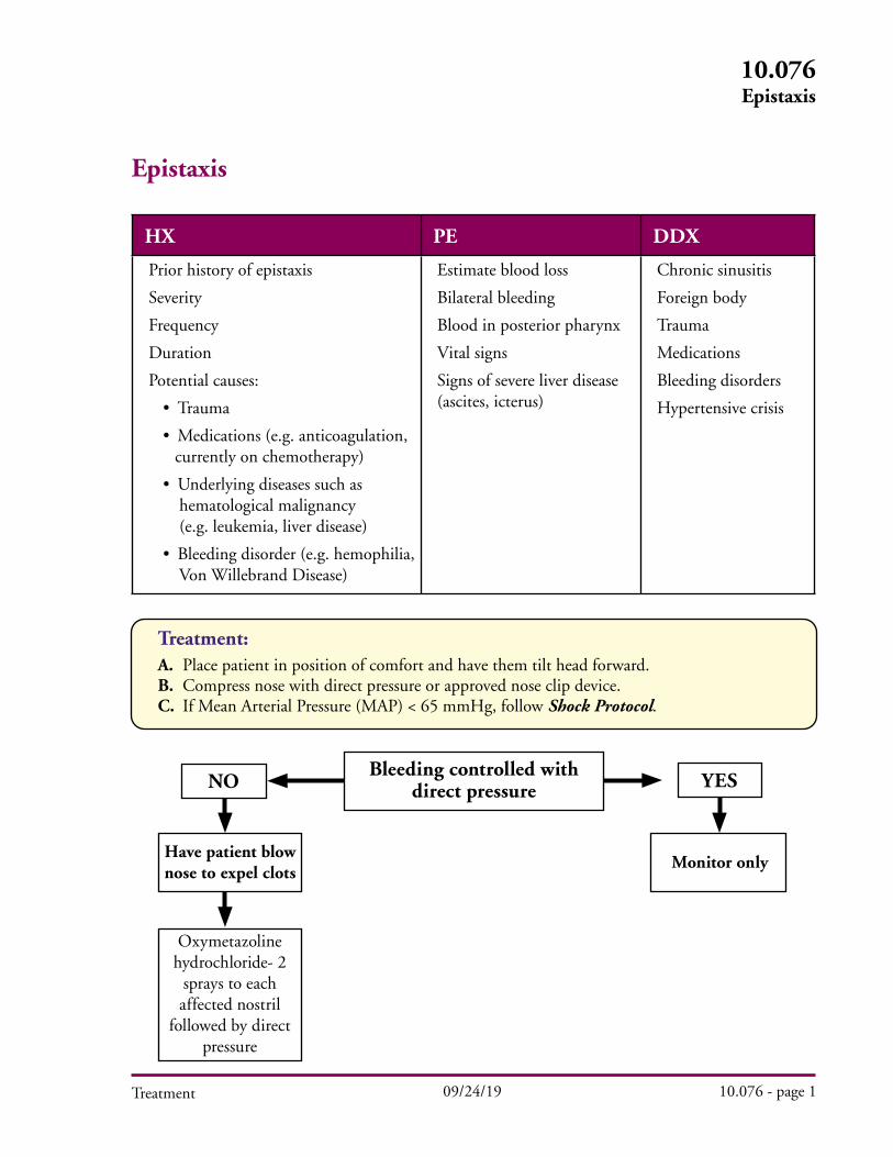
10.076Epistaxis
09/24/19 10.076 - page 1
Epistaxis
HX PE DDX
Prior history of epistaxis
Severity
Frequency
Duration
Potential causes:
• Trauma
• Medications (e.g. anticoagulation, currently on chemotherapy)
• Underlying diseases such as hematological malignancy (e.g. leukemia, liver disease)
• Bleeding disorder (e.g. hemophilia, Von Willebrand Disease)
Estimate blood loss
Bilateral bleeding
Blood in posterior pharynx
Vital signs
Signs of severe liver disease (ascites, icterus)
Chronic sinusitis
Foreign body
Trauma
Medications
Bleeding disorders
Hypertensive crisis
Treatment:A. Place patient in position of comfort and have them tilt head forward.B. Compress nose with direct pressure or approved nose clip device.C. If Mean Arterial Pressure (MAP) < 65 mmHg, follow Shock Protocol.
YESNO
Have patient blow nose to expel clots
Oxymetazoline hydrochloride- 2
sprays to each affected nostril
followed by direct pressure
Monitor only
Bleeding controlled with direct pressure
Treatment
10.076Epistaxis
revised 10/25/19 10.076 - page 2
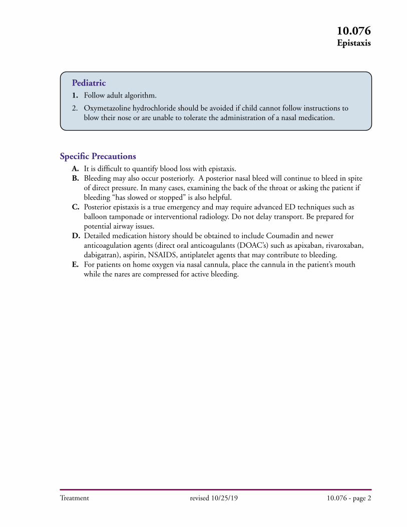
Pediatric 1. Follow adult algorithm.
2. Oxymetazoline hydrochloride should be avoided if child cannot follow instructions to blow their nose or are unable to tolerate the administration of a nasal medication.
Specific Precautions A. It is difficult to quantify blood loss with epistaxis. B. Bleeding may also occur posteriorly. A posterior nasal bleed will continue to bleed in spite of direct pressure. In many cases, examining the back of the throat or asking the patient if bleeding “has slowed or stopped” is also helpful. C. Posterior epistaxis is a true emergency and may require advanced ED techniques such as balloon tamponade or interventional radiology. Do not delay transport. Be prepared for potential airway issues. D. Detailed medication history should be obtained to include Coumadin and newer anticoagulation agents (direct oral anticoagulants (DOAC’s) such as apixaban, rivaroxaban, dabigatran), aspirin, NSAIDS, antiplatelet agents that may contribute to bleeding. E. For patients on home oxygen via nasal cannula, place the cannula in the patient’s mouth while the nares are compressed for active bleeding.
revised 08/01/18 10.077 - Page 1Treatment
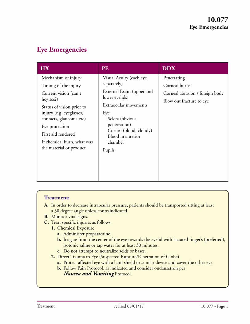
Eye Emergencies
HX PE DDX
Mechanism of injury
Timing of the injury
Current vision (can t hey see?)
Status of vision prior to injury (e.g. eyeglasses, contacts, glaucoma etc)
Eye protection
First aid rendered
If chemical burn, what was the material or product.
Visual Acuity (each eye separately)
External Exam (upper and lower eyelids)
Extraocular movements
Eye Sclera (obvious penetration) Cornea (blood, cloudy) Blood in anterior chamber
Pupils
Penetrating
Corneal burns
Corneal abrasion / foreign body
Blow out fracture to eye
Treatment:A. In order to decrease intraocular pressure, patients should be transported sitting at least a 30 degree angle unless contraindicated.B. Monitor vital signs.C. Treat specific injuries as follows: 1. Chemical Exposure a. Administer proparacaine. b. Irrigate from the center of the eye towards the eyelid with lactated ringer’s (preferred), isotonic saline or tap water for at least 30 minutes. c. Do not attempt to neutralize acids or bases. 2. Direct Trauma to Eye (Suspected Rupture/Penetration of Globe) a. Protect affected eye with a hard shield or similar device and cover the other eye. b. Follow Pain Protocol, as indicated and consider ondansetron per Nausea and Vomiting Protocol.
10.077Eye Emergencies
revised 08/01/18 10.077 - Page 2Treatment
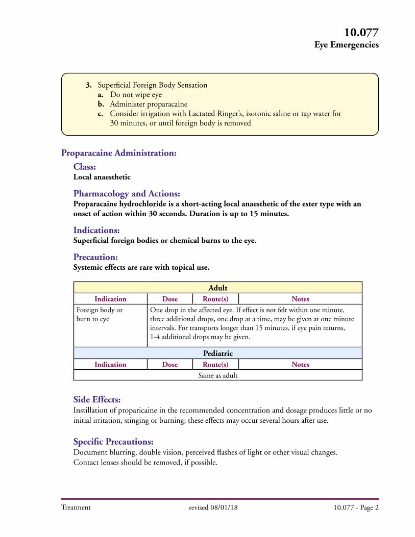
3. Superficial Foreign Body Sensation a. Do not wipe eye b. Administer proparacaine c. Consider irrigation with Lactated Ringer’s, isotonic saline or tap water for 30 minutes, or until foreign body is removed
Proparacaine Administration:
Class: Local anaesthetic
Pharmacology and Actions: Proparacaine hydrochloride is a short-acting local anaesthetic of the ester type with an onset of action within 30 seconds. Duration is up to 15 minutes.
Indications: Superficial foreign bodies or chemical burns to the eye.
Precaution: Systemic effects are rare with topical use.
AdultIndication Dose Route(s) Notes
Foreign body or burn to eye
One drop in the affected eye. If effect is not felt within one minute, three additional drops, one drop at a time, may be given at one minute intervals. For transports longer than 15 minutes, if eye pain returns, 1-4 additional drops may be given.
PediatricIndication Dose Route(s) Notes
Same as adult
Side Effects: Instillation of proparicaine in the recommended concentration and dosage produces little or no initial irritation, stinging or burning; these effects may occur several hours after use.
Specific Precautions: Document blurring, double vision, perceived flashes of light or other visual changes. Contact lenses should be removed, if possible.
10.077Eye Emergencies
Treatment
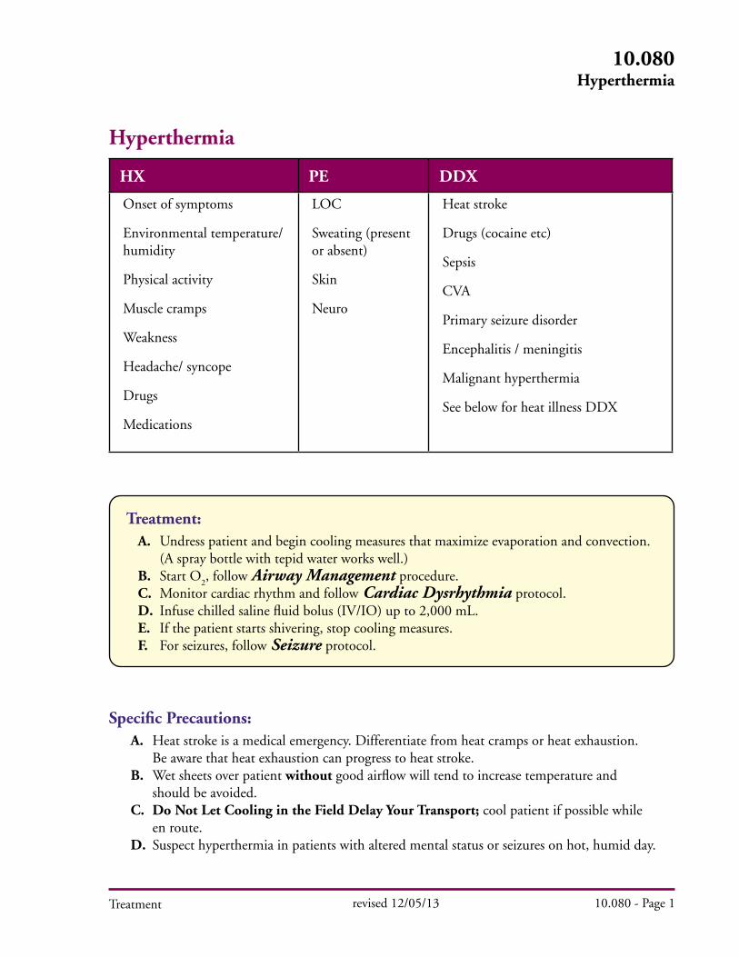
Hyperthermia
HX PE DDX
Onset of symptoms
Environmental temperature/ humidity
Physical activity
Muscle cramps
Weakness
Headache/ syncope
Drugs
Medications
LOC
Sweating (present or absent)
Skin
Neuro
Heat stroke
Drugs (cocaine etc)
Sepsis
CVA
Primary seizure disorder
Encephalitis / meningitis
Malignant hyperthermia
See below for heat illness DDX
Treatment: A. Undress patient and begin cooling measures that maximize evaporation and convection. (A spray bottle with tepid water works well.) B. Start O
2, follow Airway Management procedure.
C. Monitor cardiac rhythm and follow Cardiac Dysrhythmia protocol. D. Infuse chilled saline fluid bolus (IV/IO) up to 2,000 mL. E. If the patient starts shivering, stop cooling measures.
F. For seizures, follow Seizure protocol.
Specific Precautions: A. Heat stroke is a medical emergency. Differentiate from heat cramps or heat exhaustion. Be aware that heat exhaustion can progress to heat stroke. B. Wet sheets over patient without good airflow will tend to increase temperature and should be avoided. C. Do Not Let Cooling in the Field Delay Your Transport; cool patient if possible while en route. D. Suspect hyperthermia in patients with altered mental status or seizures on hot, humid day.
revised 12/05/13 10.080 - Page 1
10.080 Hyperthermia

Treatment
Hypothermia
HX PE DDX
Environmental Exposure(submersion, cold environmental)
Underlying medical conditions Elderly Infants, neonates Sepsis Shock Starvation Endocrine (diabetes, hypothyroid) Medications Spinal cord injury Burns
LOC
Presence or absence of spontaneous respiration, oxygen saturation (if obtainable)
Pulse (rate)
ECG (underlying rhythm wide/narrow QRS)
Etiology: Increased heat loss (environment, burns, prolonged extrication etc) OR Decrease heat production (starvation, age extremes etc)
Severity of hypothermia best assessed by:Mental status Orientated: Mild Confused, disorientated: Moderate Comatose: SevereECG QRS duration Narrow: Mild Sinus brady: Moderate Severe bradycardia (<40) Wide QRS: Severe
Consider underlying medical conditions if no environmental factors.
Treatment: A. Start O
2, follow Airway Management procedure with the following exception:
1. Manage airway with BVM. 2. If oral intubation is necessary, proceed carefully. a. If jaw is difficult to open, use BVM. b. Paralytics should not be used in these patients. B. Remove all wet clothing as soon as possible and provide patient with warm blankets. Place patient in a heated environment as soon as possible. C. Start IV/IO as needed, if possible infuse warmed IV/IO fluids (99° to 113° F). D. Patients who are profoundly hypothermic, (Patient “A”), may require pump rewarming; call OLMC for direction. E. Apply AED or cardiac monitor, if available, and use the following guidelines. 1. Patient “A” — Disorganized ECG rhythm, no pulses, follow Arrest Algorithm for cardiac arrest: a. CPR is advised for these patients. b. Call OLMC for direction regarding resuscitation and before administering any medications. 2. Patient “B” — Organized ECG, with or without palpable pulses, handle gently. F. No CPR or pacing if patient is bradycardic, call OLMC for direction regarding resuscitation and before administering any medications.
revised 1/99 10.090 - Page 1
10.090 Hypothermia
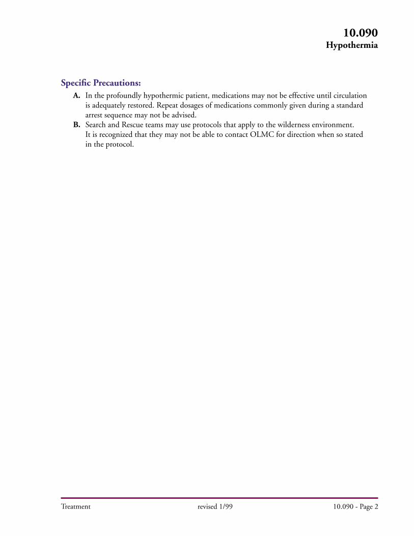
Treatment
Specific Precautions: A. In the profoundly hypothermic patient, medications may not be effective until circulation is adequately restored. Repeat dosages of medications commonly given during a standard arrest sequence may not be advised. B. Search and Rescue teams may use protocols that apply to the wilderness environment. It is recognized that they may not be able to contact OLMC for direction when so stated in the protocol.
revised 1/99 10.090 - Page 2
10.090 Hypothermia
Treatment
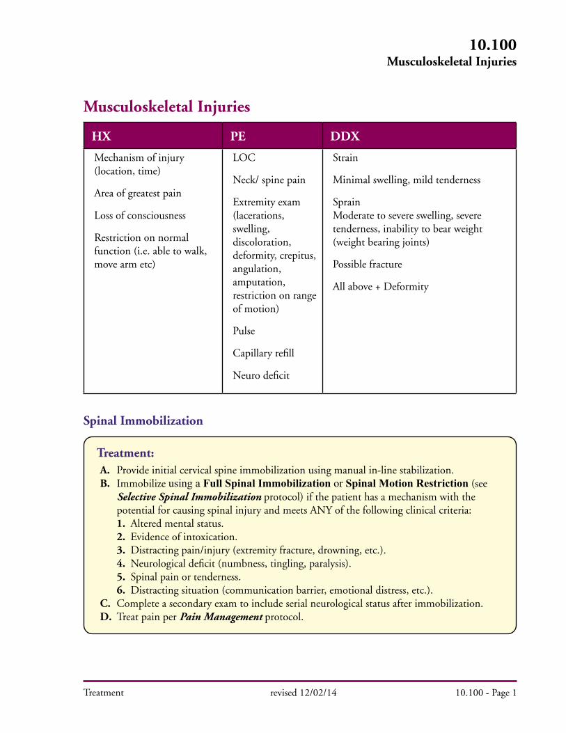
Musculoskeletal Injuries
HX PE DDX
Mechanism of injury (location, time)
Area of greatest pain
Loss of consciousness
Restriction on normal function (i.e. able to walk, move arm etc)
LOC
Neck/ spine pain
Extremity exam (lacerations, swelling, discoloration, deformity, crepitus, angulation, amputation, restriction on range of motion)
Pulse
Capillary refill
Neuro deficit
Strain
Minimal swelling, mild tenderness
SprainModerate to severe swelling, severe tenderness, inability to bear weight (weight bearing joints)
Possible fracture
All above + Deformity
Spinal Immobilization
Treatment: A. Provide initial cervical spine immobilization using manual in-line stabilization. B. Immobilize using a Full Spinal Immobilization or Spinal Motion Restriction (see Selective Spinal Immobilization protocol) if the patient has a mechanism with the potential for causing spinal injury and meets ANY of the following clinical criteria:
1. Altered mental status.2. Evidence of intoxication.3. Distracting pain/injury (extremity fracture, drowning, etc.).4. Neurological deficit (numbness, tingling, paralysis).5. Spinal pain or tenderness.6. Distracting situation (communication barrier, emotional distress, etc.).
C. Complete a secondary exam to include serial neurological status after immobilization. D. Treat pain per Pain Management protocol.
10.100 Musculoskeletal Injuries
revised 12/02/14 10.100 - Page 1
Treatment
Specific Precautions: A. If any immobilization techniques cause an increase in pain or neurological deficits, immobilize patient in the position found or position of greatest comfort. B. Carefully assess the patient’s respiratory status during transport. Loosen straps as needed to avoid respiratory compromise. C. Comorbid age factors (< 12 or > 65 yrs) may impact the EMS Provider’s ability to assess the patient’s perception and communication of pain. A conservative approach to immobilizing these patients is strongly recommended. D. Patients in the third trimester of pregnancy should have the right side of the backboard elevated six inches. E. Pad backboards for all inter-facility transports. Consider padding backboards for prolonged scene transports. F. If sports injury, immobilize patient per the Sports Equipment Removal procedure.
Amputation: A. If amputation is above the wrist or ankle, enter the patient into the Trauma System. B. Cover stump or partial amputation with sterile dressing, saturate with sterile Normal Saline and cover with dry dressing. 1. Partial amputations should be splinted in anatomical position to avoid torsion and angulation. 2. Control bleeding by direct pressure, indirect pressure and/or elevation, hemostatic dressings and/or tourniquet. C. Wrap severed part in sterile dressing, place in plastic bag or wrap in plastic and keep dry. 1. Place bag in ice water combination without salt, if available. 2. Time is of the greatest importance to assure viability, if the transport time will be prolonged due to extrication or other circumstances, consider sending the amputated part ahead to be surgically prepared for reimplantation.
Sprains, Possible Fractures and Dislocations: A. Check for pulses, movement and sensation (PMS) in the extremity distal to the injury site both before and after immobilization. B. If pulses are absent distal to fracture/dislocation, apply axial traction to bring extremity into a more normal anatomical position. Once pulse is restored, immobilize extremity. C. For suspected pelvic fracture, follow Pelvic Wrap procedure if indicated. D. Elevate and apply ice or cold packs if time and extent of other injuries allow.
10.100 Musculoskeletal Injuries
revised 10/25/17 10.100 - Page 2

Note: • If an improvised tourniquet is present before medical provider arrival, place a commercial tourniquet per protocol and remove improvised tourniquet if operationally feasible.
Treatment
Pediatric Considerations: 1. Small children may require extra padding under the shoulders. a. Children require extra padding behind the T-spine and shoulders and are best immobilized on a pediatric backboard. b. If using an adult backboard: i. Since the pediatric patient is at risk of sliding from side to side on a backboard, it is recommended that the EMS Provider place rolled up blankets or other dense, soft support material on both sides of the pediatric patient prior to securing the chest and hip straps. ii. The location of the straps on the backboard may have to be adjusted so they securely hold the pediatric patient in place and do not compress the abdomen. 2. Fentanyl dose for children < 40 kg: 1 microgram/kg IV, IO, or IN. May repeat with 0.5-1 microgram/kg every 3-5 minutes as needed to a MAX total dose of 4 micrograms/kg. Or, fentanyl 1-2 micrograms/kg IM; may repeat every 15 minutes to a MAX total dose of 4 micrograms/kg. If > 40 kg follow adult dosing.
10.100 Musculoskeletal Injuries
revised 07/18/17 10.100 - Page 3
Open Fractures: A. Control bleeding by direct pressure, indirect pressure and/or elevation, hemostatic dressings and/or tourniquet. B. Apply sterile dressing. C. Saturate with sterile Normal Saline. D. Cover with dry dressing. E. If the fracture/dislocation is open or involves a joint, splint in place unless neurovascular compromise is present distal to the fracture site.
Femur Shaft Fracture: Apply traction splint for immobilization.
Pain Control for Isolated Extremity Injuries: A. Consider fentanyl 25-100 micrograms IV/IO/IN; may repeat every 3-5 minutes as needed to a MAX of 400 micrograms. Or, fentanyl 25-100 IM; may repeat every 15 minutes to a MAX of 400 micrograms.
Treatment
HX PE DDX
Onset, duration, total number
Blood, bile?
Associated symptoms (abdominal pain, headache, dizziness, pain, neuro symptoms)
Pregnancy
Medications, allergies
LOC
Neuro deficits
Abdominal exam
Ataxia
CNS (migraine, CVA)
Vestibular (vertigo, dizziness, middle ear)
Cardiac: Acute MI
Eye (blurred vision)
GI (gastroenteritis)
Pregnancy
Severe pain (MI, renal stone, fracture, trauma)
Medication
Treatment:
A. Start O2, follow Airway Management procedure, as indicated.
B. Start IV if needed; if shock syndrome is present follow Shock protocol. C. Consider fluid challenge in patients exhibiting signs of dehydration. D. Consider offering patient an isopropyl alcohol swab and allowing the patient to self- administer the swab by inhalation. Emphasize slow deep inhalation. May be repeated up to 2 times (total of 3 administrations) but should not delay the administration of ondansetron. E. Administer 8 mg ondansetron orally dissolving tablets (Zofran ODT) or 8 mg ondansetron slow IV push over 2 minutes or IM. 1. If nausea and/or vomiting are inadequately controlled after 10 minutes, may repeat ondansetron for a total of 2 doses. 2. If ondansetron unavailable or patient has a known allergy to ondansetron, consider prochlorperazine (Compazine) 5-10 mg slow IV push over 2 minutes or IM (pediatric 2.5 mg IM ONLY). If extrapyramidal reactions are observed, administer diphenhydramine 25-50 mg IV/IM (1 mg/kg pediatric). 3. If ondansetron and prochlorperazine (Compazine) unavailable or if the patient has a known allergy to ondansetron and prochlorperazine, administer diphenhydramine 25-50 mg IV/IM (1 mg/kg pediatric). F. If patient continues to vomit administer fluid challenge and consider other causes.
revised 07/17/18 10.110 - Page 1
10.110 Nausea and Vomiting
Nausea and Vomiting
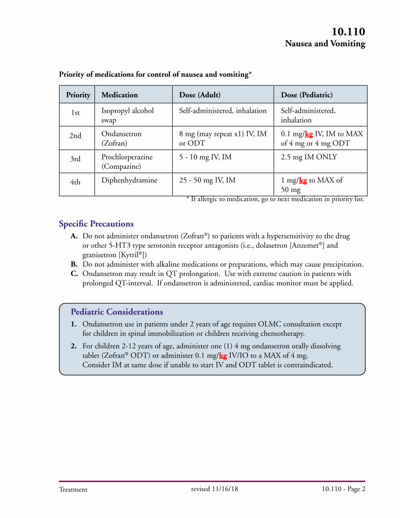
Priority Medication Dose (Adult) Dose (Pediatric)
1st Isopropyl alcohol swap
Self-administered, inhalation Self-administered, inhalation
2nd Ondansetron (Zofran)
8 mg (may repeat x1) IV, IM or ODT
0.1 mg/kg IV, IM to MAX of 4 mg or 4 mg ODT
3rd Prochlorperazine(Compazine)
5 - 10 mg IV, IM 2.5 mg IM ONLY
4th Diphenhydramine 25 - 50 mg IV, IM 1 mg/kg to MAX of 50 mg
Priority of medications for control of nausea and vomiting*
* If allergic to medication, go to next medication in priority list.
Treatment
Specific Precautions A. Do not administer ondansetron (Zofran) to patients with a hypersensitivity to the drug or other 5-HT3 type serotonin receptor antagonists (i.e., dolasetron [Anzemet] and granisetron [Kytril]) B. Do not administer with alkaline medications or preparations, which may cause precipitation. C. Ondansetron may result in QT prolongation. Use with extreme caution in patients with prolonged QT-interval. If ondansetron is administered, cardiac monitor must be applied.
Pediatric Considerations 1. Ondansetron use in patients under 2 years of age requires OLMC consultation except for children in spinal immobilization or children receiving chemotherapy.
2. For children 2-12 years of age, administer one (1) 4 mg ondansetron orally dissolving tablet (Zofran ODT) or administer 0.1 mg/kg IV/IO to a MAX of 4 mg. Consider IM at same dose if unable to start IV and ODT tablet is contraindicated.
10.110 Nausea and Vomiting
revised 11/16/18 10.110 - Page 2
Treatment
Neonatal Resuscitation
HX PE DDX
Painful bleeding in mother (Abruptio Placentae)
Prolonged rupture of membranes
Maternal fever, hypertension, edema, seizures
Meconium-stained fluid
Prolapsed cord
APGAR score
Initial questions:• Amniotic fluid clear of meconium?
• Breathing or crying?
• Good muscle tone?
• Pink color?
• Term infant?
Management priorities:• Provide warmth
• Clear, open airway
• Dry, stimulate infant
• Oxygen
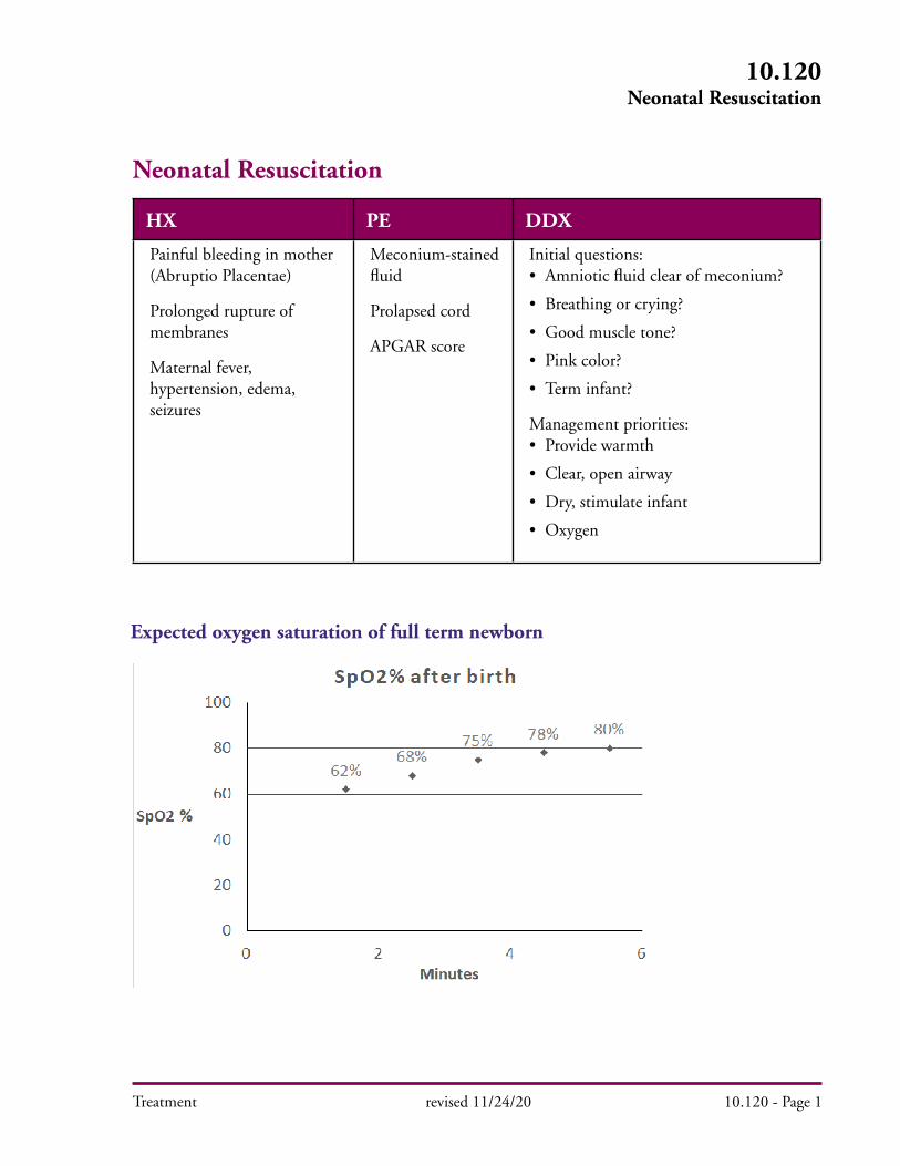
revised 11/24/20 10.120 - Page 1
10.120Neonatal Resuscitation
Expected oxygen saturation of full term newborn
Treatment
10.120Neonatal Resuscitation
revised 11/30/20 10.120 - Page 2
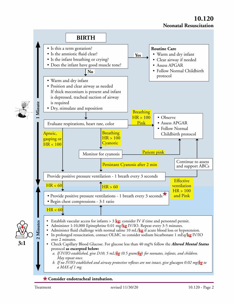
BIRTH
• Is this a term gestation?• Is the amniotic fluid clear?• Is the infant breathing or crying?• Does the infant have good muscle tone?
Routine Care• Warm and dry infant• Clear airway if needed• Assess APGAR• Follow Normal Childbirth protocol
• Observe• Assess APGAR• Follow Normal Childbirth protocol
Evaluate respirations, heart rate, color
Monitor for cyanosis
Provide positive pressure ventilation - 1 breath every 3 seconds
• Provide positive pressure ventilations - 1 breath every 3 seconds• Begin chest compressions - 3:1 ratio
Continue to assess and support ABCs
Consider endotracheal intubation.
Breathing HR > 100Cyanotic
Apneic,gasping orHR < 100
1 M
inut
e2
Min
utes
Yes
No
Breathing HR > 100
Pink
Patient pink
Effective ventilationHR > 100 and Pink
Persistant Cyanosis after 2 min
HR > 60HR < 60
HR < 60
3:1
• Warm and dry infant• Position and clear airway as needed If thick meconium is present and infant is depressed, tracheal suction of airway is required• Dry, stimulate and reposition
• Establish vascular access for infants > 3 kg; consider IV if time and personnel permit.• Administer 1:10,000 Epinephrine 0.01 mg/kg IV/IO. Repeat every 3-5 minutes.• Administer fluid challenge with normal saline 10 mL/kg if acute blood loss or hypotension.• In prolonged resuscitation, contact OLMC to consider sodium bicarbonate 1 mEq/kg IV/IO over 2 minutes.• Check Capillary Blood Glucose. For glucose less than 40 mg% follow the Altered Mental Status protocol as excerpted below: a. If IV/IO established, give D10; 5 mL/kg (0.5 gram/kg) for neonates, infants, and children. May repeat once. b. If no IV/IO established and airway protective reflexes are not intact, give glucagon 0.02 mg/kg to a MAX of 1 mg.
Treatment
Meconium Aspiration: Meconium in the amniotic fluid can be aspirated resulting in a potentially fatal course or requiring high-pressure ventilation and resulting chronic lung disease. Many of these complications can at least be attenuated, if not prevented, by suctioning meconium from the airway PRIOR to ventilating. This can be emotionally difficult to do when confronted with a depressed, blue, bradycardic neonate, but direct tracheal suctioning through the ET tube should be considered part of establishing a patent airway in these neonates.
A. With all infants who have passed meconium, as soon as the baby’s head is delivered (before delivery of the shoulders), using a l0 French or larger suction catheter, suction the mouth, pharynx and nose. B. After delivery, proceed with intubation for all infants who are depressed and have passed meconium or any infant passing thick, particulate meconium. 1. Check blood glucose and follow Altered Mental Status and Coma protocol. 2. Procedure: a. Suction the mouth, nose and posterior pharynx, using a 10 French or larger catheter hooked to machine suction, when the head is delivered and again after the rest of the infant has been delivered.
b. Secure protected airway, intubate the infant with the appropriately sized endotracheal tube and suction with a meconium suction adapter or use a specially designed meconium aspiration catheter/endotracheal tube such as a Neovac type device.
c. Suction should not last more than 3 to 5 seconds. i. Do not suction with your own mouth! ii. Use the portable machine suction or wall suction if available.
3. In an infant with severe asphyxia, clinical judgment should be used to determine the number of intubation attempts. It may not be possible to clear the trachea of all meconium before initiating other resuscitation measures.
10.120Neonatal Resuscitation
revised 11/20/20 10.120 - Page 3
Notes and PrecautionsA. Do not use Atropine in neonatal resuscitation.B. If meconium is lightly stained and infant is vigorous (strong respiratory effort, good muscle tone, heart rate > 100 bpm) endotracheal suctioning should not be performed.C. 100% oxygen should not be used to initiate resuscitation. Begin resuscitation with room air and add supplemental oxygen if infant remains cyanotic or oxygen saturation less than 70% after 2 minutes. Expected oxygen saturation immediately after full newborn birth ranges between 70% to 85% within the first 5 minutes. Most full or near term infants will have oxygen saturation of 90% by 5 minutes. If supplemental oxygen is needed after 5 minutes, attempt to keep oxygen saturation between 90% to 95%.D. An infant may need resuscitation if intrapartum risk factors for asphyxia are present (prolapsed cord, painful bleeding, prolonged rupture of membranes, maternal fever, multiple births, abnormal presentation, maternal hypo-hypertension or seizure).E. To monitor HR, either auscultate heart tones or use 3-lead ECG. Palpation of HR is unreliable and intermittent.
Treatment
HX PE DDX
Last menstrual period
Pregnancy • Single or multiple • Due date • Abdominal pain/ contractions (timing/ duration) • Ruptured membranes
Seizures
Hypertension
Vaginal bleeding
Past medical /OB Hx
Hypertension
Edema
Abdominal exam
Vaginal bleeding
If possibility of delivery exists, inspect perineum for bleeding, fluid (not color), crowning or abnormal presentation
Do not perform a digital exam
Vaginal bleeding
Early pregnancy
Ectopic pregnancy
Spontaneous abortion
Late pregnancy
Abruptio placenta
Placenta previa
HTN/headache/edema/seizures
Eclampsia/pre-eclampsia
General Treatment: 1. If multiple, or abnormal birth, consider second transport unit. 2. Start O
2 in all abnormal deliveries. Follow Airway Management procedure.
3. If in third trimester, transport on left side (pillow under right hip, or, if on backboard, tilt right side of board up 6 inches) to keep uterine pressure off inferior vena cava unless delivery is imminent. 4. Start IV/IO as needed. Vital signs may not be a reliable indicator of shock or respiratory distress in the pregnant patient. 5. Acute onset severe hypertension in pregnant and postpartum women. a. Includes all pregnant women and up to 6 weeks postpartum with symptoms. b. Symptoms include headache, visual disturbances, chest discomfort, shortness of breath, confusion, abdominal pain. c. Notify receiving hospital of patients with a sustained elevation in BP ≥ 140 mmHg systolic and/or ≥ 100 mmHg diastolic are present for at least 15 minutes or more. d. Initiate treatment (if feasible) if sustained elevation in BP > 160 mmHg systolic and/or ≥ 110 mmHg diastolic (either one or both) persists for at least 15 minutes or more. Treatment options: Labetalol (if available) a. Administer Labetalol 10 mg slow IV push over 1-2 minutes. b. Target systolic BP 140-150 mmHg and diastolic BP 90-100 mmHg. c. Labetalol may be repeated twice (up to 3 total doses) every 15 minutes (doubling doses if needed depending on effect of preceding dose; e.g. 1st dose - 10mg, 2nd dose - 20mg, 3rd dose - 40mg). d. Stop administration if HR < 60 bpm or other adverse effects. e. Maximum dose of Labetalol is 70 mg. f. Formulation 4 mL (5 mg/mL total 20 mg) Carpuject Sterile cartridge. 6. Seizures (eclampsia) follow Seizure protocol. a. Consult OLMC for administration of Magnesium Sulfate.
10.130OB/GYN Emergencies
revised 09/29/20 10.130 - Page 1
OB/GYN Emergencies
Treatment
Childbirth: A. Normal child birth 1. Use sterile or clean technique. 2. Guide and control – but do not retard or hurry – delivery. 3. Check for cord around baby’s neck and gently remove if found. 4. After delivery, assess infant per Neonatal Resuscitation protocol. Assess infant using APGAR at time of birth and five minutes later. Documentation should describe the infant using criteria rather than giving a numerical score. If no resuscitation is needed (term infant, breathing or crying, good muscle tone), proceed as below. 5. Do not suction infant’s nose and mouth unless there is meconium present and the infant is depressed; or there is a need to clear the airway. 6. Briefly dry infant and place on mother’s chest, in skin-to-skin contact. Cover both with a clean, dry blanket. 7. At 1 to 3 minutes after delivery, clamp and cut the umbilical cord about 6 inches from infant. If resuscitation is needed, cord may be clamped and cut as soon as necessary. 8. Do not delay transport to deliver the placenta. After the placenta has delivered, gently externally massage uterus to encourage contraction and prevent bleeding. 9. If mother has significant postpartum hemorrhage (> 500 mL), continue uterine massage, treat for shock, and update receiving facility. 10. Unless infant needs treatment, keep on mother’s chest for transport. Monitor vital signs of mother and infant during transport.
B. Abnormal Childbirth 1. General: a. Transport to nearest appropriate hospital. b. Give receiving hospital earliest possible notification. c. Contact OLMC for advice. d. Transport position as in General Treatment #3 above. 2. Breech Presentation (buttocks first): a. If delivery is imminent, prepare the mother as usual and allow the buttocks and trunk to deliver spontaneously, then support the body while the head is delivered. b. If the head is not delivered within 3 minutes, suffocation can occur: i. Place your gloved hand in the vagina, with your palm toward the baby’s face. ii. Form a “V” with your fingers on either side of the baby’s nose and push the vaginal wall away from the baby’s face to create airspace for breathing. c. Assess for presence of pulse in umbilical cord, if presenting. 3. Prolapsed cord: a. With a gloved hand, gently attempt to push the baby up the vagina several inches. b. Do not attempt to push the cord back. c. Assess for presence of pulse in umbilical cord. d. Transport patient in position that best ensures adequate fetal circulation and is safe.
10.130OB/GYN Emergencies
revised 09/25/20 10.130 - Page 2
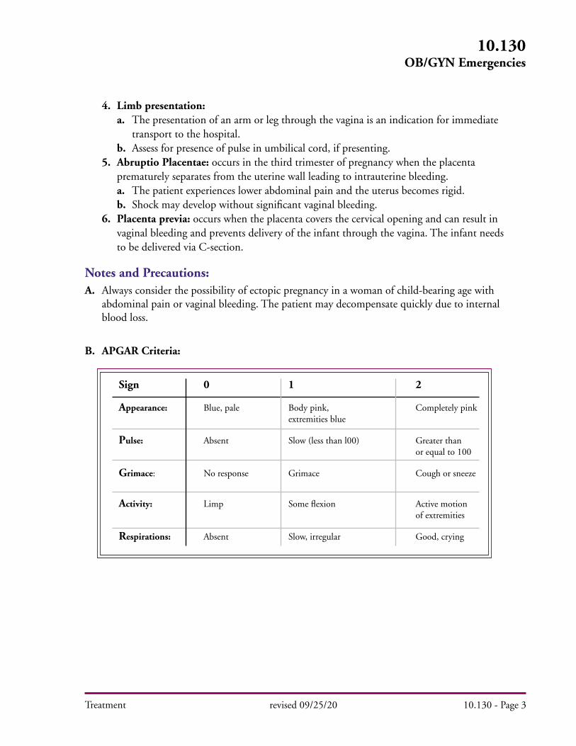
Treatment
Notes and Precautions:A. Always consider the possibility of ectopic pregnancy in a woman of child-bearing age with abdominal pain or vaginal bleeding. The patient may decompensate quickly due to internal blood loss.
B. APGAR Criteria:
Sign 0 1 2
Appearance: Blue, pale Body pink, Completely pink extremities blue
Pulse: Absent Slow (less than l00) Greater than or equal to 100
Grimace: No response Grimace Cough or sneeze
Activity: Limp Some flexion Active motion of extremities
Respirations: Absent Slow, irregular Good, crying
10.130OB/GYN Emergencies
revised 09/25/20 10.130 - Page 3
4. Limb presentation: a. The presentation of an arm or leg through the vagina is an indication for immediate transport to the hospital. b. Assess for presence of pulse in umbilical cord, if presenting. 5. Abruptio Placentae: occurs in the third trimester of pregnancy when the placenta prematurely separates from the uterine wall leading to intrauterine bleeding. a. The patient experiences lower abdominal pain and the uterus becomes rigid. b. Shock may develop without significant vaginal bleeding. 6. Placenta previa: occurs when the placenta covers the cervical opening and can result in vaginal bleeding and prevents delivery of the infant through the vagina. The infant needs to be delivered via C-section.
Treatment
10.135Pain Management
Pain Management
ACUTE CHRONIC PRINCIPLES OF MANAGEMENT
Is a symptom of illness or injury
Serves a biological purpose
Is associated with identifiable pathology
Is present for less than 6 months
Is the problem
Has no biological function
May or may not be associated with identifiable pathology
Is present for more than6 months
Pain is best treated early
Untreated or under treated pain produces more pain
Analgesia and a search for the cause of the pain should happen simultaneously
Pain scales should be used routinely
Treatment: For acute pain and uncontrolled chronic pain: A. Consider oxygen per Airway Management protocol. B. Monitor vital signs and level of consciousness, consider ECG monitor. C. Establish venous access if indicated. Determine location of pain and severity using numeric scale (1-10) or Faces scale. D. Consider and treat underlying cause of pain. E. Use non-pharmacological pain management (i.e., position of comfort, hot/cold pack, elevation, splinting, padding, wound care, therapeutic calming and communication). F. Administer pain medication: 1. Mild to moderate pain (Ex: 1-4 on Pain scale) a. Acetaminophen 1000mg or b. Ibuprofen 600 mg. 2. Moderate to severe pain (Ex: 5-8 on Pain scale) a. Ketorolac 15 mg IV or 30 mg IM (2-64 years old) 3. Severe pain (Ex: 9-10 on Pain scale) a. Ketorolac 15 mg IV or 30 mg IM -OR- b. Fentanyl 25-100 micrograms IV/IO/IN; may repeat every 3-5 minutes as needed to a MAX of 400 micrograms. Or, fentanyl 25-100 IM; may repeat every 15 minutes to a MAX of 400 micrograms. -OR- c. Sufentanil 5-10 micrograms IV/IO; may repeat every 3-5 minutes as needed to a MAX of 40 micrograms. Or, sufentanil 5-10 micrograms IM; may repeat every 15 minutes as needed to a MAX of 40 micrograms. -OR-
revised 06/20/18 10.135 - Page 1
Treatment
Pediatric Considerations: 1. Mild to Moderate pain: a. If no contraindication to oral medication, consider acetaminophen 15 mg/kg PO to a MAX of 1000 mg or ibuprofen 10 mg/kg PO to a MAX of 600 mg, if available. 2. Mild to Moderate pain: a. Ketorolac dose for pediatrics (<40 kg) is 0.5 mg/kg IV (MAX dose of 15 mg) or 1 mg/kg IM (MAX dose of 30 mg). Do not exceed adult dose. Do not repeat. 3. Severe pain: a. Fentanyl 1-2 micrograms/kg IM; may repeat every 15 minutes to a MAX total dose of 4 micrograms/kg. Do not exceed adult dosing. -OR- b. Sufentanil 0.1 micrograms/kg IV/IO/IM; may repeat every 3-5 minutes as needed to a MAX of 10 micrograms. Do not exceed adult dosing. -OR- c. For children under 20 kg, morphine 0.1 mg/kg IV or IM. May repeat every 3-5 minutes. Do not exceed adult dosing. 4. Contact OLMC if MAX dose of either medication is reached without adequate pain management.
10.135Pain Management
revised 09/24/19 10.135 - Page 2
d. Morphine 2-8 mg IV/IO every 3-5 minutes to a MAX of 20 mg. Or, morphine 5-10 mg IM; may repeat 5 mg every 10-15 minutes to a MAX of 20 mg -OR- e. Ketamine 15 mg IV/IO/IM (Administer one time dose of midazolam 2.5 mg IV/IO/IM in adults to prevent/treat negative emergence reaction.) Do not administer pain medications if any of the following are present: a. Respiratory distress or O
2 saturation of < 90%
b. Known allergy to pain medications c. Altered mental status d. Systolic blood pressure of < 100 mm/Hg G. Obtain a full set of vital signs and pain scale rating prior to and after each administration of pain medication.

Treatment
Faces Pain Scale
0No Hurt
2Hurts
Little Bit
4Hurts
Little More
6Hurts
Even More
8Hurts
Whole Lot
10Hurts Worst
10.135Pain Management
revised 11/17/20 10.135 - Page 3
Pain
Mild to
Moderate
Severe
Moderate to
Severe
Acetaminophen 1000 mg PO-OR-
Ibuprofen 600 mg PO
Ketorolac 15 mg IV or 30 mg IM
-OR-Fentanyl 25-100 micrograms
IV/IO/IM (MAX of 400 micrograms)
-OR-Sufentanil 5-10 micrograms
IV/IO/IM (MAX of 40 micrograms)
-OR-Morphine 2-8 mg
IV/IO/IM (MAX of 20 mg)
-OR-Ketamine 15 mg IV/IO/IM (with midazolam 2.5 mg)
Ketorolac 15 mg IV or 30 mg IM
Acetaminophen 15 mg/kg PO(MAX of 1000 mg)
-OR-Ibuprofen 10 mg/kg PO
(MAX 600 mg)
Fentanyl 1-2 micrograms/kg IV/IO/IM
(MAX of 4 micrograms/kg)-OR-
Sufentanil 0.1 micrograms/kg IV/IO/IM
(MAX of 10 micrograms)-OR-
If <20 kg, Morphine 0.1 mg/kg IV/IO/IM
Ketorolac 0.5 mg/kg IV or 1 mg/kg IM
(MAX of 30 mg)
1 - 4
9 - 10
5 - 8
Medication Dose(Adult)
Medication Dose(Pediatric)
PainScale
Treatment
Poisons and Overdoses
HX PE DDX
Type of ingestion or exposure
What, when, how much
Multiple patients with similar symptoms
Reason for ingestion (accidental or intentional)
Action by bystanders
Previous similar events
LOC
Pupils
Breath (odor)
Temperature (hyper/hypothermic)
Neuro status
ECG (rate, rhythm, QRS duration)
See toxidromes
Treatment: [Consider HAZMAT Response]
A. Consider use of any of the following protocols:1. Hazardous Materials -- Multiple Toxic Exposure2. Hazardous Materials Treatment, if trained and authorized3. Mass Casualty Incident4. Staging for High Risk Response
B. External Contamination:1. Protect medical personnel.2. Remove contaminated clothing.3. Brush off any solid material from the skin.4. Flush contaminated skin and eyes with copious amounts of water.
C. Internal Ingestion:1. Assess and support ABCs.2. Start O
2, follow Airway Management procedure.
3. If ingestion is Aspirin (ASA) and/or Tylenol (APAP) only and is less than 2 hours old, administer 1 gram/kg activated charcoal if available (MAX dose of 100 grams). 4. For all other ingestions less than 2 hrs old contact OLMC for consideration of activated charcoal. 5. For ingestions more than 2 hrs old, activated charcoal is not indicated. 6. Start IV/IO if needed and follow Shock protocol.
revised 11/08/18 10.140 - Page 1
10.140Poisons and Overdoses
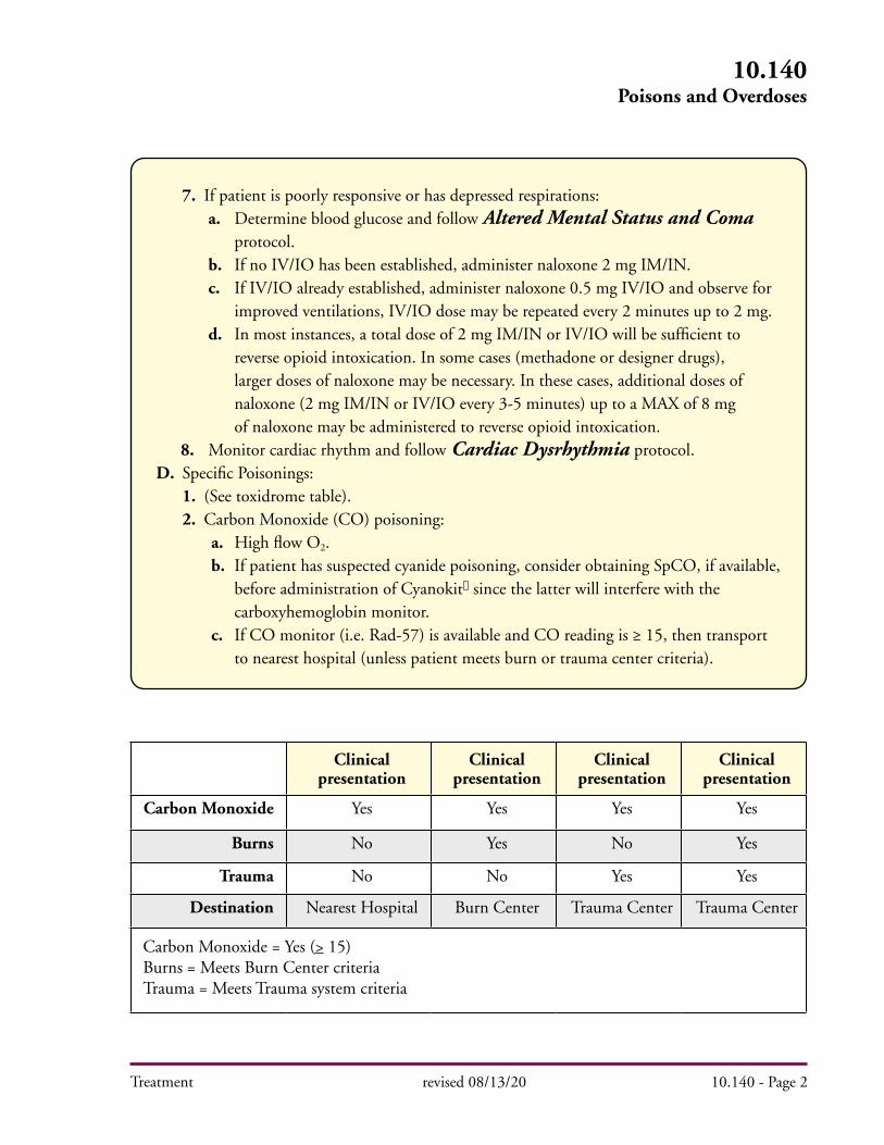
Clinical presentation
Clinical presentation
Clinical presentation
Clinical presentation
Carbon Monoxide Yes Yes Yes Yes
Burns No Yes No Yes
Trauma No No Yes Yes
Destination Nearest Hospital Burn Center Trauma Center Trauma Center
Carbon Monoxide = Yes (> 15)Burns = Meets Burn Center criteriaTrauma = Meets Trauma system criteria
Treatment
7. If patient is poorly responsive or has depressed respirations:a. Determine blood glucose and follow Altered Mental Status and Coma protocol.
b. If no IV/IO has been established, administer naloxone 2 mg IM/IN. c. If IV/IO already established, administer naloxone 0.5 mg IV/IO and observe for improved ventilations, IV/IO dose may be repeated every 2 minutes up to 2 mg. d. In most instances, a total dose of 2 mg IM/IN or IV/IO will be sufficient to reverse opioid intoxication. In some cases (methadone or designer drugs), larger doses of naloxone may be necessary. In these cases, additional doses of naloxone (2 mg IM/IN or IV/IO every 3-5 minutes) up to a MAX of 8 mg of naloxone may be administered to reverse opioid intoxication. 8. Monitor cardiac rhythm and follow Cardiac Dysrhythmia protocol. D. Specific Poisonings:
1. (See toxidrome table).2. Carbon Monoxide (CO) poisoning:
a. High flow O2. b. If patient has suspected cyanide poisoning, consider obtaining SpCO, if available, before administration of Cyanokit since the latter will interfere with the carboxyhemoglobin monitor. c. If CO monitor (i.e. Rad-57) is available and CO reading is ≥ 15, then transport to nearest hospital (unless patient meets burn or trauma center criteria).
10.140Poisons and Overdoses
revised 08/13/20 10.140 - Page 2

Treatment
Pediatric Considerations: [Consider HAZMAT Response]
1. Consider possibility of neglect or abuse.2. Determine blood glucose and follow Altered Mental Status & Coma protocol.
3. Activated charcoal dose is 1 gram/kg (MAX dose of 100 grams). 4. Naloxone dose is 0.1 mg/kg, MAX 2 mg per dose. 5. IV/IO Atropine dose, per OLMC, may be very high in children that have orally ingested organophosphate poisons.
Specific Precautions: A. Inhalation poisoning, SLUDGE symptoms (salivation, lacrimation, urination, defecation, gastrointestinal symptoms and emesis), or acid/alkali exposure may be dangerous to rescuers. B. Do not neutralize acids or alkalis.
revised 11/08/18 10.140 - Page 3
10.140Poisons and Overdoses
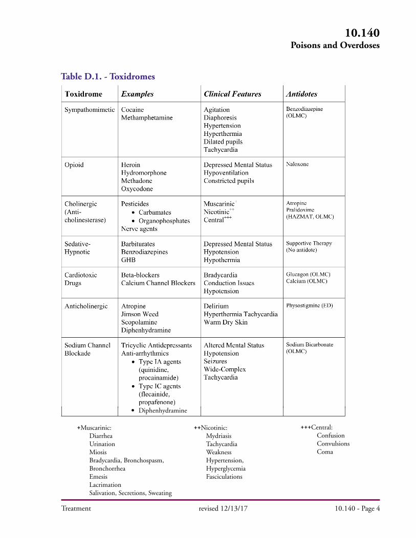
Treatment
10.140Poisons and Overdoses
revised 12/13/17 10.140 - Page 4
+++Central: Confusion Convulsions Coma
++Nicotinic: Mydriasis Tachycardia Weakness Hypertension, Hyperglycemia Fasciculations
+Muscarinic: Diarrhea Urination Miosis Bradycardia, Bronchospasm, Bronchorrhea Emesis Lacrimation Salivation, Secretions, Sweating
Table D.1. - Toxidromes
• Diphenhydramine

Treatment
Psychiatric and Behavioral Disorders
HX PE DDX
Recent crises
Behavioral change
Suicide/homicidal ideation
Past medical, psych history
Medications
Drugs
ETOH
Pupils
Orientation
Mental status exam appearance, behavior orientation affect/ mood, thought (delusions, illogical etc), hallucinations (visual/auditory) memory (recent/past)
Confusion
Metabolic (electrolyte)
Infectious (encephalitis, sepsis)
Neurologic (CVA, tumor)
Medication
Drug abuse
Psych
Treatment: A. Immediate danger to medical personnel or patient:
1. Follow Staging for High Risk Response protocol.2. Protect yourself and others.3. Summon law enforcement.
B. No evidence of immediate danger to medical personnel or patient:1. Assess ABCs. Follow Airway Management procedure, if needed.2. Assess orientation and level of consciousness, follow Altered Mental Status and Coma protocol if indicated.
C. General approach to the patient:1. Show self-confidence and convey concern for the patient.2. One EMS Provider should establish rapport and interact with the patient.
3. Do not stay alone with the patient. Have enough help to restrain the violent patient. See Patient Restraint (Physical) procedure.
4. If chemical restraint is indicated, see Patient Restraint (Chemical) procedure.
Choice of Destination A. Voluntary patient: 1. Hospital destination is determined by patient preference. 2. If the patient has no preference, transport to Unity Hospital or Portland Providence.B. Involuntary patient, patients on Directors Hold or Police Officer Mental Hold: 1. Patients of 9-1-1 incidents, transported by ambulance, must be evaluated at a licensed hospital Emergency Department.
revised 11/19/18 10.150 - Page 1
10.150Psychiatric and Behavioral Disorders
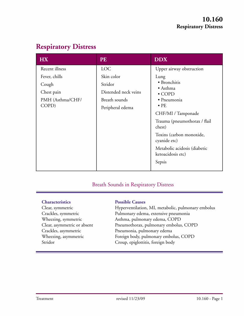
Treatment
Respiratory Distress
HX PE DDX
Recent illness
Fever, chills
Cough
Chest pain
PMH (Asthma/CHF/COPD)
LOC
Skin color
Stridor
Distended neck veins
Breath sounds
Peripheral edema
Upper airway obstruction
Lung • Bronchitis • Asthma • COPD • Pneumonia • PE
CHF/MI / Tamponade
Trauma (pneumothorax / flail chest)
Toxins (carbon monoxide, cyanide etc)
Metabolic acidosis (diabetic ketoacidosis etc)
Sepsis
Breath Sounds in Respiratory Distress
Characteristics Possible CausesClear, symmetric Hyperventilation, MI, metabolic, pulmonary embolusCrackles, symmetric Pulmonary edema, extensive pneumoniaWheezing, symmetric Asthma, pulmonary edema, COPDClear, asymmetric or absent Pneumothorax, pulmonary embolus, COPDCrackles, asymmetric Pneumonia, pulmonary edemaWheezing, asymmetric Foreign body, pulmonary embolus, COPDStridor Croup, epiglottitis, foreign body
revised 11/23/09 10.160 - Page 1
10.160Respiratory Distress

Treatment
10.160Respiratory Distress
revised 09/24/19 10.160 - Page 2
Treatment:
A. Start O2, follow Airway Management procedure, as indicated. Use pulse oximeter.
B. Start IV/IO as needed. C. Monitor cardiac rhythm and follow Cardiac Dysrhythmia protocol. D. Treat underlying cause as follows:
1. Upper Airway (croup, epiglottitis, anaphylaxis, foreign body):a. Obstructed airway procedures for complete obstruction.
b. Treat anaphylaxis per Anaphylaxis and Allergic Reaction protocol. c. Foreign body: Remove using laryngoscopy if complete obstruction. d. Complete Obstruction: If you cannot effectively ventilate the patient and they are deteriorating, consider cricothyrotomy. 2. Respiratory Distress Unknown Etiology: a. Consider nebulized albuterol (1 unit dose), may repeat as needed.
b. If a second and/or third treatment is needed, add ipratropium (1 unit dose) to albuterol treatment.
3. Pulmonary Edema: a. Sit patient upright. b. If BP less than 100 mm/Hg: Treat possible cardiogenic shock. See Shock protocol. c. If BP greater than 100 mm/Hg: i. Nitroglycerine 0.4 mg SL (spray, tablet or powder), repeat nitroglycerine every 3-5 minutes.
NOTES:
Do not administer nitroglycerine without OLMC if patient has taken Viagra® or other similar drugs in the last 24 hours or Cialis® (tadalafil) within last 48 hours.
ii. If the patient remains in severe respiratory distress (e.g., unable to speak more than one or two words, low O
2 saturation [< 90 %], RR > 40)
start CPAP if available. iii. Consider albuterol 2.5 mg by nebulizer. May repeat as needed. iv. Furosemide (if SBP > 100 mmHg and fluid overload state [JVD, rales, peripheral edema, hypertension]). a. If patient is not currently taking furosemide, administer 20 mg IV/IO. b. If patient is taking furosemide, administer 40 mg IV/IO. d. If severe respiratory distress and patient not tolerating CPAP, administer IV/IO nitroglycerine 1 mg. May repeat once in 5 minutes. i. If patient is tolerating CPAP but still with severe respiratory distress, continue CPAP and administer IV/IO nitroglycerine 1 mg. May repeat once in 5 min.
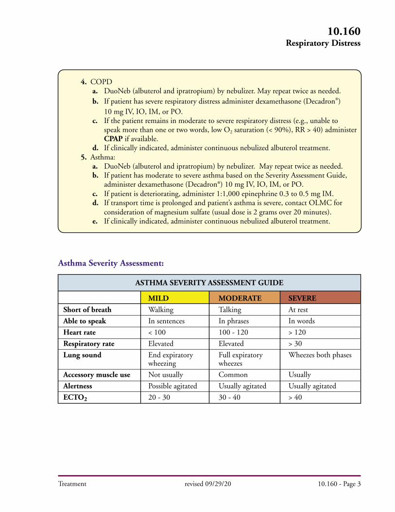
Treatment
10.160Respiratory Distress
revised 09/29/20 10.160 - Page 3
4. COPDa. DuoNeb (albuterol and ipratropium) by nebulizer. May repeat twice as needed.b. If patient has severe respiratory distress administer dexamethasone (Decadron) 10 mg IV, IO, IM, or PO.c. If the patient remains in moderate to severe respiratory distress (e.g., unable to speak more than one or two words, low O2 saturation (< 90%), RR > 40) administer CPAP if available.d. If clinically indicated, administer continuous nebulized albuterol treatment.
5. Asthma:a. DuoNeb (albuterol and ipratropium) by nebulizer. May repeat twice as needed.b. If patient has moderate to severe asthma based on the Severity Assessment Guide, administer dexamethasone (Decadron) 10 mg IV, IO, IM, or PO.c. If patient is deteriorating, administer 1:1,000 epinephrine 0.3 to 0.5 mg IM.d. If transport time is prolonged and patient’s asthma is severe, contact OLMC for consideration of magnesium sulfate (usual dose is 2 grams over 20 minutes).e. If clinically indicated, administer continuous nebulized albuterol treatment.
Asthma Severity Assessment:
Short of breath
Able to speak
Heart rate
Respiratory rate
Lung sound
Accessory muscle use
Alertness
ECTO2
ASTHMA SEVERITY ASSESSMENT GUIDE
MILD
Walking
In sentences
< 100
Elevated
End expiratory wheezing
Not usually
Possible agitated
20 - 30
MODERATE
Talking
In phrases
100 - 120
Elevated
Full expiratory wheezes
Common
Usually agitated
30 - 40
SEVERE
At rest
In words
> 120
> 30
Wheezes both phases
Usually
Usually agitated
> 40
Treatment
10.160Respiratory Distress
revised 11/16/18 10.160 - Page 4
Pediatric Patients:1. Upper Airway Obstruction a. In patients 6 months to 6 years of age with audible stridor at rest, administer 5 mL epinephrine 1:1,000 or racemic epinephrine 11.25 mg (0.5 mL) diluted in 2.5 mL normal saline, nebulized. May repeat once. Contact OLMC for additional dosing. b. Suspected anaphylaxis or foreign body: i. Treat anaphylaxis and foreign body obstruction per adult guidelines. c. Suspected croup, epiglottis, or laryngeal edema: The usual cause of respiratory arrest in children with croup, epiglottitis or laryngeal edema is exhaustion, not complete obstruction. i. If suspected croup, administer dexamethasone per peds guide. ii. If the child deteriorates, perform BVM. iii. If unable to ventilate with BVM, perform intubation. iv. If intubation unsuccessful and unable to ventilate effectively, perform needle cricothyrotomy.2. Asthma a. Administer albuterol and ipratropium per adult guidelines. b. If patient is deteriorating administer 1:1,000 epinephrine 0.01 mg/kg IM (MAX dose 0.5 mg). Contact OLMC for additional doses. c. If patient has Moderate to Severe asthma based on Asthma Severity Assessment Guide and is not improving with treatment, consider dexamethasone 0.6 mg/kg IV/IO/IM/PO up to 10 mg.3. Acute Bronchiolitis (< 2 years old) Mild-moderate respiratory distress (see Infant Respiratory Distress Guide below) a. Administer oxygen via blow-by, nasal cannula or mask to keep SpO
2 > 92%.
Monitor EtCO2 if available.
b. If nasal secretions and/or congestion, use nasal suction with adapter if available. If secretions are thick, may use normal saline to loosen. c. If wheezing, administer albuterol 2.5 mg via nebulizer. If improvement, may use every 10 minutes. Discontinue if patient’s heart rate is > 200. d. If patient worsens and is still wheezing, administer epinephrine 5 mL of 1:1000 via nebulizer. If improvement, may use every 10 minutes. Discontinue if patient’s heart rate is > 200. e. If unable to keep SpO
2 > 92% with oxygen or patient has continued significant work
of breathing despite treatment: i. 30-90 days old: titrate High Flow Nasal Cannula (pediatric) Oxygen (HFNCO) starting at 2 LPM up to 4 LPM. ii. Greater than 90 days old: titrate High Flow Nasal Cannula Oxygen up to 6 LPM.4. Severe respiratory distress (see Infant Respiratory Distress Assessment Guide below) a. Suction nares as described above. b. Initiate high flow nasal cannula oxygen as described above with EtCO
2 monitoring.
c. If wheezing, administer 5 mL epinephrine 1:1,000 via nebulizer. If improvement may use every 10 minutes. Discontinue if patients heart rate is > 200.

Treatment revised 09/14/16 10.160 - Page 5
10.160Respiratory Distress
Key Considerations:Speed of onset, recent illness/infection, fever, chills or productive cough, medications and allergies, distended neck veins, peripheral edema, lung sounds, medical history (including asthma, CHF, COPD, pneumonia).
Respiratory Rate ≤ 2 months 2-12 months 1-2 years
Retractions
Dyspnea
Auscultation
INFANT RESPIRATORY DISTRESS ASSESSMENT GUIDE
MILD
≤ 60 ≤ 50 ≤ 40
Subcostal or intercostal
1 of: difficulty feeding, decreased vocalization, or agitation
End-expiratory wheeze only
MODERATE
61-69 51-59 41-44
2 of: subcostal, intercostal, substernal retractions, OR nasal flaring
2 of: difficulty feeding, decreased vocalization, or agitation
Expiratory wheeze only
SEVERE
≥ 70 ≥ 60 ≥ 45
3 of: subcostal, intercostal, substernal, suprasternal, supraclavicular retractions, OR nasal flaring OR head bobbing
Stops feeding, no vocalization OR drowsy and confused
Inspiratory and expiratory wheezing OR diminished breath sounds OR both
d. Prepare for positive pressure ventilation with BVM and intubation for apnea, EtCO
2 > 55 or inability to maintain SpO
2 > 85%.
5. BRUE (Brief Resolved Unexplained Event) An episode that is frightening to the observer and is characterized by some combination of apnea, color change (cyanosis or pallor) change in muscle tone (flaccid or rigid), and choking or gagging.
a. BRUE is a group of symptoms, not a disease process. b. BRUEs are most common in infants under one year of age, but may occur up to two years of age. They are more frequent in premature infants and infants with other health conditions such as cystic fibrosis, bronchiolitis and congenital heart disease. c. Serious underlying causes can include pneumonia, bronchiolitis, seizure, sepsis, intracranial hemorrhage, and meningitis. d. Many infants will have returned to normal by the time EMS arrives. e. Consider non-accidental trauma. f. Transport via ALS to an ED capable of treating critically ill infants even if the infant currently appears in no distress.
Treatment
Seizures
HX PE DDX
Seizure • Onset, duration • Type (grand mal, focal etc.) • Fever • Urine/fecal incontinence
History • Diabetes • Head trauma • Pregnancy • Previous seizure • Current medications (including compliance) • Other (drug/ETOH, fever) • Toxic exposure
LOC
O2 saturation
Active seizure (focal/ grand mal)
Neuro deficits
Evidence of trauma
HypoxiaHypoglycemia Febrile Trauma CNS (CVA, meningitis, tumor)Drugs/ODIdiopathicETOH withdrawalElectrolyte disturbanceCardiac arrestEclampsiaShock
Treatment: A. History of seizure with current altered mental status (postictal) or witnessed seizure.
1. Move hazardous objects away from patient and protect head; restrain only if necessary.2. Start O
2; follow Airway Management procedure.
3. Place patient on left side for transport.4. Continue assessment and document level of consciousness every 5 minutes.5. Monitor cardiac rhythm and follow Cardiac Dysrhythmias protocol.
B. If patient is in status seizure (continuous seizure or repetitive seizures without regaining consciousness): Proceed as in “A” above, and do the following:
1. If no IV/IO access, administer midazolam 10 mg IM/IN. If seizure continues, may repeat in 3-5 minutes with an additional 10 mg midazolam IM/IN dose.2. If still seizing, establish IV/IO access. Administer midazolam 2.5-5.0 mg IV/IO. If seizure continues, in 2-3 minutes administer 5-10 mg midazolam IV/IO. If still in status seizures, contact OLMC for possible midazolam infusion. 3. If IV/IO is immediately available, administer midazolam 2.5-5.0 mg IV/IO. If seizure continues, in 2-3 minutes administer 5-10 mg midazolam IV/IO. If still in status seizures, contact OLMC for possible midazolam infusion.
4. Monitor patient’s respiratory status closely after midazolam administration. 5. Check blood glucose and treat per Altered Mental Status and Coma protocol.
revised 01/27/21 10.170 - Page 1
10.170Seizures
CAUTION: All of these dosages may cause apnea and hypoventilation in a large proportion of patients. Please apply oximeter, EtCO2 and cardiac monitor when feasible.
C. Transport may be unnecessary if the patient: 1. Clears completely and is fully oriented within 20 minutes after arrival of EMS responders, and 2. Is taking prescribed medications, and 3. Has a physician, and 4. Is experiencing the usual frequency of seizures. D. If patient is not transported, have the patient (or guardian) sign an Information Form and document the patient’s mental status (see Non-Transport procedure). E. The patient should always be encouraged to contact a physician as soon as possible. F. All first-time seizure patients require medical evaluation by a physician. Contact OLMC if patient refuses transport.
Treatment
Pediatric Considerations: 1. If patient is in status seizure (continuous seizure or repetitive seizures without regaining consciousness): a. If no IV/IO access, administer midazolam: i. <13 kg = 0.3 mg/kg IM/IN ii. 13-40 kg = 5 mg IM/IN iii. >40 kg = 10 mg IM/IN b. If still seizing, in 3-5 minutes repeat midazolam using same IM/IN dose as above. If patient continues seizing in 3-5 minutes, establish IV/IO. c. Administer 0.1 mg/kg IV/IO (single MAX dose of 5 mg IV/IO). If patient continues seizing in 2-3 minutes, repeat using double the initial IV/IO dose (single MAX dose of 10 mg on second dose). If patient in recurrent seizure, consult OLMC and consider midazolam infusion. d. If IV/IO already established, administer 0.1 mg/kg IV/IO (single MAX dose of 5 mg IV/IO). If patient continues seizing in 2-3 minutes, repeat using double the initial IV/IO dose. If patient in recurrent seizure, consult OLMC and consider midazolam infusion.
2. Febrile seizures is a seizure accompanied by a fever and are generally found between the ages of 6 months through 5 years of age. They are usually short in duration. Febrile seizures occur in 2-5% of all children and make up the most common convulsive event in children younger than 5 years. Simple febrile seizures are defined as primary generalized seizures that last for less than 15 minutes and do not recur within 24 hours. Complex febrile seizures are defined as focal, prolonged (15 minutes), and/or recurrent within 24 hours. 3. Febrile seizures are generally found between the ages of 1-6 and are usually short in duration. 4. If, on arrival, the patient is not actively seizing (postictal) an IV is not required. 5. Check blood glucose and treat per Altered Mental Status and Coma protocol. 6. All hypoglycemic or first-time pediatric seizure patients should be transported.
10.170Seizures
revised 02/10/21 10.170 - Page 2
CAUTION: All of these dosages may cause apnea and hypoventilation in a large proportion of patients. Please apply oximeter, EtCO2 and cardiac monitor when feasible.
Treatment
Specific Precautions: A. Remember to check for a pulse once a seizure terminates. Seizure activity may be the first sign of cerebral hypoxia or dysrhythmia. B. New onset seizures in a pregnant woman, especially in the third trimester, may indicate toxemia of pregnancy that is life threatening to the mother and fetus. C. Avoid administering vecuronium or rocuronium to patient with status seizures as these may mask continuing seizure activity. D. New onset seizures in any patient need medical evaluation.
10.170Seizures
revised 02/10/21 10.170 - Page 3
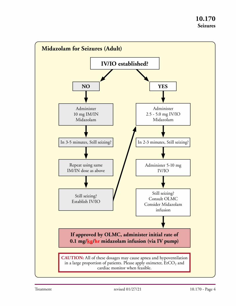
Treatment
10.170Seizures
revised 01/27/21 10.170 - Page 4
YES
In 2-3 minutes, Still seizing?In 3-5 minutes, Still seizing?
NO
Administer10 mg IM/INMidazolam
Repeat using same IM/IN dose as above
Still seizing?Establish IV/IO
Administer2.5 - 5.0 mg IV/IO
Midazolam
Administer 5-10 mg IV/IO
Still seizing?Consult OLMC
Consider Midazolam infusion
If approved by OLMC, administer initial rate of 0.1 mg/kg/hr midazolam infusion (via IV pump)
Midazolam for Seizures (Adult)
IV/IO established?
CAUTION: All of these dosages may cause apnea and hypoventilation in a large proportion of patients. Please apply oximeter, EtCO2 and
cardiac monitor when feasible.
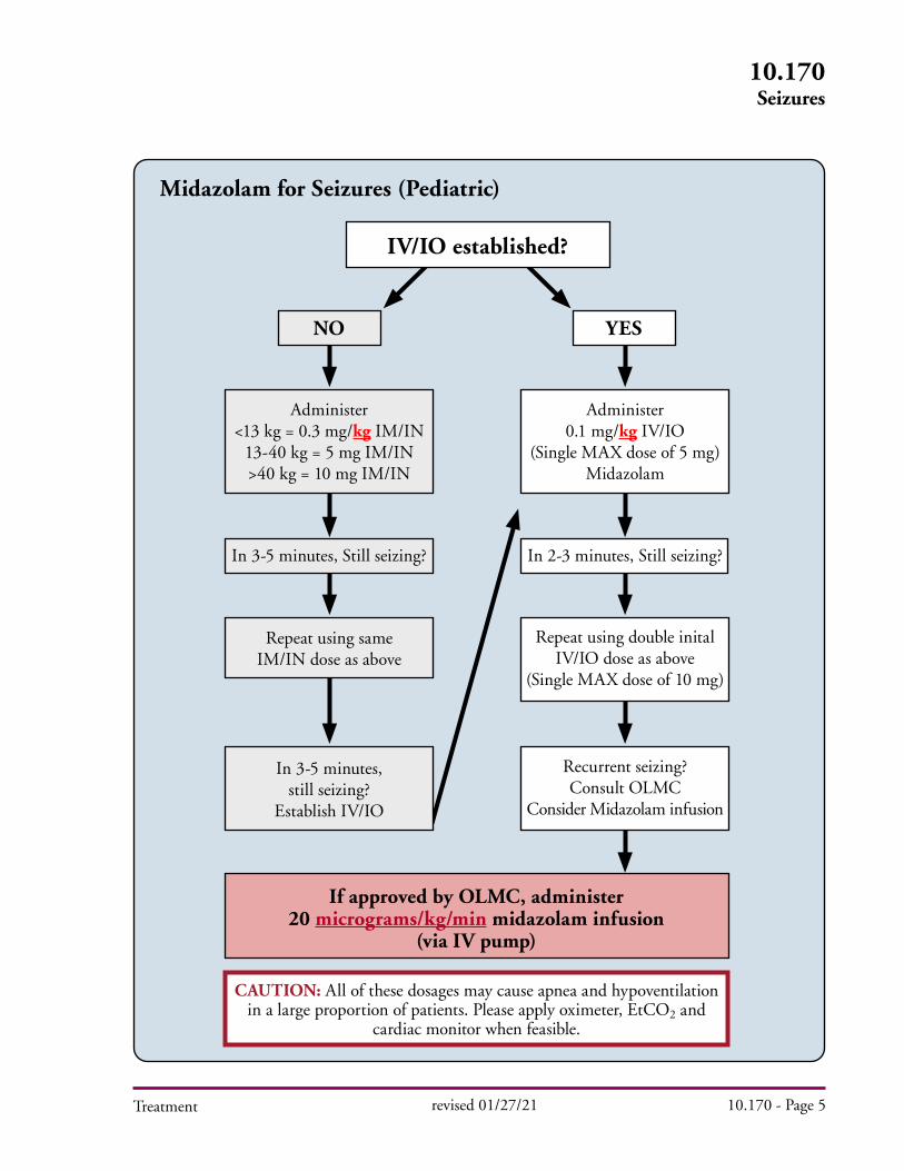
Treatment
10.170Seizures
revised 01/27/21 10.170 - Page 5
YESNO
Administer<13 kg = 0.3 mg/kg IM/IN
13-40 kg = 5 mg IM/IN>40 kg = 10 mg IM/IN
Repeat using sameIM/IN dose as above
In 3-5 minutes,still seizing?
Establish IV/IO
Administer0.1 mg/kg IV/IO
(Single MAX dose of 5 mg)Midazolam
Repeat using double inital IV/IO dose as above
(Single MAX dose of 10 mg)
Recurrent seizing?Consult OLMC
Consider Midazolam infusion
Midazolam for Seizures (Pediatric)
IV/IO established?
If approved by OLMC, administer 20 micrograms/kg/min midazolam infusion
(via IV pump)
CAUTION: All of these dosages may cause apnea and hypoventilation in a large proportion of patients. Please apply oximeter, EtCO2 and
cardiac monitor when feasible.
In 2-3 minutes, Still seizing?In 3-5 minutes, Still seizing?
Treatment
10.175Sepsis
Sepsis
HX PE DDX
Known or suspected infection
Weak immune system
Malnutrition
Recent surgery
Current indwelling catheter
Altered LOC
Hypotension
Hyperthermia
Tachycardia
Increased respiratory rate
Hypovolemia
Cardiogenic shock
Pulmonary embolus
Anaphylaxis
Adverse effect of medication
Adrenal Insufficiency
revised 11/29/18 10.175 - Page 1
Treatment: A. Perform primary and secondary exam. B. Maintain SpO2 above 95%. C. Establish large bore vascular access, if possible D. If MAP is >65 mmHg (SBP >90 mmHg) and septicemia is suspected, initiate 500-1000mL fluid bolus (10-20 ml/kg for pediatrics). E. If MAP is <65 mmHg (SBP < 90 mmHg), treat per SHOCK protocol: 1. Target is MAP > 65 mmHg F. Check blood glucose. G. Notify the receiving hospital with “Sepsis Alert” if all items below are present: 1. Suspected infection + 2. EtCO
2 is < 25mmHg
AND 3. Two or more qSOFA criteria
Notes and Precautions:A. Sepsis is a rapidly progressing, life threatening condition due to systemic infection. Sepsis must be recognized early and treated aggressively to prevent progression to shock and death.
B. The purpose of a “Sepsis Alert” is to provide pre-arrival emergency department notification in order to facilitate rapid assessment and treatment of a suspected severe sepsis patient.
C. For age appropriate pediatric vital signs, refer to Pediatric Guide.
qSOFAPhysical Exam ScoreHypotension 1 MAP <65 mmHg (SBP <90 mmHg)
Altered LOC 1
Tachypnea ≥ 22/min 1
Treatment
Shock
HX PE DDX
Trauma
Hemorrhage (external / internal)
Chest pain / SOB
Fever / sepsis
Allergy / anaphylaxis
Environmental (e.g. heat)
Dehydration
Fever
LOC
Capillary refill
Skin appearance (rash, swelling, trauma)
Neck veins
Lung sounds
Abdominal tenderness
Pelvis
Extremity fracture
Neuro
Hypovolemic
Cardiac
Septic
Anaphylactic
Obstructive (PE/pneumothorax/pericardial tamponade)
Spinal
Treatment: A. Do not delay transport. B. Start O
2, follow Airway Management procedure.
C. Start IV/IO. D. Determine type of shock and treat as follows: 1. Hypovolemic Shock: a. Control external bleeding with direct pressure, elevation, tourniquet, and/or hemostatic dressings. b. Administer 500-1000 mL fluid challenge to maintain a MAP > 65 mmHg (SBP > 90 mmHg). Repeat fluid boluses if continued signs of shock and no pulmonary edema. c. For shock secondary to trauma or suspected AAA, do not over resuscitate. Goal is MAP 55-65 mmHg (SBP 70-90 mmHg) for trauma patients without head trauma. For patients with head trauma, maintain MAP > 65 mmHg (SBP 90 mmHg) 2. Cardiogenic Shock: a. If suspected cardiac event follow Chest Pain protocol. b. Monitor cardiac rhythm and follow Cardiac Dysrhythmia protocol. c. Administer 250-500 mL fluid challenge to maintain a MAP > 65 mmHg (SBP of > 90 mmHg). Repeat once if continued signs of shock and no pulmonary edema. MAX of 1000 mL. d. Consider push dose epinephrine until norepinephrine infusion is administered.
revised 09/25/20 10.180 - Page 1
10.180Shock
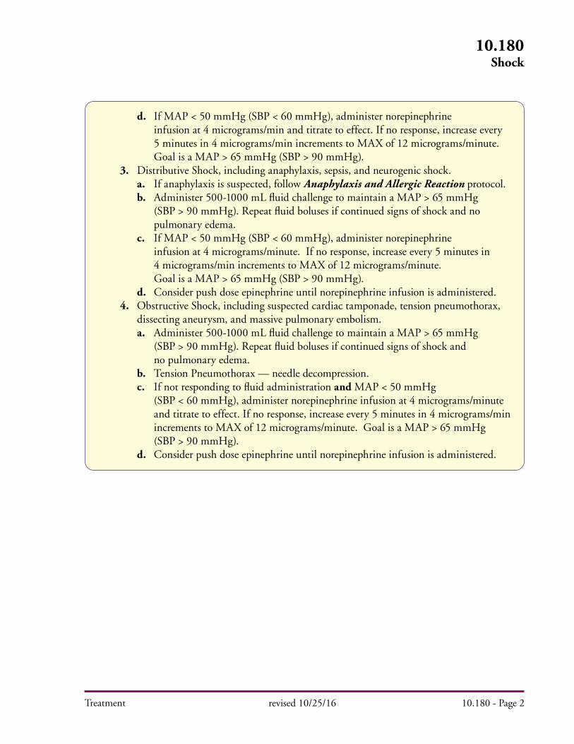
Treatment
d. If MAP < 50 mmHg (SBP < 60 mmHg), administer norepinephrine infusion at 4 micrograms/min and titrate to effect. If no response, increase every 5 minutes in 4 micrograms/min increments to MAX of 12 micrograms/minute. Goal is a MAP > 65 mmHg (SBP > 90 mmHg). 3. Distributive Shock, including anaphylaxis, sepsis, and neurogenic shock. a. If anaphylaxis is suspected, follow Anaphylaxis and Allergic Reaction protocol. b. Administer 500-1000 mL fluid challenge to maintain a MAP > 65 mmHg (SBP > 90 mmHg). Repeat fluid boluses if continued signs of shock and no pulmonary edema. c. If MAP < 50 mmHg (SBP < 60 mmHg), administer norepinephrine infusion at 4 micrograms/minute. If no response, increase every 5 minutes in 4 micrograms/min increments to MAX of 12 micrograms/minute. Goal is a MAP > 65 mmHg (SBP > 90 mmHg). d. Consider push dose epinephrine until norepinephrine infusion is administered. 4. Obstructive Shock, including suspected cardiac tamponade, tension pneumothorax, dissecting aneurysm, and massive pulmonary embolism. a. Administer 500-1000 mL fluid challenge to maintain a MAP > 65 mmHg (SBP > 90 mmHg). Repeat fluid boluses if continued signs of shock and no pulmonary edema. b. Tension Pneumothorax — needle decompression. c. If not responding to fluid administration and MAP < 50 mmHg (SBP < 60 mmHg), administer norepinephrine infusion at 4 micrograms/minute and titrate to effect. If no response, increase every 5 minutes in 4 micrograms/min increments to MAX of 12 micrograms/minute. Goal is a MAP > 65 mmHg (SBP > 90 mmHg). d. Consider push dose epinephrine until norepinephrine infusion is administered.
10.180Shock
revised 10/25/16 10.180 - Page 2
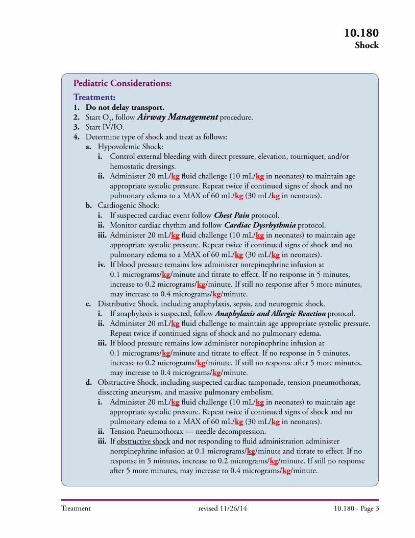
Treatment
10.180Shock
revised 11/26/14 10.180 - Page 3
Pediatric Considerations:
Treatment:1. Do not delay transport.2. Start O
2, follow Airway Management procedure.
3. Start IV/IO.4. Determine type of shock and treat as follows: a. Hypovolemic Shock: i. Control external bleeding with direct pressure, elevation, tourniquet, and/or hemostatic dressings. ii. Administer 20 mL/kg fluid challenge (10 mL/kg in neonates) to maintain age appropriate systolic pressure. Repeat twice if continued signs of shock and no pulmonary edema to a MAX of 60 mL/kg (30 mL/kg in neonates). b. Cardiogenic Shock: i. If suspected cardiac event follow Chest Pain protocol. ii. Monitor cardiac rhythm and follow Cardiac Dysrhythmia protocol. iii. Administer 20 mL/kg fluid challenge (10 mL/kg in neonates) to maintain age appropriate systolic pressure. Repeat twice if continued signs of shock and no pulmonary edema to a MAX of 60 mL/kg (30 mL/kg in neonates). iv. If blood pressure remains low administer norepinephrine infusion at 0.1 micrograms/kg/minute and titrate to effect. If no response in 5 minutes, increase to 0.2 micrograms/kg/minute. If still no response after 5 more minutes, may increase to 0.4 micrograms/kg/minute. c. Distributive Shock, including anaphylaxis, sepsis, and neurogenic shock. i. If anaphylaxis is suspected, follow Anaphylaxis and Allergic Reaction protocol. ii. Administer 20 mL/kg fluid challenge to maintain age appropriate systolic pressure. Repeat twice if continued signs of shock and no pulmonary edema. iii. If blood pressure remains low administer norepinephrine infusion at 0.1 micrograms/kg/minute and titrate to effect. If no response in 5 minutes, increase to 0.2 micrograms/kg/minute. If still no response after 5 more minutes, may increase to 0.4 micrograms/kg/minute. d. Obstructive Shock, including suspected cardiac tamponade, tension pneumothorax, dissecting aneurysm, and massive pulmonary embolism. i. Administer 20 mL/kg fluid challenge (10 mL/kg in neonates) to maintain age appropriate systolic pressure. Repeat twice if continued signs of shock and no pulmonary edema to a MAX of 60 mL/kg (30 mL/kg in neonates). ii. Tension Pneumothorax — needle decompression. iii. If obstructive shock and not responding to fluid administration administer norepinephrine infusion at 0.1 micrograms/kg/minute and titrate to effect. If no response in 5 minutes, increase to 0.2 micrograms/kg/minute. If still no response after 5 more minutes, may increase to 0.4 micrograms/kg/minute.
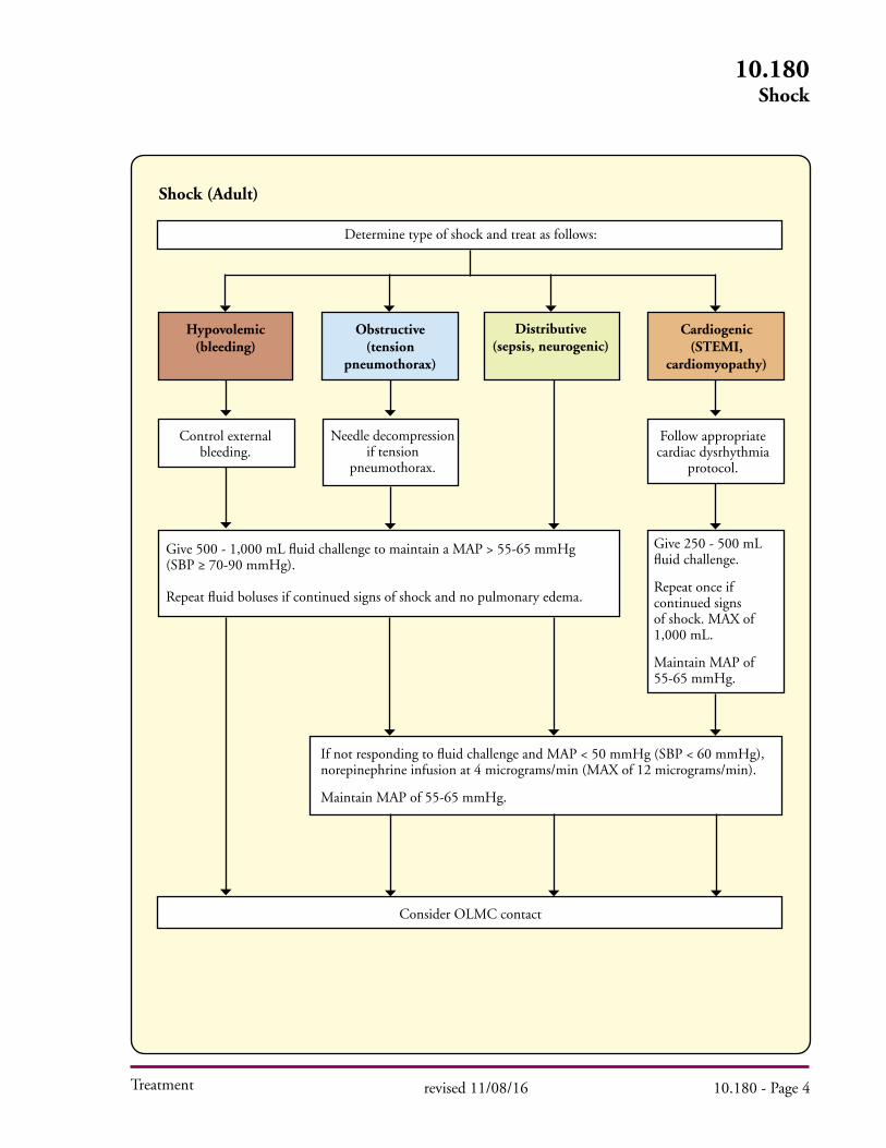
Treatment
10.180Shock
revised 11/08/16 10.180 - Page 4
Hypovolemic (bleeding)
Cardiogenic(STEMI,
cardiomyopathy)
Distributive(sepsis, neurogenic)
Determine type of shock and treat as follows:
Control external bleeding.
Follow appropriate cardiac dysrhythmia
protocol.
Give 500 - 1,000 mL fluid challenge to maintain a MAP > 55-65 mmHg (SBP ≥ 70-90 mmHg).
Repeat fluid boluses if continued signs of shock and no pulmonary edema.
Give 250 - 500 mL fluid challenge.
Repeat once if continued signs of shock. MAX of 1,000 mL.
Maintain MAP of 55-65 mmHg.
If not responding to fluid challenge and MAP < 50 mmHg (SBP < 60 mmHg), norepinephrine infusion at 4 micrograms/min (MAX of 12 micrograms/min).
Maintain MAP of 55-65 mmHg.
Consider OLMC contact
Shock (Adult)
Needle decompression if tension
pneumothorax.
Obstructive (tension
pneumothorax)

Treatment
10.180Shock
revised 11/26/14 10.180 - Page 5
Determine type of shock and treat as follows:
Follow appropriate cardiac dysrhythmia
protocol.
Give 20 mL/kg fluid challenge (10 mL/kg in neonates) to maintain age appropriate systolic pressure. Repeat twice if continued signs of shock and no pulmonary edema to a MAX of 60 mL/kg (30 mL/kg in neonates).
Consider OLMC contact
Lowest normal pediatric pressure by age:i. Less than one month: > 60 mmHg.ii. One month to 1 year: > 70 mmHg.iii. Greater than 1 year: 70 + 2x age in years.
Shock (Pediatric)
Needle decompression if tension
pneumothorax.
Cardiogenic(STEMI,
cardiomyopathy)
Distributive(sepsis, neurogenic)
Control external bleeding
Hypovolemic (bleeding)
Obstructive (tension
pneumothorax)
If not responding to fluid challenge, norepinephrine infusion at 0.1 micrograms/kg/minute. If no response in 5 min, increase to 0.2 micrograms/kg/minute. After 5 more min may increase to 0.4 micrograms/kg/minute.
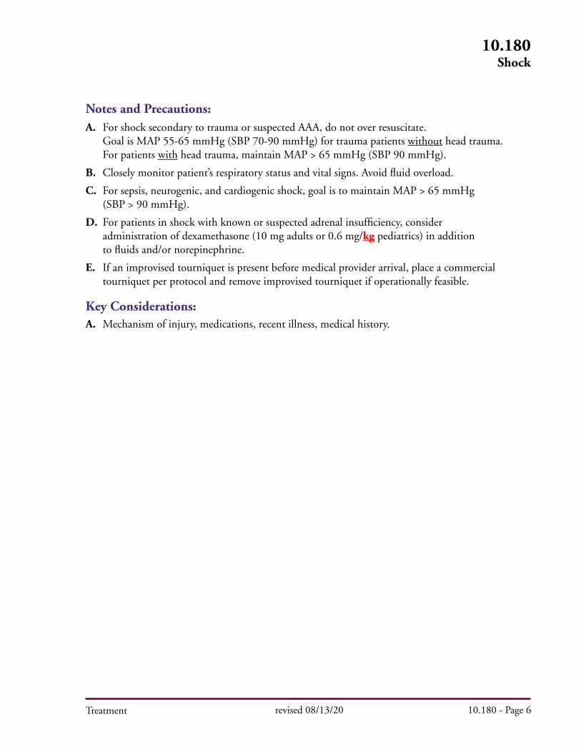
Treatment
10.180Shock
revised 08/13/20 10.180 - Page 6
Notes and Precautions:A. For shock secondary to trauma or suspected AAA, do not over resuscitate. Goal is MAP 55-65 mmHg (SBP 70-90 mmHg) for trauma patients without head trauma. For patients with head trauma, maintain MAP > 65 mmHg (SBP 90 mmHg).
B. Closely monitor patient’s respiratory status and vital signs. Avoid fluid overload.
C. For sepsis, neurogenic, and cardiogenic shock, goal is to maintain MAP > 65 mmHg (SBP > 90 mmHg).
D. For patients in shock with known or suspected adrenal insufficiency, consider administration of dexamethasone (10 mg adults or 0.6 mg/kg pediatrics) in addition to fluids and/or norepinephrine.
E. If an improvised tourniquet is present before medical provider arrival, place a commercial tourniquet per protocol and remove improvised tourniquet if operationally feasible.
Key Considerations:A. Mechanism of injury, medications, recent illness, medical history.
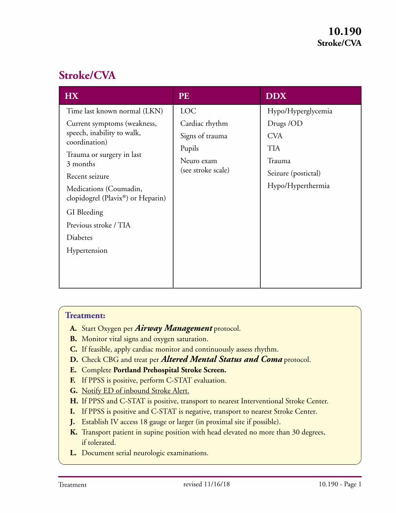
Treatment revised 11/16/18 10.190 - Page 1
10.190Stroke/CVA
Treatment: A. Start Oxygen per Airway Management protocol. B. Monitor vital signs and oxygen saturation. C. If feasible, apply cardiac monitor and continuously assess rhythm. D. Check CBG and treat per Altered Mental Status and Coma protocol. E. Complete Portland Prehospital Stroke Screen. F. If PPSS is positive, perform C-STAT evaluation. G. Notify ED of inbound Stroke Alert. H. If PPSS and C-STAT is positive, transport to nearest Interventional Stroke Center. I. If PPSS is positive and C-STAT is negative, transport to nearest Stroke Center. J. Establish IV access 18 gauge or larger (in proximal site if possible). K. Transport patient in supine position with head elevated no more than 30 degrees, if tolerated. L. Document serial neurologic examinations.
Stroke/CVA
HX PE DDX
Time last known normal (LKN)
Current symptoms (weakness, speech, inability to walk, coordination)
Trauma or surgery in last 3 months
Recent seizure
Medications (Coumadin, clopidogrel (Plavix) or Heparin)
GI Bleeding
Previous stroke / TIA
Diabetes
Hypertension
LOC
Cardiac rhythm
Signs of trauma
Pupils
Neuro exam (see stroke scale)
Hypo/Hyperglycemia
Drugs /OD
CVA
TIA
Trauma
Seizure (postictal)
Hypo/Hyperthermia
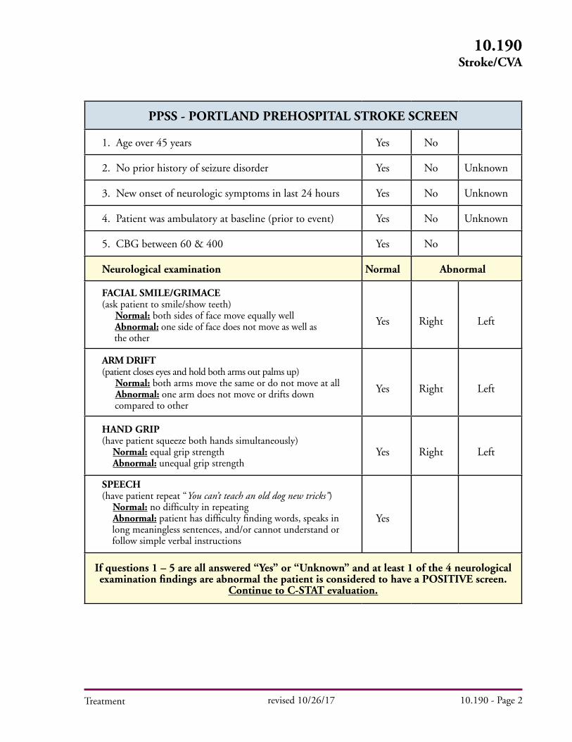
Treatment revised 10/26/17 10.190 - Page 2
10.190Stroke/CVA
PPSS - PORTLAND PREHOSPITAL STROKE SCREEN
1. Age over 45 years Yes No
2. No prior history of seizure disorder Yes No Unknown
3. New onset of neurologic symptoms in last 24 hours Yes No Unknown
4. Patient was ambulatory at baseline (prior to event) Yes No Unknown
5. CBG between 60 & 400 Yes No
Neurological examination Normal Abnormal
FACIAL SMILE/GRIMACE(ask patient to smile/show teeth) Normal: both sides of face move equally well Abnormal: one side of face does not move as well as the other
Yes Right Left
ARM DRIFT(patient closes eyes and hold both arms out palms up) Normal: both arms move the same or do not move at all Abnormal: one arm does not move or drifts down compared to other
Yes Right Left
HAND GRIP(have patient squeeze both hands simultaneously) Normal: equal grip strength Abnormal: unequal grip strength
Yes Right Left
SPEECH(have patient repeat “You can’t teach an old dog new tricks”) Normal: no difficulty in repeating Abnormal: patient has difficulty finding words, speaks in long meaningless sentences, and/or cannot understand or follow simple verbal instructions
Yes
If questions 1 – 5 are all answered “Yes” or “Unknown” and at least 1 of the 4 neurological examination findings are abnormal the patient is considered to have a POSITIVE screen.
Continue to C-STAT evaluation.
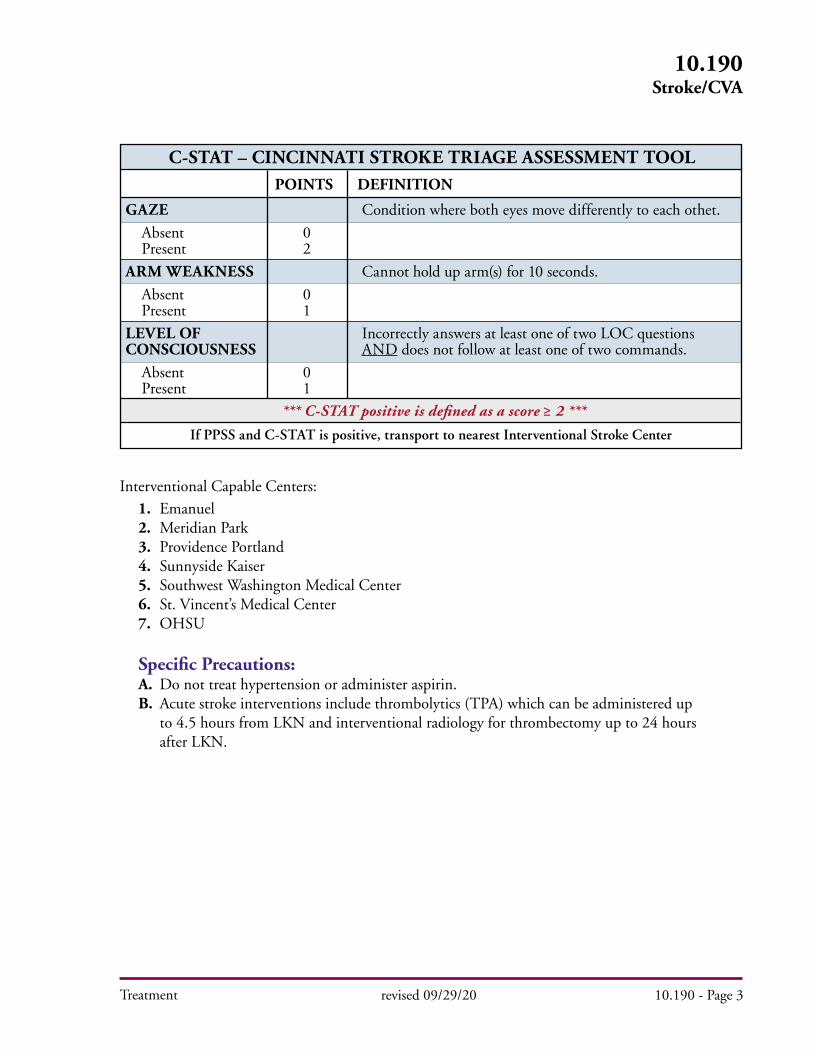
Treatment
Interventional Capable Centers: 1. Emanuel 2. Meridian Park 3. Providence Portland 4. Sunnyside Kaiser 5. Southwest Washington Medical Center 6. St. Vincent’s Medical Center 7. OHSU
Specific Precautions: A. Do not treat hypertension or administer aspirin. B. Acute stroke interventions include thrombolytics (TPA) which can be administered up to 4.5 hours from LKN and interventional radiology for thrombectomy up to 24 hours after LKN.
GAZE Condition where both eyes move differently to each othet.
Absent 0 Present 2
ARM WEAKNESS Cannot hold up arm(s) for 10 seconds.
Absent 0 Present 1
LEVEL OF Incorrectly answers at least one of two LOC questions CONSCIOUSNESS AND does not follow at least one of two commands.
Absent 0 Present 1
*** C-STAT positive is defined as a score ≥ 2 ***
POINTS DEFINITION
C-STAT – CINCINNATI STROKE TRIAGE ASSESSMENT TOOL
revised 09/29/20 10.190 - Page 3
10.190Stroke/CVA
If PPSS and C-STAT is positive, transport to nearest Interventional Stroke Center
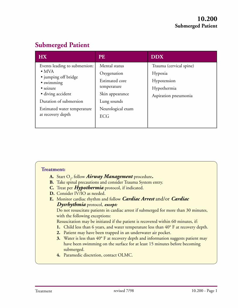
Treatment revised 7/98 10.200 - Page 1
10.200Submerged Patient
Treatment: A. Start O
2, follow Airway Management procedure.
B. Take spinal precautions and consider Trauma System entry. C. Treat per Hypothermia protocol, if indicated. D. Consider IV/IO as needed.
E. Monitor cardiac rhythm and follow Cardiac Arrest and/or Cardiac Dysrhythmia protocol, except:
Do not resuscitate patients in cardiac arrest if submerged for more than 30 minutes, with the following exceptions: Resuscitation may be initiated if the patient is recovered within 60 minutes, if: 1. Child less than 6 years, and water temperature less than 40° F at recovery depth. 2. Patient may have been trapped in an underwater air pocket. 3. Water is less than 40° F at recovery depth and information suggests patient may have been swimming on the surface for at least 15 minutes before becoming submerged. 4. Paramedic discretion, contact OLMC.
Submerged Patient
HX PE DDX
Events leading to submersion: • MVA • jumping off bridge • swimming • seizure • diving accident
Duration of submersion
Estimated water temperature at recovery depth
Mental status
Oxygenation
Estimated core temperature
Skin appearance
Lung sounds
Neurological exam
ECG
Trauma (cervical spine)
Hypoxia
Hypotension
Hypothermia
Aspiration pneumonia
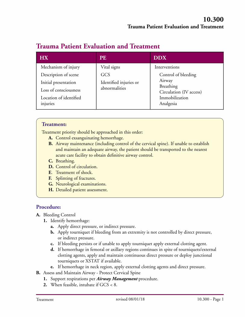
Treatment
HX PE DDX
Mechanism of injury
Description of scene
Initial presentation
Loss of consciousness
Location of identified injuries
Vital signs
GCS
Identified injuries or abnormalities
Interventions
Control of bleeding Airway Breathing Circulation (IV access) Immobilization Analgesia
10.300Trauma Patient Evaluation and Treatment
revised 08/01/18 10.300 - Page 1
Procedure:A. Bleeding Control 1. Identify hemorrhage: a. Apply direct pressure, or indirect pressure. b. Apply tourniquet if bleeding from an extremity is not controlled by direct pressure, or indirect pressure. c. If bleeding persists or if unable to apply tourniquet apply external clotting agent. d. If hemorrhage in femoral or axillary regions continues in spite of tourniquets/external clotting agents, apply and maintain continuous direct pressure or deploy junctional tourniquets or XSTAT if available. e. If hemorrhage in neck region, apply external clotting agents and direct pressure.B. Assess and Maintain Airway - Protect Cervical Spine 1. Support respirations per Airway Management procedure. 2. When feasible, intubate if GCS < 8.
Treatment:Treatment priority should be approached in this order: A. Control exsanguinating hemorrhage. B. Airway maintenance (including control of the cervical spine). If unable to establish and maintain an adequate airway, the patient should be transported to the nearest acute care facility to obtain definitive airway control. C. Breathing. D. Control of circulation. E. Treatment of shock. F. Splinting of fractures. G. Neurological examinations. H. Detailed patient assessment.
Trauma Patient Evaluation and Treatment
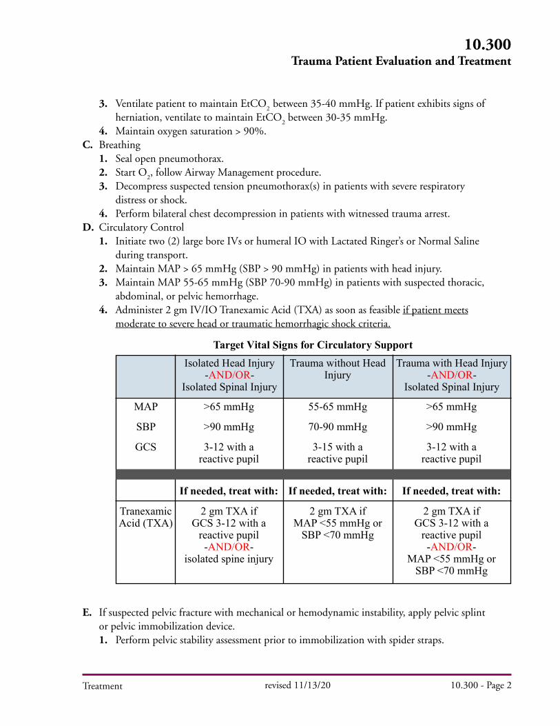
Treatment
3. Ventilate patient to maintain EtCO2
between 35-40 mmHg. If patient exhibits signs of herniation, ventilate to maintain EtCO
2 between 30-35 mmHg.
4. Maintain oxygen saturation > 90%.C. Breathing 1. Seal open pneumothorax. 2. Start O
2, follow Airway Management procedure.
3. Decompress suspected tension pneumothorax(s) in patients with severe respiratory distress or shock. 4. Perform bilateral chest decompression in patients with witnessed trauma arrest.D. Circulatory Control 1. Initiate two (2) large bore IVs or humeral IO with Lactated Ringer’s or Normal Saline during transport. 2. Maintain MAP > 65 mmHg (SBP > 90 mmHg) in patients with head injury. 3. Maintain MAP 55-65 mmHg (SBP 70-90 mmHg) in patients with suspected thoracic, abdominal, or pelvic hemorrhage. 4. Administer 2 gm IV/IO Tranexamic Acid (TXA) as soon as feasible if patient meets moderate to severe head or traumatic hemorrhagic shock criteria.
E. If suspected pelvic fracture with mechanical or hemodynamic instability, apply pelvic splint or pelvic immobilization device. 1. Perform pelvic stability assessment prior to immobilization with spider straps.
10.300Trauma Patient Evaluation and Treatment
revised 11/13/20 10.300 - Page 2
Target Vital Signs for Circulatory SupportIsolated Head Injury
-AND/OR- Isolated Spinal Injury
Trauma without Head Injury
Trauma with Head Injury-AND/OR-
Isolated Spinal Injury
MAP
SBP
GCS
Tranexamic Acid (TXA)
>65 mmHg
>90 mmHg
3-12 with a reactive pupil
If needed, treat with:
2 gm TXA if GCS 3-12 with a
reactive pupil -AND/OR-
isolated spine injury
55-65 mmHg
70-90 mmHg
3-15 with a reactive pupil
If needed, treat with:
2 gm TXA if MAP <55 mmHg or
SBP <70 mmHg
>65 mmHg
>90 mmHg
3-12 with a reactive pupil
If needed, treat with:
2 gm TXA if GCS 3-12 with a
reactive pupil -AND/OR-
MAP <55 mmHg or SBP <70 mmHg

Note: • In a situation where the mechanism of injury appears inconsistent with the patient’s and not severe enough to induce traumatic arrest, consider a primary medical cause for the patient’s cardiac arrest. • Traumatic arrest is approached in a distinct fashion from medical cardiac arrest. TXA is not for cardiac or traumatic arrest. • DO NOT allow chest compressions to interfere with addressing the reversible causes of the arrest (such as needle decompression). • If the patient is being transported undergoing CPR, the use of a mechanical CPR device is appropriate when available. • Consider early transport for patients with pregnancy > 22 weeks. Notify receiving hospital that patient is pregnant. • If an improvised tourniquet is present before medical provider arrival, place a commercial tourniquet per protocol and remove improvised tourniquet if operationally feasible. • Suspected isolated spinal cord injury (i.e. Neurogenic Shock - injury to spinal cord alone with motor loss and hypotension) should be treated with vasopressors rather than volume and/or TXA. (Ex: diving injury with suspected cervical spine injury)(Ex: diving injury with suspected cervical spine injury). • Isolated falls from standing most frequently results in head injury and not hemorrhagic shock.
F. Moderate to Severe Head Trauma (GCS 3-12 with a reactive pupil) 1. Administer 2 gm IV/IO Tranexamic Acid (TXA) as soon as feasible.
G. Perform fracture immobilization per protocol. 1. For isolated penetrating head, neck, or torso trauma, immobilization of the cervical spine is unnecessary unless there is overt neurologic deficit or an adequate physical examination cannot be performed, e.g., a patient with altered mental status or a patient with distracting injury.H. Prevent hypothermia. 1. If a patient deteriorates to cardiac arrest during transport, perform CPR and notify the trauma facility.
Treatment
10.300Trauma Patient Evaluation and Treatment
revised 11/13/20 10.300 - Page 3
Inclusion Criteria Exclusion Criteria
- Adult ≥15 years old- ≥ 50 kg if age unknown- Blunt or penetrating injury- ≤ 1 hour from of injury- IV/IO establishedPhysiological criteria:- GCS between 3 - 12 with a reactive pupil
-AND/OR-- MAP < 55 mmHg (SBP < 70 mmHg)
- GCS = 3 with no reactive pupil- >1 hour from time of injury or time unknown- Any chest compressions (manual or mechanical)- Seizures, MI, Stroke, or Dialysis (head injury only)- Known or suspected pregnancy- Drowning- Hanging