Embed Size (px)
Citation preview
2017
October 4
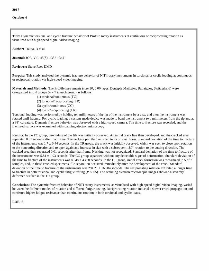
Title: Dynamic torsional and cyclic fracture behavior of ProFile rotary instruments at continuous or reciprocating rotation as
visualized with high-speed digital video imaging
Author: Tokita, D et al.
Journal: JOE, Vol. 43(8): 1337-1342
Reviewer: Steve Rees DMD
Purpose: This study analyzed the dynamic fracture behavior of NiTi rotary instruments in torsional or cyclic loading at continuous
or reciprocal rotation via high-speed video imaging
Materials and Methods: The ProFile instruments (size 30, 0.06 taper; Dentsply Maillefer, Ballaigues, Switzerland) were
categorized into 4 groups (n = 7 in each group) as follows:
(1) torsional/continuous (TC)
(2) torsional/reciprocating (TR)
(3) cyclic/continuous (CC)
(4) cyclic/reciprocating (CR)
Torsional loading was performed by holding ten millimeters of the tip of the instrument by a vise, and then the instrument was
rotated until fracture. For cyclic loading, a custom-made device was made to bend the instrument two millimeters from the tip and at
a 38° curvature. Dynamic fracture behavior was observed with a high-speed camera. The time to fracture was recorded, and the
fractured surface was examined with scanning electron microscopy.
Results: In the TC group, unwinding of the file was initially observed. An initial crack line then developed, and the cracked area
separated 0.01 seconds after that frame. The necking part then returned to its original form. Standard deviation of the time to fracture
of the instruments was 1.7 0.44 seconds. In the TR group, the crack was initially observed, which was seen to close upon rotation
to the noncutting direction and to open again and increase in size with a subsequent 180 rotation to the cutting direction. The
cracked area then separated 0.01 seconds after that frame. Necking was not recognized. Standard deviation of the time to fracture of
the instruments was 5.81 1.93 seconds. The CC group separated without any detectable signs of deformation. Standard deviation of
the time to fracture of the instruments was 80.48 43.60 seconds. In the CR group, initial crack formation was recognized in 5 of 7
samples, and, in these cracked specimens, file separation occurred immediately after the development of the crack. Standard
deviation of the time to fracture of the instruments was 294.21 168.04 seconds. The reciprocating rotation exhibited a longer time
to fracture in both torsional and cyclic fatigue testing (P < .05). The scanning electron microscopic images showed a severely
deformed surface in the TR group.
Conclusion: The dynamic fracture behavior of NiTi rotary instruments, as visualized with high-speed digital video imaging, varied
between the different modes of rotation and different fatigue testing. Reciprocating rotation induced a slower crack propagation and
conferred higher fatigue resistance than continuous rotation in both torsional and cyclic loads.
LOE: 5
Title: Assessment of a cavity to optimize ultrasonic efficiency to remove intraradicular posts
Author: Graca I et al.
Journal: JOE Vol. 43 (8): 1350-1353.
Reviewed by: Stephanie Serrano DDS
Purpose: The study assessed an in vitro protocol for the removal of cast metal posts using ultrasonic vibration in multirooted teeth
by drilling a cavity in the coronal portion of the post followed by ultrasound application in the cavity.
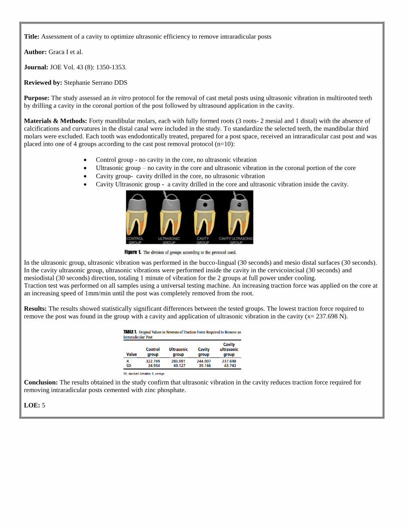
Materials & Methods: Forty mandibular molars, each with fully formed roots (3 roots- 2 mesial and 1 distal) with the absence of
calcifications and curvatures in the distal canal were included in the study. To standardize the selected teeth, the mandibular third
molars were excluded. Each tooth was endodontically treated, prepared for a post space, received an intraradicular cast post and was
placed into one of 4 groups according to the cast post removal protocol (n=10):
Control group - no cavity in the core, no ultrasonic vibration
Ultrasonic group – no cavity in the core and ultrasonic vibration in the coronal portion of the core
Cavity group- cavity drilled in the core, no ultrasonic vibration
Cavity Ultrasonic group - a cavity drilled in the core and ultrasonic vibration inside the cavity.
In the ultrasonic group, ultrasonic vibration was performed in the bucco-lingual (30 seconds) and mesio distal surfaces (30 seconds).
In the cavity ultrasonic group, ultrasonic vibrations were performed inside the cavity in the cervicoincisal (30 seconds) and
mesiodistal (30 seconds) direction, totaling 1 minute of vibration for the 2 groups at full power under cooling.
Traction test was performed on all samples using a universal testing machine. An increasing traction force was applied on the core at
an increasing speed of 1mm/min until the post was completely removed from the root.
Results: The results showed statistically significant differences between the tested groups. The lowest traction force required to
remove the post was found in the group with a cavity and application of ultrasonic vibration in the cavity (x= 237.698 N).
Conclusion: The results obtained in the study confirm that ultrasonic vibration in the cavity reduces traction force required for
removing intraradicular posts cemented with zinc phosphate.
LOE: 5
Title: Pyrosequencing analysis of cryogenically ground samples from primary and secondary/persistent endodontic infections
Author: Keskin, C et al.
Source: Journal of Endodontics, Volume 43(8):1309 – 1316
Reviewer: Parth Karia DMD
Purpose: This report characterized the microbial communities of primary endodontic infections (PEI) and secondary/persistent
endodontic infections (SPEI) using high-throughput pyrosequencing from the pulverized samples
Materials & Methods: Inclusion criteria: patients whose teeth were planned for extraction because of prosthetic reasons or their
refusal for endodontic treatment or nonsurgical endodontic retreatment. Exclusion criteria: patients with systemic disorders,
progressive periodontal disease, pregnancy/lactation, or those receiving antibiotic treatment in last 3 months. PEI group: 20 teeth
showing primary endodontic infection with a diagnosis of asymptomatic apical periodontitis or a chronic apical abscess. SPEI
group: 20 teeth that were treated with RCT at least 2 years earlier and showed PARL with PEI and were diagnosed as post-
treatment apical periodontitis. Two endodontists verified all periapical lesions and only selected teeth with PAI > 3. All samples
had intact coronal restoration that had no exposure of obturation material to the oral cavity. Control: a 1st premolar tooth, with no
caries, restoration, pulpal or periapical pathology that was planned for extraction for orthodontic purpose. The procedures were as
follows:
Rinse mouth with 0.12% Chlorhexidine gluconate mouthwash
Atraumatic extraction under strict aseptic conditions and flushed with sterile saline
Attached soft tissue removed with a sterile #15 periodontal curette
Outer tooth surface irrigated with 30% H2O2, followed by 5.25% NaOCl and 5% Sodium thiosulfate
Teeth decoronated with sterile diamond discs and stored at -20°Celsius
All samples were transferred to polycarbonate vials, immersed in liquid nitrogen and crushed in a cryogenic
grinder
These root powders were then preincubated with a lysis buffer to obtain optimal DNA yield
Extracted DNA were eluted with 50 µl elution buffer and quantified via a NanoDrop spectrophotometer and
Qubit
Pyrosequencing was performed and identification of the bacteria and archae was done through the database of the
National Center for Biotechnology Information
Results: Detection included 15 phyla, 160 genera and 368 species. No significant difference between PEI and SPEI was found
regarding the diversity and richness of operational taxonomic units at the phyla, genera, and species levels. A novel archael species,
Candidatus Nitrosoarchaeum limnia from the Thaumarchaeota phylum, was detected for the first time in root canals of teeth with
PEI.
Conclusion: SPEI has a microbial community as diverse as that of PEI
LOE: 5
Title: Antibacterial properties of chitosan nanoparticles and propolis associated with calcium hydroxide against single- and
multispecies biofilms: An in vitro and in situ study
Author: del Carpio-Perochena A et al.
Journal: JOE Vol 43(8): 1332-1336
Reviewer: Lauren Shin, DDS
Purpose: Propolis is a product of honeybees, which is known for its antibacterial, antifungal, and healing properties. Chitosan is a
nontoxic cationic natural biopolymer that has significant antibacterial activity. The aim of this study was to evaluate the efficacy of
chitosan nanoparticles (CNPs) and ethanolic propolis extract (EPE) incorporated into calcium hydroxide paste to kill bacterial
biofilms.
Materials and Methods:
First stage: In Vitro Study: Mandibular premolars were instrumented and irrigated, sectioned into 40 4mm discs and autoclaved.
Each sample was placed in a tube and inoculated with E. faecalis suspension and centrifuged 4 times and then incubated for 21
days. The samples were divided into 4 groups (n=10) for different treatments and then incubated for 7 and 14 days:
Group 1 – distilled water
Group 2 – Calcium hydroxide
Group 3 – Calcium hydroxide + CNPs
Group 4 – Calcium hydroxide + EPE
After the incubation periods, the colony forming units (CFUs) from the dentinal shavings post treatment were calculated. The pH of
the experimental pastes was also measured at 0, 1, 2, 7, and 14 days.
Second Stage: In Situ Study: Thirty-two sterile human dentin blocks were fixed into a palatal orthodontic device to allow intraoral
dentin infection for 48 hours. The samples were divided into the same treatment groups as the in vitro study and were immersed in
the pastes and incubated for 7 and 14 days (n=8). After the incubations periods, the remaining biofilm was dyed to visualize live
and dead bacteria with a multichannel confocal microscope.
Results:
First stage: In Vitro Study: No significant pH variation of pastes through time. The averages for Groups 2, 3, and 4, were 11.35,
11.58, and 11.4 respectively. Log CFU/mL:
Group 1: Control – 8.61 (7 days) and 8.0 (14 days)
Group 2: Ca(OH)2 – 2.04 (7 days) and 2.52 (14 days)
Group 3: Ca(OH)2 + CNP – 0.49 (7 days) and 0.09 (14 days)
Group 4: Ca(OH)2 + EPE – 1.36 (7 days) and 2.7 (14 days)
Second Stage: In Situ Study: Unlike the first stage, the Ca(OH)2 groups did not decrease the percentage of live cells (65.77% at 7
days and 62.87% at 14 days) compared with control (96.1% P>0.5). Ca(OH)2 + CNP group showed highest percentages of
bacterial death; 30.73% at 7 days to 12.23% at 14 days. The Ca(OH)2 + EPE group showed significant antibacterial
activity in 7 days (23.93%) but the percentage raised after 14 days (59.34%).
Conclusion: Incorporating CNPs into calcium hydroxide paste has the potential of increasing its antibacterial activity. Calcium
hydroxide with EPE paste was not able to maintain its antibacterial efficacy through time
LOE: 5
Title: Detectability of middle mesial root canal orifices by troughing technique in mandibular molars: A micro-computed
tomographic study
Authors: Keles A et. al.
Journal: Journal of Endodontics, Vol. 43(8):1329
Reviewer: Ruoxue Feng DMD
Purpose: Middle mesial canals (MMCs): also known as accessory mesial or mesiocentral root canals are additional mesial root
canal between the mesiobuccal and mesiolingual canals. Their location is deep within the isthmus or a developmental groove
between orifices of the mesiobuccal and mesiolingual root canals. They appear 0.26 – 46.2%, depending on ethnicities and ages of
the patients. Azim AA et al (2015): talked about a procedure that aided in finding the MMCs by preparing a groove with a 2mm
depth and 1 mm diameter in the mesioapical direction between the orifices of the mesiobuccal and mesiolingual root canals
To measure the orifice depth of middle mesial canals (MMCs) and evaluate the detectability of orifices using troughing preparation
with the aid of micro-CT imaging at different troughing levels
Materials and Methods: Mesial roots of mandibular molars were mounted and scanned on a high-resolution micro-CT system.
Slices of the image were obtained with an 11-megapixel camera. Scanning was performed with 180 rotation around the vertical
axis with a camera. Data was reconstructed using NRecon software. CTAn and Data Viewer software were used to present the root
canal configuration of each root. Eighty-five roots showing MMCs were selected for studying. MMC configurations were classified
according to Pomeranz et al:
Independent: where there were 3 independent root canals extending from the chamber to the root apex.
Confluent: when an MMC joined either the MB or ML root canals in its trajectory toward the apex.
Fin: when the MMC orifice was connected to either the MB or ML canal orifices but ended as separate foramina.
The distance between MMC orifices and CEJs was measured. The number and percentage of frequencies of each configuration
were calculated. Chi-square test was used to calculate the significance level at 5%.
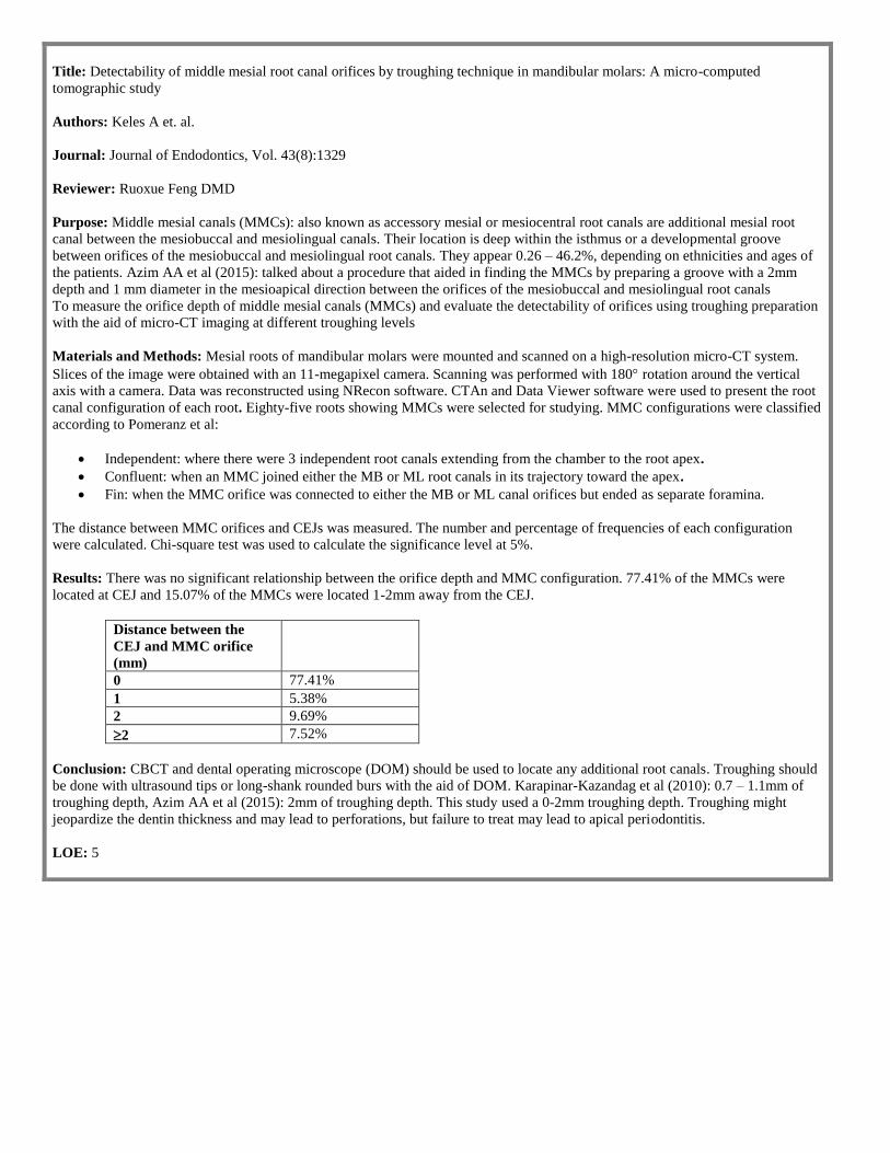
Results: There was no significant relationship between the orifice depth and MMC configuration. 77.41% of the MMCs were
located at CEJ and 15.07% of the MMCs were located 1-2mm away from the CEJ.
Distance between the
CEJ and MMC orifice
(mm)
0 77.41%
1 5.38%
2 9.69%
2 7.52%
Conclusion: CBCT and dental operating microscope (DOM) should be used to locate any additional root canals. Troughing should
be done with ultrasound tips or long-shank rounded burs with the aid of DOM. Karapinar-Kazandag et al (2010): 0.7 – 1.1mm of
troughing depth, Azim AA et al (2015): 2mm of troughing depth. This study used a 0-2mm troughing depth. Troughing might
jeopardize the dentin thickness and may lead to perforations, but failure to treat may lead to apical periodontitis.
LOE: 5
October 11
Title: A comparative study of ProTaper Universal and ProTaper Next used by undergraduate students to prepare root canals
Author: Aleman A, et al.
Journal: JOE, Vol. 43(8):1364
Reviewer: Rachel Mitrani DDS
Purpose: ProTaper Next® (PTN) is the latest variation of the ProTaper® rotary systems, and is made of M-Wire. Tulsa Dentsply
claims that files using M-Wire NiTi feature deeper flutes and smaller core diameters which adds to overall flexibility, yet still
demonstrates a greater resistance to cyclic fatigue. The ProTaper Universal® (PTU) system is made of conventional NiTi alloy.
Additionally, PTN has a cross section of an off-center rectangular design that allows for a “swaggering movement” of the file
during rotation. This study determined whether final-year undergraduate dental students achieved better shaping outcomes using the
ProTaper Next® system to prepare root canals for the first time compared with the existing ProTaper Universal® system on which
they had been trained.
Materials & Methods Each student prepared two simulated S-shaped canals contained within endodontic training blocks with an
apical diameter of 0.15mm and 0.02 taper. The first block was prepared with PTN, the second with PTU. Alcohol was used for
canal irrigation, and SlickGel was used as a lubricant. Preparation with PTU was completed up to F2 (size 25, 0.08 taper), whereas
preparation with PTN was completed to X2 (size 25m, 0.06 taper. Pre-op, intra-op, and post-op images of the canals were acquired
using a video camera imagine system. For PTU, an additional image was taken after use of the F1 file. Measurements were taken of
the amount of resin removed 1mm from preparation end-point, inner side of apical curvature (Max-AC), and the minimal canal
width between the two curvatures (Min-W). An assessment was made of the presence and location of canal aberrations (ledges).
A questionnaire was administered to each student regarding their opinions and preferences of each of the two rotary systems.
Results: The amount of resin removed at Max-AC was significantly larger with PTN. The Min-W values were significantly larger
for PTU compared with PTN. All of the canals without ledges were over-instrumented beyond the determined working length.
Significantly more ledges were created with PTU compared with PTN (30% vs. 0%). Canal transportation occurred in 5 canals with
PTU and 8 canals with PTN. The mean preparation time of PTU was significantly longer than PTN (12 vs. 8 minutes). More middle
constrictions were created with PTN compared to PTU. The middle constriction refers to an elbow-like aberration resulting from
the tendency of instruments to straighten curved root canals. The students perceived that the number of files of the PTU system was
significantly higher than that of PTN. The students recommended the use of PTN over PTU.
Discussion: A greater number of canal ledges occurred when PTU was used, compared to PTN. This difference can be attributed
to instrument flexibility. PTN is made of M-Wire, which is more flexible, while PTU is made of NiTi. Additionally, PTU has a
convex triangular cross-section and PTN has a rectangular cross section. The greater taper of the PTU system combined with its
alloy composition and cross-sectional shape result in a stiffer instrument that is more prone to creating ledges. As for student
preferences, there was a higher level of perceived difficulty when using PTU compared with PTN. More students selected PTN as
the system they would use in the future, and stated reasons such as “decreased preparation time” and “lower number of files.”
Conclusion: A further study should seek to evaluate patient satisfaction and clinical outcomes in relation to board certification.
LOE: 5
Title: Free active chlorine in sodium hypochlorite solutions admixed with octenidine, SmearOFF, chlorhexidine, and EDTA.
Author: Krishnan U et al.
Journal: JOE, Vol.43(8):1354-1359.
Reviewer: Reza Akahaven DMD
Purpose: The therapeutic effects of sodium hypochlorite (NaOCl) solutions are dependent on the levels of free available chlorine
(FAC). Mixing these solutions with irrigants can result in significant reductions in FAC. Although the effect of some irrigants on
FAC is known, the effect of other commonly used irrigants is not. Thus, the therapeutic ramifications of the concurrent use of these
on the efficiency of NaOCl solutions is not known. This study measured levels of free available chlorine (FAC) when mixing
NaOCl with Octenidine, SmearOFF™, Chlorhexidine, and EDTA.
Material and Methods: Aliquots of 5.2% (w/v) NaOCl solutions were admixed in proportions of 90:10, 80:20, and 50:50 with the
following irrigants: octenidine dihydrochloride (OCT); SmearOFF ™ (Vista Dental Products, Racine, WI), 17% EDTA; and 0.2%,
2%, and 5% chlorhexidine (CHX) solutions. Changes in FAC were measured by iodometric titration. Statistical differences between
means were determined.
Results: OCT appeared not to affect FAC and was significantly different than all other irrigants, except for 90:10 and 80:20
mixtures of low concentration (0.2%) CHX. CHX solutions showed a marked concentration- and mixture proportion-dependent
detrimental effect on FAC. The reduction of FAC between different concentrations of CHX was statistically significant in 80:20
and 50:50 proportions, with 50:50 mixtures of 5% CHX having the greatest influence. Mixtures containing even small proportions
of SmearOFF™ or EDTA exhibited significant losses in FAC.
Conclusion: OCT has little effect on FAC and can be used concurrently with NaOCl solutions. Higher concentrations of CHX
significantly affect FAC. Their combined use with NaOCl solutions should be avoided. EDTA and SmearOFF™ should not be
mixed with NaOCl solutions.
LOE: 5

Title: Long term success of nonvital, immature permanent incisors treated with a mineral trioxide aggregate plug and adhesive
restorations: A case series from a private endodontic practice
Author: Ree M, et. al.
Journal: JOE, Vol. 43(8):1370
Reviewer: Laura Kim DDS
Material and Methods:
83 teeth in 72 patients (age range 7-27 years) with nonvital immature roots were treated by apical MTA plug and
an adhesive restoration of composite resin and fiber posts (45/83)
Most of these teeth had pulpal necrosis and apical periodontitis due to traumatic dental injuries
Apical size of size #70 or larger was considered for apical MTA plug technique
At the first appointment the teeth were gently debrided mechanically (using LightSpeed® rotary files) and
chemically (using sodium hypochlorite 5%). The apical foramen size was gauged, and if larger than #70, MTA
plug was determined to be used (not placed during this appointment). Calcium hydroxide was placed and the
tooth was temporized.
At the second appointment (3-4 weeks later) calcium hydroxide was removed with ultrasonically activated 5%
sodium hypochlorite and 17% EDTA. If pt was asymptomatic and a dry canal could be obtained, treatment
proceeded with the placement of the MTA plug. 4-5mm of ProRoot MTA was placed in the apical portion of the
root with Dovgan MTA gun. Thick paper points were used to condense the MTA. When the solid apical plug
was obtained, a metal plugger was placed against the MTA and activated with indirect ultrasonic energy. A moist
cotton pellet was placed and tooth was temporized. If the patient was symptomatic calcium hydroxide was
replaced and the patient was rescheduled for 3-4 weeks later.
At the third appointment the full set of MTA was verified. If clinical crown was discolored, internal bleaching
was done using sodium perborate for 2 weeks (34/83 cases). After color returned to normal, 10% sodium
ascorbate was used for 3 minutes to reverse the oxidizing effect of sodium perborate (interferes with bonding).
Restoration was completed by backfilling canals with Resilon™ or warm gutta percha and then adhesive
restoration or fiber post. In 40 of 83 cases 1 or more quartz fiber posts were inserted and bonded against set
MTA. Fiber post placement protocol : Dentin of the root canal walls were sandblasted with microetcher; thickest
fiber post that fits passively was inserted, and if there was still space, additional posts were inserted; the chamber
was prepared with etch, primer and dual cure bonding agent; posts were soaked in 24% hydrogen peroxide and
bonded in the canal using LuxaCore®; the posts were cut back 2mm under the cavosurface margin and covered
with composite.
Patients were contacted for recall appointments via email/ phone.
The reasons given for declined post op appointment was due to lack of time and lack of symptoms
Results:
• 69/83 were available for follow-up after 5-15 years (mean follow-up time was 8.29 years)
• 96% (66/69) were considered healed
• 3/69 had periapical radiolucency at recall
• 6 teeth which showed preop radiographic sign of replacement resorption or invasive cervical resorption were
considered healed due to lack of symptoms and resolution of periradicular radiolucency
• 22/69 had slight to moderate yellow discoloration of the clinical crown
• 8/69 had gray discoloration
• None of the teeth were found to have sustained fractures
Conclusion: MTA plug and adhesive restoration technique is a viable and predictable treatment approach for long-term success of
nonvital immature teeth, with 96% success rate.
LOE: 4
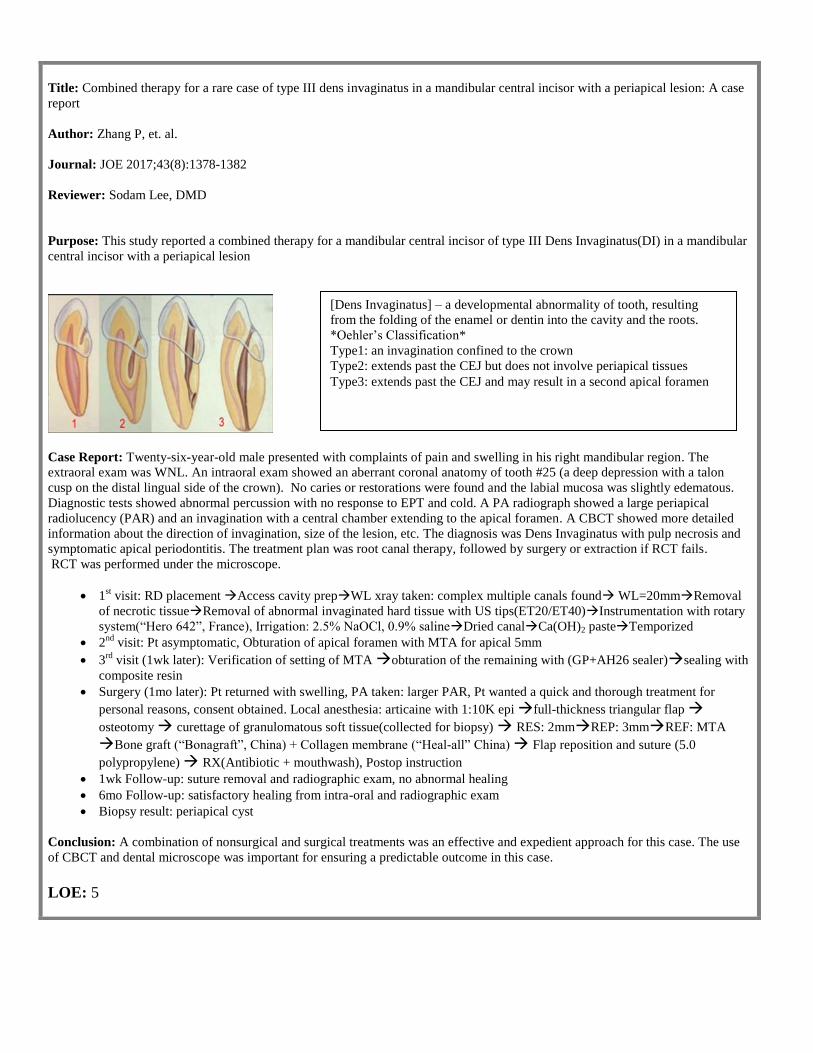
Title: Combined therapy for a rare case of type III dens invaginatus in a mandibular central incisor with a periapical lesion: A case
report
Author: Zhang P, et. al.
Journal: JOE 2017;43(8):1378-1382
Reviewer: Sodam Lee, DMD
Purpose: This study reported a combined therapy for a mandibular central incisor of type III Dens Invaginatus(DI) in a mandibular
central incisor with a periapical lesion
Case Report: Twenty-six-year-old male presented with complaints of pain and swelling in his right mandibular region. The
extraoral exam was WNL. An intraoral exam showed an aberrant coronal anatomy of tooth #25 (a deep depression with a talon
cusp on the distal lingual side of the crown). No caries or restorations were found and the labial mucosa was slightly edematous.
Diagnostic tests showed abnormal percussion with no response to EPT and cold. A PA radiograph showed a large periapical
radiolucency (PAR) and an invagination with a central chamber extending to the apical foramen. A CBCT showed more detailed
information about the direction of invagination, size of the lesion, etc. The diagnosis was Dens Invaginatus with pulp necrosis and
symptomatic apical periodontitis. The treatment plan was root canal therapy, followed by surgery or extraction if RCT fails.
RCT was performed under the microscope.
1st visit: RD placement Access cavity prepWL xray taken: complex multiple canals found WL=20mmRemoval
of necrotic tissueRemoval of abnormal invaginated hard tissue with US tips(ET20/ET40)Instrumentation with rotary
system(“Hero 642”, France), Irrigation: 2.5% NaOCl, 0.9% salineDried canalCa(OH)2 pasteTemporized
2nd
visit: Pt asymptomatic, Obturation of apical foramen with MTA for apical 5mm
3rd
visit (1wk later): Verification of setting of MTA obturation of the remaining with (GP+AH26 sealer)sealing with
composite resin
Surgery (1mo later): Pt returned with swelling, PA taken: larger PAR, Pt wanted a quick and thorough treatment for
personal reasons, consent obtained. Local anesthesia: articaine with 1:10K epi full-thickness triangular flap osteotomy curettage of granulomatous soft tissue(collected for biopsy) RES: 2mmREP: 3mmREF: MTA
Bone graft (“Bonagraft”, China) + Collagen membrane (“Heal-all” China) Flap reposition and suture (5.0
polypropylene) RX(Antibiotic + mouthwash), Postop instruction
1wk Follow-up: suture removal and radiographic exam, no abnormal healing
6mo Follow-up: satisfactory healing from intra-oral and radiographic exam
Biopsy result: periapical cyst
Conclusion: A combination of nonsurgical and surgical treatments was an effective and expedient approach for this case. The use
of CBCT and dental microscope was important for ensuring a predictable outcome in this case.
LOE: 5
[Dens Invaginatus] – a developmental abnormality of tooth, resulting
from the folding of the enamel or dentin into the cavity and the roots.
*Oehler’s Classification*
Type1: an invagination confined to the crown
Type2: extends past the CEJ but does not involve periapical tissues
Type3: extends past the CEJ and may result in a second apical foramen
Title: Cyclic fatigue resistance of Reciproc Blue, Reciproc, ad WaveOne Gold reciprocating instruments
Author: Keskin C et al.
Journal: JOE Volume 43(8): 1360-1363
Reviewer: Adnan Kazim DMD
Purpose: This study compared the cyclic fatigue resistance of Reciproc® Blue R25 with Reciproc® R25 and WaveOne® Gold
Materials and Methods:
Fifteen Reciproc® Blue R25, fifteen Reciproc® R25, and fifteen WaveOne® Gold were inspected under a stereomicroscope
to verify instruments were free of visible defects and irregularities.
Cyclic fatigue testing was performed in a stainless steel artificial canal with an inner diameter of 1.5mm and a curvature
angle of 60 degrees with a curvature radius of 5mm.
All instruments were used with a VDW endodontic motor that were used under the file setting in the motor
Synthetic oil was used to reduce friction of the instruments (WD-40)
The instruments were operated with a 3mm/s axial movement until fracture.
Time to Fracture (TF) and length of fractured fragment was recorded.
Two fractured instruments from each group were analyzed under scanning electron microscope.
Data was analyzed using Shapiro-Wilk test, 1-way analysis of variance, and Kruskal Wallis and Dunn tests and SPSS with
5% selected as the significance level.
Results: Reciproc® Blue R25 instruments were associated with the highest cyclic fatigue resistance values among all instruments
(P<.05). The cyclic fatigue resistance of WaveOne® Gold was significantly higher than that of Reciproc® R25. There was no
significant difference in the man length of the fractured fragments
Conclusion: The effect of thermal treatment of Reciproc® Blue alloy on cyclic fatigue resistance is evident in the current study
because Reciproc® and Reciproc® Blue have identical tapers, cross sectional shapes, and operational modes. Thermally treated
NiTi instruments show phase transformation changes that enhance the flexibility and cyclic fatigue resistance. Apart from
metallurgic difference of these files, cross sectional shape might also contribute to difference. WaveOne® Gold has a parallelogram
shaped cross section whereas Reciproc® has an S-shaped cross section. There is not a consensus on the effect of the cross sectional
shape on its cyclic fatigue resistance. Some studies indicate the cross-sectional dimension is a more important factor in cyclic
fatigue resistance.
LOE: 5
October 18
Title: AAE position statement: AAE guidance on the use of systemic antibiotics in endodontics
Journal: JOE Volume 43(9): 1409 – 1413
Reviewer: Steve Rees DMD
Purpose: This document is intended to present the available evidence related to prescribing antibiotics, highlight appropriate
clinical recommendations and identify gaps in knowledge for which personal judgment is the best guide for assessing risks and
benefits in this practice.
Overall risks and benefits of prescribing systemic antibiotics: Up to 50% of all antibiotics are prescribed or used incorrectly.
C. difficile was responsible for almost half a million infections and was associated with approximately 29,000 deaths in 2011.
Among the antibiotics prescribed for endodontic infections clindamycin, amoxicillin, cephalosporins are commonly associated with
C. difficile infection. Each year at least two million people in the U.S. become infected with multidrug resistant bacteria and 23,000
deaths have been attributed to these infections.
Use of adjunctive antibiotics in addition to adequate debridement and surgical drainage: The key to successful management
of infection of endodontic origin is adequate debridement of the infected root canal and drainage for both soft and hard tissue.
Localized soft tissue swelling of endodontic origin should be incised and drained concurrently with canal debridement. Adjunctive
antibiotics are not effective in preventing or ameliorating signs and symptoms in cases with irreversible pulpitis, symptomatic
apical periodontitis, or localized acute apical abscess, when adequate local debridement, medication and incision for drainage (if
indicated) have been achieved. In cases with spreading infections, the practitioner should use the shortest effective course of
antibiotics and minimize the use of broad-spectrum antibiotics. Most practitioners prescribe 3-7 days of antibiotics, but some
evidence suggests that perhaps 2-3 day courses may be successfully used as adjunctive therapies
Use of antibiotics in the absence of adequate debridement and surgical drainage: Supplemental antibiotics following adequate
debridement and drainage in cases of localized endodontic infections are ineffective. The standard of care is to prescribe primary or
adjunctive antibiotics, in conjunction with local debridement and surgical drainage, for patients who have spreading infections or
systemic involvement (fever, malaise, cellulitis and/or lymphadenopathies). It is not known whether systemic antibiotic therapy
would provide sufficient relief of symptoms and prevention of spread of infection to warrant a prescription in cases where the
practitioner is not able to render local debridement and drainage or in complex cases where the efficacy of local treatment may not
be completed.
Comparison of the efficacy of different types, dosage and duration of antibiotics: Patients who are immunocompromised or
have predisposing conditions such as previous endocarditis should be medicated as a prophylactic measure. Penicillin VK and
amoxicillin, both beta-lactam antibiotics, are the first line of antibiotics chosen as adjunct therapeutic agents in endodontics in the
U.S. Therapies lasting 7 days with amoxicillin have been shown to increase the population of resistant strains. Approximately 30%
of severe dento-alveolar infections have strains resistant to penicillin-like drugs. Amoxicillin may be combined with clavulanic acid
(125 mg bid or tid), which is a beta-lactamase inhibitor, to increase the susceptibility of penicillin resistant strains. This
combination has been shown to be 100% effective against cultivable endodontic bacteria, increasing the spectrum of amoxicillin in
persistent infections. Clindamycin is the first drug of choice for patients with a history of hypersensitivity to penicillin drugs. It has
been shown to be effective against 75% of cultivable endodontic pathogens. GI disturbances are the most common side effect with
an eight-fold increased risk of developing C. difficile infection than with the use of penicillin. Patients with a history of penicillin
allergy and severe GI reactions to clindamycin require alternative antibiotics such as macrolides (azithromycin), quinolones
(moxifloxacin) or tetracyclines. Unfortunately, endodontic pathogens have lesser susceptibility to these alternatives with increased
prevalence of resistant strains. Studies show that beta-lactam antibiotics are the optimal drugs for endodontic pathogens, because
there is very little bacterial resistance to amoxicillin with clavulanic acid. Studies have demonstrated more resistance to
clindamycin (which is typically the drug of choice for penicillin-allergic patients). Therefore, in penicillin-allergic patients, other
drugs such as moxifloxacin or azithromycin should be considered.
LOE: 5
Title: Influence of cone-beam computed tomography on endodontic retreatment strategies among general dental practitioners and
endodontists
Author: Rodriguez G, et. al.
Journal: JOE Vol. 43 (9): 1433-1437
Reviewed by: Stephanie Serrano DDS
Purpose: This study determined the impact of cone-beam computed tomographic (CBCT) imaging on clinical decision making
among general dental practitioners and endodontists after failed RCT. A second objective was to assess the self-reported level of
difficulty in making a treatment choice before and after viewing a preoperative CBCT scan.
Materials & Methods: Study participants included 120 male and female clinicians who varied in age and clinical experience. The
examiners were comprised of 80 general dental practitioners and 40 endodontists. Case Selection included eight cases randomly
selected from a list of patients who received a CBCT scan in order to complete their diagnosis. The cases included endodontically
treated teeth diagnosed as symptomatic apical periodontitis, acute apical abscess, or chronic apical abscess and teeth with definitive
and adequate coronal restorations (without coronal leakage) were selected. To standardize terminology used, examiners were
gathered in small groups and briefed on treatment alternatives to failed RCT: nonsurgical retreatment, apical surgery, intentional
reimplantation and extraction. In the first session, the examiners were given the details of each case, including at least two clinical
photographs, two peri-apical and one bitewing radiographs and were asked to choose one of the proposed treatment alternatives.
Examiners were also asked to assess the difficulty of making a decision on a scale from 1 to 5 (1 and 2 = easy decision, 3 =
moderate decision, and 4 and 5= difficult decision). One month later, the examiners reviewed randomly the same eight cases with
the additional information from the CBCT data. Examiners selected the most appropriate treatment plan and level of decision-
making difficulty for the 8 CBCT scans.
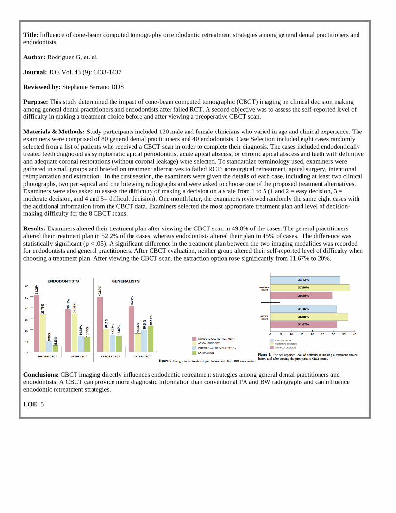
Results: Examiners altered their treatment plan after viewing the CBCT scan in 49.8% of the cases. The general practitioners
altered their treatment plan in 52.2% of the cases, whereas endodontists altered their plan in 45% of cases. The difference was
statistically significant (p < .05). A significant difference in the treatment plan between the two imaging modalities was recorded
for endodontists and general practitioners. After CBCT evaluation, neither group altered their self-reported level of difficulty when
choosing a treatment plan. After viewing the CBCT scan, the extraction option rose significantly from 11.67% to 20%.
Conclusions: CBCT imaging directly influences endodontic retreatment strategies among general dental practitioners and
endodontists. A CBCT can provide more diagnostic information than conventional PA and BW radiographs and can influence
endodontic retreatment strategies.
LOE: 5
Title: Further treatments of root-filled teeth in the swedish adult population: A comparison of teeth restored with direct and indirect
coronal restorations
Author: Dawson V et al.
Journal: JOE Vol 43(9): 1428-1432
Reviewer: Lauren Shin, DDS
Purpose: This study evaluated the frequencies of nonsurgical retreatment, root-end surgery, extraction, and further restoration in
relation to a direct versus an indirect restoration during a 5-year follow-up period of teeth root-filled and restored within six months
in Sweden in 2009.
Materials and Methods: A search was performed for treatment codes corresponding to root filling of one to four canals in the
SSIA database in the year of 2009. These root-filled teeth were tagged and tracked in the database for five years. The teeth were
divided into three groups depending on the type of coronal restoration received within six months of completion of the root canal:
indirect restoration, direct restoration, unknown. Further treatment provided was identified within five years including extraction,
nonsurgical retreatment, root-end surgery, direct restoration and indirect restoration. The frequencies of initial nonsurgical
retreatment, initial root-end surgery, extraction, and further restorative procedures were calculated for all teeth root filled during
2009 with respect to the type of coronal restoration.
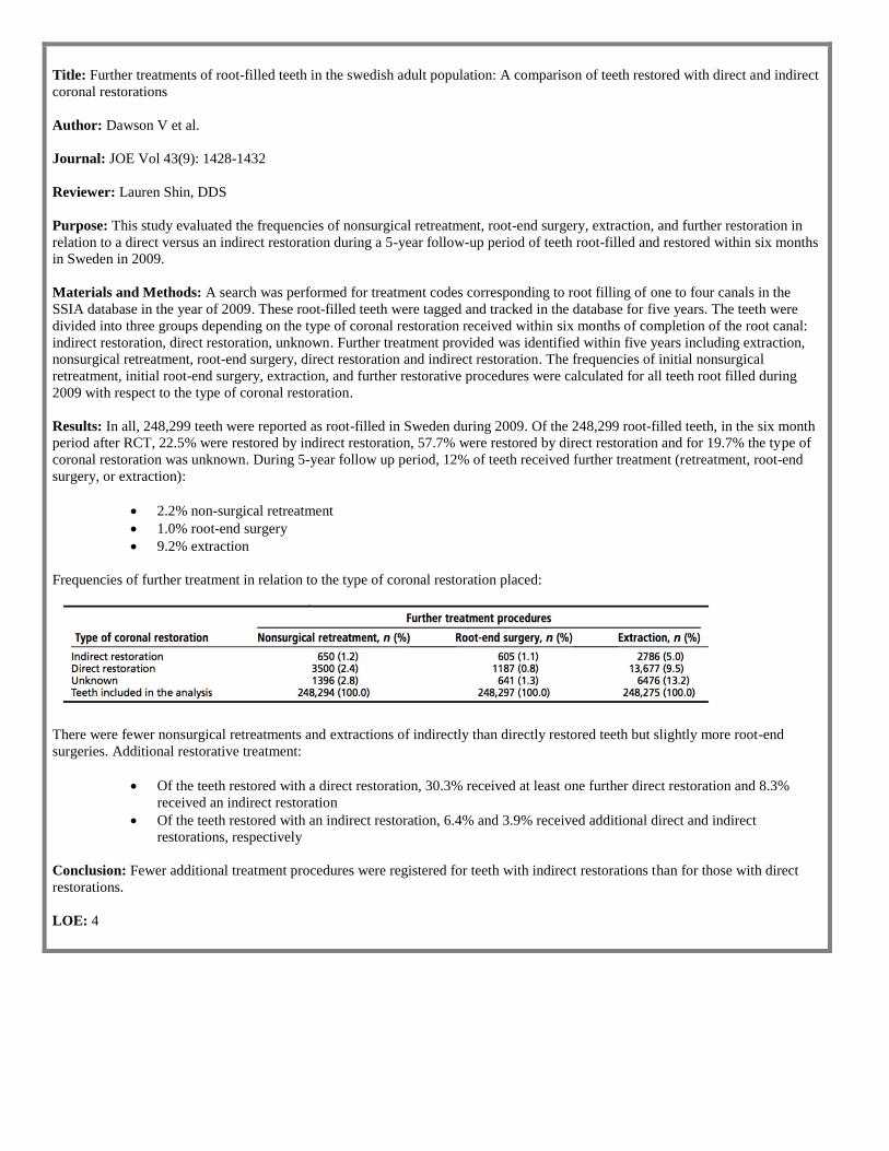
Results: In all, 248,299 teeth were reported as root-filled in Sweden during 2009. Of the 248,299 root-filled teeth, in the six month
period after RCT, 22.5% were restored by indirect restoration, 57.7% were restored by direct restoration and for 19.7% the type of
coronal restoration was unknown. During 5-year follow up period, 12% of teeth received further treatment (retreatment, root-end
surgery, or extraction):
2.2% non-surgical retreatment
1.0% root-end surgery
9.2% extraction
Frequencies of further treatment in relation to the type of coronal restoration placed:
There were fewer nonsurgical retreatments and extractions of indirectly than directly restored teeth but slightly more root-end
surgeries. Additional restorative treatment:
Of the teeth restored with a direct restoration, 30.3% received at least one further direct restoration and 8.3%
received an indirect restoration
Of the teeth restored with an indirect restoration, 6.4% and 3.9% received additional direct and indirect
restorations, respectively
Conclusion: Fewer additional treatment procedures were registered for teeth with indirect restorations than for those with direct
restorations.
LOE: 4
Title: Partial pulpotomy in mature permanent teeth with clinical signs indicative of irreversible pulpitis: A randomized clinical trial
Author: Taha N, et. al.
Journal: JOE, Vol. 43(9):1417
Reviewer: Ruoxue Feng DMD
Purpose: Partial pulpotomy includes the removal of 2-3mm from the inflamed coronal pulp beneath a pulpal exposure followed by
placement of a suitable agent over the remaining coronal pulp and a restoration that provides a hermetic seal. Studies have shown
that vital pulp therapy needs to be restricted to young or asymptomatic teeth because cariously exposed pulps in mature teeth are
capable of regeneration. Studies have shown that spontaneous or severe preoperative pain does not always indicate that the pulp has
no regenerative capability. American Academy of Pediatric Dentistry guidelines suggested MTA as a more favorable material than
calcium hydroxide (CH). This study explored the outcome of partial pulpotomy in mature teeth clinically diagnosed with
irreversible pulpitis using MTA compared with CH and monitored clinically and radiographically up to two years.
Materials and Methods:
• 50 teeth from referred patient pool were selected based on inclusion criteria presented in table 1.
• Clinical diagnosis of irreversible pulpitis was confirmed for all 50 teeth.
• Procedure steps were :Anesthesia, rubber dam, disinfection, caries excavation, partial pulpotomy, coin toss test to
randomly allocate teeth for MTA or CH, 27 teeth had MTA and 23 teeth and CH placement
• MTA group: vitrobond and final restoration after one week, CH group: immediate vitrobond and final restoration
• Clinical success is defined as no history of spontaneous pain or discomfort except during the first few days after treatment
and a functional tooth with no pain or discomfort on chewing or eating, a positive response to cold test, no tenderness to
percussion or palpation, normal grade I mobility, and no swelling or sinus tract.
• Radiographical success is defined as no intraradicular pathosis, internal resorption, or root resorption and perioapical index
<3 according to Østravik et al.
• Radiographs were evaluated by an experienced endodontist which was blinded to the capping material by masking the
crown of the tooth at 2 separate occasions
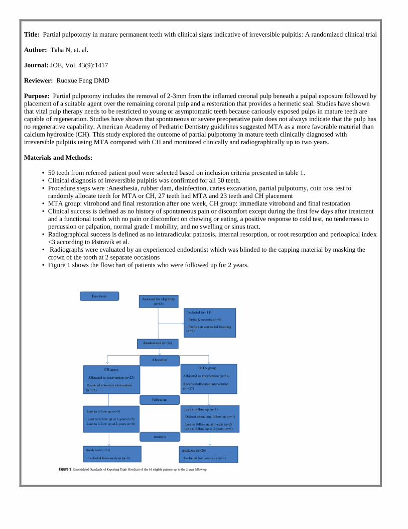
• Figure 1 shows the flowchart of patients who were followed up for 2 years.
• Fisher exact test was performed for material and sex.
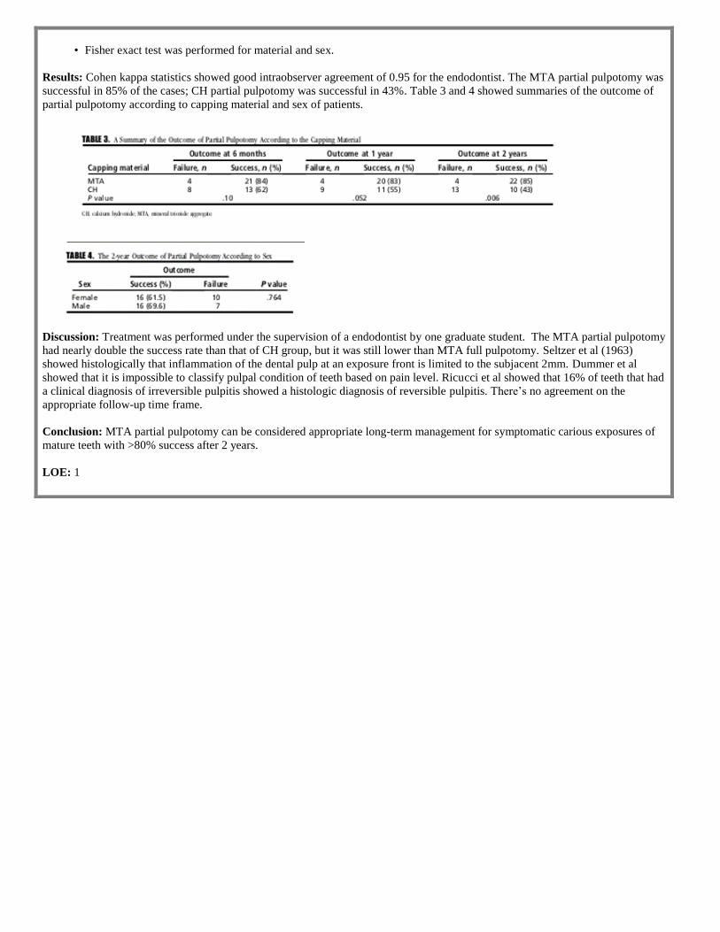
Results: Cohen kappa statistics showed good intraobserver agreement of 0.95 for the endodontist. The MTA partial pulpotomy was
successful in 85% of the cases; CH partial pulpotomy was successful in 43%. Table 3 and 4 showed summaries of the outcome of
partial pulpotomy according to capping material and sex of patients.
Discussion: Treatment was performed under the supervision of a endodontist by one graduate student. The MTA partial pulpotomy
had nearly double the success rate than that of CH group, but it was still lower than MTA full pulpotomy. Seltzer et al (1963)
showed histologically that inflammation of the dental pulp at an exposure front is limited to the subjacent 2mm. Dummer et al
showed that it is impossible to classify pulpal condition of teeth based on pain level. Ricucci et al showed that 16% of teeth that had
a clinical diagnosis of irreversible pulpitis showed a histologic diagnosis of reversible pulpitis. There’s no agreement on the
appropriate follow-up time frame.
Conclusion: MTA partial pulpotomy can be considered appropriate long-term management for symptomatic carious exposures of
mature teeth with >80% success after 2 years.
LOE: 1
Title: Effect of the simultaneous working length control during root canal preparation on postoperative pain
Author: Arslan H et al.
Source: Journal of Endodontics, Vol 43(9):1422-1427
Reviewer: Parth Karia DDS
Purpose: This study evaluated the effect of simultaneous length control during root canal preparation on postoperative pain
compared with separate working length determination and root canal preparation
Methods: Inclusion data included healthy patients with symptomatic apical periodontitis and symptomatic irreversible pulpitis in
first or second molar teeth, Visual analogue scale (VAS) value of either preoperative pain or pain on percussion of >50 and a lack
of periapical lesion. Exclusion data included those with systemic diseases, swelling, sinus tract or preoperative palpation pain, those
having bruxism or clenching, a severely damaged tooth, severe periodontal disease in related tooth or resorption in related tooth,
having previously undergone RCT in related tooth; having taken analgesics during past 24 hours or absence of opposing tooth to
the related tooth. Forty-four patients were chosen who got treatment from July - Dec 2016. Sixteen patients were divided into two
groups randomly were as part of pilot study. Twenty- eight more patients that were randomly selected were added making it total of
42 patients.
Control group: separate WL determination and root canal preparation
Experimental group: simultaneous WL control during root canal preparation (Reciproc instrument inserted into the
handpiece of Gold Reciproc motor obturation with single cone and followed by composite restoration)
Variables (age, gender, tooth number and preoperative pain on VAS) were measured. The pain level on days 1, 3, 5 and 7, and the
analgesic intake (400mg Ibuprofen) after the procedure and initial/final percussion pain were recorded. Statistical analysis done
with Chi-squared test, independent samples t test and Mann-Whitney U test
Results: No statistically significant difference found in groups in relation to demographics, preoperative pain, preoperative pain on
percussion, and postoperative pain on days 3, 5, and 7. Only significant difference was found on postoperative pain on day one.
Two patients in control group needed to use analgesics post-treatment but none in experimental group
Conclusion: Simultaneous length control during root canal preparation as a nonpharmacologic strategy for reducing postoperative
pain is a beneficial technique for preventing postoperative pain
LOE: 2
October 25
Title: An evidence-based review of the efficacy of treatment approaches for immature permanent teeth with pulp necrosis
Authors: Kahler B et al
Journal: J Endod 43 (7): 1052-1057
Reviewer: Laura Kim DDS
Purpose: This study investigated available evidence to support the premise of achieving further root maturation using regenerative
endodontic procedures (REPs) for immature necrotic teeth as well as examining the reported outcomes for calcium hydroxide
apexification, MTA apical barrier technique, and REPs.
Materials and Methods: A systematic search of English language publications was performed on online databases, Scopus
PubMed and Web of Science. Search terms used: Dental pulp, regenerative endodontic therapy, revascularization, revitalization.
Inclusion criteria were: Studies written in English language, performed in humans with a sample of 5 or more teeth, involving
immature necrotic permanent teeth treated with REPs, and performed quantitative assessment of root length and/or width and/or
apical diameter changes
Results: Of the 368 studies identified, 6 cohort studies satisfied the inclusion criteria and were examined for root maturation after
REPs and of these, 4 assessed and compared clinical outcomes between different treatment approaches. The tooth survival rate from 4 studies with follow-up times of 12-36 months (REPs- 98.6% and Calcium hydroxide apexification/ MTA barrier- 88.6%)
Clinical success rate of REPs from 2 studies, follow up time of 14-21 months (REP – 89.7% and Calcium hydroxide apexification/
MTA barrier- 100%). The percentage change of root wall width was greater in the REP group (28.2%) compared with the MTA
apexification group (0.0%) and the calcium hydroxide apexification group (1.5%) [Jeeruphan 2012]. The percentage change in root
length was significantly greater in the REP group (14.9%) compared with the MTA (6.1%) and calcium hydroxide apexification
groups (0.4%) [Jeeruphan 2012].
Conclusion: The results of this study show similar outcomes for both treatment modalities, suggesting that REPs could be
considered as a first treatment option in the treatment of immature teeth with pulp necrosis. However, further root maturation is
variable for teeth treated with REPs.
LOE: 1
Title: Furcation perforation: Periradicular tissue response to biodentine as a repair material by histopathologic and indirect
immunofluorescence analyses
Authors: Silva L et al
Journal: J Endod 43 (7): 1137-1142
Reviewer: Laura Kim DDS
Purpose: This study evaluated the response of periradicular tissues after sealing of furcation perforation with Biodentine, mineral
trioxide aggregate (MTA, positive control) and gutta-percha (negative control) in dogs’ teeth.
Materials and Methods:
30 teeth from 3 dogs with furcation involvement were accessed, cleaned/shaped and obturated
Teeth were endodontically treated and perforated at furcation and divided into three groups: Biodentine (14/30), MTA
(10/30), and Gutta percha (6/30)
After 120 days, maxillas and mandibles were removed and sectioned and were examined under the microscope for new
mineralized tissue formation and the thickness and the number of inflammatory cells as well as bone resorption at the
perforation site
Results: MTA presented the highest frequency of complete sealing of furcation perforations. Biodentine and MTA induced repair
by formation of mineralized tissue sealing totally or partly the furcation perforation in almost all cases: Biodentine- 92.9%, MTA-
88.9% and Gutta percha- 0%. MTA induced complete sealing of furcation by mineralized tissue with more frequency (77.8% vs.
35.8% in Biodentine). MTA induced the formation of a significantly thicker tissue than Biodentine. Biodentine and MTA showed
no bone resorption in 100% of the cases. Gutta percha group showed bone resorption was present in 100% of the cases.
Conclusion: Biodentine and MTA provided good histopathologic results and can be considered as adequate furcation perforation
repair materials.
LOE: 5

Title: Endodontic treatment in single and multiple visits: An overview of systematic reviews
Authors: Moreira M et al.
Journal: JOE Vol. 43 (6): 864-870 Reviewer: Rachel Mitrani DDS
Purpose: The effectiveness of endodontic treatment regarding the number of sessions to complete the therapy is still controversial.
This was an overview of published systematic reviews (SR) comparing endodontic treatment in single and multiple visits.
Materials & Methods: A systematic search was performed in the MEDLINE/Pubmed and Cochrane databases of articles
published up until August 18, 2016, without language restriction. Eligibility Criteria: 1) Systematic reviews, 2) A focus on
endodontic techniques in single or multiple visits. The phases of eligibility and analysis of risk of bias were conducted by 2 or 3
independent and calibrated examiners. A fourth examiner was consulted to resolve inconsistencies. Assessment of Multiple
Systematic Reviews was used to evaluate the risk of bias of the included SR’s, which were assessed according to the risk to develop
knowledge and the existing knowledge gap. Main characteristics extracted from the SR’s: Healing rates, success, and clinical
complications during and after endodontic treatment.
Results: Twenty SR’s were initially identified, and eight were included in the analysis. Of these, six showed low to moderate risk
of bias and were suitable to provide strong clinic evidence on the topic.
Discussion: Contraindications for RCT in a single visit are still debatable. This treatment modality is justified by some authors who
agree that one-visit RCT prevents inter-appointment leakage via the temporary coronal sealing, and reinfection of the root canal
system. Opponents of single-visit RCT argue that multi-visit RCT is necessary to ensure no pain or post-operative complications.
Additionally, with multi-visit RCT, there is a greater likelihood of achieving microbiological reduction levels compatible with
tissue repair via the use of intra-canal medication.
The results of this systematic review showed that single and multiple visits produce similar repair or success rates
regardless of the pre-condition of the pulp and peri-apex. The apical periodontitis sub-group showed a slight positive trend towards
a decreased incidence in post-op complications, and a higher effectiveness and efficiency for a single session. The higher frequency
of post-op complications in multiple visits may be due to repeated mechanical, chemical, or micro-biological injury to the peri-
apical tissue.
LOE: 1
Title: Fracture strength of endodontically treated teeth with different access cavity designs
Authors: Plotino G et al.
Journal: JOE 43 (6): 995-1000
Reviewer: Rachel Mitrani DDS
Purpose: It has been suggested that the removal of tooth structure needed for access cavity preparation may undermine the strength
of the tooth to fracture under functional loads. Extended preparation of endodontic access cavities critically reduces the amount of
sound dentin. This study compared (in vitro) the fracture strength of root-filled and restored teeth with traditional endodontic cavity
(TEC), conservative endodontic cavity (CEC), or ultra-conservative “ninja” endodontic cavity (NEC) access.
Materials & Methods: One hundred sixty extracted maxillary and mandibular premolars and molars were selected. The teeth were
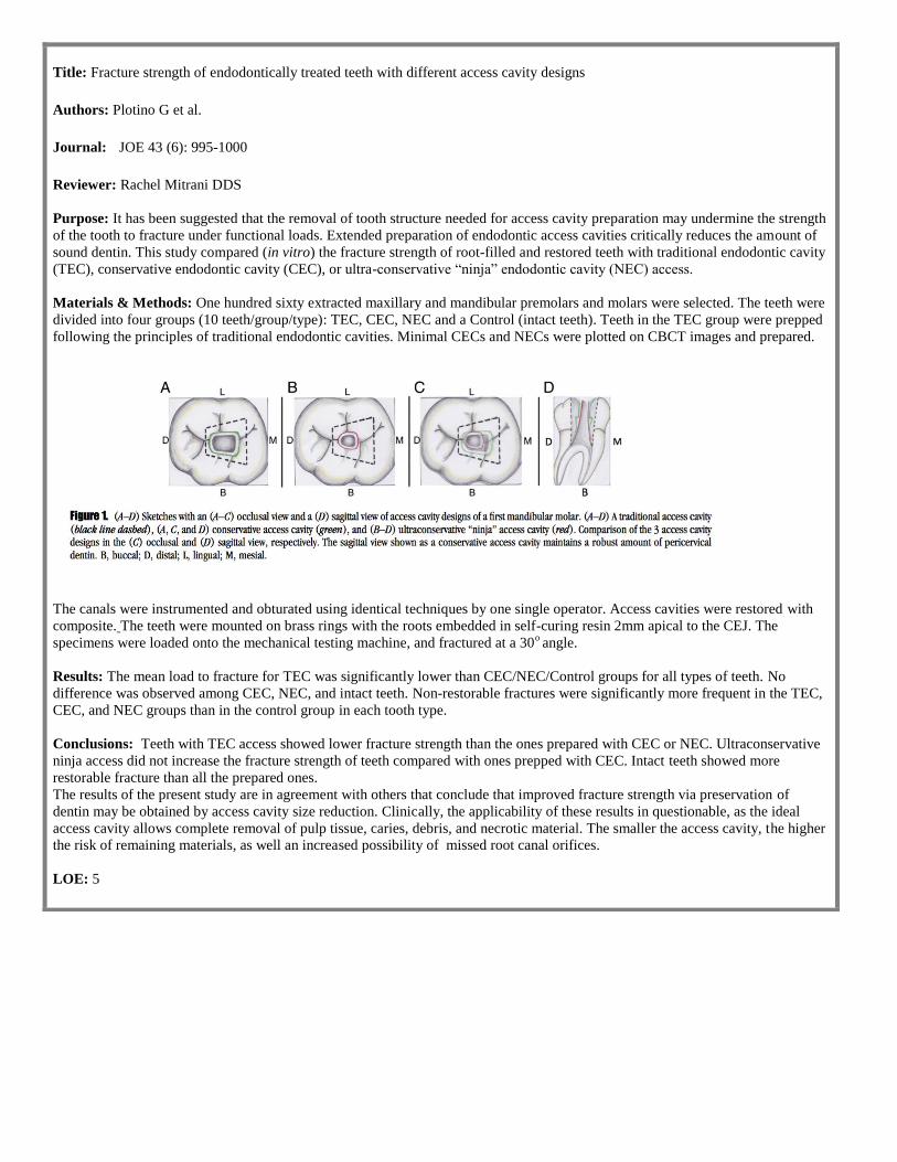
divided into four groups (10 teeth/group/type): TEC, CEC, NEC and a Control (intact teeth). Teeth in the TEC group were prepped
following the principles of traditional endodontic cavities. Minimal CECs and NECs were plotted on CBCT images and prepared.
The canals were instrumented and obturated using identical techniques by one single operator. Access cavities were restored with
composite. The teeth were mounted on brass rings with the roots embedded in self-curing resin 2mm apical to the CEJ. The
specimens were loaded onto the mechanical testing machine, and fractured at a 30o angle.
Results: The mean load to fracture for TEC was significantly lower than CEC/NEC/Control groups for all types of teeth. No
difference was observed among CEC, NEC, and intact teeth. Non-restorable fractures were significantly more frequent in the TEC,
CEC, and NEC groups than in the control group in each tooth type.
Conclusions: Teeth with TEC access showed lower fracture strength than the ones prepared with CEC or NEC. Ultraconservative
ninja access did not increase the fracture strength of teeth compared with ones prepped with CEC. Intact teeth showed more
restorable fracture than all the prepared ones.
The results of the present study are in agreement with others that conclude that improved fracture strength via preservation of
dentin may be obtained by access cavity size reduction. Clinically, the applicability of these results in questionable, as the ideal
access cavity allows complete removal of pulp tissue, caries, debris, and necrotic material. The smaller the access cavity, the higher
the risk of remaining materials, as well an increased possibility of missed root canal orifices.
LOE: 5
Title: Association of end-stage renal disease with radiographically and clinically diagnosed apical periodontitis: A hospital-based
study.
Author: Khalighinejad N. et al.
Journal: J Endod. 43(9):1438-1441
Reviewer: Reza Akhavan DMD
Purpose: This study evaluated the prevalence of apical periodontitis (AP) and endodontic treatment in patients with end-stage renal
disease (ESRD) as compared with patients with no history of ESRD.
Material and Methods: In this cross-sectional study, 40 patients diagnosed with non-diabetic ESRD were included. The control
group consisted of 40 age-matched and sex-matched healthy individuals. Digital panoramic radiographs were exposed on patients
in both the experimental and control groups. The number of remaining teeth and the prevalence of nonsurgical and/or surgical root
canal treatment were evaluated. In addition, the presence of AP in all teeth and endodontically treated teeth (ETT) was recorded.
Logistic regression was used to determine the possible association between ESRD and AP.
Results and Conclusion: Apical periodontitis in at least one tooth was found in 29 of the patients with ESRD (73%) and in 16 of
the control patients (40%) (odds ratio [OR] = 3.9, P < .05). In 21 (52%) patients with ESRD in the experimental group, at least one
ETT was diagnosed with AP. In the control group, 11 (28%) individuals had AP affecting at least one of the ETT (OR = 2.9, P <
.05). Adjusted for the number of teeth and endodontic treatment, ESRD was significantly associated with the presence of AP (OR =
2.6). Apical periodontitis was significantly more prevalent in the experimental group. This may suggest that ESRD could
possibly alter the pathogenesis of AP. However, these findings do not confirm the presence of any cause-and-effect relationship
between these conditions.
LOE: 5