Embed Size (px)
Citation preview

1
ALPORT SYNDROME: CLINICAL AND MOLECULAR STUDY
OF PORTUGUESE FAMILIES
MARIA JOÃO NABAIS SÁ
Tese de doutoramento em Patologia e Genética Molecular
2013

2

3
MARIA JOÃO NABAIS SÁ
ALPORT SYNDROME:
CLINICAL AND MOLECULAR STUDY OF PORTUGUESE FAMILIES
Tese de Candidatura ao grau de Doutor em Patologia e Genética Molecular submetida ao Instituto de Ciências Biomédicas Abel Salazar da Universidade do Porto.
Orientador – Doutor João Paulo Ferreira da Silva Oliveira
Categoria – Professor Associado
Afiliação – Faculdade de Medicina da Universidade do Porto
Co-orientador – Doutora Filipa Abreu Gomes de Carvalho
Categoria – Professora Auxiliar com Agregação
Afiliação – Faculdade de Medicina da Universidade do Porto
Apoios financeiros da Fundação para a Ciência e a Tecnologia (SFRH/SINTD/60054/2009; HMSP-ICJ/SAU-ICT/0003/2009; Unidade de Investigação e Desenvolvimento em Nefrologia – FCT-725) no âmbito do III Quadro Comunitário de Apoio e da Sociedade Portuguesa de Nefrologia através de bolsa de financiamento de Projecto de Investigação Científica de 2011.

4

5
Aos meus pais e irmão.

6

7
“Basic science could both inform and be informed by clinical genetics.”
Charles Joseph Epstein, 2011
A todos os que contribuem diariamente para esta ponte.

8

9
Agradecimentos Acknowledgements
Aos doentes e familiares, que são “o vento que faz mover as minhas
asas”.
Ao Professor Doutor João Paulo Oliveira e à Professora Doutora
Filipa Carvalho, por supervisionarem o trabalho desta tese e por
continuamente desenvolverem a minha autonomia e estimularem
uma colaboração estreita com médicos e grupos de investigação
dedicados às áreas científicas versadas desta tese. Ao Professor
Doutor Alberto Barros, Diretor do Departamento de Genética da
Faculdade de Medicina da Universidade do Porto (FMUP), por ter
acolhido parte do trabalho experimental desta tese.
A todos os médicos que deram voz aos doentes neste projecto, em
particular à Drª. Susana Sampaio, à Drª. Fernanda Carvalho, à
Professora Doutora Ângela Carneiro e ao Dr. Sérgio Silva Estrela. À
NephroCare – Portugal, por toda a colaboração. Ao Professor Doutor
Manuel Pestana, Diretor do Serviço de Nefrologia do Hospital São
João (HSJ) e Director da Unidade de I&D de Nefrologia e Doenças
Infecciosas – INEB (I3S) da Universidade do Porto, pelo apoio
incondicional.
A todos quem, no HSJ, nomeadamente nos Serviços de Arquivo
Clínico, de Nefrologia, de Anatomia Patológica, de Oftalmologia, de
Otorrinolaringologia e da consulta externa de Genética, assim como
na FMUP, incluindo as Secretarias, a Biblioteca, o Laboratório de
Imunohemoterapia, os Departamentos de Informática, de Estatística
e de Genética, tão bem conhecem a importância do seu contributo
para este projecto. À Susana Alves e Ana Paula Neto, por toda a
dedicação.
À FCT, ao Programa Harvard Medical School-Portugal, à Sociedade
Portuguesa de Nefrologia, ao Departamento de Genética e à
Unidade de I&D de Nefrologia e Doenças Infecciosas – INEB (I3S) da
Universidade do Porto, pela confiança depositada, assim como pela
divulgação e financiamento do projecto.

10
Agradecimentos Acknowledgements
À Professora Doutora Graça Porto, impulsionadora do desafio.
À Drª. Ana Maria Fortuna, Directora da Unidade de Genética Médica
do Centro de Genética Médica Doutor Jacinto Magalhães/Centro
Hospitalar do Porto, e à Drª. Gabriela Soares, Orientadora do
Internato em Genética Médica, por confiarem no percurso pioneiro
que escolhi.
À Professora Doutora Maria do Carmo Fonseca, por me ensinar a
construir o caminho, do sonho à sua concretização.
To Guy Froyen and Nathalie Firemans, for their genuine
collaboration.
To Frances Flinter, who inspires me and shows me that better has no
limit. To Helen Storey and Mato Nagel, who committed their efforts
with the aim of this project and continue to persue it, in a global
perspective.
A todos os meus amigos, cuja coragem sempre espelharam.
To Arjan de Brouwer, for teaching me that treasure is everywhere.
À minha família, a quem sempre me acompanha e a quem já partiu
para seguir com outro olhar esta jornada.

11
Resumo
A análise genética molecular dos genes COL4A5, COL4A4 e COL4A3
genes é a melhor abordagem não invasiva disponível para
estabelecer o diagnóstico, tratamento, prognóstico e aconselhamento
genético de nefropatias associadas ao colagéneo tipo IV – síndrome
de Alport e hematúria familiar benigna / nefropatia de membrana basal
fina. Neste estudo multicêntrico nacional, foram estudadas 65 famílias
não relacionadas com o diagnóstico clínico de síndrome de Alport ou
de nefropatia de membrana basal fina. Foram identificadas mutações
patogénicas no gene COL4A5 em 22 famílias (22/60; 37%), que
confirmaram o diagnóstico de síndrome de Alport ligada ao
cromossoma X. Foi identificada uma nova correlação genótipo-
fenótipo, dado ter sido detectada uma deleção que envolve o gene
COL4A5, mas não o gene COL4A6, numa família com leiomiomatose
difusa associada a síndrome de Alport ligada ao cromossoma X. Este
resultado sugere que a deleção dos exões 5’ do gene COL4A6 e da
região promotora comum aos genes COL4A5_COL4A6 não é
essencial para o desenvolvimento de leiomiomatose difusa nestes
doentes. Foi efetuada sequenciação directa dos genes COL4A3 e
COL4A4 em cinco famílias com padrão de hereditariedade
autossómico, como abordagem de primeira linha, e em 35 famílias
com padrão de hereditariedade incerto e nas quais não foram
detectadas mutações patogénicas no gene COL4A5. Foram
detectadas mutações patogénicas nos genes COL4A3 ou COL4A4
em 25 famílias (25/40; 63%), confirmando o diagnóstico de síndrome
de Alport autossómica ou de nefropatia de membrana basal fina.
Observou-se uma proporção semelhante de famílias com síndrome
de Alport ligado ao cromossoma X e de famílias com síndrome de
Alport autossómica ou com nefropatia de membrana basal fina, sendo
as prevalências de 37% (22/62) e de 40% (25/62), respetivamente. No
seu conjunto estes resultados sugerem que a abordagem laboratorial
por método de sequenciação de ADN de próxima geração será mais
eficaz e menos dispendiosa para diagnóstico genético molecular da
síndrome de Alport na população portuguesa.
Palavras-chave: Síndrome de Alport; COL4A3; COL4A4; COL4A5.

12

13
Abstract
Molecular genetic analysis of the COL4A5, COL4A4 and COL4A3
genes is the best non-invasive available approach for diagnosis,
treatment, prognosis, and genetic couseling of collagen IV-related
nephropathies – Alport syndrome and thin basement membrane
nephropathy. In this national multicenter study, 65 unrelated families
with a clinical diagnosis of Alport syndrome or thin basement
membrane nephropathy were studied. Pathogenic COL4A5 mutations
were identified by direct sequencing and multiplex-ligation dependent
amplification in 22 families (22/65; 34%), confirming a diagnosis of X-
linked AS. A novel genotype-phenotype correlation was disclosed, as
a deletion involving COL4A5, but not COL4A6, was detected in a
family with diffuse leiomyomatosis associated with X-linked AS. This
result suggests that the deletion of the 5’ exons of COL4A6 and of the
common COL4A5_COL4A6 promoter region is not essential for the
development of diffuse leiomyomatosis in patients with Alport
syndrome and diffuse leiomyomatosis. COL4A3 and COL4A4 were
analyzed by direct sequencing in five families with autosomal
inheritance pattern, as a first-tier approach, and in 35 families with an
unclear inheritance pattern and no pathogenic mutations in the
COL4A5 gene. Pathogenic mutations in COL4A3 or COL4A4 were
detected in 25 unrelated families (25/40; 63%), confirming the
diagnosis of autosomal Alport syndrome or thin basement membrane
nephropathy. A similar proportion of families with X-linked Alport
syndrome and of families with autosomal Alport syndrome or with thin
basement membrane nephropathy was observed, with prevalences of
37% (22/62) and 40% (25/62), respectively. Altogether, these results
suggest that Next-Generation Sequencing would be a more effective
and less expensive approach to the genetic molecular study of Alport
syndrome in the Portuguese population.
Keywords: Alport syndrome; COL4A3; COL4A4; COL4A5.

14

15
Publicações
Publications
Artigos em revistas internacionais
Papers in international journals
Nabais Sá MJ, Fieremans N, de Brouwer AP, Sousa R, Teixeira E Costa F, Brito MJ, Carvalho F, Rodrigues M, Teixeira de Sousa F, Felgueiras J, Neves F, Carvalho A, Ramos U, Vizcaíno JR, Alves S, Carvalho F, Froyen G, Oliveira JP. “Deletion of the 5'exons of COL4A6 is not needed for the development of diffuse leiomyomatosis in patients with Alport syndrome.” J Med Genet. 2013 Nov;50(11):745-53. Nabais Sá MJ, Sampaio S, Oliveira A, Alves S, Pinto de Moura C, Estrela Silva S, Castro R, Augusto Araújo J, Rodrigues M, Neves F, Seabra J, Soares C, Gaspar MA, Tavares I, Freitas L, Couto Sousa T, Castro Henriques A, Teixeira e Costa F, Morgado E, Teixeira Sousa F, Pratas e Sousa J, Gomes da Costa A, Filipe R, Garrido J, Montalban J, Ponce P, Alves R, Faria B, Carvalho MF, Pestana M, Carvalho F, Oliveira JP. “Collagen type IV-related nephropathies in Portugal: spectrum of pathogenic COL4A5 mutations and clinical characterization of 22 families.” Manuscript submitted to Journal of Medical Genetics (Manuscript ID: jmedgenet-2014-102471). Nabais Sá MJ, Storey H, Flinter F, Nagel M, Sampaio S, Castro R, Augusto Araújo J, Gaspar MA, Soares C, Oliveira A, Castro Henriques A, Gomes da Costa A, Pinto Abreu C, Ponce P, Alves R, Pinho L, Estrela Silva S, Pinto de Moura C, Mendonça L, Carvalho MF, Pestana M, Alves S, Carvalho F, Oliveira JP. “Collagen type IV-related nephropathies in Portugal: spectrum of pathogenic COL4A3 and COL4A4 mutations and clinical characterisation of 25 families.” Manuscript submitted to Journal of Medical Genetics (Manuscript ID: jmedgenet-2014-102472).

16

17
Comunicacõ es em encontros cientificos internacionais Communications in international meetings
Maria João Nabais Sá, Susana Alves, Helen Storey, Frances Flinter, Patrick J Willems,Filipa Carvalho, João P Oliveira on behalf of the investigators of the Portuguese Alport syndrome Collaborative Study. Molecular characterization of 65 Portuguese families with clinical diagnosis of collagen type IV glomerulopathies. 50th ERA-EDTA Congress, 18th-21st May 2013, Istanbul, Turkey. (Poster Presentation)
Maria João Nabais Sá, Rita Sousa, Fernando Teixeira e Costa, Fernanda Carvalho, Susana Alves, Filipa Carvalho, João Paulo Oliveira. A large deletion confined to COL4A5 causes Alport syndrome and diffuse leiomyomatosis. American Society of Human Genetics 62nd Annual Meeting, 6-10th November 2012, San Francisco, U.S.A. (Poster Presentation)
Maria João Nabais Sá, Susana Alves, Filipa Carvalho, João Paulo Oliveira on behalf of the Portuguese Alport Syndrome Collaborative Study. COL4A5 mutational analysis of 51 unrelated Portuguese patients with Alport syndrome – preliminary report. European Society of Human Genetics 2012, 23rd-26th June 2012, Nürnberg, Germany. (Poster Presentation)
Comunicacõ es em encontros cientificos nacionais Communications in national meetings
Maria João Nabais Sá, Susana Alves, Filipa Carvalho, João Paulo Oliveira on behalf of the Portuguese Alport Syndrome Collaborative Study. Alport syndrome: clinical and molecular study of Portuguese families – preliminary results. Encontro Renal 2012, 28th-31rst March 2012, Vilamoura, Portugal. (Poster presentation)
Maria João Nabais Sá, Susana Sampaio, Ana Oliveira, Isabel Tavares, Ana Paula Neto, Susana Alves, Filipa Carvalho, João Paulo Oliveira. X-linked Alport syndrome: a novel mutation shared by two apparently unrelated Portuguese families. 15ª Reunião da Sociedade Portuguesa de Genética Humana, 10th-12th November 2011, Lisboa, Portugal. (Poster presentation)

18

19
Table of Contents
Chapter 1 Introduction 25
1. Alport syndrome 25
1.1. Diagnosis: Historical perspective 25
1.2. Prevalence 29
1.3. Clinicopathologic features 29
1.3.1. Impaired renal function 30
1.3.2. Impaired renal and skin structure 34
1.3.3. Sensorineural hearing loss 37
1.3.4. Ocular abnormalities 38
1.3.5. Diffuse leiomyomatosis 41
1.3.6. Differential Diagnosis 44
1.4. Molecular genetics and pathogenesis 46
1.4.1. Type IV collagen coding genes 46
1.4.2. Type IV collagen α-chains and protomers 48
1.4.3. Type IV collagen networks 48
1.5. Genotype-phenotype correlations 50
2. Significance, hypothesis and objectives of the stud y 54
Chapter 2 Patients and Methods 59
1. Study cohort 59
1.1. Units of analysis 61
1.2. Units of observation 62
1.3. Nature of data 62
1.4. Origin of data 63

20
2. Study design 64
2.1. Clinical data collection and analysis 65
2.2. Molecular data collection and analysis 67
2.3. Genotype-phenotype correlation: Analysis techniques 68
Chapter 3 Results 71
1. Cohort assembly: Results of the recruitment process 71
2. Clinical and molecular characterization of patients with clinical diagnosis or
suspicion of Alport syndrome in Portugal 79
2.1. Patients with pathogenic COL4A5 and COL4A6 mutation s 80
2.1.1. Collagen type IV-related nephropathies in Portugal: spectrum of
pathogenic COL4A5 mutations and clinical characterization of 22 fami lies
(Manuscript 1) 81
2.1.2. Deletion of the 5’exons of COL4A6 is not needed for the development
of diffuse leiomyomatosis in Alport syndrome patien ts (Manuscript 2) 123
2.2. Patients with pathogenic COL4A3 and COL4A4 mutation s 156
2.2.1. Collagen type IV-related nephropathies in Portugal: spectrum of
pathogenic COL4A3 and COL4A4 mutations and clinical characterisation of 25
families (Manuscript 3) 157
Chapter 4 Discussion 193
1. Aim 1: Molecular analysis of COL4A5, COL4A4 and COL 4A3 in Portugal 193
1.1. The epidemiology of pathogenic COL4A5, COL4A4 and C OL4A3 mutations in
Portugal is different from other countries 193
1.1.1. High detection rate of pathogenic COL4A5 , COL4A4 and COL4A3
mutations 193
1.1.2. The prevalence of pathogenic COL4A5 mutations is similar to that of
pathogenic COL4A4 and COL4A3 mutations in Portugal 196

21
1.1.3. Documentation of pathogenic COL4A5 , COL4A4 and COL4A3
mutations 198
1.1.4. Pathogenicity of the novel COL4A5 , COL4A4 and COL4A3 variants 199
2. Aim 2: Clinical characterization of Portuguese pati ents 206
2.1. Phenotype of the 65 genetic probands 206
2.1.1. Microscopic hematuria and thinning of the GBM were hallmarks 206
3. Aim 3: A novel genotype-phenotype correlation among other correlations 210
3.1. Deletion of the common COL4A5/COL4A6 promotor and 5 ’ exons of COL4A6 is not
needed for development of diffuse leiomyomatosis in X-linked Alport syndrome 210
3.2. An unknown family history of hematuria, CKD and hea ring loss is not synonymous of
a negative family history 211
3.3. Kidney biopsy with ultrastructure examination is re commended to clarify the etiology
of urinary abnormalities 212
3.4. Skin biopsy is helpful in determining the inheritan ce pattern 213
3.5. The mutation status influences the severity of the phenotype 213
3.6. The type of mutation influences the severity of the phenotype 214
3.7. Deliniation of the natural history of the disease i n our sample 214
3.8. Patients without known mutations: differences and s imilarities 219
3.9. Diagnostic criteria defined for XLAS by Flinter and colleagues (1988) also enable the
diagnosis of patients with ARAS 223
4. Short term impact of the study: Massive parallel se quencing is recommended to
simultaneously study the COL4A5, COL4A4 and COL4A3 genes 225
5. Long term impact of the study: Future perspectives 227
6. Main recommendations 229
Conclusion 232
References 233
Protocol P1 Clinical protocol 249
Protocol P2 Molecular protocol 253

22
Index 259
List of figures 259
List of tables 260
Appendix I Clinical and molecular characterization of patients (CD) 262
23
Abbreviations
ACE: Angiotensin Converting Enzyme
ADAS: Autosomal Dominant Alport Syndrome
α1(IV) chain: alpha-1 chain of collagen type IV
α2(IV) chain: alpha-2 chain of collagen type IV
α3(IV) chain: alpha-3 chain of collagen type IV
α4(IV) chain: alpha-4 chain of collagen type IV
α5(IV) chain: alpha-5 chain of collagen type IV
α6(IV) chain: alpha-6 chain of collagen type IV
Anti-GBM: Anti-Glomerular Basement Membrane
ARAS: Autosomal Recessive Alport Syndrome
ARB: Angiotensin Receptor Blocker
AS: Alport Syndrome
ATS-DL: Alport Syndrome and Diffuse Leiomyomatosis
BFH: Benign Familial Hematuria
BM: Basement Membrane
CFHR5: Complement Factor H-related Protein 5
CKD: Chronic Kidney Disease
CRF: Chronic Renal Failure
DNA: Deoxyribonucleic Acid
EBM: Epidermal Basement Membrane
ECASCA: European Community Alport Syndrome Concerted Action
ESRD: End-Stage Renal Disease
GBM: Glomerular Basement Membrane
GFND: Glomerulopathy with Fibronectin Deposits
GFR: Glomerular Filtration Rate
HL: Hearing Loss
Hz: Hertz (audiometric measurement)
MPS: Massive Parallel Sequencing
NGS: Next-Generation Sequencing

24
OCT: Optical Coherence Tomography
pCr: Plasma Creatinine Measurement
PCR: Polymerase Chain Reaction
RBC: Red Blood Cell
RCE: Recurrent Corneal Erosion
RNA: Ribonucleic Acid
RRT: Renal Replacement Therapy
SNHL: Sensorineural Hearing Loss
TBMN: Thin Basement Membrane Nephropathy
UTR: Untranslated Region
XLAS: X-linked Alport Syndrome

25
Chapter 1 Introduction
1. Alport syndrome
1.1. Diagnosis: Historical perspective
In 1927, Arthur Cecil Alport recognized that the association of hematuria
and sensorineural deafness, reported in a previously studied family, belong to a
unique clinical entity (Alport 1927), which in time became known as Alport
syndrome (AS) (Williamson 1961). This family had been the subject of several
successive observations (Guthrie 1902; Kendall 1912; Hurst 1923; Alport 1927).
Guthrie described the familial occurrence of hematuria of unknown cause, which
he called hereditary hematuria, since it was observed in two consecutive
generations of the family and it was congenital or presented during childhood
(Guthrie 1902). Hematuria occurred in both genders, was microscopic and/or
macroscopic, was persistent or recurrent and was prone to paroxysmal
exacerbations concomitantly with flue-like symptoms. Phenotypic heterogeneity
was emphasized by reporting that affected males manifested uremia and died at a
younger age, while females were usually mildly affected (Hurst 1923; Eason J
1924; Alport 1927). Eason mentioned that, in this family, a non-affected female did
not transmit the disorder, while affected females and another non-affected female
did; and that infections by non-haemolytic streptococcus predisposed to the
exacerbation of the kidney disease (Eason J 1924). Hurst had noted the
occurrence of deafness in three patients of the family, but he did not make a
comment on this (Hurst 1923). However, Authur C. Alport who, observing “nerve”
deafness in both affected males and females, recognized auditory loss as an
integral sign of this hereditary disease (Alport 1927). Perkoff and colleagues
published a detailed study of a much larger Mormon family from Utah with similar
clinical features and hypothesized for the first time a mechanism of inheritance for
the disease – a partially sex-linked dominant inheritance with crossing-over
between X and Y chromosomes, which was subsequently contested (Perkoff,
Stephens et al. 1951; Stephens, Perkoff et al. 1951; Perkoff, Nugent et al. 1958).
Ocular manifestations were first reported as part of the syndrome in 1954

26
(Reyersbach and Butler 1954; Sohar 1954; Sohar 1956). As there were no specific
diagnostic criteria for the diagnosis of AS until 1988, a variety of clinically
heterogeneous inherited nephritis were included in the AS phenotype for many
years and a multiplicity of hypotheses were posed regarding the inheritance of AS
(Crawfurd 1988).
The pathophysiology of AS was clarified by the observation of characteristic
ultrastructural changes in the glomerular basement membrane (GBM) of the
kidney (Kinoshita, Osawa et al. 1969; Hinglais, Grunfeld et al. 1972; Spear and
Slusser 1972; Churg and Sherman 1973). In the late 60s, Kefalides had identified
a unique collagen in the GBM (Kefalides 1966; Kefalides 1968). Three identical α-
chains were suggested to form the scaffold of the GBM (Kefalides 1971). With
advances in electron microscopy in the early 1970s, several laboratories reported
typical ultrastructural changes in the GBM of AS patients, namely focal or diffuse
thinning, thickening and splitting of the GBM containing granular material
(Kinoshita, Osawa et al. 1969; Hinglais, Grunfeld et al. 1972; Spear and Slusser
1972; Churg and Sherman 1973). These findings lead Spear to hypothesize that
mutations in the gene for this unique structural GBM component could cause AS
(Spear 1973). Two additional observations were fundamental to the recognition of
a possible type IV collagen defect as the cause of AS: (i) the Goodpasture (human
anti-GBM) antigen, which was later known to be directed against the globular
domain of collagen type IV (Wieslander, Barr et al. 1984), did not bind GBM of
patients with AS (Olson, Anand et al. 1980; McCoy, Johnson et al. 1982); and (ii)
the development of post-transplant glomerulonephritis in AS patients was due to
the synthesis of antibodies directed against collagen type IV chain epitopes
(Milliner, Pierides et al. 1982; Kashtan, Fish et al. 1986), more specifically
antibodies directed against α3 and α5 collagen type IV chains (Fleming, Savage et
al. 1988; Kleppel, Kashtan et al. 1989; Kleppel, Santi et al. 1989; Savage, Noel et
al. 1989; vd Heuvel, Schroder et al. 1989; Kashtan, Butkowski et al. 1990).
Framing the reported findings, Flinter and colleagues (1988) proposed four
criteria for the clinical diagnosis of classic AS, also known as X-linked Alport
syndrome (XLAS). An individual with unexplained hematuria would probably have
XLAS if, at least, three of the following four criteria were present: (i) Positive family
history of hematuria with or without chronic renal failure (CRF); (ii) Typical

27
ultrastructural GBM changes in a renal biopsy specimen; (iii) High-tone
sensorineural deafness; (iv) Characteristic ophthalmological signs (lenticonus
and/or macular flecks) (Flinter, Cameron et al. 1988).
It was not until 1990 that molecular genetic techniques enabled the
elucidation of the genetic basis of XLAS. By immunohistochemical analysis, α5(IV)
chains were shown to localize to the GBM of the kidney and the COL4A5 gene,
coding for α5(IV) chain of collagen type IV, was found to be located to Xq22
(Hostikka, Eddy et al. 1990; Myers, Jones et al. 1990; Pihlajaniemi, Pohjolainen et
al. 1990). The same locus had previously been identified by linkage analysis
(Atkin, Hasstedt et al. 1988; Brunner, Schroder et al. 1988; Szpiro-Tapia, Bobrie et
al. 1988). In addition, changes in the COL4A5 sequence were identified in patients
with AS, giving support to its X-linked inheritance pattern (Barker, Hostikka et al.
1990). In 1994, some families with AS were found to carry mutations in the
COL4A3 or COL4A4 genes, located to the autosomal chromosome 2 (2q36-q37),
molecularly demonstrating an autosomal recessive pattern of inheritance
(Mochizuki, Lemmink et al. 1994) and proving the locus heterogeneity of AS.
Diagnostic criteria were further extended by Gregory and colleagues (1996), who
suggested two additional clinical criteria, namely the association of diffuse
leiomyomatosis with AS (Cochat, Guibaud et al. 1988) and immunohistochemical
absence of α3-5(IV) chains in the GBM or in the epidermal basement membrane
(EBM) (Gregory, Terreros et al. 1996) (Table I). Finally, molecular genetic testing
was also included as a major diagnostic criterion. Indeed, since the clinical
diagnosis of AS is challenging due to the great spectrum of symptoms, the
identification of pathogenic COL4A5, COL4A4 or COL4A3 mutation(s) became the
most informative diagnostic criterion, also providing support for the prognosis,
treatment and prevention of AS.

28
Table I. Diagnostic criteria of Alport syndrome. Flinter et al . (1988) * Gregory et al . (1996) ** Hematuria Unexplained hematuria
(obligatory criterion) Persistent hematuria, without evidence of another possibly inherited nephropathy such as thin GBM disease, polycystic kidney disease, or IgA nephropathy
Family history Hematuria and /or CRF Family history of nephritis or unexplained hematuria, in a first-degree relative of the index case or in a male relative linked through any number of females
ESRD - Gradual progression to ESRD, in the index case or at least two family members
Ultrastructural GBM abnormalities
Thinning of the GBM; Thickening of the GBM; Splitting of lamina densa; Electron-dense bodies in lacunae of lamina densa
Widespread GBM ultrastructural abnormalities, in particular thickening, thinning, and splitting
Immuno-histochemical GBM abnormalities
- Immunohistochemical evidence of complete or partial lack of the α3(IV), α4(IV), and α5(IV)-chain, in the GBM, EBM or both
Hearing loss Bilateral high-tone SNHL Bilateral SNHL in the 2,000-8,000 Hz range: the hearing loss develops gradually, is not present in early infancy, and commonly presents before the age of 30 years
Ophthalmological abnormalities
Anterior lenticonus; Macular flecks
Ocular lesions, including anterior lenticonus, posterior subcapsular cataract, posterior polymorphous dystrophy, and retinal flecks
Leiomyomatosis - Diffuse leiomyomatosis of oesophagus, female genitalia, or both
[Absence of] Hematologic abnormalities †
- Macrothrombocytopenia or granulocytic inclusions
Genetic molecular analysis
- A COL4A3, COL4A4, or COL4A5 mutation
* Combination of unexplained hematuria and at least three of the four criteria must be observed for clinical diagnosis of AS to be made. ** Combination of at least four of the ten criteria must be observed for diagnosis of AS to be made in an individual without family history; probable diagnosis of AS is made if family history is positive and one additional criterion is observed; definitive diagnosis of AS is made if family history is positive and two additional criteria are observed. CRF: Chornic renal failure. ESRD: End-stage renal disease. GBM: Glomerular basement membrane. EBM: epidermal basement membrane. SNHL: sensorineural hearing loss. † This criterion is now known to be diagnostic of Epstein and Fechtner syndromes, caused by a pathogenic MYH9 mutation; thus excluding the diagnosis of Alport syndrome.

29
1.2. Prevalence
AS is one of the most frequent hereditary causes of end-stage renal
disease (ESRD). The most widely used estimate of the prevalence of AS is
1:5,000 live births, based on the finding of about 300 cases in Utah and southern
Idaho in a population of 1,500,000 people (Hasstedt and Atkin 1983). According to
the United States Renal Data System (USRDS), approximately 0.2% of adults and
3% of children in the United States with ESRD carry a diagnosis of AS (United
States Renal Data System. Annual Data Report 2008. USRDS - ADR. Available at
http://www.usrds.org/adr.htm. Accessed June 19, 2013). In Europe, AS accounts
for 0.56% of patients starting renal replacement therapy (RRT) (Rigden, Mehls et
al. 1996; Levy and Feingold 2000; System, Health et al. 2010). The incidence of
AS was found to be 1:53,000 in Finland (Pajari, Kaariainen et al. 1996), and
1:17,000 in southern Sweden (Persson, Hertz et al. 2005).
1.3. Clinicopathologic features
“For decades, the syndrome was diagnosed solely based on
its clinical manifestations.”
(Khoshnoodi, Pedchenko et al. 2008)
Collagen type IV-related nephropathies – a group of kidney diseases
encompassing AS and Benign Familial Hematuria / Thin Basement Membrane
Nephropathy (BFH/TBMN; MIM#141200) –, are hereditary glomerulopathies with
three different inheritance patterns: X-linked, autosomal recessive and autosomal
dominant. X-linked AS (XLAS; MIM#301050) explain approximately 80-85% of
families with AS (Flinter, Cameron et al. 1988), while autosomal recessive AS
(ARAS; MIM#203780) occurs in about 10-15% of families. An autosomal dominant
inheritance pattern has been observed in around 5% of families with AS (ADAS;
MIM#104200).
Since intrafamilial variability occurs, following examination of the proband
and relatives, at least three out of four clinical criteria, as defined by Flinter and
colleagues (1988) should be present (Flinter, Cameron et al. 1988) (Table I).
Methodic investigation searching for a positive family history is important, as

30
patients are often not aware of the presence or severity of the disease in relatives.
Urinalysis, audiological and ophthalmological examinations of parents and other
first-degree relatives of a patient with unexplained hematuria will often be
informative (Savige, Gregory et al. 2013). When diagnostic criteria are satisfied
within a family, the pedigree should be studied to define the mode of inheritance of
AS. When facing a patient with clinical diagnosis of AS and without evidence of
family history of hematuria or CRF, both the occurrence of a de novo pathogenic
COL4A5 mutation, which is observed in 10-15% of males with XLAS, and ARAS
should be considered (Kruegel, Rubel et al. 2013).
1.3.1. Impaired renal function
X-linked Alport syndrome in males
In XLAS, males often present with urinary abnormalities during childhood,
the renal function gradually declines and ESRD is reached in late adolescence or
young adulthood. In one third of males affected with XLAS, macrohematuria is
observed during the course of an infectious event or a physical effort, at an
average age of 3 years old (Flinter 1997) and all affected males have persistent
microhematuria. These observations make hematuria the most prominent familial
trait of the syndrome (Flinter, Cameron et al. 1988; Jais, Knebelmann et al. 2000).
As a consequence, a 5 years old boy with consistent absence of hematuria in
repeated urinalysis is very unlikely to develop AS (Flinter, Maher et al. 2003).
Nearly all affected males develop proteinuria and become nephrotic, developing
hypertension during adolescence (Grunfeld 1985; Kashtan and Michael 1996).
Proteinuria does not precede hematuria (Firth and Hurst 2007). However, it may
be the first sign to be noticed, as well as hearing loss, hypertension, end-stage
renal disease (ESRD) or leiomyomatosis (Jais, Knebelmann et al. 2000). In the
end, virtually all males with XLAS will develop ESRD. Two phenotypic types of AS
were initially defined, according to the age of onset of ESRD: a “juvenile” type, in
which male patients reach ESRD at an average age of 31 years or lower, and an
“adult” type, in which ESRD develops only after 31 years (Schneider 1963). Extra-
renal involvement was reported to be more frequently observed in “juvenile” type.
However, with increasing knowledge in genotype-phenotype correlations, this

31
classification was rendered to more reliable diagnostic methods for genetic
counselling, management and prognosis. Although inter- and intra-familial
variability exists regarding the age at ESRD, 51%, 76.5% and 94% of males
patients carrying a pathogenic COL4A5 mutation reach ESRD before 20, 31 and
41 years old, respectively (Jais, Knebelmann et al. 2000).
X-linked Alport syndrome in females
Among females with XLAS, the clinical course is extraordinarily variable,
presumably due to the X-chromosome inactivation (Rheault 2012). Somatic
mosaicism in XLAS may also account for some cases of apparent non-penetrance
in female carriers (Bruttini, Vitelli et al. 2000; Plant, Boye et al. 2000). More than
90% of females with molecularly confirmed XLAS manifested microscopic
hematuria (Jais, Knebelmann et al. 2003). However, since it may be intermittent in
the great majority of females, several urinalyses should be performed before
excluding its occurrence. Macroscopic hematuria presents in around 35% of
females with XLAS at an average age of 9 years and one-third develop
hypertension (usually in middle-age) (Flinter 1997). Approximately 12% of females
with XLAS develop ESRD before age 40 years, increasing to 30% by age 60 years
and 40% by age 80 years (Jais, Knebelmann et al. 2003). Accordingly, whenever
XLAS is molecularly confirmed in a female relative and she is being considered for
living related kidney donation for transplant, the risk of the donor developing
impaired renal function needs to be discussed (Kashtan 1990; Sessa, Pietrucci et
al. 1995; Kashtan 2009; Rheault 2012). Proteinuria should be an exclusion
criterion and even female donors with isolated microhematuria should be aware of
an increased risk of CRF (Gross, Weber et al. 2009; Savige, Gregory et al. 2013).
Autosomal recessive Alport syndrome: patients and c arriers
In ARAS, the course of the disease is similar to the affected males with
XLAS, in both males and females. In particular, most patients who are affected
with ARAS develop significant proteinuria in late childhood or early adolescence
and ESRD before age 30 years (Kashtan and Michael 1996). An autosomal
recessive inheritance pattern is suggested by parental consanguinity, by evidence

32
of equally severely affected male and female siblings at a young age and by
occurrence of severe manifestations of the disorder in only one generation.
The parents of affected individuals with ARAS may develop isolated
microscopic hematuria, although not manifesting hearing loss or ocular signs of
the disease (Dagher, Buzza et al. 2001). Still, ARAS carriers show normal
urinalysis in approximately half of the cases. A diagnosis of TBMN is established,
in an ARAS carrier, when besides presenting with microscopic hematuria,
associated or not with mild proteinuria, a thin GBM is visualized by electron
microscopy on renal biopsy, an ultrastructural finding also observed as a normal
variant in 5-10% of the population (Firth and Hurst 2007). TBMN is genetically
heterogeneous, being caused by heterozygous pathogenic mutations in the
COL4A3 and COL4A4 in 40-50% of the affected families and being transmitted in
an autosomal dominant pattern, where male-to-male transmission may occur
(Temme, Peters et al. 2012). Although TBMN was initially thought to be benign
with excellent prognosis, the reason why it was previously called BFH, the work of
two independent groups revealed that it was not indeed a benign disorder and that
it should be the subject of annual long term follow-up (Voskarides, Damianou et al.
2007; Marcocci, Uliana et al. 2009). In some families with TBMN, it may be
observed progression to CRF. In autosomal dominant AS (ADAS), age of onset of
ESRD is later and progression may be more insidious than in ARAS (Kashtan and
Michael 1996; Flinter, Maher et al. 2003). In summary, the spectrum of renal
manifestations in ARAS carriers is wider and less benign than previously thought,
as these individuals may express a gradient in severity of urinary abnormalities
(hematuria with or without proteinuria) and renal function deterioration that may
progress with age to ESRD. So, similarly to what is observed in XLAS females,
due to the risk of progressive chronic kidney disease (CKD) with increasing age,
long-term yearly follow-up of patients with BFH/TBMN by a nephrologist is advised
(Temme, Peters et al. 2012).
Treatment of renal manifestations
Several therapeutic approaches proved effective in the treatment and
prevention of renal manifestations. Pharmacologic inhibition of the renin-
angiotensin-aldosterone system by using angiotensin-converting enzyme (ACE)

33
inhibition and angiotensin receptor blockade (ARB), tested in AS animal models
and in clinical trials, has been shown to delay renal function impairment (Grodecki,
Gains et al. 1997; Gross, Beirowski et al. 2003; Gross, Schulze-Lohoff et al. 2004).
ACE inhibitors and/or ARBs should be administered to patients with AS who have
proteinuria with or without hypertension. Both classes of drugs apparently help to
reduce proteinuria by decreasing intraglomerular pressure. In children with AS and
CKD, the decline in renal function is lower in those receiving ACEI and intensified
blood-pressure control (Wuhl, Mehls et al. 2004; Wuhl, Trivelli et al. 2009).
Moreover, the inhibition of angiotensin II, a growth factor that is implicated in
glomerular sclerosis, can slow sclerotic progression. Furthermore, molecular
genetic diagnosis before the onset of proteinuria would theoretically allow early
effective medical intervention (Gross and Kashtan 2009). Also in females with
XLAS who consider kidney donation, the risk of developing CRF might be
minimized by post-transplant nephroprotective strategies (Gross, Weber et al.
2009). Cyclosporine treatment is not recommended due to its nephrotoxicity and
the transient effect on proteinuria reduction (Charbit, Gubler et al. 2007; Massella,
Muda et al. 2010).
Dialytic therapy and, ultimately, kidney transplantation, are usually offered to
patients developing ESRD secondary to AS (Kashtan, McEnery et al. 1995;
Haberal, Emiroglu et al. 2001; Byrne, Budisavljevic et al. 2002). This is due to
several reasons: (i) renal transplantation significantly increases AS patients
survival (Gobel, Olbricht et al. 1992; Karakayali, Pehlivan et al. 2008); (ii) recurrent
disease does not occur in the transplanted kidney; (iii) the allograft survival rate in
these patients is similar to that in patients with other renal diseases. However,
post-transplant anti-glomerular basement membrane (anti-GBM) antibody
nephritis, although rare, occurs in 1-5% of transplanted AS patients (Milliner,
Pierides et al. 1982; Brainwood, Kashtan et al. 1998), increasing the risk of
recurrence of this complication in this group of patients.
Biological treatment is under investigation and includes mainly two types:
gene and cell based therapies. GBM reparation is the principal aim of biological
treatment, since renal disease may be the only life-threatening complication of AS.
Absence of life-threatening extra-renal manifestations turns the GBM of AS
patients into the key target for gene therapy for several reasons: (i) the therapy

34
can be targeted solely at the kidney, with a specific circulatory system, decreasing
systemic side effects; (ii) the turnover of type IV collagen is fairly slow, with a more
than one year half-life. A lesson that was learnt from carriers is that concomitant
expression of abnormal gene product is not an obstacle for gene therapy. Despite
encouraging results in animal model studies, several issues need to be overtaken
before translation of gene therapy into clinical practice (Heikkila, Tryggvason et al.
2000), including: (i) availability of an appropriate gene delivery system into cells of
renal glomeruli; (ii) expression of the delivered type IV collagen gene in those
cells; (iii) proper post-translational modifications and folding of the respective α(IV)
chains which facilitate intracellular association into an α3α4α5(IV) triple helical
molecule; (iv) incorporation of those heterotrimers, extracellulary, into the GBM,
which could restore the deteriorated GBM structure (Heikkila, Tibell et al. 2001);
(v) greater knowledge of the regulation of the genes requiring treatment
(Tryggvason, Heikkila et al. 1997). Regarding cell based therapy, bone marrow-
derived stem cells were shown to improve renal histology and function in COL4A3
knockout mice (Prodromidi, Poulsom et al. 2006; Sugimoto, Mundel et al. 2006;
LeBleu, Sugimoto et al. 2009). Mesenchymal stem cell therapy however was not
proved to delay onset of end-stage renal disease (Ninichuk, Gross et al. 2006).
The available experimental data on biological treatment antecipate a further
therapeutic approach to AS patients, as soon as risks are addressed and prove to
be minimal (Gross and Kashtan 2009). Furthermore, success of clinical trials will
benefit from further efforts in gathering detailed information about the natural
history of the disease, in order to show treatment efficacy by identifying a change
in the disease natural course. The “European Alport Registry” and the “Alport
Syndrome Treatments and Outcomes Registry (ASTOR)” aim to be large
databases of AS patients followed in multiple centers, in Europe and North
America, respectively. The active maintenance of both databases will be decisive
for the development of future clinical trials (www.clinicaltrials.gov).
1.3.2. Impaired renal and skin structure
Renal biopsy from AS patients reveals characteristic, although not specific
immunohistochemical and ultrastructural changes (Mazzucco, Barsotti et al. 1998;
Mazzucco, De Marchi et al. 2002). Indications for renal biopsy are controversial;

35
however, when there is a clear familial segregation of microscopic hematuria,
intermittent or persistent (>6 months), obtaining a kidney biopsy from an affected
individual may be extremely informative, preventing the need of additional kidney
biopsies in other affected family members.
Focal thinning of the lamina densa of the GBM in childhood is the first
ultrastructural change, however it may be mistaken with TBMN, which may have a
benign course (Grunfeld 1985). A renal biopsy of a patient with AS, performed at
an older age, usually shows progressive widespread of characteristic
ultrastructural findings of AS (Cangiotti, Sessa et al. 1996). Often indicated as
pathognomonic, AS specific GBM changes include areas of GBM thinning and
thickening, splitting and splintering of its lamina densa and entrapment of electron-
dense granules within electron lucent areas of the GBM (Hinglais, Grunfeld et al.
1972; Spear and Slusser 1972) (Figure 1). The severity of GBM splitting correlates
with degree of proteinuria and is indicative of progressive disease course (Rumpelt
1980; Basta-Jovanovic, Venkataseshan et al. 1990). Due to the diagnostic and
prognostic information given by electron microscopy examination of a renal biopsy
from a patient suspected of having AS, it is crucial that pathologists search for
ultrastructural changes, specifically those typical of AS. Light microscopy is helpful
solely in excluding diferential diagnosis (Pirson 1999).
Immunohistochemical analyses of collagen type IV expression in renal or
skin biopsies are helpful diagnostic criteria. In the majority of males with XLAS,
α5(IV) chains are lacking in the GBM. It is often found that α3(IV) and α4(IV) chain
are simultaneously absent, since inability to synthesize α5 (IV) chains interferes
with the assembling of α3α4α5 heterotrimers. In XLAS females, due to random
inactivation of one X chromosome during the embryonic period, some cells will
express α5(IV) chains necessary for the assembly of α3α4α5 triple helix of type IV
collagen (Pirson 1999). As a result of the mosaic expression of α5(IV) chains,
females (heterozygotes) show a patchy distribution of the collagen triple helix, in
contrast with males (hemizygotes) who present a homogeneous co-absence of α-
chains. Immunohistochemical analysis distinguishes XLAS from ARAS, because,
in the latter, although there is absence of α3(IV) and α4(IV) chains, α5 (IV) chains
are produced (Pirson 1999). Although not detectable in the GBM, they are
visualized in Bowman’s capsule and tubuli basement membranes. The failure of

36
anti-GBM antibodies to bind to the GBM in patients with AS can be diagnostically
useful, but has limited sensitivity and specificity (Mazzucco, Barsotti et al. 1997).
Figure 1. Photograph of the GBM ultrastructure of a patient with ARAS (25
years), showing alternating thinning (Tn), thickening (Tc) and lamellation (L) of the
GBM with electrondense bodies (EB) surrounded by a halo [courtesy of Dr.
Susana Sampaio, Unit of Renal Morphology, Department of Nephrology, São João
Hospital Centre, Porto, Portugal].
In skin biopsies of individuals affected with XLAS, expression of α5(IV)
chains in the EBM is similar to the observed in GBM. Absence of α5(IV)
expression in the EBM of a male patient, or clearly mosaic α5(IV) expression in a
female, suggests the diagnosis of XLAS. However, a discordant pattern with
α5(IV) present in the EBM and absence in the GBM has been reported (Naito,
Nomura et al. 1997). Importantly, normal expression of collagen type IV chains in
the EBM does not exclude AS. Normal skin reactivity to α5(IV) chains antigens
occurs in: (i) EBM of patients with ARAS, since only α3(IV) or α4(IV) chains are
lacking (Gubler, Knebelmann et al. 1995); (ii) patients carrying a pathogenic
COL4A5 mutation which allows skin expression of α5(IV) chains. So, although

37
defective, all α3, α4 and α5(IV) chains may be present in X-linked and autosomal
AS. Therefore, when α5(IV) chains are detected in the EBM, it is advisable to
perform a renal biopsy to confirm whether the typical ultrastructural features of AS
are observed (Kashtan 1999). On the other hand, immunohistochemical studies
may be particularly helpful when standard electron microscopy examination of the
GBM has given equivocal results (Flinter, Maher et al. 2003). The combination of
electron microscopy and immunohistochemical studies enabled a diagnosis of AS
in 92% of a cohort of 108 Italian patients (Mazzucco, Barsotti et al. 1998).
1.3.3. Sensorineural hearing loss
In the majority of XLAS and ARAS families, hematuria and CRF are
accompanied by sensorineural hearing loss (SNHL) in the range of 2,000-8,000
Hz. According to Jais and colleagues, the proportion of XLAS males with SNHL is
approximately 79% (Jais, Knebelmann et al. 2000). In XLAS females, SNHL is
less frequent (Jais, Knebelmann et al. 2003). High-frequency SNHL may be
asymmetrical, but is often bilateral. It is not present at birth and becomes apparent
in late childhood or early adolescence, at an average age of 11 years old, in male
patients with XLAS and patients with ARAS, and only exceptionally before 20
years old in females with XLAS (Flinter 1997; Jais, Knebelmann et al. 2003).
SNHL may begin in adulthood in some families with autosomal dominant and X-
linked inheritance patterns of AS (Barker, Pruchno et al. 1996).
SNHL is associated with the deterioration of renal function (Alves and
Ribeiro Fde 2008). Consequently, its progression may be slowly and not easily
perceptible during childhood, rapid in teens (especially in XLAS males and ARAS)
and often reaches a plateau (Grunfeld 1985). SNHL may extend to lower
frequencies and interfere with frequencies of conversational speech (Myers and
Tyler 1972). However, patients do not usually become completely deaf. Hearing
aids often show limited effectiveness (Flinter, Maher et al. 2003). Occasionally,
there is some improvement in hearing post-transplant (Mitschke, Schmidt et al.
1975; McDonald, Zincke et al. 1978), but this may be secondary to the treatment
of uremia (Mitschke, Schmidt et al. 1977). Since tonal audiometry detects SNHL
before it is self-noticed, it should be performed in the evaluation of the affected
patient and first degree relatives with suspected AS. Likewise, formal assessment

38
of hearing is indicated in any patient with unexplained hematuria. Conversely, a
patient with SNHL with childhood or later onset should be investigated for urinary
hematuria and CRF, as occasionally awareness of hearing problems may precede
recognition of hematuria or other evidence of renal disease, and hearing
impairment in members of families with AS is always accompanied by evidence of
renal involvement (Flinter, Maher et al. 2003; Alves and de 2005; Alves and
Ribeiro Fde 2008).
1.3.4. Ocular abnormalities
Ocular signs were observed in up to 72% of males and 38% of females with
XLAS (Jais, Knebelmann et al. 2000; Dagher, Buzza et al. 2001; Jais,
Knebelmann et al. 2003). Autosomal dominant inherited forms of AS are usually
not associated with the characteristic ophthalmological signs, although ARAS may
be (Colville, Savige et al. 1997; Colville and Savige 1997). The most frequent
ocular findings associated with AS are central or peripheral dot-and-fleck
retinopathy and anterior lenticonus (Sabates, Krachmer et al. 1983; Hentati,
Sellami et al. 2008), which were observed in 30% and 15-20% of patients,
respectively (Pirson 1999). They are not congenital, usually presenting in
adolescents or young adults, in parallel with the decline in renal function (Jais,
Knebelmann et al. 2000; Flinter, Maher et al. 2003). Lenticonus and retinopathy
were first noted in 14 and 11 year olds, respectively (Zhang, Colville et al. 2008).
Lesions of the cornea, as corneal endothelial vesicles (similar to posterior
polymorphous dystrophy) and recurrent corneal erosion (RCE), occur more rarely
(Snyers, Rhys et al. 1996; Bower, Edwards et al. 2009; Seymenoglu and Baser
2009). Patients with posterior polymorphous dystrophy should be examined for
renal abnormalities and hearing loss (Teekhasaenee, Nimmanit et al. 1991). RCE
has to be considered as an additional ocular manifestation of AS, with a
prevalence of 15 to 21%. Ophthalmologists should also be aware of this
association, when confronted with a patient suffering from non-traumatic RCE.
(Snyers, Rhys et al. 1996; Rhys, Snyers et al. 1997). The demonstration of a dot-
and-fleck retinopathy in any individual with a family history of AS or with end-stage
renal disease is diagnostic of AS. The presence of anterior lenticonus or posterior
polymorphous corneal dystrophy in any individual is highly suggestive of the

39
diagnosis of AS (Colville and Savige 1997). Additional ocular features described in
XLAS include other corneal dystrophies, microcornea, arcus, iris atrophy,
cataracts, spontaneous lens rupture, spherophakia, posterior lenticonus, poor
macular reflex, fluorescein angiogram hyperfluorescence, electrooculogram and
electroretinogram abnormalities, and retinal pigmentation (Colville and Savige
1997). These findings underline the great variation of ocular disorders related to
AS (Herwig, Eter et al. 2011). Consequently, the assistance of an interested
ophthalmologist is invaluable in the diagnosis of AS (Savige and Colville 2009).
The main lens abnormalities reported in patients with AS are anterior
lenticonus, cataracts and spontaneous anterior capsular rupture. Anterior
lenticonus was considered by some authors a pathognomonic sign of AS (Cheong,
Kashtan et al. 1994), as more than 90% of cases of anterior lenticonus are
associated with AS (Junk, Stefani et al. 2000). Consequently, any patient
presenting with anterior lenticonus should be methodically investigated for
microhematuria, CRF, SNHL and family history of any of these manifestations
(Hild, Walter et al. 2009). Often bilateral and manifesting in early adulthood around
the time the kidneys fail, anterior lenticonus is a conical or spherical protrusion of
the anterior surface into the anterior chamber (Jacobs and Meire 2000; Junk,
Stefani et al. 2000). Conversely, posterior lenticonus, often occurring in young
children, unilaterally, sporadically and without association with systemic features,
is not considered a specific sign of AS. Only rarely it is found bilaterally in patients
with AS (Bhatnagar, Kumar et al. 1990; Sukhija, Saini et al. 2003; Vedantham,
Rajagopal et al. 2005; Al-Mahmood, Al-Swailem et al. 2010). An “oil droplet” reflex
is a typical sign of anterior lenticonus observed by direct ophthalmoscopy; slit lamp
and biomicroscopy examination detect both anterior and posterior lenticonus
(Jacobs and Meire 2000; Junk, Stefani et al. 2000). Anterior lenticonus may lead
to visual impairment due to progressive myopia, anterior capsular cataract, or
spontaneous rupture of the anterior lens (Olitsky, Waz et al. 1999; Flinter, Maher
et al. 2003; Hild, Walter et al. 2009).
Retinal changes have been recognized for more than 60 years in patients
with AS and may occur alone or in combination with anterior lenticonus or lens
opacities (Fehmers and Croughs 1964; Reynolds 1964; Unger and Rother 1964).
Retinal abnormalities include a perimacular dot-and-fleck retinopathy and a

40
peripheral fleck retinopathy, which might occur independently of each other; a 'dull
macular reflex' or 'lozenge', when the perimacular flecks are confluent; and, rarely,
a macular hole caused by retinal thinning (Savige and Colville 2009). The macular
lesions observed in patients with AS are typically characterized by yellowish or
whitish areas, which may vary from mainly pigmentary (“dots”) to mainly adjoining
depigmented areas (“flecks”) within the macular and midperipheral regions of the
retina (McCarthy and Maino 2000). In XLAS and ARAS the peripheral retinopathy
occurs even when the central retinopathy is absent (Shaw, Colville et al. 2007).
Clinicians must be aware that the "lozenge" or "dull macular reflex" described in
AS is not a normal variant but reflects a severe, almost confluent perimacular dot
and fleck retinopathy. This sign is useful diagnostically and also prognostically,
since it is associated with early-onset renal failure (Colville, Wang et al. 2009). In
contrary to the anterior lenticonus, retinal changes are rarely associated with
visual impairment (Spraul and Lang 2000). Macular flecks in AS are not
associated with demonstrable retinal dysfunction (Gehrs, Pollock et al. 1995).
Retinal abnormalities will be more evidently seen using a hand-held
ophthalmoscope (fundoscopy), which may be complemented or replaced by
bilateral ocular fundus photography (retinography), with the advantage of being
analysed by several professionals at another location and/or time (for example,
when monitoring the progression of the retinopathy). Careful retinal examination
and photography that includes the periphery is a safe and inexpensive method that
may help in the diagnosis of AS especially in carriers of X-linked disease (Shaw,
Colville et al. 2007). Optical coherence tomography (OCT) has the powerful
advantage that measurements can be performed over a period of time to
longitudinally study disease progression (staging of ocular disease). Temporal
macular thinning is a prominent sign associated with XLAS, suggesting that OCT
measurements are essential in the diagnosis and prognosis of the disease. The
pathological basis for the retinal abnormalities of XLAS remains to be established
(Ahmed, Kamae et al. 2013). Hence, retinal OCT is a powerful technique to track
the early stages of this disease in order to contribute to a better understanding of
pathogenesis.
Although the identification of ocular signs of AS is not mandatory for the
clinical diagnosis of the disease, eye examination (particularly of adult individuals)
41
may recognize abnormalities of the retina, the lens and the cornea which are
typical of AS, enabling diagnosis and prognosis of the disease (Hentati, Sellami et
al. 2008; Zhang, Colville et al. 2008; Xu, Zhang et al. 2010). Furthermore,
diagnosis of ocular manifestations is crucial for an early surveillance and effective
therapeutic intervention (Blaise, Delanaye et al. 2003). Regarding the treatment of
the principal causes of visual impairment in patients with AS: (i)
phacoemulsification and intraocular lens implantation, rather than correction of
refractive errors, is a safe and efficient therapeutic choice for the management of
anterior lenticonus secondary to AS, with or without associated cataract (Chung,
Lin et al. 2007) (Zare, Rajabi et al. 2007) (Aslanzadeh, Gharabaghi et al. 2008)
(Liu, Tan et al. 2008) (Seymenoglu and Baser 2009); (ii) routine treatment of
cataracts is adequate (Kashtan 1993); (iii) assure cornea protection from minor
trauma to avoid recurrent corneal erosions (Kashtan 1993).
1.3.5. Diffuse leiomyomatosis
Alport syndrome and diffuse leiomyomatosis (ATS-DL; MIM#308940) is a
rare clinical association of benign nodular smooth muscle tumors of esophagus,
tracheo-bronchial tree, and genital tract with XLAS manifestations (Miner 1999).
This clinical entity was first described by Garcia-Torres in 1975 at the Sixth
International Congress of Nephrology (Firenze, Italy), but only published in 1983
(Garcia Torres and Guarner 1983). It is likely that, at least, two formerly
published reports already referred to this association (Johnston, Clagett et al.
1953; Kenney 1953). Diffuse leiomyomatosis, although rare, is a significant
extra-renal cause of morbidity occurring in XLAS patients, both in children and in
adults (Van Loo, Vanholder et al. 1997). ATS-DL has been regarded as a
contiguous gene deletion syndrome, caused by deletions partially involving the
COL4A5 and COL4A6 genes.
Leiomyomatosis is fully penetrant, both in males and females, while XLAS
is fully penetrant in males, but has a lower penetrance in females (Dahan, Heidet
et al. 1995). At least one female has already been reported who presented with DL
but without signs of nephropathy, in whom a typical causative mutation of ATS-DL
was found (Dahan, Heidet et al. 1995). Therefore, females with apparently isolated
leiomyomatosis can be heterozygous for ATS-DL, their sons having a 50% risk of

42
developing ATS-DL, their daughters having a 50% risk of developing
leiomyomatosis in association with variable degrees of severity of renal and extra-
renal AS manifestations (Dahan, Heidet et al. 1995).
The clinical expression of ATS-DL is variable, even within the same family
(Van Loo, Vanholder et al. 1997). The prognosis of ATS-DL depends as much on
the location of the leiomyomas as on the nephropathy (Cochat, Guibaud et al.
1988). Typically, leiomyomatosis affects gastrointestinal, respiratory and female
reproductive systems. The esophageal component is usually the first manifestation
(Le Bras, David et al. 1998), being reported as earlier as 30 months of age
(Cochat, Guibaud et al. 1988). Gastrointestinal symptoms can include dysphagia,
odynophagia and retrosternal pain, regurgitation, and bleeding. (Lerone, Dodero et
al. 1991). The diagnosis of esophageal leiomyomatosis may be delayed or may be
mistaken as achalasia, based on the clinical presentation and the results of
radiographic studies, endoscopy, and manometry (Leborgne, Le Neel et al. 1989;
Legius, Proesmans et al. 1990; Rabushka, Fishman et al. 1991; Garcia-Torres and
Orozco 1993). Leiomyoma of the esophagus must be included in the differential
diagnosis of the mediastinal masses or when achalasia is considered as a
diagnostic possibility in children (Bourque, Spigland et al. 1989).
Leiomyoma is the most common benign tumor of the esophagus (Bourque,
Spigland et al. 1989). Leiomyomatosis associated with XLAS may be differentiated
from sporadic esophageal leyomiomas, regarding the following clinical
characteristics: (1) Age of onset: leiomyomas associated with AS are usually
diagnosed in the pediatric population (mean age 14 years, range 4 to 20
years)(Bourque, Spigland et al. 1989), as opposed to isolated leiomyomas, usually
diagnosed in the adult population (mean age 44 years; no patient was <12 years
of age)(Seremetis, Lyons et al. 1976); (2) Sex ratio: in ATS-DL, leiomyomatosis
appears 1.71 times more often in females than in males (Bourque, Spigland et al.
1989), while in the general population the female:male ratio is 1:1.9 (Seremetis,
Lyons et al. 1976); (3) Extent of lesions: in ATS-DL, localized lesions are found in
only 9%, whereas the diffuse form predominates in 91% (Bourque, Spigland et al.
1989), as opposed to the occurrence of only one tumor of variable size in the
absence of ATS-DL (Seremetis, Lyons et al. 1976); (4) Location of lesions: in ATS-
DL, the entire esophagus may be involved 35% of the time, and encroachment on

43
the cardia or upper stomach occurs in 70% (Bourque, Spigland et al. 1989),
whereas, in a localized form of leiomyomatosis, the preferential location was the
lower third of the esophagus, situated intramurally. Of note, the early age of onset
of leiomyomatosis and its diffuse distribution are in keeping with its genetic
etiology.
The genital manifestations are usually confined to women, typically
involving the perineum. Because unusual, a combination of leiomyoma of vulva
and esophagus (MIM#150700) should prompt search for other features of AS in
the female patient herself or in male relatives (Cochat, Guibaud et al. 1988).
Lesions can extend posteriorly to involve the rectum (Lerone, Dodero et al. 1991)
and anteriorly to the vesical collar. Urethral leiomyomas have also been reported.
In addition, uterine leiomyomas may occur in these patients (Cooper, Patterson et
al. 1999).
Tracheobronchial leiomyomatosis is barely symptomatic. Histologic
examination of relevant autopsy samples of three ATS-DL patients, disclosed
extensive leiomyomas engulfing the trachea and stem bronchi (Cochat, Guibaud
et al. 1988). Nonetheless, when symptomatic, it usually presents as paroxysmal
dyspnea, mimicking asthma, and it may be lethal due to bronchospasm (Le
Bras, David et al. 1998). When there is tracheobronchial involvement, careful
endoscopic examination probably should be carried out because of the risk of
sudden death (Cochat, Guibaud et al. 1988). Potentially fatal pulmonary
complications have been observed in several patients and were attributed to
tracheobronchial localization of leiomyomas (Garcia Torres and Guarner 1983;
Van Loo, Vanholder et al. 1997). This diagnosis should be considered not only
in the presence of esophageal leiomyomas in the child but also when hereditary
nephritis is associated with digestive or respiratory signs (Cochat, Guibaud et al.
1988).
In summary, a timely diagnosis is important, in view of the morbidity and
the inheritance pattern of the disease (Van Loo, Vanholder et al. 1997). Patients
with AS should be examined for signs of leiomyomatosis and vice versa (Van
Loo, Vanholder et al. 1997). Visceral leiomyomatosis is exceptional in children
outside of the context of ATS-DL. In view of the important clinical and genetic
implications, renal function and urinary status should be controlled in any patient

44
with esophageal leiomyomatosis. Conversely, the possibility of ATS-DL should
be considered in AS patients with dysphagia (Van Loo, Vanholder et al. 1997).
1.3.6. Differential Diagnosis
The most common hereditary cause of hematuria progressing to ESRD is
AS. However, clinical diagnosis of AS remain challenging due to age-dependent
and gender-related expression of manifestations. In children, differentiation
between TBMN and AS can be difficult, because both disorders manifest by
persistent hematuria and thin GBM at that age (Lemmink, Schroder et al. 1997).
Additional etiologies of glomerular hematuria include a number of chronic
glomerulopathies (Table II). In the child with no known family history of hematuria,
the most likely diagnoses are IgA nephropathy, TBMN, AS, and
membranoproliferative glomerulonephritis (Kashtan 1993). In addition, glomerular
disorders including IgA nephropathy and focal segmental glomerulosclerosis may
occur in individuals with TBMN (Norby and Cosio 2005). In a child with family
history of hematuria and/or CKD, the causes of familial hematuria are TBMN and
AS, MYH9-related disorders – previously known as Epstein syndrome, Fechtner
syndrome, May-Hegglin anomaly, Sebastian syndrome (Arrondel, Vodovar et al.
2002) –, CFHR5-Related Dense Deposit Disease (DDD), glomerulopathy with
fibronectin deposits and occasional familial cases of IgA nephropathy. Molecular
genetics is crucial in discriminating the precise etiologic diagnosis when a patient
presents with familial hematuria progressing to ESRD (Deltas, Pierides et al.
2013).

45
Table II . Differential diagnosis of hereditary nephritis syndromes of glomerular origin.
Collagen IV-related nephropathies MYH9-Related Disorders CFHR5 Nephropathy Glomerulopathy with fibronectin deposits
Alport syndrome
TBMN ATS-DL ATS-MR / AMME
Epstein syndrome
Fechtner syndrome
C3 Glomerulonephritis
Gene(s) COL4A3 COL4A4 COL4A5
COL4A3 COL4A4
Contiguous deletion of COL4A5 and COL4A6
Contiguous deletion including COL4A5
MYH9 MYH9 CFHR5 FN1
Genetic loci 2q36.3 Xq22.3
2q36.3 Xq22.3 Xq22.3 22q12.3 22q12.3 1q31.3 2q35
Inheritance pattern
AR, AD, XL AD XL XL AD AD AD AD
Hematuria progressing to ESRD
Frequent
Rare
Frequent
Frequent
+ + + +
Hearing loss + - + + + + - - Ocular signs + - + + + + + - Ultrastructural findings in the GBM
Alternate thinning and thickening, lamellation, electron-dense bodies
Thinning Alternate thinning and thickening, lamellation, electron-dense bodies
Alternate thinning and thickening, lamellation, electron-dense bodies
Irregular thickness of the GBM
Irregular thickness of the GBM
Segmental, discontinuous, or diffuse pattern of dense deposits in the lamina densa
Mesangial and subendothelial deposits of fibrils or microtubules
Other features
- - Diffuse leiomyomatosis
Mental retardation Midface hypoplasia Elliptocytosis
Congenital macrothrombocytopenia
Congenital macrothrombocytopenia Leukocyte Inclusions
Immunohistochemical studies (kidney biopsy): C3-positive, usually in the absence of immunoglobulin deposition in the GBM
Immunohistochemical studies (kidney biopsy): fibronectin staining in the mesangium and GBM
TBMN: thin basement membrane nephropathy; ATS-DL: Alport syndrome and diffuse leiomyomatosis; ATS-MR / AMME: Alport syndrome with mental retardation, midface hypoplasia and elliptocytosis; XL: X-linked; AR: autosomal recessive; AD: autosomal dominat; + : Clinical sign present; - : Clinical sign absent; GBM: glomerular basementmembrane; ESRD: end-stage renal disease.

46
1.4. Molecular genetics and pathogenesis
“In 1990, the role of basement membranes in human disease was established by
the identification of COL4A5 mutations in Alport's syndrome.”
(Van Agtmael and Bruckner-Tuderman 2010)
1.4.1. Type IV collagen coding genes
Genetic defects in genes that code for three of six collagen type IV α-chains –
COL4A3, COL4A4 and COL4A5 – are the common genetic basis for a
heterogeneous group of disorders, ranging from AS to TBMN (Table III). Pathogenic
mutations in COL4A5 result in X-linked inheritance of XLAS, ATS-DL and AMME
complex. Pathogenic mutations in COL4A3 and/or COL4A4 are the cause of the
autosomal recessive and the autosomal dominant forms of collagen IV-related
nephropathies, while pathogenic mutations in COL4A1 (MIM*120130) and COL4A2
(MIM*120090) are not associated with any of those disorders.
Table III. Location of the human genes coding for α3, α4, α5 and α6 chains of type IV
collagen.
Gene MIM number Genetic locus
Genomic coordinates (GRCh37) Protein
COL4A3 MIM*120070 2q36.3 2: 228,029,280 - 228,179,507 α3(IV) chain COL4A4 MIM*120131 2q36.3 2: 227,867,426 - 228,029,274 α4(IV) chain COL4A5 MIM*303630 Xq22.3 X: 107,683,073 - 107,940,774 α5(IV) chain COL4A6 MIM*303631 Xq22.3 X: 107,398,836 - 107,682,703 α6(IV) chain
These six evolutionarily related mammalian genes code for a family of six long,
highly homologous polypeptides, designated α1(IV) to α6(IV), with three structurally
different domains: (i) an amino-terminal 7S domain – thus named for being the
sedimentation coefficient of the isolated crosslinked tetramer following bacterial
collagenase treatment of a basement membrane (BM) (Timpl, Risteli et al. 1979;
Risteli, Bachinger et al. 1980) –, containing a peptide signal and being essential for
interchain crosslinking of four triple-helical molecules through dissulfide bonds and
lysine-hydroxylysine crosslinks; (ii) a central collagenous domain, containing glycine-
X-Y repetitions (where X and Y are proline or lysine residues, extensively hydroxy-

47
and glycosylated) separated by a variable number of interruptions, which are
characteristic of each α-chain and which are responsible for their flexibility within the
network and for serving as cell-binding sites and interchain crosslinking; and (iii) C-
terminal noncollagenous (NC1) domain, where assembly of heterotrimers starts and
dimerization of collagen type IV heterotrimers occurs (Table IV) (Hudson, Tryggvason
et al. 2003; Khoshnoodi, Pedchenko et al. 2008). The crosslinked tetramer is heavily
glycosylated making it resistant to collagenase activity (Risteli, Bachinger et al.
1980).
Table IV. Structure of the human α3, α4, α5 and α6 chains of type IV collagen.
Alpha (IV)
chain
Chain size after
translation*
Collagenous domain
size*
NC1 domain
size*
Number of interruptions in the collagenous
domain
Cysteine residues
(NC1 domain; 7S+collagenous
domains) α3(IV) 1670 1410 232 23 12; 12 α4(IV) 1690 1421 231 26 12; 20 α5(IV) 1685 1430 229 22 12; 8 α6(IV) 1691 1417 228 25 12; 9
* Number of amino acids. Adapted from (Khoshnoodi, Pedchenko et al. 2008)
The genes encoding the α1(IV) to α6(IV) chains are organized pairwise with
head-to-head orientation, the COL4A1 – COL4A2 located at chromosome position
13q34, the COL4A3 – COL4A4 located at chromosome position 2q36.3, and the
COL4A5 – COL4A6 located at chromosome position Xq22.3. Due to the genes and
respective α(IV)-chains similarity, COL4A1, COL4A3 and COL4A5 were grouped into
α1-like group, while COL4A2, COL4A4 and COL4A6 belong to α2-like group. Based
on sequence analysis from several mammalian species, Zhou et al. (1994)
suggested that these gene pairs evolved from a unique gene through three
consecutive gene duplications (Zhou, Ding et al. 1994). Another characteristic that
makes type IV collagen unique in the large collagen superfamily is the fact that each
gene pair shares a bidirectional promoter (Sugimoto, Oohashi et al. 1994; Heikkila
and Soininen 1996). The bifunctional promoter of COL4A5 and COL4A6 regulates
the expression of α5(IV) and α6(IV) chains in a distinct cell-specific fashion (Sund,
Maeshima et al. 2005). Despite extensive studies and identification of the regulatory

48
elements in the promoter regions, the regulation of the differential expression
collagen IV genes is not well understood (Khoshnoodi, Pedchenko et al. 2008).
1.4.2. Type IV collagen α-chains and protomers
The α(IV) chains associate in three different types of triple helical molecules –
α1α1α2(IV), α3α4α5(IV), α5α5α6(IV) – that make up the building blocks of the
collagen type IV network in BMs (Hudson, Kalluri et al. 1994; Boutaud, Borza et al.
2000; Borza, Bondar et al. 2001; Hudson 2004). The α(IV) heterotrimers (which are
called protomers) assemble in the endoplasmic reticulum, a process starting as result
of the specific interaction between three NC1 domains that form robust disulfide
bridges and progressing toward the N terminus (Boutaud, Borza et al. 2000;
Khoshnoodi, Cartailler et al. 2006). Finally, heterotrimers are secreted to the
extracellular space, where they interact to form a basket weave collagen type IV
network. Dimerization of protomers occurs by the interaction of C-terminal domains of
two heterotrimers, while the N-terminal domain participates in tetramerization of the
collagen type IV heterotrimers. Other interactions occur between protomer-protomer
and protomer-extracellular matrix molecules (Vandenberg, Kern et al. 1991;
Khoshnoodi, Pedchenko et al. 2008).
1.4.3. Type IV collagen networks
Collagen type IV is a major structural component of human BMs (Khoshnoodi,
Pedchenko et al. 2008). The BM is a thin layer of specialized extracellular matrix
found basolaterally to all epithelial and endothelial cell monolayers in the body, which
compartmentalize tissues and provide important signals for the differentiation of the
cells they support. BMs are important for the regulation of cell adhesion, migration
and development and for tissue regeneration and wound healing; are a reservoir of
growth factors and enzymes; and constitute a molecular sieve, which is a particularly
important function in the GBM (Khoshnoodi, Pedchenko et al. 2008). In BMs, α(IV)-
chains form a covalently-stabilized supramolecular network polymer, self-assembled
through complex interactions between the triple helical α(IV)-chain monomers.
Detailed analyses of high-resolution electron micrographs revealed that collagen
molecules assemble to form irregular polygonal networks held together
predominantly by overlapping lateral interactions along the triple-helical domain as

49
well as the N-terminal and C-terminal end-domains (Vandenberg, Kern et al. 1991).
Supercoil formation by supramolecular twisting is a characteristic feature of lateral
associations which provides further stability to the BM collagenous network
(Vandenberg, Kern et al. 1991), important for its function in a complex molecular
network. Through interactions with specific cellular receptors such as integrins, the
BM collagen IV networks not only provide structural support to the cells and tissues,
but they also affect their biological fate during and after development (Khoshnoodi,
Pedchenko et al. 2008).
The expression of α(IV) chains is tightly regulated, both temporally and spatially
(Butkowski, Wieslander et al. 1989; Miner and Sanes 1994; Sugimoto, Oohashi et al.
1994; Yoshioka, Hino et al. 1994; Gubler, Knebelmann et al. 1995; Ninomiya,
Kagawa et al. 1995; Peissel, Geng et al. 1995). Unlike the 28 other different types of
the large collagen superfamily, type IV collagen occurs only in the BMs. Networks
composed of α1α1α2(IV)-α1α1α2(IV) hexamers are ubiquitously distributed in tissues
and the first to be expressed during fetal life. It was observed that type IV collagen
composition of the GBM changes during glomerular development and maturation
(Abrahamson, Hudson et al. 2009). In some BMs, including the GBM and the tubular
BM in the kidneys, the alveolar BM in the lungs, and ocular and the cochlear BMs,
these α1α1α2(IV)-α1α1α2(IV) networks are gradually replaced, since early in the fetal
development and through adult life, by networks composed of α3α4α5(IV)-
α3α4α5(IV) hexamers (Butkowski, Wieslander et al. 1989). In the BMs of the skin,
tracheobronchial tree, Bowman’s capsule, and of smooth muscle cells in the
esophagus and other parts of the gastrointestinal tract, bladder and uterus, a network
of α1α1α2(IV)-α5α5α6(IV) hexamers is expressed after the embryonic period. In
comparison to the α1α1α2(IV) network, the α3α4α5(IV) network is more highly
crosslinked by inter- and intrachain disulfide bonds (Hudson, Kalluri et al. 1992) and
the switch to a predominant expression of α3(IV), α4(IV) and α5(IV) chains confers to
the GBM a greater resistance to proteolysis and to the age-related increase of the
capillary glomerular pressure. In summary, while the α1(IV) and α2(IV) chains are
present in the BM of all tissues, the remainder α(IV) chains are expressed in tissues
where its greater structural resistance is required. The α3(IV), α4(IV), and α5(IV)
chains are expressed in the GBM of the kidney, lung, testis, and eye, whereas the

50
α5(IV) and α6(IV) chains are found in the BM of skin, smooth muscle, and the kidney
(Khoshnoodi, Pedchenko et al. 2008).
The developmental switch is abnormal in AS (Kalluri, Shield et al. 1997). In
males with XLAS or ATS-DL, the α1(IV) and α2(IV) are the predominant α(IV)-chains
identified by immunohistochemistry of the GBM, a pattern resembling the early
stages of glomerular development. Most of these patients have no GBM expression
of α3(IV), α4(IV) and α5(IV), but some COL4A5 mutations allow partial formation of
α3α4α5(IV) networks, resulting in less severe phenotypes. It has been suggested
that pathogenic mutations of α3(IV), α4(IV) or α5(IV) might impair the folding and
assembly of the α(IV) heterotrimers, resulting in their rapid degradation inside the cell
and arrest of the normal developmental switch, leading to the persistence of
α1α1α2(IV) networks in the mature GBM (Thorner, Zheng et al. 1996; Kalluri, Shield
et al. 1997; Harvey, Zheng et al. 1998). Absence of α6(IV) has also been noted in
renal BMs and the EBMs of XLAS patients, suggesting that the α6(IV) chain cannot
assemble into these BMs without the α5(IV) chain (Hino, Takemura et al. 1996; Miner
1999). Irrespective of which gene is affected, mutations that alter normal expression,
posttranslational modifications or assembly of the α3α4α5(IV) protomers will cause
an arrest in the developmental switch and persistence of the α1α1α2(IV) network in
the GBM (Gunwar, Ballester et al. 1998). The differences in their intrinsic physical
and biological properties most likely underlie the inability of the α1α1α2(IV) network
to compensate for the lack of the α3α4α5(IV) network in patients with AS (Gunwar,
Ballester et al. 1998).
1.5. Genotype-phenotype correlations
The collagen IV-related nephropathies are caused by mutations in any of the
COL4A5, COL4A4 and COL4A3 genes (Barker, Hostikka et al. 1990). The
association of diffuse leiomyomatosis and Alport syndrome (ATS-DL) results from a
contiguous gene deletion with partial loss of the 5’ end of COL4A5 and the first two
exons of the COL4A6 gene (Cochat, Guibaud et al. 1988; Zhou, Mochizuki et al.
1993). The association of Alport syndrome with mental retardation, midface
hypoplasia and elliptocytosis (ATS-MR or AMME; MIM#300194) is another
contiguous gene deletion syndrome on the same chromosomal locus but, in contrast

51
to ATS-DL, extends in telomeric direction rather than centromerically (Jonsson,
Renieri et al. 1998; Meloni, Vitelli et al. 2002; Rodriguez, Bhat et al. 2010).
More than 700, 60 and 90 pathogenic mutations have already been reported
respectively in COL4A5, COL4A4 and COL4A3 [The Human Gene Mutation
Database at the Institute of Medical Genetics in Cardiff, 2008. (Available at:
http://www.hgmd.cf.ac.uk/ac/gene.php?geneCOL4A5. Accessed March 9, 2014)].
Other locus-specific databases are: (i) The Alport syndrome COL4A5 variant
database (http://arup.utah.edu/database/ALPORT/ALPORT_display.php) (Crockett,
Pont-Kingdon et al. 2010); (ii) COL4A5 homepage - Collagen, type IV, alpha
(https://grenada.lumc.nl/LOVD2/COL4A/home.php?select_db=COL4A5) (Savige, Ars
et al. 2013). Pathogenic COL4A5 mutations are found in approximately 80-85% of
families with clinical diagnosis of AS, while a minority carries pathogenic mutations in
COL4A3 or COL4A4. Pathogenic COL4A5, COL4A4 and COL4A3 mutations are
often unique to each family. The nonprivate mutations are probably explained by
common ancestors (Barker, Pruchno et al. 1996). Pathogenic mutations are
distributed throughout these genes and include mostly small mutations, as missense,
nonsense, frameshift and splice site mutations, but also large deletions in 10-15% of
families. Pathogenic COL4A5 mutations occurred de novo in 10-18% of families
(Knebelmann, Breillat et al. 1996; Lemmink, Schroder et al. 1997; Inoue, Nishio et al.
1999; Plant, Green et al. 1999; Jais, Knebelmann et al. 2000). COL4A5 molecular
testing should be performed in any patient meeting at least two clinical diagnostic
criteria of AS; COL4A3 and COL4A4 analysis should be considered primarily if
autosomal inheritance is suspected and if a COL4A5 mutation is not detected
(Hanson, Storey et al. 2010). Initially, genealogical, clinical, histopathological data,
and the relative frequencies of collagen IV-related nephropathies should guide
molecular genetic analysis, which can be quite expensive and time consuming
(Artuso, Fallerini et al. 2012). More recently, the possibility to simultaneously study
COL4A5, COL4A4 and COL4A3 by Next Generation Sequencing (NGS) prior to a
renal or skin biopsy, not only confirms information on the family history and clinical
features, but it even adds reliable information on the natural history of the disease
(Artuso, Fallerini et al. 2012).

52
Large genotype-phenotype correlations studies in European and North
American populations showed that age of onset of ESRD, hearing loss detected by
audiometry and typical ocular signs in males with XLAS is associated with the type of
mutation in COL4A5 gene. Although pathogenic COL4A5 mutation types were
grouped differently in different studies, genotype-phenotype correlations were similar.
Age of onset of ESRD was earlier and hearing loss and ocular changes were more
frequent in patients with mutations which lead to premature end of translation (large
rearrangements, frameshift and premature stop mutations) than in patients with in-
frame deletions/insertions or nucleotide substitutions (missense mutations) (Jais,
Knebelmann et al. 2000; Gross, Netzer et al. 2002; Bekheirnia, Reed et al. 2010).
Furthermore, independently of type of mutation, mutations occurring closer to the 5’
end of COL4A5 gene were suggested to be significantly correlated with younger age
at onset of ESRD, presence of hearing loss and occurrence of ocular changes
(Bekheirnia, Reed et al. 2010). A possible explanation may be the production of
smaller proteins, in case of emergent truncated collagen IV α-chains, since
translation begins in direction 5’ → 3’ of the gene. However, glycine substitutions in
exons 1-20 were associated with a less severe phenotype (Gross, Netzer et al.
2002). In females heterozygous for COL4A5 mutations no significant genotype-
phenotype correlations were identified, but a high risk of progression to ESRD (50%
at the age of 40) was emphasized, with hearing loss and proteinuria being adverse
prognostic factors (Jais, Knebelmann et al. 2003).
Type of mutations in the COL4A5 gene was shown to have great impact in
management and genetic counseling of males with XLAS, since it influences
development of ESRD, hearing loss and ocular abnormalities (Gross, Netzer et al.
2002). Gross and colleagues (2002) analyzed the genotype of XLAS patients by
dividing mutations in seven types: 5’ glycine substitutions (class 1), 3’ glycine
substitutions (class 2), in-frame mutations (class 3), splice donor mutations (class 4),
splice acceptor mutations (class 5), frameshift and premature stop mutations (class
6), and large rearrangements (class 7) (Gross, Netzer et al. 2002). Genotype-
phenotype analysis clustered patients with class 4, 6 and 7 mutations in a group with
mean age of onset of ESRD of 19.8 ± 5.7 years (also referred as “truncated protein
group”, since these mutations are predicted to prevent the synthesis or lead to the

53
synthesis of a truncated protein); class 2, 3 and 5 mutations formed a group with
mean age of onset of ESRD 25.7 ± 7.2 years (also called “altered protein structure
group”); patients with class 1 mutations developed ESRD at an older age (30.1 ± 7.2
years) (Gross, Netzer et al. 2002). Percentages of patients with hearing loss and
typical ocular signs were also higher in “truncated protein group” than in “altered
protein structure group” (80% versus 65-70% and 40% versus 30%, respectively).
Furthermore, in this meta-analysis, Gross and colleagues (2002) suggested a
classification of the phenotype based on the genotype: (i) severe phenotype (type S:
ESRD at ~20 years of age, 80% hearing loss, 40% ocular lesions), which included
patients with class 4, 6 and 7 mutations; (ii) moderate-severe phenotype (type MS:
ESRD at ~26 years of age, 65% hearing loss, 30% ocular lesions), which included
patients with class 2, 3 and 5 mutations; (iii) moderate phenotype (type M: ESRF at
~30 years of age, 70% hearing loss, 30% ocular lesions), which included patients
with class 1 mutations (Gross, Netzer et al. 2002).
If a mutation known to be the cause of AS was previously identified in the
proband, a family member presenting with hematuria should be tested, independently
of the need of further clinical tests (Flinter, Maher et al. 2003). Women carrying a
pathogenic COL4A5 gene mutation have 50% (1/2) probability of transmitting the
same mutation either to female or male offspring. Affected males do not transmit a
COL4A5 mutation to their sons, but all his daughters will inherit the pathogenic
mutation. Germline mosaicism of pathogenic COL4A5 mutations was previously
reported, and, apart from de novo mutations, it may explain the non-penetrance in
female carriers without affected ancestors. Germline mosaicism increases the
recurrence risk of XLAS in the offspring of the molecular mosaic male or female
(Bruttini, Vitelli et al. 2000). When both parents are carriers of either COL4A3 or
COL4A4 mutations, the couple has a 25% (1/4) probability of having an affected
descendent. On the other hand, 50% of the progeny of such couples will be
heterozygous for one the mutations carried by the parents and should receive
appropriate long term clinical follow up (Frasca, Onetti-Muda et al. 2005; Marcocci,
Uliana et al. 2009; Temme, Peters et al. 2012). Carrier status detection and specific
reproductive options, as prenatal diagnosis (PND) and preimplantation genetic
diagnosis (PGD), are available after gene mutation identification.

54
Almost one century has passed since the publication of the seminal Arthur C.
Alport’s clinical report and much has been learned from subsequent research, largely
through molecular biology approaches in the last three decades. AS should be
suspected when facing a young patient with hematuria or CRF, especially in the
presence of family history of glomerulonephritis. Audiologic and ophthalmologic
evaluations are not only diagnostic tools, but also predictive factors of general
prognosis of kidney function (Bekheirnia, Reed et al. 2010). Since its availability, the
molecular diagnosis of collagen IV-related nephropathies became a preferential
method to confirm the etiologic diagnosis and the mode of transmission of these
disorders in each family, offering a non-invasive diagnostic approach and making
pre-symptomatic diagnosis available. Moreover, an earlier genetic diagnosis of a
patient or a carrier has significant implications in the treatment, family genetic
counselling, and reproductive options. The genetic diagnosis enables the treatment
decision-making to be timely and standardized, allowing for earlier therapeutic
interventions that may favorably impact on the clinical prognosis (Temme, Peters et
al. 2012). Since intra- and inter-familial phenotypic variability is observed, the
identification of pathogenic COL4A5, COL4A4 or COL4A3 mutations differentiates
between X-linked and autosomal inheritance, enabling the recurrence risk to be
accurately estimated and avoiding kidney donation by potential kidney donors who
are at-risk of CRF (Gross, Weber et al. 2009). Finally, once the disease-causing
mutation is identified, reproductive options became broader for the patients and their
relatives.
2. Significance, Hypothesis and Objectives of the S tudy
The increasing number of publications describing effective therapeutic options
for AS and the requirement of the underlying molecular defect characterization for
precise diagnosis, genetic counseling and appropriate therapy (Savige, Gregory et al.
2013) determined the need for identifying pathogenic mutations in the genes known
to cause collagen IV-related nephropathies in the Portuguese population of patients
with AS. Although not being considered an innovative methodology, the molecular
genetic analysis of those genes in a never previously genotyped population was

55
expected to reveal novel data on the molecular pathology of this spectrum of
disorders, due to their locus and allelic heterogeneity. Concomitant clinical
characterization of index cases and family members would be needed to confirm the
pathogenicity of the novel DNA variants. Moreover, detailed knowledge on the
molecular pathology of collagen IV-related nephropathy in the Portuguese population
would guide the implementation of a molecular genetics study strategy adjusted to
Portuguese patients with clinical diagnosis of AS, for use in clinical practice.
The detection of previously unreported as well as reported pathogenic variants
in the COL4A5, COL4A4 and COL4A3 genes in a cohort of well clinically
characterized patients with AS would enable the extension of genotype-phenotype
correlations, clarifying the pathogenic mechanisms involved in collagen type IV
related disorders, while having a direct utility in the clinical practice by (i) confirming
the clinical diagnosis, (ii) reducing the time and invasiveness of the diagnostic
process, (iii) assisting in choosing an adequate treatment and surveillance of
patients, (iv) supporting the genetic counseling including the identification of at risk
relatives, (v) preventing kidney donation between affected family members,
essentially by estimating the risk of living-related female kidney donors developing
CKD, and (vi) increasing the available reproductive options of couples at-risk of
having affected offspring. Eventually, the identification of a more restricted group of
patients with clinical features within the spectrum of collagen IV-related
nephropathies but no detectable pathogenic variants in the COL4A5, COL4A4 and
COL4A3 genes would be formed, in whom clinical and molecular genetic analyses
should be persued with the aim to investigate new genotype-phenotype associations.
A first challenge of pioneering the study of families with AS in Portugal was the
lack of knowledge regarding the number and phenotypic variation of Portuguese
patients affected with collagen type IV glomerulopathy, which lead to the need of
adopting broad inclusion criteria. The phenotypic variability, as well as the locus and
allelic genetic heterogeneity underlying AS, would raise further challenges in
distinguishing pathogenic from benign molecular changes. After identification of
genetic defects in the COL4A5, COL4A4 or COL4A3 genes, molecular variations
underlying AS in the population of Portuguese patients were further characterized by
(i) studying first degree relatives of the proband and other at-risk, affected and

56
unaffected family members, (ii) investigating for the presence of the variation in
Portuguese healthy controls and in described European-American control
populations, (iii) predicting in silico the effect of novel genetic variants in the structure
and function of the corresponding protein using bioinformatic tools. In the end, the
relevance of the implementation of the clinical and molecular study of Portuguese
families with AS would be (i) to determine the COL4A5, COL4A4 and COL4A3
mutation detection rate in Portuguese AS patients and (ii) to broadly describe the
genetic epidemiology of AS in Portugal.
Consequently, a largely exploratory research project was undetaken, based on
the following research questions (Figure 2):
Research question 1: Are the type of pathogenic mutations in the genes
known to cause collagen IV-related nephropathies – COL4A3, COL4A4, COL4A5
and COL4A6 – and their frequency among Portuguese families with AS similar to
other cohorts?
Hypothesis 1: The type and frequency of pathogenic COL4A3, COL4A4,
COL4A5 and COL4A6 mutations in Portuguese families are different from mutations
found in other countries.
Aim 1: To identify pathogenic mutations in the COL4A3, COL4A4, COL4A5
and COL4A6 genes in Portuguese families with clinical diagnosis of AS and compare
them with other previously identified pathogenic mutations.
Research question 2: Are clinical features in Portuguese individuals with
pathogenic mutations in the genes known to cause collagen IV-related nephropathy
similar to clinical features seen in other cohorts of patients with collagen IV-related
nephropathy?
Hypothesis 2: The phenotype of Portuguese individuals with pathogenic
mutations identified in the genes known to cause collagen IV-related nephropathy is
similar to the phenotype observed in other cohorts.
Aim 2: To perform the clinical characterization of Portuguese individuals with
pathogenic mutations identified in the genes known to cause collagen IV-related
nephropathy and compare their phenotype with other cohorts.

57
Research question 3: Is the association between genotype and phenotype in
Portuguese individuals with pathogenic mutations identified in the genes known to
cause collagen IV-related nephropathy similar to the genotype-phenotype association
seen in other cohorts of patients with collagen IV-related nephropathy?
Hypothesis 3: The association between the genotype and the phenotype in
Portuguese individuals with mutations identified in the genes known to cause
collagen IV-related nephropathy is similar to the genotype-phenotype association
observed in other cohorts.
Aim 3: To establish genotype-phenotype correlations in a Portuguese cohort of
individuals with mutations identified in the genes known to cause collagen IV-related
nephropathy and compare it with other cohorts.

58
Figure 2. Overview of the research project, showing the aims 1, 2 and 3 and the research areas investigated in each manuscript.
GBM: Glomerular basement membrane.

59
Chapter 2 Patients and Methods
1. Study cohort The study cohort consisted of patients with a clinical diagnosis of AS, treated or
followed at clinical nephrology, transplantation or dialysis clinics of public hospitals or
treated in private hemodialysis clinics in Portugal. Since few mutations in Portuguese
patients with AS had already been reported (namely patients included in the
European Community Alport Syndrome Concerted Action – ECASCA) (Jais,
Knebelmann et al. 2000; Jais, Knebelmann et al. 2003), the cohort assembly was
also expected to be a largely exploratory process of making an inventory of
Portuguese families with AS and of recognition of the disease causing mutations in
each family. The research project was approved by the Health Ethics Committee of
São João Hospital Centre (CHSJ), Porto, Portugal.
A closed cohort was gathered between the 1st January 2009 and the 30th June
2012. Index cases were assembled by simple random sampling, while family
members were identified by chain-referral sampling (Table V). Probands were
referred for molecular study of the COL4A5, COL4A4 and COL4A3 genes based on
the occurrence of at least one of the four clinical criteria proposed by Flinter (Flinter,
Cameron et al. 1988). This cut-off inclusion criterion would enable the constitution of
a cohort of patients with hereditary nephropathy, facilitating the enrollment of carriers
of XLAS or ARAS and patients with early manifestations of XLAS or ARAS, besides
affected probands with well-founded clinical diagnosis of XLAS or ARAS. Informed
consent for participation in this research project, and subsequent collection of clinical
data and biological samples was obtained from 178 individuals who fulfilled the
eligibility criteria, belonging to 65 unrelated Portuguese families.
Table V. Eligibility criteria for selection of the study sample.
Subjects eligible for the research study:
� Index cases with history of hematuria of unknown etiology and one or more of the clinical criteria of AS proposed by Flinter et al. (1988)
� Family members of index cases with clinical signs of AS or ≥ 18 years without clinical signs of AS

60
The specific characteristics of the sample that made it reliable as a set of one or
more examples of the unit of observation are:
- The gross or less specific (versus fine or with highly specificity) selection of
families with collagen IV-related nephropathy (AS or TBMN);
- The study of COL4A5, COL4A4 and COL4A3 genes in all, but three, of the 65
selected families (Figure 3).
In three probands without a pathogenic COL4A5 mutation, it was decided not to
perform the molecular analysis of COL4A3 and COL4A4 within this research project,
as: (i) in one family, there was insufficient available clinical data (family 59); (ii) in two
families, clinical manifestations were not characteristic of AS (family 27 and 58).
Figure 3. Research study flowchart.

61
1.1. Units of analysis
To operationalize the impact of the genotype (exposure) on the phenotype
(outcome) of Portuguese families with collagen IV-related nephropathies, one unit of
analysis of the phenotype, one unit of analysis of the genotype and one unit of
analysis of the genotype-phenotype correlation were determined.
The elected phenotypic unit of analysis was the individual. The levels of analysis
of the phenotype are the function and, when available, the structure of three main
organs affected by pathogenic COL4A5, COL4A4 and COL4A3 mutations – kidney,
ear and eye.
The selected genotypic unit of analysis was the disease causing mutation. The
levels of analysis chosen, or the scales of molecular unit of analysis, are the mutation
status and the type of the mutation. Mutation statuses are hemizygous, homozygous,
compound heterozygous and heterozygous. Mutations types were previously
proposed to be aggregated in two or more groups: type M – moderate – versus type
MS – moderate/severe – versus type S – severe (Gross, 2002), truncating versus
non-truncating (Hertz, 2009). Other levels of analysis may be the exon, intron or
region (5’ versus 3’) in the gene where the mutation occurred, the class of aminoacid
that was changed, the domain of the protein the mutation affected, etc.
The selected unit of analysis to characterize the phenotypic effect of each
mutation was the group of patients with similar genetic background, i.e. identical
mutation status or identical mutation type, as mutations in the three know genes that
cause AS are usually private, i.e. unique to a single family.
The major benefit of using a group of patients with identical mutation status or
identical mutation type as a unit of analysis is because it corresponds to the cohort of
patients that express the phenotypic effect of a specific mutation. Additionally, by
identifying a first patient in a family, and usually the more severely affected, it is
relatively easy to localize, contact and characterize other family members carrying
the same pathogenic mutations. Family members are generally motivated to have the
cause of the disease confirmed by a genetic test and to understand the inheritance
pattern and transmission risks of the disease in the family. The limitation of choosing
families as units of analysis is the expected intra-familial and inter-familial variability

62
and the possibility that it may not be feasible to enroll several patients by family to
increase the number of observations of the phenotype of a particular pathogenic
mutation. Consequently, there is the risk that, if only one member of a family is
studied, an outlier may be characterized, not being sufficient to adequately
characterize the phenotypic effect of the mutation.
The levels of analysis of the genotype-phenotype correlation are the function
and, when available, the structure of three main organs affected by pathogenic
COL4A5, COL4A4 and COL4A3 mutations – kidney, ear and eye – by mutation
status or mutation type.
1.2. Units of observation
The unit of observation about which data were collected to characterize the unit of
analysis was the individual, including both affected and non-affected family members
of each family. At a molecular level, the affected individual is the “perfect” proxy for
the family, because it is expected that all affected family members will carry the same
mutation(s). At the phenotypic level, the benefit of collecting data on the individual as
a proxy for the clinical characterization of the family is to identify and increase the
number of observed outcomes occurring as the result of a single mutation (and many
times unique, i.e. not previously identified or reported). Consequently, it will enable:
- Observation of the phenotypic effect of hemizygous or heterozygous
pathogenic COL4A5 or COL4A5_COL4A6 mutations;
- Observation of the phenotypic effect of truncating and non-truncating
pathogenic COL4A5 mutations;
- Observation of the phenotypic effect of homozygous, compound heterozygous
or heterozygous COL4A3 or COL4A4 mutations.
In each individual, data were collected on renal function and ultrastructure,
audiologic function and ophthalmologic structure and function.
1.3. Nature of data
The qualitative data collected in the research study included words (interviews,
hospital clinical files and questionnaires), drawings (pedigrees), images (audiograms)

63
and photographs (renal biopsies, skin biopsies, OCT). Molecular data was also
classified qualitatively (mutation type, mutation status).
The quantitative data collected in the research project included age of
participants, biochemical analytes in blood (plasma creatinine concentration, pCr)
and urinalysis results (red blood cells (RBC) count and urine, protein excretion), and
the estimated glomerular filtration rate (eGFR).
Ultimately, the research was quantitative, because, independently of the nature of
the data collected, the analyses involved statistic treatment of data.
1.4. Origin of data
The primary data of the research project included (Figure 4):
- results of blood and urine laboratory analyses;
- images of audiologic exams (tonal and high frequency audiograms);
- photographs obtained during ophthalmologic examination;
- photographs of the GBM ultrastructure obtained on electron microscopy
examination of kidney biopsies;
- mutations identified in each patient, i.e. data that were collected by
implementation of the molecular genetics analysis of COL4A5 gene and by
purchasing the molecular genetic testing of COL4A3 and COL4A4 from DNA
diagnostic laboratories in the United Kingdom and Germany.
Expected benefits of the primary data were to review, catalogue and organize the
clinical data according to the criteria chosen in the research project, and to describe
novel pathogenic COL4A5, COL4A4 and COL4A3 mutations in a population that had
never been genetically screened for collagen IV-related nephropathies.
The secondary data (data that were originally collected elsewhere) included:
- data collected on patient’s interviews;
- data collected from the patient’s medical records, namely clinical diaries, pre-
dialysis medical reports, referral information;
- data provided through a two-phase questionnaire filled by each patient’s
assistant physician.

64
Expected benefits of secondarily collected data were the reduction of costs and
time savings; limitations are those inherent to retrospective data collection, including
incompleteness and dispersal.
Figure 4. Overview of the molecular (exposures) and clinical (outcomes) variables.
2. Study design
An observational (descriptive) study followed by an analytic study was the
selected research strategy. A retrospective multicentric cohort study was considered
the most efficient to satisfactorily reach the established aims, which were to identify
pathogenic COL4A5, COL4A4 and COL4A3 mutations, to perform the clinical
characterization and to investigate the genotype-phenotype correlations in
Portuguese individuals with clinical diagnosis of AS or TBMN. Multicentric enrolment,
enabling the study of a random sample of patients from the whole country (or at least
from several districts and different medical care settings) would enhance the
generalizability (external validity) of the study. The retrospective nature of the study
design was appropriate for a disease of low prevalence (Levy and Feingold 2000),
with outcomes that progress during decades. Furthermore, follow-up time, effort and
economic resources were effectively saved.

65
2.1. Clinical data collection and analysis
Both clinical data collection techniques and data analyses techniques were
selected for this specific research strategy to meet the required scientific quality
criteria.
Clinical data: Collection techniques
The clinical data collection techniques adopted in this research were: (i)
clinical interviews with the patient and, when required, with family members (CHSJ,
Porto, and University Hospitals, Coimbra); (ii) review of medical records (CHSJ,
Porto, and University Hospitals, Coimbra); (iii) questionnaires with data retrieved from
interviews and clinical files (Protocol P1); (iv) photographs of ultrastructure of GBM
obtained by electron microscopy and of immunohistochemistry analysis of glomerular
and EBM obtained by light microscopy.
The photographic and clinical archives of the kidney biopsy databases of the
Pathology Department of CHSJ (Porto) and of the Unit of Renal Morphology of Curry
Cabral Hospital (Lisboa) – which are the only two sites performing electron
microscopy of kidney biopsies in Portugal –, were systematically reviewed in
collaboration with their curator nephrologists, Dr. Susana Sampaio and Dr. Fernanda
Carvalho, respectively. By this approach, a total of 26 patients from 22 unrelated
families were eventually enrolled in the study cohort through the CHSJ. From 22
patients there were electron microscopy photographs of kidney biopsies available for
review, allowing to characterize in detail the ultrastructural GBM pathology. In all
biopsied patients, light microscopy examination had disclosed only non-specific
features. In addition, electron microscopy photographs of the kidney biopsies of two
patients enrolled through sites in the Lisbon region were reviewed at the Curry
Cabral Hospital.
Questionnaires were requested at two different time points: (i) a first
standardized questionnaire was fulfilled at enrollment; (ii) a customized second
questionnaire, containing questions regarding incomplete answers or fields left blank
in the first questionnaire, was fulfilled at a later time, with the objective of updating or
detailing the clinical evaluation of participants, and reduce the missing data, since
66
some patients had renal, audiological and/or ophthalmical investigation requested
after the enrollment in the research project. The structured questionnaires on each
patient’s phenotype was completed and retrieved by the participant’s nephrologist
and/or geneticist.
Clinical data: Analysis techniques
The data selected to answer the research question #2, i.e. to define the natural
history of the disease in the study cohort were: (i) pedigree analysis (including family
history of hematuria, ESRD or SNHL), which enabled inference of the inheritance
pattern in each family; (ii) clinical and laboratory manifestations of renal disease,
CKD progression and need for RRT, as evaluated by urinalysis (for ascertainement
of occurrence and age at diagnosis of hematuria and proteinuria), blood analysis
(serial pCr measurements for ascertainement of occurrence and age at diagnosis of
CKD), assessement of blood pressure (for ascertainement of occurrence and age at
diagnosis of hypertension), as well as age at onset of RRT; (iii) renal pathology, as
specifically described by ultrastructural examination of kidney biopsies
(ascertainement of occurrence and age at diagnosis of the typical GBM features of
AS – thinning, thickening, lamellation and electron-dense bodies); (iv) history of
hearing loss and of specialized otolaryngology investigation, namely the occurrence
of self-perceived hearing loss and age at self-perception of hearing loss, and whether
confirmation and characterization of SNHL by audiogram was obtained (following
bilaterally normal otoscopic and tympanogram examinations); (v) presence of typical
ocular signs, as specifically diagnosed by ophthalmoscopy, fundoscopy and OCT
(ascertainement of occurrence and age at diagnosis of anterior lenticonus, dot-and-
fleck maculopathy and cataracts) (Supplementary table S1 of manuscript 1).
The CKD-EPI equation (Levey, Stevens et al. 2009) or the four variable MDRD
equation (Levey, Bosch et al. 1999) (calculators accessed online at http://mdrd.com/)
were used, as appropriate, to compute eGFR from each available pCr value.

67
2.2. Molecular data collection and analysis
Molecular data: Collection techniques
The molecular data obtained in this study were generated by the following
laboratory approaches: (i) polymerase chain reaction (PCR); (ii) direct DNA Sanger
sequencing by capillary electrophoresis; (iii) multiplex ligation-dependent probe
amplification (MLPA); (vi) fragment length analysis; (v) high-resolution X
chromosome-specific microarray-based comparative genomic hybridization (array-
CGH); (vi) quantitative PCR (qPCR). The molecular genetic analysis of the COL4A5
gene was developed in the Department of Genetics of the Faculty of Medicine of the
University of Porto (Porto, Portugal) (Protocol P2). Distinct methods were applied for
identification of different types of mutation in COL4A5 gene: PCR amplification and
direct sequencing of genomic DNA for detection of point mutations and MLPA for
detection of large rearrangements. The molecular genetic analyses of the COL4A3
and COL4A4 genes were performed in the DNA Laboratory, GSTS Pathology, Guy’s
& St Thomas’ Hospital Foundation Trust (London, UK), in collaboration of with Hellen
Storey and Prof. Frances Flinter, and in the Center for Nephrology and Metabolic
Diseases (Weisswasser, Germany), in collaboration of Dr. Mato Nagel. The high-
resolution array-CGH and qPCR were performed at the Human Genome Laboratory
from the Department of Human Genetics of the VIB Center for the Biology of Disease
(Leuven, Belgium), under the supervision of Prof. Guy Froyen.
Molecular data: Analysis techniques
The molecular data analysis techniques adopted to answer the research
question #1 are the following theoretical knowledge and bioinformatic tools: (i) the
pathogenicity of previously described pathogenic variants was supported by clinical
and molecular data from the literature; (ii) the pathogenicity of the previously
unreported variants was presumed from the type of mutation, and was also
supported by intrafamilial genotype-phenotype correlations, following genotyping of
at-risk first degree relatives of the probands, as well as the absence of the DNA
sequence variant in 150 X-chromosomes of Portuguese healthy controls or in more

68
than 8,000 alleles from more than 4000 European-Americans unrelated individuals
listed on the Exome Variant Server (Exome Variant Server); (iii) the pathogenicity of
the previously unreported nonsense, splicing and frameshift variants was presumed
from the type of mutation, with such mutations regarded as disease-causing; (iv) the
possible impact of novel point mutations upon the structure and function of the
corresponding protein was predicted in silico with the bioinformatic tools
“MutationTaster” (http://www.mutationtaster.org/), “MutPred”
(http://mutpred.mutdb.org/), “PolyPhen-2” (http://genetics.bwh.harvard.edu/pph2/)
and SNPs&GO (http://snps.biofold.org/snps-and-go/snps-and-go.html) (Calabrese,
Capriotti et al. 2009; Adzhubei, Schmidt et al. 2010; Thusberg, Olatubosun et al.
2011; Adzhubei, Jordan et al. 2013); (v) the NNSPLICE software
(http://www.fruitfly.org/seq_tools/splice.html) was used to predict the effect of intronic
variants on mRNA splicing; (vi) to confirm whether the mutation identified in the
proband was inherited or de novo, molecular testing of the proband’s parents was
performed, whenever possible; (vii) genotyping using four microsatellite polymorphic
markers flanking the COL4A5 gene (DXS1120, DXS1105, DXS1210, DXS456) was
performed to track its inheritance across the generations of the pedigree available for
study and to test whether two apparently unrelated families with a same COL4A5
mutation shared a common haplotype.
Although it had been initially planned to perform mRNA extraction and cDNA
synthesis, as a confirmatory method of the presumably pathogenic splicing effect of
variants in the nucleotide sequence on the aminoacid sequence of the COL4A5,
COL4A4 and COL4A3 proteins, this method was replaced by bioinformatic tools
analysis (Thusberg, Olatubosun et al. 2011). Besides the consistency of prediction
pathogenic mutations in the genomic DNA into proteins, the use of bioinformatic tools
was also justified to additionally overcome the refusal of patients to undergo skin or
kidney biopsy and save time and financial resources required to implement and
perform the analysis of mRNA.
2.3. Genotype-phenotype correlation: Analysis techn iques
The analysis techniques adopted to determine genotype-phenotype
associations were: (i) quasi-statistics (interpretation of frequencies); (ii) bivariate

69
statistical analysis (two variables). The predictor (independent) variable selected for
the genotype-phenotype correlation analyses was the mutation status: hemizygosity,
homozygosity, compound heterozygosity and heterozygosity. Pathogenic COL4A5
mutations were also divided into two groups, according to its type: non-truncating
mutations (glycine substitutions, in-frame mutations, splice acceptor mutations) and
truncating mutations (nonsense mutations, frameshift mutations, splice donor
mutations, and large rearrangements). Clinical data were censored at the date the
last information collection. Nominal and numeric dependent variables included in the
four main groups of outcomes – (i) of renal function; (ii) of renal structure; (iii)
audiological; (iv) ophthalmological – were analyzed using non-parametric tests:
Mann-Whitney test for comparison of continuous unpaired data, Fisher’s exact test
and chi-square test for comparison of proportions between the two independent
groups and log rank test for comparison of time to an event between two groups. Chi-
square for trend was used to test for the presence of a trend in the
COL4A5/COL4A4/COL4A3 mutation detection rate as related to the number of
diagnostic criteria identified in each patient. The 95% confidence intervals (95%CI)
for prevalence estimates were computed by the modified Wald method (Agresti and
Coull 1998). The data were analyzed using the following statistics software
packages: IBM® SPSS® Statistics version 21 (SPSS IBM; New York, NY, U.S.A) and
GraphPad Prism® version 5 (GraphPad Software, Inc.; La Jolla, CA, U.S.A.).

70

71
Chapter 3 Results
In the first part of this chapter, the results of the patient enrollment
process are presented and discussed. The second part consists of the scientific
manuscripts that have been generated by this research project. Manuscript #1
reports the results of the comprehensive mutational screening of COL4A5 that
was carried out as first tier approach to genetic diagnosis in all probands with a
clinical diagnosis or suspicion of AS, who belonged to families in whom X-linked
inheritance could not be excluded by pedigree analysis. Manuscript #2
describes a novel genotype-phenotype correlation identified in a family with
ATS-DL, raising the question that this disorder may not be a contiguous gene
deletion syndrome, as it is usually described. Manuscript #3 reports the results
of the mutational analyses of COL4A3 and COL4A4 genes by Sanger
sequencing, in patients without a pathologic COL4A5 mutation identified on the
first tier genetic study, or who belonged to families with unequivocal evidence of
autosomal inheritance of kidney disease.
1. Cohort assembly: Results of the recruitment proc ess
Nationwide patient enrollment through public hospit als
In order to guarantee the largest possible nationwide enrollment of
patients, 28 nephrology departments and two medical genetics departments of
29 public hospitals – located in fifteen of the eighteen districts of mainland
Portugal and in the two autonomous Atlantic islands regions of Azores and
Madeira –, were formally invited to participate in this study. Kidney patients
living in the three mainland districts where the local hospitals do not have
nephrology services are referred to hospitals in neighboring districts but, for the
purpose of this study, the geographic origin of each enrolled patient was defined
as the district of residence.
Overall, 51 family probands and 102 at-risk relatives were eventually
enrolled in this study through 11 nephrology departments and two medical

72
genetics departments of 12 public hospitals, located in 11 of the mainland
districts and in the autonomous region of Madeira. (These data are summarized
in Table VI; see also the Supplementary figure S3 of manuscript #1). Of note,
the two medical genetics departments contributed with 25 probands and 53 at-
risk relatives, an observation that underscores the importance of the
collaboration between nephrologists and clinical geneticists for effective delivery
of comprehensive medical care to patients and families with hereditary kidney
diseases, such as the collagen IV-related nephropathies.
As shown in Table VI, the major single institutional contributors to the
enrollment of cases in the study cohort were the Department of Human
Genetics and the Department of Nephrology of CHSJ. The CHSJ is a large
tertiary care and university hospital whose catchment area for those two
specialties is the northwest region of Portugal, comprising part of the district of
Porto and the districts of Braga and Viana do Castelo, with a global resident
population of about 2,500.000 people. Twenty three probands were enrolled
through the clinic of hereditary kidney diseases of the Department of Human
Genetics, and respectively the peritoneal dialysis and the kidney transplant
clinics of the Department of Nephrology further contributed with 2 and 3
probands.
As summarized in Table VII, the nephrology departments of three major
hospitals – in Lisbon (Curry Cabral Hospital), Porto and Azores –, originally
expressed their interest in participating in this study but ultimately did not refer
any patients for genetic analyses. Some patients with AS followed at the Curry
Cabral Hospital had been previously enrolled in the ECASCA study and did not
consent to participate in this research project. Eight of the invited nephrology
departments (29%) replied that they were not currently caring for any patients
with a diagnosis of AS and other 5 (18%) did not formally reply to the invitation,
three of them having later informed the research team that they had no patients
diagnosed with AS. In the southern districts of Portalegre, Beja and Évora there
were no patients diagnosed with AS known to nephrologists of the local public
hospitals.

73
Table VI. Number of participants enrolled in public hospitals, by geographic district (n=153).
District Hospital Local researcher Probands Family members
Total of participants
Vila Real Hospital São Pedro, Vila Real Dr. Rui Castro 5 12 17 Braga Hospital de Braga Dr. Carlos Soares 3 4 7 Porto Centro Hospitalar de São João, Porto Dr. João Paulo Oliveira 28 48 79 Hospitala de Vila Nova de Gaia Dr. Joaquim Seabra 3 0 Coimbra Hospitais da Universidade de Coimbra Dr. Jorge Pratas e Sousa,
Dr. Luís Freitas 2 10 12
Viseu Hospital de São Teotónio, Viseu Dr.ª Tânia Couto Sousa, Dr. Jesus Garrido
1 0 1
Castelo Branco Hospital Amato Lusitano, Castelo Branco Dr. Rui Alves Filipe 1 0 1 Lisboa Hospital Santa Maria, Lisboa Dr. António Gomes da Costa 1 1 18 Hospital Dona Estefânia, Lisboa Dr.ª Márcia Rodrigues 2 14 Setúbal Hospital Garcia de Orta, Almada Dr. Fernando Teixeira e Costa 1 3 4 Faro Hospital de Faro Dr.ª Elsa Morgado 1 2 3 R.A. Madeira Hospital dos Marmeleiros, Funchal Dr. José Augusto Araújo 3 8 11 Total 51 102 153
R.A.: autonomous region.

74
Table VII. Invited non-participating hospitals in the research study, by geographic district (n=17).
District Hospital Local Contact Patients with Alport syndrome Bragança Hospital de Bragança Dr.ª Fátima Ramos Unknown (no reply) Braga Hospital de Fafe Dr. Joaquim Pinheiro Unknown (no reply) Porto Hospital de Santo António, Porto Dr.ª Manuela Almeida Yes (number not determined) Instituto Português de Oncologia, Porto Dr. Alfredo Loureiro None Hospital Pedro Hispano, Matosinhos Dr. José Maximino None Aveiro Hospital Infante D. Pedro, Aveiro Dr.ª Carmen do Carmo None Coimbra Hospital dos Covões, Coimbra Dr. Armando Carreira None Santarém Hospital de Santarém Dr. João Bispo Unknown (no reply) Hospital Rainha Santa Isabel, Torres Novas Dr. Sequeira Andrade Unknown (no reply) Lisboa Hospital Curry Cabral, Lisboa Dr. Fernando Nolasco Yes (number not determined) Hospital Santa Cruz, Carnaxide Dr. José Diogo Barata Unknown (no reply) Hospital Professor Doutor Fernando Fonseca, Amadora/Sintra Dr. Luís Incháustegui None Setúbal Hospital de São Bernardo, Setúbal Dr. José Vinhas Unknown (no reply) Portalegre Hospital Dr. José Maria Grande, Portalegre Dr. António Sousa None Beja Hospital José Joaquim Fernandes, Beja Dr. Carlos Pires None Évora Hospital Espírito Santo, Évora Dr. João Aniceto None R.A. Açores Hospital de Santo Espírito, Angra do Heroísmo Dr.ª Lurdes Dias Yes (number not determined)
R.A.: autonomous region.

75
Enrollment of patients from hemodialysis clinics
For a disease like AS, progressing to ESRD at relatively early ages in a
substantial proportion of patients, it is expected that a large fraction of them be
on RRT at any point in time. In Portugal, about 64% of the more than 17,500
patients on RRT are on hemodialysis; only about 6.6% of the patients on dialytic
RRT are on peritoneal dialysis (http://www.spnefro.pt/comissoes_gabinetes/
Gabinete_registo_2012/registo_2012.pdf); and only 10.7% of the patients on
chronic hemodialysis are cared for in public hospitals, the remainder being
treated at private for-profit dialysis facilities. Therefore, despite the risk of
ascertainement bias towards more severe AS clinical phenotypes, case
identification through private hemodialysis clinics would be an important step in
the research plan, to ensure a thorough nationwide ascertaiment of families. To
this end, the two major networks of hemodialysis clinics operating in Portugal –
Diaverum and NephroCare / Fresenius Medical Care – were formally invited to
collaborate. In addition, the Portuguese Society of Nephrology (SPN) publicized
this research project on their internet site. Of the two hemodialysis networks,
only NephroCare ultimately approved their participation in the study. Taking into
consideration the geographic distribution of the hemodialysis clinics of the two
networks, the non-participation of Diaverum may have affected a thorough
ascertainment of AS patients in the central-western region of the country. On
the other hand, an AS patient on chronic hemodialysis at a Diaverum facility in
Lisbon, self-referred to the research team after having read the announcement
posted at the SPN website and was enrolled as the proband of her family.
The results of patient enrollment through hemodialysis clinics are
summarized in Table VIII: a total of 14 family probands were identified in 12
dialysis clinics, from northern and central mainland districts. The importance of
having involved dialysis companies in this study is evident in the following
results: (i) the probands of all the families identified in the districts of Santarém
and Guarda were enrolled through NephroCare dialysis centers; (ii) even in
districts where affected families had been identified through public hospitals,
additional probands were enrolled through private hemodialyis clinics; (iii)

76
several affected relatives of probands enrolled through public hospitals were
recruited in private hemodialysis clinics; (iv) no patients diagnosed with AS were
receiving dialysis treatment in the southern districts of Portalegre, Beja and
Évora, confirming the findings in the local public hospitals.

77
Table VIII. Number of participants enrolled in hemodialysis clinics, by geographic district (n=25).
District Dialysis Clinic Local researcher Probands Family members Total Braga Nephrocare, Braga Dr. António Castro Henriques 1 1 2 Porto Uninefro, Santo Tirso Dr.ª Isabel Tavares 2 0 4 Diaverum, Paredes Dr.ª Ana Oliveira / Dr.ª Liliana Pinho 0 2 Coimbra Nephrocare, Coimbra Dr. Jorge Pratas e Sousa 0 1 1 Viseu Nephrocare, Viseu Dr. Rui Alves / Dr. Carlos Miguel Almeida Botelho 1 0 1 Guarda Nephrocare, Guarda Dr.ª Tânia Couto Sousa / Dr. Bernardo Faria 1 4 5 Castelo Branco Nephrocare, Covilhã Dr. José Montalban / Dr.ª Ana Bernardo 1 0 1 Santarém Nephrocare, Santarém Dr. Fernando Neves / Dr. Adelino Carvalho 3 1 6 Nephrocare, Montijo Dr. Francisco Teixeira de Sousa / Dr.ª Joana Felgueiras 1 1 Lisboa Nephrocare, Lumiar Dr. Pedro Ponce 1 0 5 Nephrocare, Restelo Dr.ª Maria Augusta Gaspar / Dr. Luís Filipe Carvalho 2 1 Diaverum, Lumiar Dr.ª Cristina Pinto Abreu 1 0 Total 14 11 25

78

79
2. Clinical and molecular characterization of patie nts with clinical diagnosis or suspicion of Alport syndrome in Portugal

80
2.1. Patients with pathogenic COL4A5 and COL4A6 mutations

81
2.1.1. Collagen type IV-related nephropathies in Po rtugal: spectrum of pathogenic COL4A5 mutations and clinical characterization of 22 families (Manuscript 1)
Manuscripts 1 and 3 were submitted simultaneously to the Journal of Medical Genetics, on the 11th April 2014 (Manuscript 1 ID: jmedgenet-2014-102471).

82
Collagen type IV-related nephropathies in Portugal: spectrum of
pathogenic COL4A5 mutations and clinical characterization of 22 fami lies
Maria João Nabais Sá1,2, Susana Sampaio2,3, Ana Oliveira3, Susana Alves1,
Carla Pinto de Moura 4,5, Sérgio Estrela Silva6, Rui Castro7, José Augusto
Araújo8, Márcia Rodrigues9, Fernando Neves10, Joaquim Seabra11, Carlos
Soares12, Maria Augusta Gaspar13, Isabel Tavares2,14, Luís Freitas15, Tânia
Couto Sousa16,17, Castro Henriques18, Fernando Teixeira e Costa19, Elsa
Morgado20, Francisco Teixeira Sousa21, Jorge Pratas e Sousa15,22, António
Gomes da Costa23, Rui Filipe24, Jesus Garrido16, José Montalban25, Pedro
Ponce26, Rui Alves27, Bernardo Faria17, Fernanda Carvalho28, Manuel
Pestana3,29, Filipa Carvalho1, João Paulo Oliveira1,2,5
1 Department of Genetics, Faculty of Medicine, University of Porto, Porto,
Portugal 2 Unit of Research and Development of Nephrology (FCT-725), Faculty of
Medicine, University of Porto, Porto, Portugal 3 Department of Nephrology, Hospital de São João, Porto, Portugal 4 Department of Otolaryngology, Hospital de São João, Porto, Portugal 5 Medical Genetics Outpatient Clinic, Hospital de São João, Porto, Portugal 6 Department of Ophthalmology, Hospital de São João, Porto, Portugal 7 Department of Nephrology, Centro Hospitalar de Trás-os-Montes e Alto
Douro, Vila Real, Portugal 8 Department of Nephrology, Hospital dos Marmeleiros, Funchal, Portugal 9 Department of Genetics, Hospital Dona Estefânia, Lisboa, Portugal 10 Dialysis Clinic of Santarém, NephroCare-Portugal, Santarém, Portugal 11 Department of Nephrology, Centro Hospitalar Vila Nova de Gaia/Espinho,
Vila Nova de Gaia, Portugal 12 Department of Nephrology, Hospital de Braga, Braga, Portugal 13 Dialysis Clinic of Restelo, NephroCare-Portugal, Lisboa, Portugal 14 Dialysis Clinic of Santo Tirso, Uninefro, Santo Tirso, Portugal
83
15 Department of Nephrology, Centro Hospitalar e Universitário de Coimbra,
Coimbra, Portugal 16 Department of Nephrology, Hospital de São Teotónio, Viseu, Portugal 17 Dialysis Clinic of Guarda, NephroCare-Portugal, Guarda, Portugal 18 Dialysis Clinic of Braga, NephroCare-Portugal, Braga, Portugal 19 Department of Nephrology, Hospital Garcia de Orta, Almada, Portugal 20 Department of Nephrology, Hospital de Faro, Faro, Portugal 21 Dialysis Clinic of Montijo, NephroCare-Portugal, Montijo, Portugal 22 Dialysis Clinic of Coimbra, NephroCare-Portugal, Coimbra, Portugal 23 Department of Nephrology, Hospital de Santa Maria, Lisboa, Portugal 24 Department of Nephrology, Hospital Amato Lusitano, Castelo Branco,
Portugal 25 Dialysis Clinic of Covilhã, NephroCare-Portugal, Covilhã, Portugal 26 Dialysis Clinic of Lumiar, NephroCare-Portugal, Lisboa, Portugal 27 Dialysis Clinic of Viseu, NephroCare-Portugal, Viseu, Portugal 28 Unit of Renal Morphology, Department of Nephrology, Hospital Curry
Cabral, Lisboa, Portugal 29 Nephrology and Infectious Diseases Research and Development Group –
INEB, University of Porto, Porto, Portugal
Corresponding author: Maria João Nabais Sá, Department of Genetics, Faculty of
Medicine, University of Porto, Alameda Prof. Hernâni Monteiro, 4200 - 319 Porto,
Portugal. E-mail: [email protected]. Telephone: +351965546341. Fax:
+351225513648.
Key words: X-linked Alport syndrome, COL4A5, molecular genetics, genotype-
phenotype correlations.

84
ABSTRACT
Background
Alport syndrome (AS) is a disorder of basement membranes caused by mutations in
the genes encoding the α3 (COL4A3), α4 (COL4A4) or α5 (COL4A5) chains of
collagen IV. Organ-selective expression of those α(IV)-chains explains the AS
phenotype including hematuria, chronic renal failure (CRF), sensorineural hearing
loss (SNHL) and ocular abnormalities. Mutations in the X-linked COL4A5 gene have
been identified in 80-85% of the families (XLAS).
Patients and Methods
Sixty-five apparently unrelated families with clinical diagnosis of AS were enrolled in
this study, aiming to describe the genetic pathology of AS in Portugal. COL4A5
molecular analysis was carried out by Sanger sequencing and multiplex-ligation
probe amplification in 60 probands who showed X-linked or unclear inheritance
patterns.
Results
Twenty-two out of the 60 probands (37%) had a pathogenic COL4A5 mutation, of
which 12 (57%) are novel and 9 (43%) were previously described. Two families
shared a missense mutation, with identical microsatellite haplotypes. In two probands
(9%), the COL4A5 mutation proved to be de novo. Males had more severe and
earlier renal and extrarenal complications, but microscopic hematuria was a constant
finding irrespective of patient gender. Truncating mutations were associated with a
younger age of onset of SNHL in males, and with a higher risk of CRF and SNHL in
females.
Discussion
Diagnosis of XLAS was genetically confirmed in a lower than expected proportion of
Portuguese families. Conclusive interpretation of these data requires reliable
information about the prevalence of autosomal AS, due to pathogenic COL4A3 or
COL4A4 mutations, in the remaining families.

85
INTRODUCTION
The eponym Alport syndrome (AS) refers to a heterogeneous group of progressive
familial hematuric nephropathies caused by pathogenic mutations in any of the α5,
α4 or α3 type IV collagen chains.(Kashtan 1993-2013; Kashtan 1999; Kashtan 2001;
Tryggvason and Patrakka 2009; Kruegel, Rubel et al. 2013) Since these α(IV)-chains
are the major components of the protein scaffold of the mature glomerular basement
membrane (GBM) in the human kidney,(Hudson, Tryggvason et al. 2003;
Khoshnoodi, Pedchenko et al. 2008) genetic defects that either impair their synthesis
or extracellular assembly in specific collagenous networks ultimately lead to chronic
kidney disease (CKD),(Cosgrove 2012; Kruegel, Rubel et al. 2013) by compromising
the normal role of the GBM as a specialized plasma filtration barrier. The structural
abnormalities of the GBM observed on electron microscopy (EM), particularly the
thickening, splitting and fragmentation of the lamina densa, are an important
diagnostic clue of AS.(Haas 2009) Overall, about 0.6% of the patients with end-stage
renal failure (ESRF) starting renal replacement therapy (RRT) in Europe had AS, of
which 22.8% were females and 6.5% were children aged less than 15 years.(Rigden,
Mehls et al. 1996) Besides CKD, high-tone sensorineural hearing loss (SNHL) and a
spectrum of ocular lesions affecting the lens, the retina, and the cornea are
additional, though less common manifestations of AS.(Crawfurd 1988; Flinter 1997;
Kashtan 2001; Savige and Colville 2009; Tryggvason and Patrakka 2009; Kruegel,
Rubel et al. 2013) In patients with hematuria of uncertain etiology, the following set of
criteria enable the diagnosis AS:(Flinter, Cameron et al. 1988) (i) family history of
macro/microscopic hematuria and/or of chronic renal failure (CRF); (ii) EM evidence
of AS on renal biopsy; (iii) high tone SNHL; (iv) characteristic ophthalmological signs
(anterior lenticonus and/or white macular flecks). When the patient, or the proband
and other affected family members between them, fulfill at least three of those
criteria, the clinical diagnosis of AS can be confidently established.
Type IV collagen is exclusive of basement membranes (BM) and comprises
six genetically distinct isoforms of α(IV)-chains, designated α1(IV) through α6(IV).
The α(IV)-chains form three different types of triple-helical protomers – α1:α1:α2(IV),
α3:α4:α5(IV) and α5:α5:α6(IV) – that combine with each other, giving rise to complex
collagenous networks that provide structural support to BM and influence cell

86
adhesion, migration, and differentiation.(Hudson, Tryggvason et al. 2003;
Khoshnoodi, Pedchenko et al. 2008) Whereas the α1:α1:α2(IV) are ubiquitously
present in all mammalian BM, the two other heterotrimers are selectively expressed
in different tissues.(Hudson, Tryggvason et al. 2003; Khoshnoodi, Pedchenko et al.
2008) The presence of α3:α4:α5(IV) in the GBM and in BM of the ear and in the eye
explains the phenotype of AS. Furthermore, the α3:α4:α5(IV) protomers, which are
more resistant to increased intraglomerular pressure and to proteolytic degradation,
gradually replace the α1:α1:α2(IV) heterotrimers in the GBM, after early embryonic
development. This is a critical step of GBM adaption to the postnatal glomerular
hemodynamic conditions and to the increased exposure to serum
proteases.(Hudson, Tryggvason et al. 2003; Khoshnoodi, Pedchenko et al. 2008)
The human α(IV)-chain genes (COL4A1 through COL4A6) have similar genomic
structures and are arranged pairwise in head-to-head orientation, COL4A1-COL4A2
on the long arm of chromosome 13, COL4A3-COL4A4 on the long arm of
chromosome 2 and COL4A5-COL4A6 on the long arm of chromosome
X.(Khoshnoodi, Pedchenko et al. 2008)
About 80-85% of AS families reportedly have the classical X-linked form of the
disease (XLAS; MIM#301050), caused by COL4A5 mutations.(Kashtan 1999;
Kashtan 2001; Tryggvason and Patrakka 2009; Kruegel, Rubel et al. 2013) XLAS is
characterized by more severe phenotype and worse prognosis in males.(Jais,
Knebelmann et al. 2000; Jais, Knebelmann et al. 2003) The remainder of cases,
which are due to mutations in COL4A43 and/or COL4A4, are inherited as autosomal
recessive (ARAS; MIM#2013780) or autosomal dominant (ADAS; MIM#104200)
disorders. In families with ARAS, males and females are equally affected and the
kidney disease is similar to that observed in males with XLAS.(Tryggvason and
Patrakka 2009) ADAS is usually quoted as the rarest form of AS,(Rumpelt 1980;
Kashtan 1999; Kashtan 2001; Tryggvason and Patrakka 2009) comprising less than
5% of the patients, but this concept has been recently challenged.(Fallerini, Dosa et
al. 2013) Besides XLAS, mutations affecting COL4A5 also occur in AS with diffuse
leiomyomatosis (ATS-DL; MIM#308940)(Garcia Torres and Guarner 1983) and in AS
with mental retardation, midface hypoplasia and elliptocytosis (AMME;

87
MIM#300194),(Jonsson, Renieri et al. 1998) two rare syndromes caused by
microdeletions involving COL4A5 and adjacent genomic regions.
More than 700 disease-causing COL4A5 mutations, most of them unique to
single families, have already been described, and about 400 more are estimated to
be unpublished.(Savige, Ars et al. 2013) In “The Human Gene Mutation
Database”(Stenson, Mort et al. 2013) (HGMD®;
http://www.hgmd.cf.ac.uk/ac/gene.php?gene=COL4A5, last accessed on March 1,
2014), missense and nonsense substitutions respectively account for 41.7% and
6.7% of all the reported COL4A5 mutations; substitutions that affect the normal
splicing of COL4A5 mRNA account for 16.8%; small insertions, deletions and
insertions/deletions, involving 20 base pairs (bp) or less, account for 22.2%; deletions
and insertions/duplications greater than 20 bp and gross complex rearrangements
account for 12.6%. Glycine substitutions in the conserved Gly-Xaa-Yaa repeat
sequence of the α5(IV) collagenous domain are the most frequent type of pathogenic
COL4A5 mutation, variably accounting for 56.2-95.2% of the missense mutations
identified in several large European(Jais, Knebelmann et al. 2000; Hanson, Storey et
al. 2011) and North-American(Bekheirnia, Reed et al. 2010) cohorts. Between 14.3-
26.1% of the pathogenic COL4A5 mutations are nonsense substitutions or small
frameshifting deletions or insertions that result in premature stop codons. In
hemizygous males, large deletions and truncating mutations are associated with
earlier progression to ESRF, higher risk of developing hearing loss before age 30
years and higher probability of expressing anterior lenticonus.(Jais, Knebelmann et
al. 2000; Gross, Netzer et al. 2002; Bekheirnia, Reed et al. 2010) Mutations located
closer to the 5’ end of the gene are also associated with more severe renal
phenotype.(Bekheirnia, Reed et al. 2010) Contrastingly, no significant genotype-
phenotype correlations could be identified in heterozygous females.(Jais,
Knebelmann et al. 2003)
In males with XLAS,(Jais, Knebelmann et al. 2000) the presenting clinical
manifestation was hematuria in 81% and isolated proteinuria in 12.5%; during the
course of the disease, a single or recurrent episodes of gross hematuria occurred in
62% of the patients and proteinuria was found in 95%; and all patients who had a
kidney biopsy examined by EM showed either typical or suggestive ultrastructural

88
GBM abnormalities. Between 88.5-94% of the patients had positive family history,
with 76-84% of the families reporting history of ESRF.(Jais, Knebelmann et al. 2000;
Bekheirnia, Reed et al. 2010; Hanson, Storey et al. 2011) The prevalence of
extrarenal diagnostic criteria of AS ranged between 67-89% for clinical hypoacusia
and/or audiologically confirmed SNHL and 30-40% for ocular lesions (lenticonus
and/or maculopathy and/or congenital or early onset cataract).(Jais, Knebelmann et
al. 2000; Bekheirnia, Reed et al. 2010; Hanson, Storey et al. 2011) At least 85% of
the females with XLAS have microscopic hematuria.(Crawfurd 1988; Flinter 1997;
Kashtan 2001) Since microscopic hematuria may be intermittent, repeat testing may
be necessary before expression of the disease can be excluded, especially in a
female. At age 40 years, the probability of having ESRF or deafness is, respectively,
90% and 80% in males and 12% and 10% in females.(Jais, Knebelmann et al. 2000;
Jais, Knebelmann et al. 2003) Overall, 30% of the heterozygous females develop
ESRD by age 60 years and an additional 10% progress to ESRD during the next two
decades of life.(Jais, Knebelmann et al. 2003)
Data about the genetics of AS in Portugal are scarce and result of a small
contribution to an European Concerted Action.(Jais, Knebelmann et al. 2000; Jais,
Knebelmann et al. 2003) Our major goals were to describe the genetic pathology and
genotype-phenotype correlations in a large cohort of Portuguese families with AS,
identified through systematic ascertainment all over the country.
PATIENTS AND METHODS
Enrollment of Patients and Families
Between 1/January/2009 and 30/June/2012, patients with clinical diagnosis of AS
who were residing in Portugal and consented to participate, as well as their affected
or at-risk relatives who accepted or requested genetic evaluation, were enrolled in
this study. In order to ensure the broadest possible geographic catchment area, all
nephrology departments and two major medical genetics departments from 29 public
hospitals throughout the country, as well as the two biggest private dialysis clinics
networks operating in Portugal, were formally invited to participate, and the study
was publicized through the internet site of the Portuguese Society of Nephrology.

89
As operationally defined herein, “genetic probands” were the first patient of
each apparently unrelated family who was enrolled in this study and underwent
molecular genetics analysis. The clinical diagnosis of AS in genetic probands was
based on the set of criteria proposed by Flinter and colleagues,(Flinter, Cameron et
al. 1988) for patients with hematuria of unknown etiology. The diagnosis of AS was
considered clinically “established” if the genetic proband, or the genetic proband and
other affected family members between them, fulfilled at least three of the diagnostic
criteria. Genetic probands presenting with less than three diagnostic criteria were
considered to have a “probable” diagnosis of AS, being also eligible for mutational
analysis. The observation of typical ultrastructural GBM abnormalities on kidney
biopsy was regarded as highly suggestive of the diagnosis of AS.(Hanson, Storey et
al. 2011)
The study protocol and informed consent documents were reviewed and
approved by the Health Ethics Commission of São João Hospital Centre (CHSJ,
Porto; Portugal).
Clinical questionnaire and definitions
Specific clinical data from each participant, as well as the relevant details of their
family histories, were collected by means of a standardized questionnaire. Clinical
outcomes assessed in the questionnaire and the corresponding definition and
measurement method are described in Supplementary table S1. Baseline clinical
assessment was performed at enrollment by the referring physician, either a
nephrologist or a clinical geneticist. Additional data were retrospectively collected by
review of archive medical records and pathology reports. In some cases, information
on the family history was completed by interviewing patient’s relatives.
Molecular genetics analyses
Genomic DNA of the participants was extracted from whole blood samples using a
commercial kit (Citomed; Lisbon, Portugal), and aliquots kept refrigerated at 4ºC until
used for molecular analysis. Blood sampling from patients on hemodialysis was done
at the beginning of a dialysis session, before the administration of heparin. Blood
samples drawn at the central laboratory (Genetics Department; Faculty of Medicine,
University of Porto – FMUP; Portugal) were processed for DNA extraction within 24
hours; blood samples drawn at other sites were sent to the central laboratory by

90
priority mail, at room temperature, and processed for DNA extraction no later than 72
hours following blood collection.
In the genetic probands, all 53 exons of COL4A5 as well as their
corresponding intronic flanking regions were polymerase chain reaction (PCR)-
amplified using previously described intronic primers and conditions (see
Supplementary table S2).(Martin, Heiskari et al. 1998) PCR products were
automatically sequenced in forward and reverse directions in an ABI Prism® 310
Genetic Analyzer (Applied Biosystems; Foster City, CA, USA), using the BigDye®
Terminator v3.1 Cycle Sequencing kit (Applied Biosystems), and the corresponding
electropherograms were visually checked for sequence variants. Subsequent
screening for a known mutation in affected or at-risk probands’ relatives was
performed by direct analysis of relevant exon.
The GenBank® identifier NM_000495.3 (National Center for Biotechnology
Information; Bethesda, MD, USA; http://www.ncbi.nlm.nih.gov/nuccore/),
corresponding to transcript variant 1 of COL4A5, that lacks the two alternatively
spliced exons located within intron 41, was used for reporting the COL4A5 variants.
In order to facilitate the comparison of our data with those of previous reports, the
alternatively spliced exons are respectively designated as 41A and 41B, according to
their original nomenclature.(Guo, Van Damme et al. 1993; Martin and Tryggvason
2001) Therefore, in comparison to the GenBank® COL4A5 genomic reference
sequence NG_011977.1, where all the exons are numbered sequentially, the exon
identifiers beyond exon 41 differ by two, the last one being numbered 51 instead of
53.
The SALSA P191-B1/192-B1 Alport Multiplex Ligation-dependent Probe
Amplification (MLPA) commercial kit (MRC-Holland; Amsterdam, The Netherlands),
which includes probes for 48 of the 53 exons of COL4A5 and for the first three exons
of COL4A6 (originally designated 1’, 1 and 2(Sugimoto, Oohashi et al. 1994)), was
used to detect large deletions/duplications involving COL4A5 and the 5’ region of
COL4A6. The MLPA assay was performed according to the recommendations of the
manufacturer (available at http://www.mrc-holland.com).
“The Human Gene Mutation Database” (HGMD®; accessed at
http://www.hgmd.cf.ac.uk/ac/gene.php?gene=COL4A5), The “Leiden Open Variation

91
Database” (accessed at
https://grenada.lumc.nl/LOVD2/COL4A/variants.php?action=search_unique&select_d
b=COL4A5), and the Alport (COL4A5) Mutation Database of the ARUP Scientific
Resource for Research and Education, University of Utah, USA (accessed at
http://www.arup.utah.edu/database/Alport/Alport_display.php) were last checked on
the March 1, 2014 for previous reports of the COL4A5 sequence variants identified in
this study.
Novel DNA sequence variants were considered pathogenic if: (i) the same
DNA variant was carried by affected relatives of the proband but was not identified in
first degree at-risk relatives with no evidence of AS; (ii) the DNA sequence variant
was not detected in a control sample of X-chromosomes (n=235); (iii) exonic single
nucleotide substitutions were predicted to be disease-causing by in silico analyses
(see Table I for details); (iv) intronic or exonic single nucleotide substitutions that
might affect mRNA splicing were predicted to be pathogenic by in silico analyses
(see Table I for details).
In apparently sporadic cases, parental genotyping was carried out, whenever
possible, to confirm whether the COL4A5 mutation identified in a proband was
inherited or de novo. In cases where the same mutation was identified in apparently
unrelated probands, the microsatellite polymorphic markers DXS1120, DXS1105,
DXS1210, DXS456, flanking the COL4A5-COL4A6 genes, were used for haplotype
analysis as previously described.(Srivastava, McMillan et al. 1999; Tazon-Vega, Ars
et al. 2007)
Whenever the comprehensive mutational analyses of COL4A5 did not reveal a
known or presumed pathogenic mutation, the patient was enrolled in a second tier
study screening for COL4A3 and COL4A4 mutations. Scanning for mutations in the
latter two genes was additionally performed in patients with novel COL4A5 variants
of uncertain pathogenicity.
Gender comparisons and genotype-phenotype correlati on analysis
For this purpose, the COL4A5 mutations were classified as non-truncating or
truncating, as formerly proposed.(Gross, Netzer et al. 2002) Demographic and
clinical data were censored at the time they were collected for each patient and the
relevant outcomes were compared between genders and between carriers of

92
truncating or non-truncating mutations within each gender. In order to minimize the
ascertainment bias, comparisons were made in a larger “phenotyping cohort”,
including not only the genetic probands (“genotyping cohort”) but also their affected
first or second degree relatives either previously identified or prospectively diagnosed
on family screening. Parametric and non-parametric statistics were used, as
appropriate, for comparisons of demographic variables and clinical outcomes. The
data were analyzed with the IBM SPSS Statistics software version 21 (IBM
Corporation; Armonk, NY, USA).
RESULTS
Sixty-five unrelated genetic probands with clinical diagnosis of AS were enrolled in
this study from all over the country (Supplementary figure S3). Comprehensive
molecular analyses of COL4A5 was carried out in 33 males (median age: 38 years)
and 26 females (median age: 36 years) but was limited to the relevant exon in a 5-
year-old boy in whom a pathogenic missense COL4A5 had already been identified as
part of his clinical diagnostic workup. In the remainder five cases, the family history
was indicative of autosomal inheritance and mutational analysis of COL4A5 was
skipped [manuscript submitted to the JMG simultaneously]. In addition to hematuria,
respectively 5 (8%), 19 (29%), 28 (43%) and 13 (20%) probands fulfilled four, three,
two and one diagnostic criteria.
COL4A5 gene mutations
Twenty-two out of the 60 probands (37%) had a pathogenic mutation in COL4A5, of
which 12 (57%) are novel and 9 (43%) have been previously described (Table
I).(Knebelmann, Breillat et al. 1996; Renieri, Bruttini et al. 1996; Martin, Heiskari et al.
1998; Plant, Green et al. 1999; Wang, Wang et al. 2005; Bekheirnia, Reed et al.
2010; Wang, Zhao et al. 2012; Sa, Fieremans et al. 2013) Eighty percent of the
probands who presented with all four diagnostic criteria, but only 1/3 of those who
presented with two or three criteria, had a pathogenic mutation in COL4A5
(Supplementary tables S4 and S5). Missense substitutions (7/21; 33%), all of them
involving glycine residues, were the most common type of mutation. The c.4342G>C
(p.Gly1448Arg) missense mutation was identified in two apparently unrelated
families, living in the same geographic area. As the microsatellite risk haplotype was

93
the same in both families (Supplementary figure S6), and additionally included an
intragenic single nucleotide polymorphism (SNP) in exon 23 (c.1584A>G), the two
families probably share a common ancestor. Other types of pathogenic COL4A5
mutations identified in our cohort were nonsense mutations (n=2), acceptor splice
site mutations (n=2), donor splice site mutations (n=4), small deletions (n=2) and
large deletions (n=4). It is of note that exon 47 harbored two distinct point mutations;
that the acceptor splice site of intron 30 harbored 2 distinct small deletions; and that
the breakpoint of two deletions confined to COL4A5 were located to intron 1. The
COL4A5 mutation was proven to be de novo by parental genotyping in 2 out of 13
(15%) genetic probands. Since the remainder 9 probands had family history of major
AS manifestations, we assume that pathogenic COL4A5 mutations occurred de novo
in 9% of probands from this cohort.
Identification of the disease-causing mutation in the genetic probands allowed
confirmation of the diagnosis of XLAS in an additional 43 patients of the 22 families,
increasing the size of the phenotyping cohort to 65 patients – 30 males (46%) and 35
females (54%), with mean ages at genotyping respectively of 36±16.8 and 42±14.8
years (Supplementary tables S7, S8, S9 and S10).
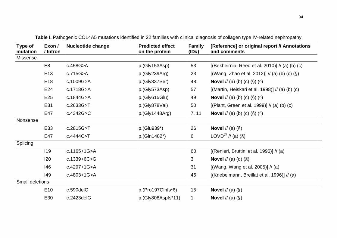
94
Table I. Pathogenic COL4A5 mutations identified in 22 families with clinical diagnosis of collagen type IV-related nephropathy.
Type of mutation
Exon / / Intron
Nucleotide change Predicted effect on the protein
Family (ID#)
[Reference] or original report // Annotations and comments
Missense
E8 c.458G>A p.(Gly153Asp) 53 [(Bekheirnia, Reed et al. 2010)] // (a) (b) (c)
E13 c.715G>A p.(Gly239Arg) 23 [(Wang, Zhao et al. 2012)] // (a) (b) (c) (§)
E18 c.1009G>A p.(Gly337Ser) 48 Novel // (a) (b) (c) (§) (^)
E24 c.1718G>A p.(Gly573Asp) 57 [(Martin, Heiskari et al. 1998)] // (a) (b) (c)
E25 c.1844G>A p.(Gly615Glu) 49 Novel // (a) (b) (c) (§) (^)
E31 c.2633G>T p.(Gly878Val) 50 [(Plant, Green et al. 1999)] // (a) (b) (c)
E47 c.4342G>C p.(Gly1448Arg) 7, 11 Novel // (a) (b) (c) (§) (^) Nonsense
E33 c.2815G>T p.(Glu939*) 26 Novel // (a) (§)
E47 c.4444C>T p.(Gln1482*) 6 LOVD@ // (a) (§)
Splicing
I19 c.1165+1G>A 60 [(Renieri, Bruttini et al. 1996)] // (a)
I20 c.1339+6C>G 3 Novel // (a) (d) (§)
I46 c.4297+1G>A
31 [(Wang, Wang et al. 2005)] // (a)
I49 c.4803+1G>A 45 [(Knebelmann, Breillat et al. 1996)] // (a) Small deletions
E10 c.590delC p.(Pro197Glnfs*6) 15 Novel // (a) (§)
E30 c.2423delG p.(Gly808Aspfs*11) 1 Novel // (a) (§)

95
I30/E31 c.2510(-1)_2525del(15bp)Δ 34 Novel Large deletions
E1-13 delE1_13 (+delE1_3,COL4A6) 33 [(Sa, Fieremans et al. 2013)] (Novel )
E2-29 delE2_29
41 [(Sa, Fieremans et al. 2013)] (Novel )
E2-51 delE2_51
19 [(Plant, Green et al. 1999; Sa, Fieremans et al.
2013)] // (^^) E31 c.2510_2554del(45bp)ΔΔ 28 Novel
E43-45 delE43_45
18 Novel
Mutations were classified according to the Human Gene Mutation Database nomenclature [www.hgmd.cf.ac.uk/ ]. Nonsense point mutations, frameshifting mutations and exonic deletions were considered pathogenic. @ Mutation reported at the LOVD – Leiden Open Variation Database [accessed at https://grenada.lumc.nl/LOVD2/COL4A/variants.php?select_db=COL4A5&action=search_all&search_Variant%2FDNA=c.4444C>T]. Bioinformatic predictions of pathogenicity for missense non-synonymous mutations, splice site intronic mutations and short (<12 nucleotides) microdeletions:
(a) MutationTaster [www.mutationtaster.org/]; (b) PolyPhen-2 [genetics.bwh.harvard.edu/pph2/ ]; (c) SIFT [sift.jcvi.org]; (d) Human Splicing Finder [http://www.umd.be/HSF/].
(§) Sequence variation not identified in 234 control X-chromosomes. (^) Pathogenic mutations reported at the same codons:
(i) p.(Gly337Asp) / c.1010G>A, reported at the Leiden Open Variation Database [LOVD; accessed at https://grenada.lumc.nl/LOVD2/COL4A/variants.php?select_db=COL4A5&action=search_all&search_Variant%2FDNA=c.1010G>A]; (ii) p.(Gly615Arg) / c.1843G>C, reported by Wang et al, 2012(Wang, Zhao et al. 2012); (iii) p.(Gly1448Ser) / c.4342G>A, reported by Wang et al. 2005(Wang, Wang et al. 2005).
(^^) Plant et al. 1999(Plant, Green et al. 1999) also reported large deletions encompassing exons 2_51 but their exact breakpoints were not determined. Δ GGTTTACATCATGGAATA. ΔΔ GTTTACATGGAATACCAGGAGAGAAGGGGGATCCAGGACCTCCTG (p.delGLHGIPGEKGDPGPP): the first nucleotide involved in this deletion is the first nucleotide of exon 31.

96
Inter-gender comparisons of clinical outcomes
Microscopic hematuria was detected in all the 65 patients of the XLAS phenotyping
cohort (Table II). History of macroscopic hematuria was significantly more common in
males than females (62% vs 17%; p=0.002). Proteinuria was more frequently
reported in males than females, but the difference did nor reach statistical
significance (96.2% vs 79%; p=0.067). Although the proportion of patients with
history of hypertension also did not differ significantly between males and females
(73% vs 53%; p=0.112), the median age at onset of hypertension was significantly
lower in the male patients (20 vs 34 years; p=0.002). Eighty-six percent of the male
patients and 62% of the females developed CRF (p=0.029). CRF was diagnosed in
males at a significantly lower age than in females (21 vs 32 years; p=0.038). At
diagnosis of CRF, eGFR was significantly lower in males (40 vs 61 ml/min/1.73m2;
p=0.003). While 77% of male patients with CRF had already started RRT, only 17%
of females with CRF were on RRT (p<0.001). Six males and three females in the
XLAS phenotyping cohort had EM photomicrographs of kidney biopsies available for
review. All of them showed the typical ultrastructural features of AS, i.e. alternate
thinning and thickening of the GBM with splitting and/or reticulation of the lamina
densa, enclosing electron-lucent areas that contain round electron-dense bodies with
diameters of up to 90 nm. Subjective hearing loss was more often reported (87% vs
46%; p=0.001) in males than in females. Likewise, anterior lenticonus and dot-and-
fleck retinopathy were more frequently diagnosed in the males (p=0.013 and
p=0.129, respectively). ATS-DL was diagnosed in a single family, in association with
a large deletion involving COL4A5, but not the COL4A6 gene.(Sa, Fieremans et al.
2013)

97
Table II. Phenotypic comparisons between hemizygous males and heterozygous
females for pathogenic COL4A5 mutations (n=65).
Hemizygotes (n=30)
Heterozygotes (n=35)
p-value
N N PHENOTYPE Age at enrolment (median (interquartile range)) 33.5 (18.5) 30 42 (14.8) 35 0.088 History of renal abnormalities History of macroscopic hematuria (%) Age at diagnosis (median (interquartile range))
61.9 5 (4)
13/21 11
17.4 3 (11.5*)
4/23 3
0.002 0.501
History of microscopic hematuria (%) Age at diagnosis (median (interquartile range))
100.0 12.5 (20.3)
25/25 22
100.0 21 (26.5)
35/35 28
a 0.219
History of proteinuria (%) Age at diagnosis (median (interquartile range))
96.2 18 (22.5)
25/26 21
78.8 24 (19)
26/33 23
0.067 0.508
History of hypertension (%) Age at diagnosis (median (interquartile range))
73.1 20 (11.5)
19/26 12
52.9 34 (22.3)
18/34 16
0.112 0.002
History of CKD stage 2 or higher (%) Age at diagnosis (median (interquartile range)) eGFR at diagnosis (median (interquartile range))
86.2 21 (10) 40 (35)
25/29 16 13
61.8 32 (17.5) 61 (26.3)
21/34 20 20
0.029 0.038 0.003
History of renal replacement therapy (%) Age at onset (median (interquartile range)) eGFR at onset (median (interquartile range))
76.7 23 (19.5) 9 (6)
23/30 21 12
17.1 40 (14) 6 (1)
6/35 6 5
<0.001 0.272 0.019
GBM ultrastructural abnormalities Age at kidney biopsy (median (interquartile range)) 20 (14.5) 6 24 (11*) 3 0.933 Thinning (%) 100.0 6/6 100.0 3/3 a Thickening (%) 100.0 6/6 100.0 3/3 a Lamellation (%) 100.0 6/6 100.0 3/3 a Electrondense bodies (%) 100.0 6/6 100.0 3/3 a Hearing loss Self-noticed or subjective (%) Age at self-noticed or subjective (median (interquartile range))
86.7 15 (30)
26/30 15
45.5 39 (19)
15/33 9
0.001 0.142
Audiogram (%) Age at diagnosis of hearing loss by audiogram (median (interquartile range))
94.1 27.5 (18)
16/17 16
91.3 41 (10)
21/23 13
0.053 0.030
Ocular abnormalities Anterior lenticonus (%) Age at diagnosis (median (interquartile range))
33.3 19 (12)
5/15 5
0.0 -
0/18 -
0.013 -
Maculopathy (%) Age at diagnosis (median (interquartile range))
50.0 25 (19)
10/20 8
26.3 44 (22)
5/19 5
0.129 0.180
Cataracts (%) Age at diagnosis (median (interquartile range))
46.2 30 (35.8)
6/13 6
15.0 56 (56*)
3/20 3
0.107 0.263
Other abnormalities Leiomyomatosis (%) Age at diagnosis (median (interquartile range))
3.3 24 (0*)
1/30 1
5.7 18 (0*)
2/35 1
1.000 0.317
GENOTYPE Truncating mutation (%) 47 14/30 37.1 13/35 0.437
SD: standard deviation. CKD: chronic kidney disease. eGFR: estimated glomerular filtration rate. GBM:
glomerular basement membrane. Age is expressed in years. eGFR is expressed in ml/min/1.73m2. * The range is
indicated instead of the interquartile range. a Statistics not computed because the proportions in the comparison
groups are the same.

98
Genotype-phenotype correlations
Among males, no statistically significant differences were found between patients
with truncating and non-truncating COL4A5 mutations in the prevalence of the major
renal manifestations of AS, or of their natural histories as captured by age or severity
of CRF at diagnosis (Supplementary table S11). Although the prevalence of SNHL
was not statistically different in the two groups, subjective hearing loss was reported
by patients with truncating mutations at a much younger age than by those with non-
truncating mutations (7 vs 36 years; p<0.001). While the difference in the
prevalences of anterior lenticonus in patients with truncating and non-truncating
mutations who underwent formal ophthalmological examination did not reach
statistical significance (50% vs 14%; p=0.282), dot-and-fleck retinopathy was
identified in a significantly higher proportion of patients with truncating mutations
(75% vs 13%; p=0.020).
Among females, CRF was diagnosed in 92.3% of those with truncating
COL4A5 mutations but only in 43% of those with non-truncating mutations (p=0.005)
(Supplementary table S12). However, the age at diagnosis of CRF did not differ
significantly (32 vs 31 years; p=0.684) and the proportion of patients who needed
RRT was similar in the two groups (15% vs 18%; p=1.000). Patients with truncating
mutations reported subjective hearing loss more frequently than those with non-
truncating mutations (69% vs 30%; p=0.027) and SNHL was also confirmed by tonal
audiogram at a significantly younger age in the former group of patients (38 vs 47
years; p=0.003). Irrespective of the type of mutation, ocular involvement was
diagnosed in only a small minority of females with AS.
DISCUSSION
Herein, we report the clinical characterization, the molecular pathology and the
genotype-phenotype correlations observed in 22 Portuguese families with genetically
confirmed XLAS, identified by mutational screening of a nationwide cohort of 65 non-
related probands in whom the diagnosis of AS was considered clinically established
or probable, according to standard clinical and pathologic criteria.(Flinter, Cameron et
al. 1988)

99
A pathogenic COL4A5 mutation was identified in only 34% (22/65) of families.
This prevalence is within the 30-67% range reported in several large studies that
have used single-strand conformation polymorphism (SSCP) analysis to screen for
COL4A5 mutations,(Hertz 2009) but is significantly lower than the 82% rate reported
with PCR amplification and direct DNA sequencing of the promoter and all the exons
of COL4A5.(Martin, Heiskari et al. 1998) Such a result cannot be attributed to
limitations of the laboratory methods that we have used to scan for mutations, as the
sensitivity of MLPA combined with direct sequencing to identify COL4A5 mutations is
higher than 95%(Hanson, Storey et al. 2011) while that of SSCP is estimated to be
around 80%.(Grompe 1993) Although we did not scan for promoter and deep intronic
mutations, missing COL4A5 mutations at those locations is an unlikely
explanation(King, Flinter et al. 2006) for the lower than expected prevalence of XLAS
observed in the Portuguese families.
The wide variety of different types of pathogenic COL4A5 mutations identified
in this study is further evidence of the effectiveness of our laboratory approach, and
their relative frequencies did not significantly differ from those reported to the
HGMD®. Surprisingly, a large deletion confined to COL4A5 was identified in a family
with ATS-DL, challenging an established concept of genotype-phenotype
correlation.(Sa, Fieremans et al. 2013) Pathogenic COL4A5 mutations were novel in
57% of families (12/21) and occurred de novo in 15% (2/13) of the probands, which is
within the expected 10-15% range.(Kashtan 2001) With the exception of a glycine
substitution (p.Gly1448Arg) shared by two apparently unrelated families from
neighboring villages, all other pathogenic COL4A5 mutations identified in our cohort
were exclusive of single families. These findings show that mutation panels designed
for the genetic diagnosis of AS should be based on population-specific data.4
The detection rates of pathogenic COL4A5 mutations in probands with
complete clinical assessment (n=22) that fulfilled one, two, three, or four diagnostic
criteria were, respectively, 0.0%, 25.0%, 14.3% and 80.0%. The corresponding
figures reported for British patients (n=101) were 9.5%, 48.5%, 82.1%, and
81.3%,(Hanson, Storey et al. 2011) which differ significantly from our own results
only for patients fulfilling three diagnostic criteria. Since the extrarenal manifestations
of AS seem to be relatively rare in patients with autosomal forms of the

100
disease,(Tryggvason and Patrakka 2009) a possible explanation for that difference
might be the higher prevalence of ADAS or ARAS in Portuguese families. Indeed,
preliminary results of second-tier mutational screening of COL4A3 and COL4A4
[manuscript submitted to the JMG simultaneously] suggest that the proportion of
XLAS in Portuguese families with AS is significantly lower than the 80-85% generally
referred in the literature.(Kashtan 1999; Tryggvason and Patrakka 2009; Kruegel,
Rubel et al. 2013) These data are much in agreement with those of a recent Italian
study(Fallerini, Dosa et al. 2013) reporting the diagnosis of XLAS in only 65% of 48
families with genetically confirmed AS, by unbiased simultaneous next generation
sequencing (NGS) of COL4A5, COL4A3 and COL4A4. In keeping with the above
argument, more than half of the Italian patients that presented with only one or two
diagnostic criteria of AS had a pathogenic mutation identified by NGS.(Fallerini, Dosa
et al. 2013)
The clinical manifestations of renal disease observed in our phenotyping
cohort did not substantially differ from the classical descriptions of the
XLAS.(Kashtan 1999; Tryggvason and Patrakka 2009) Microscopic hematuria was
the most consistent diagnostic clue, being present in all cases, irrespective of
gender. Although history of macroscopic hematuria was much more frequent in
males, the first episode typically occurred before school age in both boys and girls.
Proteinuria was also highly prevalent in males and females but its diagnosis usually
followed the detection of hematuria by several years. The risks of developing
progressive CRF and of needing RRT were considerably higher in males;
nevertheless, CRF was diagnosed in 62% of the heterozygous females, at young
adult age in many cases. Only two-thirds of the patients in our genotyping cohort had
undergone audiological assessment and only approximately 58% had been referred
for specialized ophthalmological examination. These data show that formal screening
for the extrarenal signs of AS is frequently overlooked in clinical practice.
Because of the genetic heterogeneity and variable clinical expression of AS,
and the incomplete penetrance of XLAS in females, clinicians should take a detailed
three-generation family history in order to try to recognize a pattern of inheritance,
before initiating genetic testing for AS.(Hanson, Storey et al. 2011) Particularly in
females, the diagnosis of microscopic hematuria requires repeated urinalysis. Not

101
screening for SNHL by audiogram, and for the typical ocular lesions by slit-lamp
ophthalmological examination, are common errors in clinical practice that should be
avoided. Indeed, failure to comprehensively assess patients for the clinical diagnostic
criteria of AS may be one of the reasons for missing the diagnosis in some cases, for
uninformative family histories and erroneous estimates of genetic risks, and for false
assumptions of de novo mutations.
In contrast with results of previous studies,(Jais, Knebelmann et al. 2000;
Gross, Netzer et al. 2002; Bekheirnia, Reed et al. 2010) we did not identify any
significant differences on the prevalence or severity of expression of renal disease
between males carrying truncating or non-truncating COL4A5 mutations, although
they had a more severe extrarenal phenotypes. On the other hand, the higher
prevalence of CRF and of hearing loss in females with truncating COL4A5 mutations
observed in our cohort was not apparent in the largest study of females with XLAS
reported so far.(Jais, Knebelmann et al. 2003) However, the relative small size of our
study cohort should caution about the generalization of the present findings.
Genetic testing in AS is useful for confirming a clinical diagnosis, informing
genetic counseling, and facilitating both prenatal and pre-implantation
diagnosis.(Bekheirnia, Reed et al. 2010) The identification of a pathogenic COL4A5
mutation is the most specific diagnostic criterion of XLAS, avoiding more invasive
and complex diagnostic procedures as kidney or skin biopsy.(Kashtan 1993-2013) In
addition, the type of mutation is of help in the estimation of the clinical
prognosis.(Jais, Knebelmann et al. 2000; Gross, Netzer et al. 2002; Bekheirnia, Reed
et al. 2010) However, molecular genetic analysis by Sanger sequencing of all 53
COL4A5 exons and their flanking intronic sequences is an expensive, time-
consuming process and several weeks are usually needed to complete a
diagnosis.(Artuso, Fallerini et al. 2012) The recently available, high-throughput NGS
methods for DNA sequencing are cheaper and have much lower turn-around times
than the conventional Sanger method. High-throughput DNA sequencing methods
are most useful for the molecular diagnosis of genetically heterogeneous disorders,
and NGS may soon become the first-tier genetic test in cases where AS is clinically
suspected, particularly in populations where the prevalence of XLAS is not much
higher than that of the autosomal forms.(Fallerini, Dosa et al. 2013) In those families

102
where the causative COL4A5 mutation is already known, genetic testing of at-risk
persons is much less expensive, allows the accurate assessment of individual
genetic risks and provides a tool for pre-symptomatic diagnosis, which is particularly
valuable in females, and for the selection of potential family donors for kidney
transplantation.(Gross, Weber et al. 2009; Kashtan 2009) Moreover, early
confirmation of the diagnosis of XLAS offers the opportunity to optimize treatment
outcomes, since pharmacologic blockade of the renin–angiotensin–aldosterone
system with angiotensin converting enzyme inhibitors or angiotensin receptor
blockers proved effective in delaying the progression of CRF in both males and
females.(Gross, Licht et al. 2012; Temme, Peters et al. 2012)

103
ACKNOWLEDGEMENTS
The authors thank the patients and their relatives for the invaluable collaboration in
this study; NephroCare – Portugal, for having allowed the nationwide ascertainment
of patients with Alport syndrome in their dialysis centers; and Dr.s Adelino Carvalho
(NephroCare, Santarém), Ana Bernardo (NephroCare, Covilhã), Carlos Miguel
Botelho (NephroCare, Viseu), Joana Felgueiras (NephroCare, Montijo), Liliana Pinho
(Dialysis clinic, Paredes) and Luís Filipe Carvalho (NephroCare, Restelo), for their
contributions to the clinical data collection of dialysis patients enrolled in this study.
The authors also thank Dr. Francis Earl Cook, from the Department of Epidemiology,
Harvard School of Public Health, Boston, MA, for his advice and supervision of the
epidemiological and statistical analyses.
This work is part of the PhD project of Maria João Nabais Sá (supervisor: João Paulo
Oliveira), to be presented at the ICBAS – “Instituto de Ciências Biomédicas Abel
Salazar” (Abel Salazar Biomedical Sciences Institute), University of Porto, Portugal.
These studies were partially supported by FEDER funds through the COMPETE –
“Programa Operacional Factores de Competitividade”, and by Portuguese national
funds through the FCT – “Fundação para a Ciência e a Tecnologia” (Foundation for
Science and Technology) within the framework of the Cooperation Agreement
between Portugal and Harvard Medical School (FCOMP-01-0124-FEDER-HMSP-
ICJ/SAU-ICT/0003/2009); by a research grant from the Portuguese Society of
Nephrology; by the Unit of Research & Development in Nephrology (FCT-725) at the
Department of Nephrology of the Faculty of Medicine, University of Porto, Portugal;
and by the São João Hospital Centre.
Parts of these data were presented as posters at the European Human Genetics
Conference 2012, Nürnberg, Germany, June 23-26, 2012 (P12.007) and at the 62nd
Annual Meeting of the American Society of Human Genetics, November 6-10, 2012
San Francisco, California (3122F).

104
Supplementary table S1. Outcome variables, definitions, measurement of outcomes, diagnostic methods, degree of evidence and time
of collection of clinical variables.
Outcomes Definition of outcomes Measurement o f outcomes Diagnostic evaluation
Degree of evidence
Time of diagnosis
Birth District District of Portugal where the patient was born Portuguese districts None I, CF, Q –
Residence District District of Portugal where the patient lived at the date of enrollment
Portuguese districts None I, CF, Q –
Affected relatives Family history of hematuria, CRF or hearing loss Exams required to evaluate the renal, audiologic and ocular involvement
Pedigree I, CF, Q At enrollment Retrospectively
Renal anomalies
History of macroscopic hematuria
Presence of red blood cell (RBC) in urine visible to the eye, as “cola-colored” urine.
Occurrence of outcome (Yes/No) Age at first event (in years)
Urinalysis I, CF, Q Retrospectively
History of microscopic hematuria
≥3 RBC visible per high power field under light microscopy examination of the centrifuged urine sediment or an equivalent value per microliter when assessed by flow cytometry
Occurrence of outcome (Yes/No) Age at first event (in years)
Urinalysis: - RBC count
Urinalysis report I, CF, Q
Retrospectively At enrollment During the study
History of proteinuria
Urinary protein excretion ≥300 mg/day or a urinary protein to creatinine ratio ≥300 mg/g, in morning spot samples. The highest recorded level of 24-hour proteinuria was graded as follows: ≤500 mg/day; 500–1000 mg/day; 1000–3500 mg/day; ≥3500 mg/day.
Occurrence of outcome (Yes/No) Age at first event (in years) Degree of proteinuria (mg/day or mg/g) at diagnosis/most recent Highest recorded level of proteinuria
Urinalysis: - Protein count
Urinalysis report I, CF, Q
Retrospectively At enrollment During the study

105
Outcomes Definition of outcomes Measurement o f outcomes Diagnostic evaluation
Degree of evidence
Time of diagnosis
History of hypertension
Systolic blood pressure ≥140 mmHg and/or diastolic blood pressure ≥90 mmHg. Patients undergoing chronic anti-hypertensive medication were also considered to have hypertension, irrespective of their blood pressure levels.
Occurrence of outcome (Yes/No) Age at first event (in years) Therapy with anti-hypertensive medication (Yes/No) Age at onset of anti-hypertensive medication (in years)
Blood pressure measurement
I, CF, Q Retrospectively At enrollment During the study
History of CRF (≥ stage 2 CKD)
Plasma creatinine (pCr) level ≥1.2 mg/dl in males and ≥0.9 mg/dl in females.*
Plasma creatinine level (mg/dl) CKD stage (≥2, according to the KDOQI clinical guidelines)
Blood analysis: - Plasma creatinine level
Blood analysis report I, CF, Q
Retrospectively At enrollment During the study
History of advanced CRF (≥ stage 3 CKD)
Plasma creatinine level >1.5 mg/dl in males and >1.2 mg/dl in females.
Plasma creatinine level (mg/dl) CKD stage (3 to 5, according to the KDOQI clinical guidelines)
Blood analysis: - Plasma creatinine level
Blood analysis report I, CF, Q
Retrospectively At enrollment During the study
Estimated glomerular filtration rate (eGFR)
eGFR computed from pCr values, namely at the diagnosis of CRF and advanced CRF (eGFR at diagnosis of stage 2 and stage 3 CKD).**
eGFR (ml/min/1.73 m2) CKD stage (≥2, according to the KDOQI clinical guidelines)
Blood analysis: - Plasma creatinine level
Blood analysis report I, CF, Q
Retrospectively At enrollment During the study
History of renal replacement therapy (RRT)
First procedure of renal replacement function (hemodialysis, peritoneal dialysis or renal transplantation), performed when the GFR reached <15 ml/min
Need of RRT (Yes/No) Age at onset of first RRT (in years) Plasma creatinine level at onset of first RRT (mg/dl) eGFR at onset of first RRT (ml/min/1.73 m2)
Blood analysis: - Plasma creatinine level
Blood analysis report I, CF, Q
Retrospectively At enrollment During the study
Time between onset of first RRT and first kidney transplant
Time from the date of onset of first RRT (dialysis) and first kidney transplant
Time (in months) None I, CF, Q
Retrospectively At enrollment During the study
Last available pCr (eGFR)
eGFR calculated from the last measurement of plasma creatinine level of the participant, to evaluate his/her CKD stage**
eGFR (ml/min/1.73 m2) CKD stage (≥2, according to the KDOQI clinical guidelines)
Blood analysis: - Plasma creatinine level
Blood analysis report I, CF, Q
Retrospectively At enrollment During the study

106
Outcomes Definition of outcomes Measurement o f outcomes Diagnostic evaluation
Degree of evidence
Time of diagnosis
Typical ultrastuctural GBM abnormalities
Characteristic ultrastructural GBM abnormalities of Alport syndrome, namely thinning, thickening, lamellation and electron-dense particles in GBM lacunae
Age at kidney biopsy (in years) GBM thinning (Yes/No) GBM thickening (Yes/No) GBM lamellation (Yes/No) Electron-dense particules in GBM lacunae (Yes/No)
Kidney biopsy with electron microscopy examination
Pathology database: - Electron microscopy photographies Kidney biopsy report (CF), Q
Retrospectively
Typical hearing loss (HL)
History of self-noticed HL
Self-perception of HL and approximated age at onset of self-noticed HL
Self-noticed HL (Yes/No) Age at onset of self-noticed HL (in years)
None I, CF, Q Retrospectively
Diagnosis of HL Diagnosis of HL and its characterization by audiogram, including uni- or bilateral HL, type of HL and degree of HL. The degree of HL was classified using standardized hearing loss categories, according to the WHO classification (Danermark, Cieza et al. 2010), as follows: dB HL < 26 (normal); 26 ≤ dB HL < 40 (Mild HL); 41 ≤ dB HL < 60 (Moderate HL); 61 ≤ dB HL < 80 (Severe HL); 80 < dB HL (Profound HL).
Diagnosis of HL (Yes/No) Type of HL Degree of HL: - Pure tone average (PTA): average of the three thresholds (in decibels, dB) at 500 Hz, 1000 Hz and 2000 Hz - Average of high frequencies: average of the two thresholds (in decibels, dB) at 4000 Hz and 8000 Hz Age at the first and the most recent audiometric evaluations (in years)
Pure tone audiogram High frequency audiogram
Audiogram Retrospectively At enrollment During the study
Typical ocular abnormalities
History of anterior lenticonus, retinopathy and
History of and age at diagnosis of anterior lenticonus, dot-and-fleck maculopathy, retinal atrophy and cataracts
Anterior lenticonus (Yes/No) Dot-and-fleck maculopathy (Yes/No)
BiomicroscopyFunduscopy OCT
Biomicroscopy photography Fundus
Retrospectively At enrollment During the study

107
Outcomes Definition of outcomes Measurement o f outcomes Diagnostic evaluation
Degree of evidence
Time of diagnosis
cataracts Retinal atrophy (Yes/No) Cataracts (Yes/No) Age at the first and the most recent ophthalmological assessment (in years)
photography I, CF, Q
Other clinical outcomes
Abnormal type IV collagen expression in EBM
Abnormal epidermal basement membrane (EBM) reactivity to a monoclonal antibody directed against the α5(IV) chain
Complete absence, discontinuous or presence of staining of the EBM with a monoclonal antibody directed against the collagen α5(IV) chain
Skin biopsy with immunohistochemical analysis of the type IV collagen expression in the EBM
EBM photography
Retrospectively During the study
History of diffuse leiomyomatosis
Gastrointestinal, tracheobronchial and female genital manifestations
Histologic diagnosis Endoscopic ultrasonography with esophageal biopsy
Pathology report I, CF, Q
Retrospectively At enrolment During the study
History of intellectual impairment
Level of education and employment Number of year in school Degree completed Job
None I, CF, Q Retrospectively At enrolment During the study
Typical dysmorphic face
Craneofacial dysmorphy described in patients with AMME complex (Meloni et al., 2002)
Mild midface hypoplasia Anteverted nares Flat nasal bridge Downward slanting palpebral fissures Peripalpebral fullness Facial hypotonia
Physical examination
Facial photography I, CF, Q
Retrospectively At enrollment

108
I: Patient interview. CF: Patient clinical file: consultation registry, pre-dialysis medical report, referral information. Q: Questionnaire filled by assistant physician. OCT: Optical Coherence Tomography. *These pCr cut-off values were selected to be roughly equivalent to the upper limits of eGFR for the diagnosis respectively of stage 2 and stage 3 chronic kidney disease (CKD), in patients with kidney damage, over the age of 18 years, according to the Kidney Disease Outcomes Quality Initiative (KDOQI) clinical practice guidelines for CKD evaluation, classification and stratification of the National Kidney Foundation (NKF; New York, NY, USA) (http://www.kidney.org/professionals/KDOQI/guidelines_ckd/p4_class_g1.htm). **The eGFR was computed from pCr values using the calculators available online at the NKF website (http://www.kidney.org/professionals/kdoqi/gfr_calculator.cfm), as appropriate. [Online calculator used at the NKF website: CKD-EPI Creatinine equation 2009]

109
Supplementary table S2. Primer sequences, annealing temperatures and product size for PCR amplification of the COL4A5 gene.†
Exons Foward Primers (5’ to 3’) Reverse Primers (5’ to 3’) Annealing
temperature
PCR
Product size
1 AAGCCTCACTGTCCCTCTC AAAGGAAGATAAAGGGACCC 60ºC 296 bp
2 GATTGTTGATTTCAGTTGAGCTGT CTTAAGTACTGAGATAGAAGCTC 51ºC 248 bp
3 TCTCAACCATGCCTGTGCTTG TGATGTGACACCTAGTCCCAC 60ºC 229 bp
4 TCACAGATGTTTACAGTAGTTTAAA GGTCTTTTCCAATTGTCTCAT 57ºC 237 bp
5(F)/6(R) GAACAATGAGTAGTAATAAATAGTG TTGTCAAGTATACTACCCAAGAT 52ºC 373 bp
7(F)/8(R) GGAAAGTGAAGGCTAATGAAC ATTATCCTATTGAAGTTGCCAGC 52ºC 644 bp
9 GCCTAATCTTTTAGTACATCTC GTACTAAAGGTTGAGGGAT 52ºC 275 bp
10 GGCGACACAAGTGAGACTTT GAATGTTGAGAATGCATTATGTTTTC 52ºC 248 bp
11(F)/13(R) AATACTATTTTGATGGGCTTT TATACATTGTGATGTGATTAC 52ºC 628 bp
14(F)/16(R) CTCCAGCTCTAACCATGTTG TTTTGTCATACTGCTTCTCT 55ºC 726 bp
17 GAAGACAATCTTTGGAG TAACTCAAGCCTGGGAGAA 52ºC 201 bp
18 TGGAAAGTTTCTCTTATATTC TGTGTTCTATCAAGAAAGAG 48ºC 224 bp
19 CAGGAGAACAAGGCTTTTCTTCTTTGCA
TT
ACATGGATTAGTAAGGATGC 52ºC 248 bp
20(F)/21(R) GAAGATCTTATCATTATCTAATG GTCTTGGGGTATGACCAT 51ºC 793 bp
22 GTGGAAATGCTGTCCCTTAG CATTCTAAAAGTATAAGCTCAAC 52ºC 266 bp
23(F)/24(R) TCAAAGCTTACGTTATTGTGT GGTGTTTATCTCAGCATCAG 55ºC 726 bp
25 GAAGAACTATTTATGGCTATATCC TTGGCTACTCATGGCTTCC 57ºC 350 bp
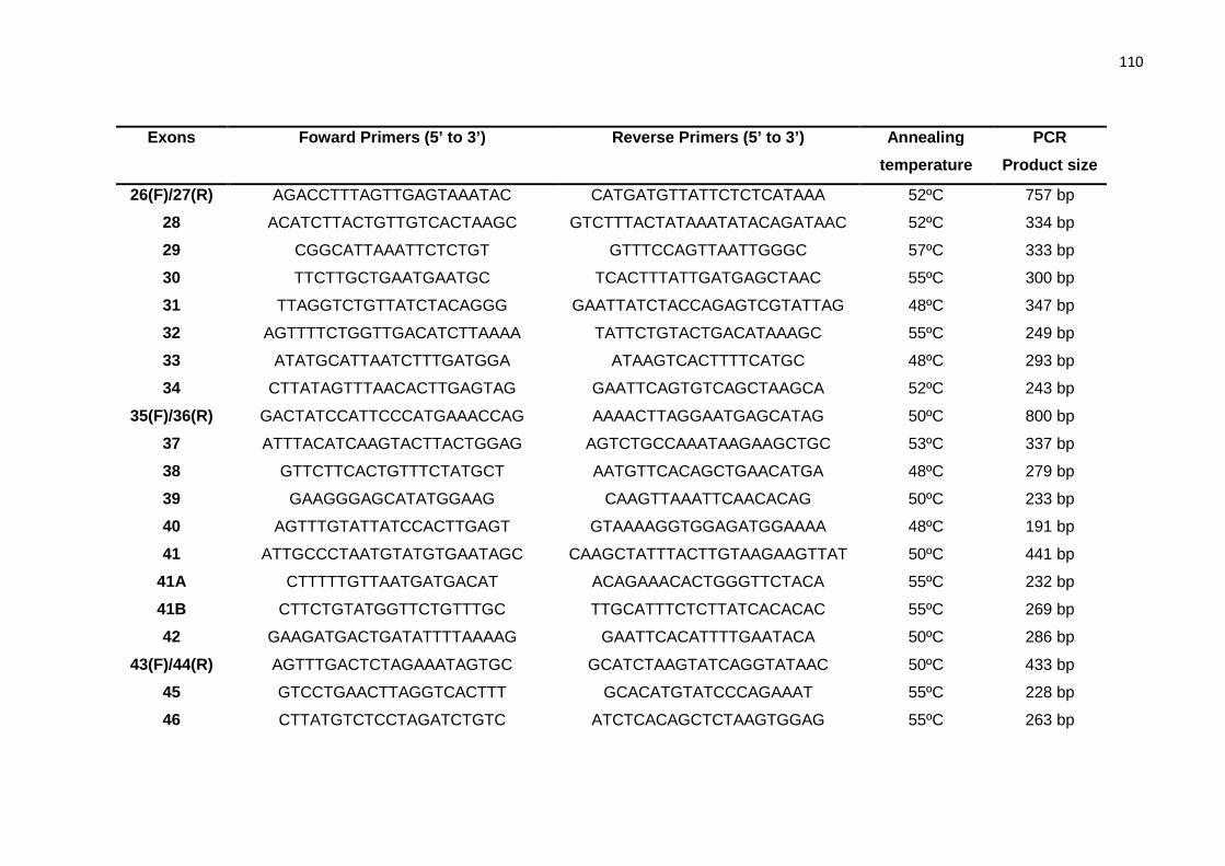
110
Exons Foward Primers (5’ to 3’) Reverse Primers (5’ to 3’) Annealing
temperature
PCR
Product size
26(F)/27(R) AGACCTTTAGTTGAGTAAATAC CATGATGTTATTCTCTCATAAA 52ºC 757 bp
28 ACATCTTACTGTTGTCACTAAGC GTCTTTACTATAAATATACAGATAAC 52ºC 334 bp
29 CGGCATTAAATTCTCTGT GTTTCCAGTTAATTGGGC 57ºC 333 bp
30 TTCTTGCTGAATGAATGC TCACTTTATTGATGAGCTAAC 55ºC 300 bp
31 TTAGGTCTGTTATCTACAGGG GAATTATCTACCAGAGTCGTATTAG 48ºC 347 bp
32 AGTTTTCTGGTTGACATCTTAAAA TATTCTGTACTGACATAAAGC 55ºC 249 bp
33 ATATGCATTAATCTTTGATGGA ATAAGTCACTTTTCATGC 48ºC 293 bp
34 CTTATAGTTTAACACTTGAGTAG GAATTCAGTGTCAGCTAAGCA 52ºC 243 bp
35(F)/36(R) GACTATCCATTCCCATGAAACCAG AAAACTTAGGAATGAGCATAG 50ºC 800 bp
37 ATTTACATCAAGTACTTACTGGAG AGTCTGCCAAATAAGAAGCTGC 53ºC 337 bp
38 GTTCTTCACTGTTTCTATGCT AATGTTCACAGCTGAACATGA 48ºC 279 bp
39 GAAGGGAGCATATGGAAG CAAGTTAAATTCAACACAG 50ºC 233 bp
40 AGTTTGTATTATCCACTTGAGT GTAAAAGGTGGAGATGGAAAA 48ºC 191 bp
41 ATTGCCCTAATGTATGTGAATAGC CAAGCTATTTACTTGTAAGAAGTTAT 50ºC 441 bp
41A CTTTTTGTTAATGATGACAT ACAGAAACACTGGGTTCTACA 55ºC 232 bp
41B CTTCTGTATGGTTCTGTTTGC TTGCATTTCTCTTATCACACAC 55ºC 269 bp
42 GAAGATGACTGATATTTTAAAAG GAATTCACATTTTGAATACA 50ºC 286 bp
43(F)/44(R) AGTTTGACTCTAGAAATAGTGC GCATCTAAGTATCAGGTATAAC 50ºC 433 bp
45 GTCCTGAACTTAGGTCACTTT GCACATGTATCCCAGAAAT 55ºC 228 bp
46 CTTATGTCTCCTAGATCTGTC ATCTCACAGCTCTAAGTGGAG 55ºC 263 bp

111
Exons Foward Primers (5’ to 3’) Reverse Primers (5’ to 3’) Annealing
temperature
PCR
Product size
47 ATGAGGTCATAATGTTTTGTC CACTTTGGCCAAGGCTACTC 55ºC 386 bp
48 TTGAAAGGCTGTTTGCTATTG ACCTAGGAGGAATATCATCAG 55ºC 364 bp
49 ATCGGCTTCCATACTAAGAAG GCAAATGACAGGGATTCCTC 55ºC 304 bp
50 GATTTGAATTTGGCCAAGCTC CATTTGTTGAGGATAAACCAT 55ºC 366 bp
51 GTCACCAAGAGAGCTACTTAACAC CATTGACGGCAGCAGTAGTAAAG 55ºC 303 bp
† Cycling conditions (adapted from (Martin, Heiskari et al. 1998)) were: initial denaturation for 5 minutes at 94ºC, followed by 35
cycles of denaturation at 94ºC for 60 seconds, annealing for 60 seconds, and extension at 72ºC for 90 seconds, ending with a final
extension at 72ºC for 10 minutes.

112
Supplementary figure S3

113
Supplementary figure S3 . Map of Portugal showing the boundaries of each of the
14 continental districts (white background) and the two autonomous Atlantic islands
regions. The names and geographic locations (black squares) of the respective
capital cities are shown only for those where probands enrolled in this study were
resident (numbers in parenthesis). Fifty-one (78.5%) of the 65 probands were
enrolled through 11 nephrology and two genetics services of public hospitals, and 14
(21.5%) were enrolled through 12 outpatient dialysis clinics. Of the 16 nephrology
departments of public hospitals that did not enroll patients, four did not reply to the
invitation to participate, two declined to participate and ten responded they had no
patients with the clinical diagnosis of Alport syndrome. Nationwide case
ascertainment through public hospitals and private dialysis clinics minimized the
negative impact that the nonparticipation in this study of some public hospitals might
have had upon the catchment of affected families in some regions of the country.
Indeed, several families could have been redundantly identified by different patients,
at different stages of kidney disease, through different sites. The average proband
catchment, per 100,000 inhabitants, was 0.62 in the continental districts and 1.12 in
the Madeira Islands. The geographic distribution of the 65 probands by area of
residence broadly paralleled the Portuguese population density, when comparing the
coastal, densely populated, and the inland, scarcely populated, regions. Possible
explanations for the lower than expected catchment in a few of the highly populated
central-western districts, including in the capital Lisbon district, are the
nonparticipation in this study of a private dialysis clinics network that is the major
provider of dialysis care in some of those districts, and the exclusion of four families
with X-linked Alport syndrome that had been previously enrolled in the European
Community Alport Syndrome Concerted Action. In contrast, the disproportionately
higher number of families identified in the northwestern districts of Braga and Porto
most probably reflects the long-term activity of an outpatient clinic dedicated to
hereditary kidney disorders operating in a major academic hospital in Porto.

114
Supplementary table S4. COL4A5 mutation detection rate per number of diagnostic criteria met, among probands tested for
COL4A5, with complete and incomplete clinical evaluation (n=60).
Diagnostic criteria COL4A5 molecular study
FH AC OC UC Probands with a pathogenic
mutation identified
Probands without a pathogenic
mutation identified
Total number of probands
Mutation detection
rate
Mutation detection rate per number of diagnostic criteria
1 criterion FH 9 4 5 9 44% 33%
AC 1
0 1 1 0% (4/12)
OC 0
0
UC 2 0 2 2 0%
2 criteria FH 14
8 6 14 57% 38%
1 0 1 1 0% (10/26)
7 1 6 7 14%
AC 1
0 1 1 0%
3 1 2 3 33%
OC 0 0
3 criteria FH AC 9
2 7 9 22% 24%
5 1 4 5 20% (4/17)
FH OC 0 0
AC OC 3 1 2 3 33%
4 criteria FH AC OC 5 4 1 5 80% 80% (4/5)
FH: family history. AC: audiologic criteria; hearing loss, with and without confirmation by audiogram. OC: ophthalmologic criteria; ocular signs, including anterior lenticonus, cataracts and retinopathy. UC: ultrastructural criteria; characteristic glomerular basement membrane changes on electron microscopy examination of kidney biopsy, including
thinning, thickening, lamellation and presence of electron-dense bodies.

115
Supplementary table S5. COL4A5 mutation detection rate per number of diagnostic criteria met, among probands tested for
COL4A5, with complete clinical evaluation (n=22).
Diagnostic criteria COL4A5 molecular study
FH AC OC UC
Probands with
a pathogenic
mutation identified
Probands without
a pathogenic
mutation identified
Total
number of
probands
Mutation
detection
rate
Mutation detection
rate per number of
diagnostic criteria
1 criterion FH 0
0 0%
AC
0
0 (0/2)
OC
0
0
UC
2 0 2 2 0%
2 criteria FH
0
0 25%
0
0 (2/8)
5 1 4 5 20%
AC
0
0
3 1 2 3 33%
OC
0 0
3 criteria FH AC
0
0 14%
4 0 4 4 0% (1/7)
FH OC
0 0
AC OC
3 1 2 3 33%
4 criteria FH AC OC
5 4 1 5 80% 80% (4/5)
FH: family history.
AC: audiologic criteria; hearing loss, with and without confirmation by audiogram.
OC: ophthalmologic criteria; ocular signs, including anterior lenticonus, cataracts and retinopathy.
UC: ultrastructural criteria; characteristic glomerular basement membrane changes on electron microscopy examination of kidney biopsy, including thinning,
thickening, lamellation and presence of electron-dense bodies.

116
Supplementary figure S6
Supplementary figure S6. Pedigrees of families 7 and 11, with X-linked
Alport syndrome segregating with the COL4A5 mutation p.Gly1448Arg, in
exon 47, showing the individual haplotypes for a set of four polymorphic
microsatellite markers adjacent to the COL4A5 and COL4A6 genes. The
numbers indicate the size of each allele, in base pairs.

117
Supplementary table S7. Correlation between genotype and phenotype in Portuguese male patients with truncating COL4A5
mutations (n=14).
a In brackets, the age at diagnosis or at last screening, in years. b In brackets, the age at start of hemodialysis (HD), peritoneal dialysis (PD) or of kidney
transplant (Tx). c In brackets, the age at kidney biopsy. d In brackets, the age at skin biopsy. < Before the age indicated within brackets. CKD: chronic kidney
disease. pCr: plasma creatinine level, in micromol/L. Conversion of micromoles/L (SI units) into mg/dL may be performed, dividing SI units by the conversion
factor 88.4. eGFR: estimated glomerular filtration rate, in mL/min/1.73m2. GBM: glomerular basement membrane. EBM: epidermal basement membrane. ND:
effect on splicing not determined by mRNA analysis. Genetic probands are indicated in bold.

118
Supplementary table S8. Correlation between genotype and phenotype in Portuguese male patients with non-truncating COL4A5
mutations (n=16).
NA: Data not available. a In brackets, the age at diagnosis or at last screening, in years. b In brackets, the age at start of hemodialysis (HD), peritoneal dialysis
(PD) or of kidney transplant (Tx). c In brackets, the age at kidney biopsy. d In brackets, the age at skin biopsy. < Before the age indicated within brackets.
CKD: chronic kidney disease. pCr: plasma creatinine level, in micromol/L. Conversion of micromoles/L (SI units) into mg/dL may be performed, dividing SI
units by the conversion factor 88.4. eGFR: estimated glomerular filtration rate, in mL/min/1.73m2. GBM: glomerular basement membrane. EBM: epidermal
basement membrane. ND: effect on splicing not determined by mRNA analysis. Genetic probands are indicated in bold.

119
Supplementary table S9. Correlation between genotype and phenotype in Portuguese female patients with truncating COL4A5
mutations (n=13).
NA: Data not available. a In brackets, the age at diagnosis or at last screening, in years. b In brackets, the age at start of hemodialysis (HD), peritoneal dialysis
(PD) or of kidney transplant (Tx). c In brackets, the age at kidney biopsy. d In brackets, the age at skin biopsy. < Before the age indicated within brackets. CKD:
chronic kidney disease. pCr: plasma creatinine level, in micromol/L. Conversion of micromoles/L (SI units) into mg/dL may be performed, dividing SI units by
the conversion factor 88.4. eGFR: estimated glomerular filtration rate, in mL/min/1.73m2. GBM: glomerular basement membrane. EBM: epidermal basement
membrane. ND: effect on splicing not determined by mRNA analysis. Genetic probands are indicated in bold.

120
Supplementary table S10. Correlation between genotype and phenotype in Portuguese female patients with truncating COL4A5
mutations (n=13).
a In brackets, the age at diagnosis or at last screening, in years. b In brackets, the age at start of hemodialysis (HD), peritoneal dialysis (PD) or of kidney
transplant (Tx). c In brackets, the age at kidney biopsy. d In brackets, the age at skin biopsy. < Before the age indicated within brackets. CKD: chronic kidney
disease. pCr: plasma creatinine level, in micromol/L. Conversion of micromoles/L (SI units) into mg/dL may be performed, dividing SI units by the conversion
factor 88.4. eGFR: estimated glomerular filtration rate, in mL/min/1.73m2. GBM: glomerular basement membrane. EBM: epidermal basement membrane. ND:
effect on splicing not determined by mRNA analysis. Genetic probands are indicated in bold.

121
Supplementary table S11. Phenotypic comparisons between hemizygous
males with truncating and non-truncating pathogenic COL4A5 mutations
(n=30).
Truncating (n=14)
Non-truncating (n=16)
p-value
N N PHENOTYPE Age at enrollment (median (interquartile range)) 28 (11.8) 14 41 (19) 16 0.046 History of renal abnormalities History of macroscopic hematuria (%) Age at diagnosis (median (interquartile range))
50.0 3.5 (10.5)
4/8 4
69.2 5 (3)
9/13 7
0.646 0.527
History of microscopic hematuria (%) Age at diagnosis (median (interquartile range))
100.0 15 (13)
12/12 9
100.0 8 (27.5)
13/13 13
a 0.969
History of proteinuria (%) Age at diagnosis (median (interquartile range))
100.0 16.5 (13)
13/13 10
92.3 23 (26)
12/13 11
1.000 0.172
History of hypertension (%) Age at diagnosis (median (interquartile range))
91.7 20 (5)
11/12 5
57.1 20 (19)
8/14 7
0.081 0.417
History of CKD stage 2 or higher (%) Age at diagnosis (median (interquartile range)) eGFR at diagnosis (median (interquartile range))
100.0 18 (7) 23 (39)
13/13 8 7
75.0 26 (10) 41.5 (35)
12/16 8 6
0.107 0.204 0.224
History of renal replacement therapy (%) Age at onset (median (interquartile range)) eGFR at onset (median (interquartile range))
78.6 21.5 (12) 9 (2.3)
11/14 10 6
75.0 25 (28) 10.5 (10)
12/16 11 6
1.000 0.512 0.936
GBM ultrastructural abnormalities Age at kidney biopsy (median (interquartile range)) 18 (14*) 3 28 (12*) 3 0.025 Thinning (%) 100.0 3/3 100.0 3/3 a Thickening (%) 100.0 3/3 100.0 3/3 a Lamellation (%) 100.0 3/3 100.0 3/3 a Electrondense bodies (%) 100.0 3/3 100.0 3/3 a Hearing loss Self-noticed or subjective (%) Age at self-noticed or subjective (median (interquartile range))
100.0 7 (6.3)
14/14 8
75.0 36 (27)
12/16 7
0.103 <0.001
Audiogram (%) Age at diagnosis of hearing loss by audiogram (median (interquartile range))
100.0 19 (15)
8/8 8
88.9 33 (15.8)
8/9 8
1.000 0.027
Ocular abnormalities Anterior Lenticonus (%) Age at diagnosis (median (interquartile range))
50.0 19 (14)
4/8 4
14.3 24 (0*)
1/7 1
0.282 0.806
Maculopathy (%) Age at diagnosis (median (interquartile range))
75.0 24 (14)
9/12 7
12.5 46 (0*)
1/8 1
0.020 0.112
Cataracts (%) Age at diagnosis (median (interquartile range))
50.0 22 (23.3)
4/8 4
40.0 56.5 (21*)
2/5 2
1.000 0.049
Other abnormalities Leiomyomatosis (%) Age at diagnosis (median (interquartile range))
7.1 24 (0*)
1/14 1
0.0 -
0/16 -
0.467 -
SD: standard deviation. CKD: chronic kidney disease. eGFR: estimated glomerular filtration rate. GBM:
glomerular basement membrane. Age is expressed in years. eGFR is expressed in ml/min/1.73m2. *The
range is indicated instead of the interquartile range. aStatistics not computed because the proportions in
the comparison groups are the same.

122
Supplementary table S12. Phenotypic comparisons between heterozygous
females with truncating and non-truncating pathogenic COL4A5 mutations
(n=35).
Truncating (n=13)
Non-truncating (n=22)
p-value
N N PHENOTYPE Age at enrollment (median (interquartile range)) 43 (16) 13 41.5 (22) 22 0.720 History of renal abnormalities History of macroscopic hematuria (%) Age at diagnosis (median (interquartile range))
22.2 1.75 (2.5*)
2/9 2
14.3 12 (-)
2/14 1
1.000 0.225
History of microscopic hematuria (%) Age at diagnosis (median (interquartile range))
100.0 26 (37)
13/13 12
100.0 21 (20.5)
22/22 16
a 0.271
History of proteinuria (%) Age at diagnosis (median (interquartile range))
92.3 19.5 (29.3)
12/13 10
70.0 27 (13)
14/20 13
0.202 0.717
History of hypertension (%) Age at diagnosis (median (interquartile range))
53.8 35 (22.3)
7/13 6
52.4 32.5 (26)
11/21 10
0.934 0.578
History of CKD stage 2 or higher (%) Age at diagnosis (median (interquartile range)) eGFR at diagnosis (median (interquartile range))
92.3 32 (14) 60 (33)
12/13 11 11
42.9 31 (21.5) 65 (23)
9/21 9 9
0.005 0.684 0.939
History of renal replacement therapy (%) Age at onset (median (interquartile range)) eGFR at onset (median (interquartile range))
15.4 38.5 (15*) 5 (0*)
2/13 2 1
18.2 40 (11.8) 6 (0.8)
4/22 4 4
1.000 0.707 0.114
GBM ultrastructural abnormalities Age at kidney biopsy (median (interquartile range)) 20.5 (7*) 2 28 (0*) 1 0.225 Thinning (%) 100.0 2/2 100.0 1/1 a Thickening (%) 100.0 2/2 100.0 1/1 a Lamellation (%) 100.0 2/2 100.0 1/1 a Electrondense bodies (%) 100.0 2/2 100.0 1/1 a Hearing loss Self-noticed or subjective (%) Age at self-noticed or subjective (median (interquartile range))
69.2 34.5 (21.3)
9/13 6
30.0 46 (7*)
6/20 3
0.027 0.407
Audiogram (%) Age at diagnosis of hearing loss by audiogram (median (interquartile range))
81.8 37.5 (14.5)
9/11 8
50.0 47 (20.5)
5/10 5
0.183 0.003
Ocular abnormalities Anterior Lenticonus (%) Age at diagnosis (median (interquartile range))
0.0 -
0/8 -
0.0 -
0/10 -
a b
Maculopathy (%) Age at diagnosis (median (interquartile range))
40.0 37 (22.3*)
4/10 4
11.1 49 (0*)
1/9 1
0.303 0.156
Cataracts (%) Age at diagnosis (median (interquartile range))
11.1 20 (0*)
1/9 1
18.2 66 (20*)
2/11 2
1.000 0.157
Other abnormalities Leiomyomatosis (%) Age at diagnosis (median (interquartile range))
15.4 18 (0*)
2/13 1
0.0 -
0/22 -
0.131 -
SD: standard deviation. CKD: chronic kidney disease. eGFR: estimated glomerular filtration rate. GBM:
glomerular basement membrane. Age is expressed in years. eGFR is expressed in ml/min/1.73m2. *The
range is indicated instead of the interquartile range. aStatistics not computed because the proportions in
the comparison groups are the same.

123
2.1.2. Deletion of the 5’exons of COL4A6 is not needed for the development of diffuse leiomyomatosis in Alport syn drome patients (Manuscript 2)
Published in J Med Genet. 2013 Nov;50(11):745-53.

124
Deletion of the 5’exons of COL4A6 is not needed for thedevelopment of diffuse leiomyomatosis in Alport syndrome patients
Maria João Nabais Sá1,2, Nathalie Fieremans3, Arjan P.M. de Brouwer4, Rita
Sousa5, Fernando Teixeira e Costa6, Maria José Brito7, Fernanda Carvalho8,
Márcia Rodrigues9, Francisco Teixeira de Sousa10, Joana Felgueiras10,
Fernando Neves11, Adelino Carvalho11, Umbelina Ramos12, José Ramón
Vizcaíno12, Susana Alves1, Filipa Carvalho1, Guy Froyen3*, João Paulo
Oliveira1,2*
1 Department of Genetics, Faculty of Medicine, University of Porto, Porto, Portugal 2 Unit of Research and Development in Nephrology (FCT-725), Faculty of Medicine, University of Porto, Porto, Portugal 3 Human Genome Laboratory, Department of Human Genetics, VIB Center for the Biology of Disease, KU Leuven, Leuven, Belgium 4 Department of Human Genetics, Institute for Genetic and Metabolic Disease, Radboud University Nijmegen Medical Centre, Nijmegen, The Netherlands 5 Department of Gastroenterology, 6 Department of Nephrology and 7 Department of Pathology, Hospital Garcia de Orta, Almada, Portugal 8 Unit of Renal Morphology, Department of Nephrology, Hospital Curry Cabral 9 Department of Genetics, Hospital Dona Estefânia, Centro Hospitalar de Lisboa Central, Lisboa, Portugal 10 Nephrocare Montijo, Fresenius Medical Care Portugal, Montijo, Portugal 11 Nephrocare Santarém, Fresenius Medical Care Portugal, Santarém, Portugal 12 Department of Pathology, Hospital de Santo António – Centro Hospitalar do Porto, Porto, Portugal * Both authors contributed equally to this project and should be considered co-last authors.
Corresponding author:
Maria João Nabais Sá, Department of Genetics, Faculty of Medicine,
University of Porto, Alameda Prof. Hernâni Monteiro, 4200 - 319 Porto,

126
ABSTRACT
Background
Alport syndrome (AS), a hereditary type IV collagen nephropathy, is a major
cause of end-stage renal disease in young people. About 85% of the cases
are X-linked (ATS), due to mutations in the COL4A5 gene. Rarely, families
have a contiguous gene deletion comprising at least exon 1 of COL4A5 and
the first exons of COL4A6, associated with the development of diffuse
leiomyomatosis (ATS-DL). We report three novel deletions identified in
families with AS, one of which challenges the current concepts on genotype-
phenotype correlations of ATS/ATS-DL.
Methods
In the setting of a multicentric study aiming to describe the genetic
epidemiology and molecular pathology of AS in Portugal, three novel COL4A5
deletions were identified in two families with ATS and in one family with ATS-
DL. These mutations were initially detected by PCR and MLPA, and further
mapped by high-resolution X chromosome-specific oligo-array and PCR.
Results
In the ATS-DL family, a COL4A5 deletion spanning exons 2 through 51, but
not extending to COL4A6, segregated with the disease phenotype. A COL4A5
deletion encompassing exons 2 through 29 was identified in one of the ATS
families. In the second ATS family, a deletion of exon 13 of COL4A5 through
exon 3 of COL4A6 was detected.
Conclusions
These observations suggest that deletion of the 5’ exons of COL4A6 and of
the common promoter of the COL4A5 and COL4A6 genes is not essential for
the development of leiomyomatosis in patients with ATS, and that
COL4A5_COL4A6 deletions extending into COL4A6 exon 3 may not result in
ATS-DL.
Key words
Alport syndrome, leiomyomatosis, COL4A5, COL4A6.

127
BACKGROUND
X-linked Alport syndrome (MIM#301050; ATS) is a childhood onset
progressive glomerulopathy with high frequency sensorineural hearing loss
(SNHL) and typical ocular signs.(Kruegel, Rubel et al. 2013) Electron
microscopy (EM) examination of kidney biopsies of ATS patients, which
remain a crucial diagnostic test for the disease,(Cosgrove 2012) shows
characteristic changes of the glomerular basement membrane (GBM),
including irregular thinning, thickening and splitting. These abnormalities are
thought to be an expression of the higher susceptibility of the GBM to
proteolytic degradation,(Khoshnoodi, Pedchenko et al. 2008) in all forms of
Alport syndrome (AS). ATS is fully penetrant and consistently severe in
hemizygous males, most of whom have reached end-stage renal disease
(ESRD) by the age of 30 years, but has lower penetrance and is clinically less
severe in heterozygous females.
In rare families, ATS is associated with diffuse leiomyomatosis
(MIM#308940; ATS-DL). Diffuse leiomyomatosis (DL) is a benign neoplastic
condition characterized by aberrant proliferation of well differentiated smooth
muscle cells, involving the gastrointestinal, tracheobronchial and female
genital tracts.(Garcia Torres and Guarner 1983) DL of the oesophagus is a
constant finding in families with ATS-DL, being the initial clinical manifestation
in most patients, frequently mimicking achalasia.(Cochat, Guibaud et al. 1988;
Bourque, Spigland et al. 1989) In contrast to sporadic oesophageal
leiomyomata, which form circumscribed and often solitary nodules, these
patients have diffuse thickening of the esophageal musculature, particularly of
the lower third. Histologically, there is extensive replacement of the normal
fibre pattern by irregular, plexiform fibres with whorl formation.(Heidet, Boye et
al. 1998) The gender-related penetrance and severity of the renal,
audiological and ocular manifestations of ATS-DL are similar to those
described in ATS. In ATS-DL, however, the esophageal involvement is
identically severe in both genders, even in females with no evidence of renal
disease, including microscopic hematuria.(Dahan, Heidet et al. 1995)

128
ATS is caused by mutations in the COL4A5 gene (MIM*303630),(Barker,
Hostikka et al. 1990) the gene encoding the alpha-5 chain of collagen type IV
(α5(IV)), which is a major structural component of the mammalian
GBM.(Khoshnoodi, Pedchenko et al. 2008) So far, more than 700 different
pathogenic mutations confined to COL4A5 have been described in affected
individuals (Human Gene Mutation Database®;
http://www.hgmd.cf.ac.uk/ac/gene.php?gene=COL4A5, last accessed on
March 9, 2014). In males, large deletions, nonsense and frameshift mutations
that lead to truncated proteins and/or mutations located closer to the 5’ end of
the gene were shown to correlate with a younger age of onset of chronic
kidney disease (CKD) and a higher risk of developing SNHL and specific
ocular changes, as compared with other types of COL4A5 mutations. In
females, no clear-cut genotype-phenotype correlations have ever been
established.(Jais, Knebelmann et al. 2003) COL4A5 is paired head-to-head
with COL4A6 (MIM*303631), at chromosome location Xq22.3, sharing a
bidirectional promoter.(Khoshnoodi, Pedchenko et al. 2008) COL4A6 encodes
the alpha-6 chain of collagen type IV (α6(IV)), which assembles with two
α5(IV) chains in long triple helical molecules. These α5(IV)/α5(IV)/α6(IV)
heterotrimers are present in the basement membrane (BM) of the skin,
smooth muscle, and of the Bowman’s capsule and distal tubules in the kidney,
but not in the GBM.(Ninomiya, Kagawa et al. 1995; Peissel, Geng et al. 1995)
Absence of α6(IV) in the GBM explains why mutations confined to COL4A6
would not cause ATS.
A contiguous gene deletion involving the 5’ exons of COL4A5 and
COL4A6 was identified in all patients with ATS-DL reported so far.(Antignac,
Zhou et al. 1992; Zhou, Mochizuki et al. 1993; Renieri, Bassi et al. 1994;
Dahan, Heidet et al. 1995; Heidet, Cohen-Solal et al. 1997; Van Loo,
Vanholder et al. 1997; Heidet, Boye et al. 1998; Ueki, Naito et al. 1998; Segal,
Peissel et al. 1999; Guillem, Delcambre et al. 2001; Mothes, Heidet et al.
2002; Wang, Ding et al. 2002; Anker, Arnemann et al. 2003; Thielen, Barker
et al. 2003; Sugimoto, Yanagida et al. 2005; Oohashi, Naito et al. 2011;
Uliana, Marcocci et al. 2011) Deletion of a 4.2 kb critical region containing

129
exon 1 of COL4A5, exons 1’, 1 and 2 of COL4A6 and the common promoter
region that regulates the expression of the two adjacent genes, is thought to
be required for the development of DL.(Thielen, Barker et al. 2003) With the
exception of a single family,(Uliana, Marcocci et al. 2011) deletions extending
beyond exon 3 of COL4A6 were associated with ATS only.(Heidet, Dahan et
al. 1995; Heidet, Cohen-Solal et al. 1997) To the best of our knowledge,
mutations involving only COL4A5 or COL4A6 have not been found in patients
with ATS-DL.
Herein, we report the clinical and molecular characterization of three
Portuguese families with ATS or ATS-DL bearing deletions of COL4A5.
Remarkably, the family with ATS-DL had a deletion confined to COL4A5, an
observation that questions the current genotype-phenotype correlation
paradigm in this disorder.
METHODS
Probands and their relatives were enrolled in a multicentric study aiming
to describe the phenotype and identify the genotype of families with AS in
Portugal. Written informed consent was obtained from all patients. Current as
well as retrospective clinical and laboratory data were collected at enrolment,
concerning typical renal, audiological and ophthalmologic manifestations of
AS. Chronic kidney disease (CKD) was defined according to the National
Kidney Foundation clinical practice guidelines
(http://www.kidney.org/professionals/KDOQI/guidelines_ckd/p4_class_g1.htm
) and the glomerular filtration rate was estimated by a standard equation
(http://www.kidney.org/professionals/kdoqi/gfr_calculator.cfm). Clinically
relevant gastrointestinal tract symptoms were investigated as appropriate.
Upper gastrointestinal workup of the proband of family 1 was detailed
elsewhere.(Sousa, Figueiredo et al. 2013)
For the purpose of inclusion in this report, we selected “large” COL4A5
deletions, defined as those involving at least 50 kb and/or 10 exons.
GenBank® reference sequences NG_011977.1 and NG_012059.2
(http://www.ncbi.nlm.nih.gov/nuccore/) were used as the reference nucleotide

130
sequences of COL4A5 and COL4A6, respectively. In order to facilitate the
comparison of our data with previous reports of deletions involving the
COL4A5 and COL4A6 genes, COL4A6 exons are numbered according to the
original description of the two alternative promoters,(Sugimoto, Oohashi et al.
1994) where exon 1' corresponds to exon 1 of reference sequence
NG_012059.2, exon 1 to exon 2, exon 2 to exon 3, and so forth. Similarly, due
to the existence of two alternatively spliced exons within intron 41 of the
COL4A5 gene, which have been termed 41A and 41B,(Guo, Van Damme et
al. 1993; Martin and Tryggvason 2001) the sequential exon numbers in
reference sequence NG_011977.1 differ from the original numbering beyond
exon 41. For this reason, the last COL4A5 exon herein numbered 51
corresponds to exon 53 in the GenBank® sequence.
Genomic DNA was extracted from peripheral blood leukocytes using a
commercial kit (Citomed; Lisbon, Portugal). PCR amplification of all exons
and adjacent intronic regions of the COL4A5 gene was performed in affected
males, using previously described primers and conditions.(Martin, Heiskari et
al. 1998) Exon 1 of the COL4A5 gene, exons 1’, 1, 2, 3 and 4 of the COL4A6
gene, and their intervening common promoter region, were amplified using
the PCR primers and conditions described in supplementary table S1.
Amplified regions were sequenced with an ABI Prism® 310 Genetic Analyzer
(Life Technologies; Foster City, CA), using the BigDye® Terminator v3.1
Cycle Sequencing kit (Life Technologies). Deletions of the COL4A5 coding
region and deletions of exons 1’, 1 and 2 of COL4A6 were further investigated
using the SALSA P191/P192 Alport Multiplex Ligation-dependent Probe
Amplification (MLPA) commercial kit (MRC-Holland; Amsterdam, The
Netherlands). The MLPA assay was performed according to the
manufacturer's protocol, available at the MRC-Holland internet site
(http://www.mrc-holland.com). The extension of deletions in family 1 was
determined by microarray-based comparative genomic hybridization (array-
CGH), using a high-resolution custom-designed X chromosome-specific 244k
oligo-array (Agilent; Santa Clara, CA), as described elsewhere.(Froyen, Van
Esch et al. 2007) Confirmation and mapping of the deletions was achieved by

131
standard PCR assay. Iterative rounds of PCR were performed at both the
proximal and distal breakpoints to narrow down the breakpoint regions, using
the primer pairs indicated in Supplementary table S2. Breakpoint positions are
reported according to the most recently updated human genome reference
sequence at the UCSC Genome Bioinformatics Site (http://genome.ucsc.edu/)
(hg19; Feb 2009). Carrier status analysis was performed by quantitative PCR
(qPCR) using the SYBRgreen relative quantitation method on a LC480
apparatus (Roche; Basel, Switzerland), as described previously.(Vandewalle,
Van Esch et al. 2009) Primer sequences used for the qPCR assays can be
obtained upon request. In order to track its inheritance across the three
generations of the pedigree available for study, the risk haplotype of the ATS-
DL family was characterized with four microsatellite polymorphic markers
flanking the COL4A5 gene (DXS1120, DXS1105, DXS1210,
DXS456).(Srivastava, McMillan et al. 1999; Tazon-Vega, Ars et al. 2007)
RESULTS
Clinical Investigation
The demographic and clinical features of patients are summarised in
table 1.
Family 1
Family 1 includes two siblings and their mother affected with ATS-DL
(figure 1-A). AS was diagnosed in the proband (III-1) when he was five years
old, based on typical EM findings on a kidney biopsy (figure 2) obtained in the
work-up of persistent urinary abnormalities. High frequency SNHL was first
documented at age six years. The patient started haemodialysis for renal
replacement therapy (RRT) at age 20, eventually receiving a kidney transplant
six months later. Typical AS retinopathy was diagnosed at age 26.
At age 25, due to persistent upper gastrointestinal symptoms, including
long-standing odynophagia, dysphagia to solids and liquids, regurgitation, and
retrosternal and epigastric pain, the proband underwent a barium
oesophagography, an oesophagogastroduodenoscopy and a stationary
manometry, whose results were consistent with the diagnosis of achalasia.

132
Unsuccessful standard surgical treatment for achalasia with Heller myotomy
and Dor fundoplication prompted further evaluation by computer tomography
(CT), which showed prominent circumferential thickening of the distal two-
thirds of the oesophagus and of the cardia (figure 3). Endoscopic ultrasound
imaging confirmed the marked increase in the thickness of the esophageal
wall, most prominently of the second endosonographic layer, suggesting the
involvement of the muscularis mucosa, and endoscopic tunnel biopsies were
diagnostic of leiomyomatosis (figures 4-A,B). As the patient suffered from
bronchial asthma since early childhood, a positron emission tomography
(PET)–CT was performed to screen for pulmonary involvement by DL. It
showed abnormal uptake of the radiotracer by the distal oesophagus, right
colon and rectum. A colonoscopy to exclude colon cancer was normal and a
rectal biopsy showed abundant smooth muscle cells but the tissue sample
was too superficial to allow the histological diagnosis of leiomyomatosis. In
the patient’s clinical setting, these results were interpreted as suggestive of
colonic and rectal involvement by DL.
The proband’s mother (II-2) has bilateral high frequency SNHL and
microscopic hematuria. She reported to have had surgeries for “achalasia” at
ages 18 and 22. At age 30, hysterectomy, right adnexectomy and left
salpingectomy were performed due to diagnostic hypothesis of uterine
fibromyoma and right adnexal mass. At age 36, she underwent total
colectomy with proctectomy for the treatment of severe long-standing
constipation, which was attributed to aganglionic megacolon. Gross
examination showed an irregularly dilated colon, reaching a maximum
diameter of 15cm, and a strikingly dilated rectum, reaching a maximum
diameter of 8 cm, with a markedly thickened muscular layer. Histologically,
however, the diagnostic hypothesis of aganglionic megacolon was not
confirmed. After the diagnosis of ATS-DL in the son, microscopic examination
of the same fragments was performed, which showed, not only the presence
of ganglion cells in the submucosa and muscle layer, but also the proliferation
of well-differentiated smooth muscle cells in the muscularis mucosa and
muscularis propria of the colon and rectum, confirming the diagnosis of DL

133
(figure 5). A barium oesophagography obtained at the age of 44, for the
investigation of recurrent complaints of dysphagia for solids and liquids,
showed significantly delayed progression of the barium swallows along the
distal two thirds of the oesophagus, especially of the liquid boluses, resulting
in their accumulation in mid-oesophagus. Endoscopic ultrasound imaging
additionally revealed thickening of the distal oesophagus, particularly of the
second layer. In view of the patient’s clinical and family history, these findings
were considered to be highly suggestive of esophageal leiomyomatosis, and
further invasive diagnostic procedures were avoided.
The proband’s sister (III-3) presented with recurrent macroscopic
hematuria and persistent urinary abnormalities in the first year of life, and
eventually reached CKD stage 3 at age 19 years. She has a long-standing
history of severe constipation, with a normal colonoscopy result before the
diagnosis of ATS-DL was suspected. At age 20, due to sporadic symptoms of
dysphagia, she underwent a barium esophagography which was consistent
with esophageal leiomyomatosis. In addition, she has microcephaly, moderate
intellectual disability and dysmorphic facial features, which have been
attributed to a de novo 13q34 terminal deletion syndrome.
The proband’s maternal grandmother (I-2) reported history of sporadic
microscopic hematuria, hypertension, SNHL and bilateral cataracts. The
proband’s brother (III-2), maternal aunt (II-3) and maternal uncle (II-4) are
asymptomatic and have no evidence of CKD.
Family 2
In family 2, a 34 years old male and his mother were diagnosed with
ATS (figure 1-B). Urinary abnormalities and CKD were detected in the
proband (III-1) at age 15 years, on the diagnostic investigation of progressive
SNHL, which had been first noticed when he was eight years old. AS was
clinically suspected but a kidney biopsy was not conclusive. Bilateral cataracts
were detected at age 25. The patient reached ESRD at age 30. The
proband’s mother (II-2) has CKD stage 3. No other cases of ESRD are known
in this family.

134
Family 3
Family 3 includes mother and son with ATS (figure 1-C). The proband
(II-2) is a female who started RRT at age 46. Urinary abnormalities had been
present since early childhood. Pure-tone audiometry screening, performed at
enrolment for the present study, revealed moderate SNHL on the right ear.
Her 18-year-old son (III-1) has persistent urinary abnormalities and bilateral
moderate SNHL since early childhood, and a right cataract was diagnosed at
age 5 years. Neither the proband nor her son reported symptoms that might
be suggestive of DL.

135
Table 1. Demographic, clinical and genetic characterization of three Alport syndrome families with COL4A5 deletions. Family 1 2 3
Patient Proband (III-1)
Sister (III-3)
Mother (II-2)
Grandmother (I-2)
Proband (III-1)
Mother (II-2)
Proband (II-2)
Son (III-1)
Genotype
Mutation del ex.2_51 [COL4A5] del ex.2_29 [COL4A5] del ex.1_13 [COL4A5] /
/ del ex.1_3 [COL4A6]
Mutation status hemizygous heterozygous heterozygous no deletion hemizygous heterozygous heterozygous hemizygous
Clinical history and phenotype
Gender, age at enrollment in years M, 26y F, 20y F, 45y F, 69y M, 34y F, 60y F, 47y M, 18y
History of macroscopic hematuriaa Yes [2] Yes [<1] No NA Yes [15] NA No No
History of microscopic hematuriaa Yes [2] Yes [<1] Yes [43] Yes [66] Yes [15] Yes [60] Yes [3] Yes [5]
History of proteinuriaa Yes [7] Yes [<1] No No Yes [15] Yes [NA] Yes [3] Yes [5]
Degree of proteinuria (mg/day) at diagnosis / most recenta 256 [7] / - - / 250 [19]+ - - NA - / 340 [60] NA NA
History of hypertensiona Yes [NA] No No Yes [NA] Yes [20] Yes [50] Yes [35] No
History of CKD stage 3 or highera Yes [NA] Yes [19] No No Yes [15] Yes [60] Yes [38] NA
pCr (eGFR) at diagnosis of CKD stage 3 NA 124 (48) - - NA 97 (51) 106 (50) NA
History of renal replacement therapyb HD [20], Tx [21] No No No HD [30], Tx
[33] No HD [46] No
Last available pCr (eGFR)†,a 97 (81) [26] 124 (48) [19] 62 (91) [44] 71 (77) [66] NA 97 (51) [60] - 44 (217) [11]
Kidney biopsyc Yes [5]* No No No Yes [NA] No No Yes [NA]
Sensorineural hearing loss‡,a Yes [6] Yes [16] Yes [40] Yes [67] Yes [8] No Yes [46] Yes [6]
Ocular signsa Yes [26] Yes [20] Yes [45] Yes [NA] Yes [25] NA NA Yes [5]
Diffuse leiomyomatosisa Yes [25] ** Yes [19] ~ Yes [<18] *** No No No No No
NA: Data not available. a In brackets, the age at diagnosis or at last screening, in years. b In brackets, the age at start of haemodialysis (HD) or of kidney transplant (Tx). c In brackets, the age at kidney biopsy. < Before the age indicated within brackets. †For patients not on HD. ‡ SNHL confirmed by audiogram in all affected patients. +Under treatment with an angiotensin-converting-enzyme inhibitor. * GBM changes shown in figure 2. pCr: plasma

136
creatinine level, in micromol/L (to convert into mg/dL divide by 88.4). eGFR: estimated glomerular filtration rate, in mL/min/1.73m2. ** DL diagnosis histologically confirmed (see figure 4); *** DL diagnosis histologically confirmed (see figure 5); ~ DL diagnosis clinically suspected.

137
Molecular Investigation
In the proband (III-1) of family 1, a COL4A5 hemizygous deletion
spanning exons 2 to 51 was detected by PCR and confirmed by MLPA
analysis. No point mutations were detected in exon 1 of COL4A5, in exons 1’,
1, 2, 3 and 4 of COL4A6, nor in the shared promoter region. High-resolution
X-chromosome array-CGH showed that there were no additional genes
included in the deletion (Supplementary figure S1). For junction analysis,
breakpoint mapping was first performed by iterative rounds of PCR. When
breakpoint regions were <3 kb, a PCR with forward primer set 2 combined
with reverse primer set 7 (Supplementary table S2) was done yielding a 4 kb
PCR product specific for the patient (Supplementary figure S2-A). A nested
PCR was then performed using the complementary sequences from reverse
primer set 2 and forward primer set 7 to yield a band of approximately 3 kb,
which was subsequently sequenced (Supplementary figure S2-B). The
COL4A5 deletion was mapped precisely to chrX:107,773,340-107,950,610,
with the proximal breakpoint located in intron 1, 9.5 kb proximal to exon 2, at
the end of a MA3-LINE1 repeat, and the distal breakpoint located 10 kb distal
to the last exon 51, within a MB5-LINE1 repeat. As both breakpoints locate in
different subfamilies of LINE1 repeats (MA3 and MB5) with no significant
homology, this junction did not likely result from non-allelic homologous
recombination (NAHR) (Supplementary figure S2-C). Mapping the breakpoint
to the nucleotide level however, cannot exclude the occurrence of additional,
but subtle rearrangements in COL4A5 or COL4A6. Regular PCR and high-
resolution X-oligo-array demonstrated that at least all other exons flanking this
deletion are neither deleted nor duplicated in the proband of family 1.
Moreover, qPCR at five different loci within COL4A6 intron 2 and one in exon
3 all revealed the expected copy number of 1.00 showing that intron 2 does
not contain any copy number variations larger than 25 kb. The MLPA and
qPCR assays showed that the proband’s mother (II-2) and sister (III-3) were
heterozygous for the same deletion but that it was not present in the
unaffected proband’s brother (III-2), maternal aunt (II-3) and maternal uncle
(II-4). Of note, this deletion was also not detected by MLPA in peripheral

138
blood genomic DNA of the proband’s maternal grandmother (I-2), who has
several clinical manifestations that might fit in the diagnosis of AS. Since
haplotype analysis showed that she transmitted the same haplotype both to
her affected daughter (II-2) and unaffected son (II-4) (Supplementary figure
S3), it might be speculated that instead the COL4A5 rearrangement having
occurred de novo in her daughter, this woman is a mosaic carrier of the
COL4A5 deletion in her gonads and other ectodermal tissues affected in AS.
In family 2, a deletion of exons 2 to 29 of the COL4A5 gene was
detected by PCR and confirmed by MLPA in the proband (II-1). The proximal
breakpoint was mapped in intron 1 in a 700 bp region 24.5 kb distal to
COL4A5 exon 1 (chrX:107,707,634-107,708,329), which contains a ME1-
LINE1 repeat. The distal breakpoint was mapped within a region of 2.9 kb in
intron 29 (chrX:107,856,319-107,858,111) also including a LINE1 of the ME1
subfamily. Although we were unable to obtain the junction sequence, the most
likely mechanism for this rearrangement is NAHR between LINE1 repeats. His
mother (II-2) carries the same COL4A5 deletion, as shown by MLPA.
In family 3, a deletion of exons 1 to 13 of COL4A5 and of exons 1’, 1 and
2 of COL4A6, was identified by MLPA in the female proband (II-2). The
deletion also includes exon 3 of COL4A6, as confirmed by PCR in her
affected son (III-1). Since COL4A6 exon 4 was retained, the proximal
breakpoint of this deletion locates in intron 3 of COL4A6. As demonstrated by
PCR in the son, the shared promoter region of both genes was also deleted.
DISCUSSION
We report the detailed molecular characterization of three deletions
involving the COL4A5 gene identified in Portuguese patients presenting with
ATS or ATS-DL. The ATS families carried either a 149 kb deletion
encompassing exons 2 to 29, or a contiguous gene deletion that included
exons 1 to 13 of COL4A5 and exons 1’ to 3 of COL4A6. According to current
paradigms of genotype-phenotype correlation in ATS, these two genetic
defects are fully consistent with the patients’ clinical phenotypes. Surprisingly,
however, in the family manifesting ATS-DL, molecular analyses revealed a

139
177 kb deletion involving exons 2 to 51 of COL4A5, which would not be
expected to cause DL. Direct sequencing of genomic DNA excluded the
presence of single nucleotide or of any other small variants in exon 1 of
COL4A5, in the intergenic common promoter region and in exons 1’, 1, 2, 3
and 4 of COL4A6. Although we did not perform pulsed field gel
electrophoresis (PFGE) for long range mapping and detection of more
complex rearrangements, a technique formerly used by other investigators to
characterize large COL4A5 (Vetrie, Boye et al. 1992) or COL4A5_COL4A6
deletions (Heidet, Dahan et al. 1995; Heidet, Cohen-Solal et al. 1997;
Guillem, Delcambre et al. 2001; Mothes, Heidet et al. 2002), cloning the
junction clearly demonstrated that the deletion in this family was a simple
rearrangement, with no inversion or insertion at the deletion breakpoints. It
was also demonstrated by high-resolution array-CGH that exons and genes
flanking the COL4A5 deletion were neither deleted nor duplicated. Although
qPCR at several locations within COL4A6 intron 2 showed the expected copy
number at all the tested loci, it does not formally exclude the presence of a
second, smaller rearrangement between the qPCR probed regions. Such a
rearrangement might be of pathogenic importance as COL4A6 intron 2 is
thought to constitute the proximal boundary of the critical region for the
development of DL in patients with ATS.(Thielen, Barker et al. 2003)
Until now, approximately 30 families with ATS-DL were found to carry
deletions comprising the 5’ regions of COL4A5 and COL4A6, all of them
including the 4.2 kb critical region (figure 6). (Antignac, Zhou et al. 1992;
Zhou, Mochizuki et al. 1993; Renieri, Bassi et al. 1994; Dahan, Heidet et al.
1995; Heidet, Dahan et al. 1995; Heidet, Cohen-Solal et al. 1997; Heidet,
Boye et al. 1998; Ueki, Naito et al. 1998; Segal, Peissel et al. 1999; Guillem,
Delcambre et al. 2001; Mothes, Heidet et al. 2002; Wang, Ding et al. 2002;
Anker, Arnemann et al. 2003; Thielen, Barker et al. 2003; Sugimoto, Yanagida
et al. 2005; Oohashi, Naito et al. 2011; Uliana, Marcocci et al. 2011) In
contrast, contiguous COL4A5_COL4A6 deletions extending upstream beyond
exon 3 of COL4A6,(Heidet, Dahan et al. 1995; Heidet, Cohen-Solal et al.
1997) or encompassing the entire COL4A5 and COL4A6 genes,(Thielen,

140
Barker et al. 2003) were identified in patients with ATS who did not develop
DL. Furthermore, intragenic COL4A5 deletions of at least one exon have been
described in more than 30 ATS families but none of these patients developed
DL, including two cases who had deletions that also involved exons 2 to 51 of
COL4A5.(Plant, Green et al. 1999) These observations led to the postulate
that the contiguous deletion of the 5’ exons of COL4A5 and COL4A6, with the
proximal breakpoint located within the intron 2 of COL4A6, was the critical
genetic defect underlying ATS-DL.(Zhou, Mochizuki et al. 1993; Antignac,
Knebelmann et al. 1994; Heidet, Dahan et al. 1995; Segal, Peissel et al. 1999;
Zheng, Harvey et al. 1999; Thielen, Barker et al. 2003).
Contiguous COL4A5_COL4A6 deletions remove the intergenic
bidirectional promoters that drive the transcription of the two
genes,(Sugimoto, Oohashi et al. 1994; Sund, Maeshima et al. 2005) thereby
preventing their normal transcription and expression. Both the α5(IV) and
α6(IV) collagen chains are absent in the BM of esophageal leiomyomata
removed from patients with ATS-DL.(Heidet, Cai et al. 1997) Lack of
expression of α5(IV) and α6(IV) in the tumour BM has also been
demonstrated in sporadic esophageal leiomyomata, in association with a
somatic deletion of the 5’ ends of COL4A5 and COL4A6, mimicking the
germline mutation in ATS-DL.(Heidet, Boye et al. 1998) These findings
suggest that the mechanisms leading to smooth muscle cell proliferation are
similar in the hereditary and sporadic leiomyomata, strengthening the case for
the pathogenic role of the specific contiguous gene deletion in ATS-
DL.(Heidet, Boye et al. 1998) Unfortunately, the expression status of α6(IV)
collagen chains in the normal esophageal smooth muscle basement
membranes of ATS patients who do not have deletions extending into
COL4A6, is not known.(Miner 1999) However, absence of α6(IV) chains from
other α6(IV)-containing BM, including of the dermal-epidermal junction and/or
of the kidney Bowman’s capsule and distal tubules, has been observed in
patients with ATS-DL (Segal, Peissel et al. 1999) or with ATS.(Ninomiya,
Kagawa et al. 1995; Peissel, Geng et al. 1995; Hino, Takemura et al. 1996;
Naito, Kawai et al. 1996; Sasaki, Zhou et al. 1998) This suggests that the

141
α6(IV) chain cannot assemble into BM without the α5(IV) chain,(Miner 1999)
an assumption supported by experimental data in the canine model of X-
linked AS.(Zheng, Harvey et al. 1999) These dogs carry a nonsense point
mutation in COL4A5 that is sufficient to prevent incorporation of the collagen
α6(IV) chain into smooth muscle BM, independent of a reduction in α6(IV)
mRNA levels. However, the lack of the α6(IV) chain does not result in any
obvious complications in the affected animals, particularly the development of
leiomyomata. This implies that the absence of the α6(IV) chain, alone or in
combination with absence of the α5(IV) chain, is not enough to cause DL, and
that some other mechanisms, other than expression of α6(IV) and its
incorporation into BM, are pathogenically relevant in this disease.(Zheng,
Harvey et al. 1999)
Several hypotheses have been proposed to explain the apparent
requirement for the proximal breakpoint of deletions associated with ATS-DL
to be located within COL4A6 intron 2: (i) gain of function of COL4A6 conferred
by deletions at that position, in a manner abrogated by more extensive
deletions,(Heidet, Cohen-Solal et al. 1997) due to activation of a cryptic
promoter, resulting in the transcription of sequences that are normally not
expressed,(Heidet, Dahan et al. 1995) or formation of pathogenic fusion
transcripts;(Thielen, Barker et al. 2003) (ii) loss of an unspecified gene or of
its regulatory elements within intron 2 of COL4A6 that might be involved in the
regulation of smooth muscle cell proliferation;(Heidet, Dahan et al. 1995; Ueki,
Naito et al. 1998) and finally (iii) alteration of the chromatin structure,
influencing the expression of neighboring genes by an epigenetic
mechanism.(Thielen, Barker et al. 2003) The detection of a COL4A6 transcript
that included exon 4, but not exon 3, in an esophageal tumour sample of a
patient with ATS-DL,(Heidet, Dahan et al. 1995) lends support to the
hypothesis that an abnormal COL4A6 mRNA transcript or truncated α6(IV)
collagen chain might be involved in the pathogenesis of DL. The epigenetic
model assumes that the critical ATS-DL region may contain a silencer
element(s) for establishment of repressed chromatin, or act as a
transcriptional insulator of genes involved in the pathogenesis of DL.(Thielen,

142
Barker et al. 2003) However, none of these hypotheses is entirely satisfactory
(Uliana, Marcocci et al. 2011) and the molecular mechanisms of the smooth
muscle overgrowth in ATS-DL patients remains to be elucidated.
Since the α5(IV) collagen chain has binding sites for numerous
components of the extracellular matrix (ECM) and interacts with multiple
integrin and non-integrin cell receptors,(Khoshnoodi, Pedchenko et al. 2008) it
is also possible that the absence or the presence of abnormal α5(IV) chains in
the BM induces changes in the structure or function of other ECM
component(s) and/or of cell surface receptors. As a result, degradation of the
physical support for ECM components provided by the BM type IV collagen
network, and the deregulation of the tissue-specific ECM-cell interactions that
coordinate intracellular signalling essential for cell growth and proliferation,
might favour the development of the leiomyomata.(Zhou, Mochizuki et al.
1993; Heidet, Cai et al. 1997; Heidet, Boye et al. 1998; Thorner, Heidet et al.
1998) Further studies are needed to test these assumptions.
The recent report of a deletion extending proximally into the intron 3 of
COL4A6 in a family with ATS-DL,(Uliana, Marcocci et al. 2011) although
leading to reconsider the location of the centromeric limit of the critical region
for DL, would still be compatible with a putative pathogenic role for the
truncated COL4A6 mRNA lacking the first three exons. Indeed, it might be
speculated that a truncated COL4A6 transcript causes the development of
leiomyomatosis by a dominant activating effect,(Heidet, Boye et al. 1998)
while the complete absence of a COL4A6 transcript does not. This hypothesis
implies the presence of a cryptic promoter within the ~90 kb large intron 3 of
COL4A6. Therefore, mapping the breakpoint sites in intron 3 in those few
reported cases with contradictory phenotypic outcomes (Heidet, Dahan et al.
1995; Heidet, Cohen-Solal et al. 1997; Wang, Ding et al. 2002; Uliana,
Marcocci et al. 2011) might provide additional proof for the existence of such
a cryptic promoter.
Our finding of a deletion confined to COL4A5 segregating with ATS-DL
further challenges the current paradigm of genotype-phenotype correlation in
this disorder, showing that contiguous deletion of the COL4A5_COL4A6

143
common promoter region and the 5’ exons of COL4A6 is not an absolute
requisite for the development of DL in ATS patients. The fact that DL
developed in family 1, carrying a COL4A5 deletion with proximal breakpoint
very close to exon 2, while it did not develop in family 2, carrying a COL4A5
deletion with proximal breakpoint much closer to exon 1, might indicate the
presence of a regulatory domain within the large intron 1 that could affect the
expression of a transcript involved in DL. Mapping the breakpoints of other
similar COL4A5 deletions, like those reported by Plant and colleagues (Plant,
Green et al. 1999) will be most important to test for this hypothesis. On the
other hand, failure to identify more ATS-DL patients with deletions confined to
COL4A5 may be due to the rarity of this disease and the difficulty in
recognizing its clinical manifestations, in the context of more common
differential diagnoses.
CONCLUSIONS
This is the first report on a family with ATS-DL carrying a deletion
confined to COL4A5, suggesting that the deletion of the 5’ exons of COL4A6
and of the common COL4A5_COL4A6 promoter region is not essential for the
development of DL in patients with ATS. Deletion breakpoint mapping might
prove essential to pinpoint the underlying molecular mechanism resulting in
ATS-DL.
Given the implications of the recognition of genotype-phenotype
correlations in ATS-DL for diagnosis and genetic counselling, the following
practical points are worth emphasising: (i) clinicians should be aware of the
full spectrum of ATS-DL manifestations, as its diagnosis requires a high
suspicion index; (ii) leiomyomatosis is often severely symptomatic, both in
males and females with ATS-DL, before patients develop advanced CKD; (iii)
besides the esophageal involvement mimicking achalasia, ATS-DL may
present with megacolon secondary to colon and anorectal involvement
(Guillem, Delcambre et al. 2001; Claus, Geboes et al. 2008); (iv) the
diagnosis of ATS-DL should be considered in any female with gastrointestinal
and gynaecological manifestations suggestive of DL, even without any clinical

144
or laboratory evidence of nephropathy, due to the risk of ATS-DL in male
offspring; and finally (v) the inclusion of primers for the exons 3 and 4 of
COL4A6 in the MLPA commercial kits for ATS is justified for a comprehensive
molecular diagnosis.

145
Acknowledgements
The authors thank all patients and their families for their invaluable
contribution to this study. This work is part of the PhD project of Maria João
Nabais Sá (supervisor: João Paulo Oliveira), to be presented at the ICBAS –
“Instituto de Ciências Biomédicas Abel Salazar” (Abel Salazar Biomedical
Sciences Institute), University of Porto, Portugal. Nathalie Fieremans is a PhD
fellow from the IWT (Agentschap voor Innovatie door Wetenschap en
Technologie), Belgium. These studies were partially supported by FEDER
funds through the COMPETE-“Programa Operacional Factores de
Competitividade”, and by Portuguese national funds through the FCT –
“Fundação para a Ciência e a Tecnologia” (Foundation for Science and
Technology) within the project FCOMP-01-0124-FEDER-HMSP-ICJ/SAU-
ICT/0003/2009; by a research grant from the Portuguese Society of
Nephrology; by the Unit of Research & Development in Nephrology (FCT-725)
at the Department of Nephrology of the Faculty of Medicine, University of
Porto, Portugal; and by the São João Hospital Centre. The authors thank
NephroCare-Portugal for the thorough ascertainent of patients with Alport
syndrome in their dialysis centers. The authors thank Laboratório Roriz, Lda
(Lisboa, Portugal) for providing archival paraffin blocks containing surgical
samples of the colon and rectum of the proband's mother of the ATS-DL
family. The authors thank the Department of Pathology of Hospital de Santo
António/Centro Hospitalar do Porto (Porto, Portugal), namely Anabela
Ferreira for providing the time and expertise required to prepare the biological
material for histological review and Dr. Francisca Costa for providing the time
and expertise for reviewing and obtaining photographs of the colectomy and
proctectomy fragments for this manuscript.
Parts of these data were presented as posters at the European Human
Genetics Conference 2012, Nürnberg, Germany, June 23-26, 2012 (P12.007)
and at the 62nd Annual Meeting of the American Society of Human Genetics,
November 6-10, 2012 San Francisco, California (3122F).

146
Figures
Figure 1
Figure 1 . Pedigrees of family 1 (A), family 2 (B), and family 3 (C).
Figure 2
Figure 2. Electron microscopy of the kidney biopsy of the proband of family 1
performed at 5 years, showing the typical glomerular basement membrane
lesions of Alport syndrome: alternating thinning and thickening, splitting, and
electron-dense bodies surrounded by a lucent halo.

147
Figure 3
Figure 3. Axial section of the lower half of the thorax from the proband of
family 1 (III-1), obtained during arterial and venous phases of a computer
tomography scan, showing “isodense to muscle”, homogeneous and
circumscribed wall thickening of the distal segment of the oesophagus with
luminal gas bubble relatively centred and symmetrical.

148
Figure 4
Figure 4. A – Light microscopy of the esophageal biopsy of the proband of
family 1 performed at 25 years, showing muscle cell proliferation arranged in
intersecting bundles (H&E (hematoxylin and eosin); x200). B –
Immunohistochemistry of the esophageal biopsy of the proband of family 1
showing desmin-positive, DOG1 (“Discovered on GIST-1”)-negative spindle
cells (desmin; x300).

149
Figure 5
Figure 5. Light microscopy of sections from the colon (A,B,C) and rectum
(D,E,F) of the proband’s mother of family 1, obtained after total colectomy with
protectomy, performed at 36 years. A – Proliferation of smooth muscle cells in
the muscularis mucosa and markedly thickened circular muscle layer of the
colon (H&E (hematoxylin and eosin); x40). B –Poorly delineated smooth
muscle proliferation of the muscular longitudinal layer and ganglion cells from
the myenteric plexus of the colon [detail from the white box is magnified in C]
(H&E; x40). C – Well-differentiated smooth muscle cells, which lost their usual
parallel orientation (H&E; x100). D – Thickened muscularis mucosa under
vertically oriented mucosal crypts of the rectum (H&E; x100). E – Smooth
muscle cell proliferation of the muscularis propria and presence of ganglion
cells from the myenteric plexus of the rectum, excluding Hirschsprung disease
[detail from the white box is magnified in F] (H&E; x40). F – Interlacing
bundles of smooth muscle cells infiltrating the myenteric plexus (H&E; x100).

150
Figure 6
Figure 6. Schematic overview of the most currently reported deletions within
COL4A5 in ATS or ATS-DL patients. The 400 kb region (107.45 Mb to 107.85
Mb; UCSC Hg19) at Xq22.3 containing the complete COL4A5 (exon 1 to 51)
and part of the COL4A6 (exon 1 to 3) genomic organization is shown above.
The seven deletions of which the junctions were mapped and sequenced are
shown first followed by 29 deletions for which only the breakpoints were
mapped. Horizontal grey bars indicate regions that were demonstrated as
deleted while the grey lines represent the regions in which the breakpoints
must be located. The first or last exon that is deleted is given by its number,
which is placed at the beginning or end of each grey bar, respectively. For
each deletion, it is mentioned whether it concerns ATS patients (underlined,
highlighted in grey) or ATS-DL patients (no highlight), followed by the
reference in which the patient(s) was described. The deletions identified in our
three families are highlighted in a box labelled ‘this study’.

151
Supplementary Material
Supplementary table S1. Primer sequences, annealing temperatures and product sizes for PCR amplification of the common promoter
region of the COL4A5 and COL4A6 genes, the exon 1 of COL4A5 and exons 1’, 1 and 2 of COL4A6.
Forward primer Reverse primer
Annealing
temperature
PCR product
size References
COL4A5 exon 1 † 5’-AAGCCTCACTGTCCCTCTC-3’ 5’-AAAGGAAGATAAAGGGACCC-3’ 60oC 296 bp Adapted from (Martin, Heiskari et al.
1998)
COL4A5-COL4A6
intergenic region † 5’-AAATTCCCGGCTGGCTCTA-3’ 5’-CAGGGAGCACCGACCAAAA-3’ 58oC 621 bp
(Sugimoto, Oohashi et al. 1994; Mothes,
Heidet et al. 2002)
COL4A6 exon 1’ † 5’-CCAGACTAGTTGACTGAGC-3’ 5’-CCCTAAGTATCTCCACAGC-3’ 58oC 1150 bp Primers designed for this study
COL4A6 exon 1 and 2 † 5’-GGCAGCTGAATCGATATCTC-3’ 5’-CTCGTGGTGAAACTCTCTGC-3’ 60oC 1218 bp Primers designed for this study
COL4A6 exon 3 †† 5'-TGAGAAAGGAGCGAGAGT-3’ 5'-GGGAGGGAAGAATCAAGT-3’ 58oC 360 bp Primers designed for this study
COL4A6 intron 3 †† 5'-GGAAAAGTGTTGAAAGGG-3’ 5'-GATAATGAAAGAGAAGGTGG-3’ 58oC 437 bp Primers designed for this study
COL4A6 exon 4 †† 5'- CACTCCTTCTGTCCACCT -3’ 5'-CCTGGCACCAAACTACTC-3’ 58oC 274 bp Primers designed for this study
† Cycling conditions were: initial denaturation for 5 minutes at 94ºC, followed by 35 cycles of denaturation at 94ºC for 60 seconds, annealing for 60 seconds, and extension at 72ºC for 90 seconds, ending with a final extension at 72ºC for 10 minutes. †† Cycling conditions were: initial denaturation for 5 minutes at 94ºC, followed by 35 cycles of denaturation at 94ºC for 30 seconds, annealing for 30 seconds, and extension at 72ºC for 40 seconds, ending with a final extension at 72ºC for 10 minutes.

152
Supplementary table S2. PCR primers to map the breakpoint regions in the proband of family 1.
N° Amplicon location a Forward primer (5’ to 3’) Reverse primer (5’ to 3’ ) Size
(bp)
Present b
Mapping the proximal breakpoint in COL4A5 intron 1
1 107,761,475 - 107,761,850 GTCCCATTTCACTATTTCC TCCCATATGTTTTTTCCCC 376 yes
2 107,770,364 - 107,770,677 CCCATGTTGTGTCGATTTA AAGTGAGATAGGGTGAAAG 314 yes
3 107,773,302 - 107,773,649 AAACATCCTACCCAACAAC TTGCCTGTTCACTCTGAT 348 no
4 107,775,848 - 107,776,325 GGGAAAAGAAGCAGTCAAA TTAGGTCAGGGTTGCATT 478 no
Mapping the distal breakpoint downstream of COL4A5
5 107,946,359 - 107,946,617 TAGAGAGGCAGGAAAGGT TGAGTAGGAGAAGGAAGTGT 259 no
6 107,948,701 - 107,949,182 TGGGGTGTTGATTTGTTG TTGTGCACTTGGATTAGG 482 no
7 107,951,237 - 107,951,625 CTGTCCTGGGCTTTTATTT CCCATCGCCACAAAAAAA 389 yes
8 107,953,812 - 107,954,214 GAAGAATAGATGCACTGGAA GGGAGCGGAAAATGGAGA 403 yes
a Positions based on UCSC Hg 19. b The column ‘Present’ indicates whether a PCR fragment was obtained (yes) or not (no).
The proximal breakpoint is located in a 2.7 kb region between positions 107,770,677 and 107,773,302.
The distal breakpoint is located in a 2.0 kb region between positions 107,949,182 and 107,951,237.

153
Supplementary figure S1
Supplementary figure S1. X chromosome-specific exome array-CGH data of
the COL4A6-COL4A5 region at Xq22 for the proband III-1 of family 1,
diagnosed with ATS-DL. The deletion (shaded area) includes exon 2 to exon 51
of COL4A5. Locations of probes are: last normal: 107,570,227; first deleted:
107,669,482; last deleted: 107,827,516; and first normal: 107,859,356.
Therefore, the proximal breakpoint region is located in intron 1 and is 9.5 kb
proximal to exon 2 while the distal breakpoint region is situated between the last
exon of COL4A5 and the IRS4 gene, being located 10 kb distal to the last exon
51. The genomic positions indicated here are based on UCSC Hg18.

154
Supplementary figure S2
Supplementary figure S2. Junction analysis of COL4A5 deletion in family 1. A.
Agarose gel analysis showing the results of the PCR with forward primer 2
combined with reverse primer 7 (Supplementary table S2). For the patient (Pat)
a 4 kb PCR product was obtained, which was absent in the control (Co). M is
the 100 bp DNA ladder. B. Agarose gel image with the 3 kb junction DNA
fragment. Nested PCR on PCR products obtained in A was done with the
complementary sequences of reverse primer 2 and forward primer 7. C.
Junction sequence from the nested PCR band. Both proximal (upper) and distal
(lower) regions are located within LINE1 sequences. However, the proximal
breakpoint is within a MA3 subtype while the distal one is located in a MB5
subtype thereby having no homology, excluding NAHR. No microhomology is
found at the junction. Mismatches at the proximal and distal regions are
highlighted in grey. Positions of the break sites are indicated and are based on
UCSC Hg19.

155
Supplementary figure S3
Supplementary figure S3. Pedigree of family 1 with haplotypes for
polymorphic markers adjacent to COL4A5-COL4A6 genes (DXS1120,
DXS1105, DXS1210, DXS456). Numbers indicate lengths of PCR products in
base pairs (bp) for different alleles of the chosen microsatellites.

156
2.2. Patients with pathogenic COL4A3 and COL4A4 mutations

157
2.2.1. Collagen type IV-related nephropathies in Po rtugal: spectrum of pathogenic COL4A3 and COL4A4 mutations and clinical characterisation of 25 families (Manuscript 3)
Manuscripts 1 and 3 were submitted simultaneously to the Journal of Medical Genetics, on the 11th April 2014 (Manuscript 3 ID: jmedgenet-2014-102472).

158
Collagen type IV-related nephropathies in Portugal: spectrum
of pathogenic COL4A3 and COL4A4 mutations and
clinical characterisation of 25 families
Maria João Nabais Sá1,2, Helen Storey3, Frances Flinter4, Mato Nagel5, Susana
Sampaio2,6, Rui Castro7, José Augusto Araújo8, Maria Augusta Gaspar9, Carlos
Soares10, Ana Oliveira11, António Castro Henriques12, António Gomes da
Costa13, Cristina Pinto Abreu14, Pedro Ponce15, Rui Alves16, Liliana Pinho11,
Sérgio Estrela Silva17, Carla Pinto de Moura 18,19, Luís Mendonça20, Fernanda
Carvalho21, Manuel Pestana6,22, Susana Alves1, Filipa Carvalho1, João Paulo
Oliveira1,2,20
1 Department of Genetics, Faculty of Medicine, University of Porto, Porto,
Portugal 2 Unit of Research and Development of Nephrology (FCT-725), Faculty of
Medicine, University of Porto, Porto, Portugal 3 DNA Laboratory, GSTS Pathology, Guy’s and St. Thomas’ Hospital National
Health Service Foundation Trust, London, UK 4 Genetics Centre, Guy's and St. Thomas' Hospital National Health Service
Foundation Trust, London, UK 5 Center for Nephrology and Metabolic Diseases, Weisswasser, Germany 6 Department of Nephrology, Hospital de São João, Porto, Portugal 7 Department of Nephrology, Centro Hospitalar de Trás-os-Montes e Alto
Douro, Vila Real, Portugal 8 Department of Nephrology, Hospital dos Marmeleiros, Funchal, Portugal 9 Dialysis Clinic, NephroCare Restelo, Fresenius Medical Care, Lisboa, Portugal 10 Department of Nephrology, Hospital de Braga, Braga, Portugal 11 Dialysis Clinic Paredes, Diaverum, Paredes, Portugal 12 Dialysis Clinic, NephroCare Braga, Fresenius Medical Care, Braga, Portugal 13 Department of Nephrology, Hospital de Santa Maria, Lisboa, Portugal 14 Dialysis Clinic Lumiar, Diaverum, Lisboa, Portugal 15 Dialysis Clinic, NephroCare Lumiar, Fresenius Medical Care, Lisboa, Portugal

159
16 Dialysis Clinic, NephroCare Viseu, Fresenius Medical Care, Viseu, Portugal 17 Department of Ophthalmology, Hospital de São João, Porto, Portugal 18 Department of Otolaryngology, Hospital de São João, Porto, Portugal 19 Medical Genetics Outpatient Clinic, Hospital de São João, Porto, Portugal 20 Department of Ophthalmology, Hospital de Braga, Braga, Portugal 21 Unit of Renal Morphology, Department of Nephrology, Hospital Curry Cabral,
Lisboa, Portugal 22 Nephrology and Infectious Diseases Research and Development Group –
INEB, University of Porto, Porto, Portugal
Corresponding author: Maria João Nabais Sá, Department of Genetics, Faculty
of Medicine, University of Porto, Alameda Prof. Hernâni Monteiro, 4200 - 319
Porto, Portugal. E-mail: [email protected]. Telephone: +351965546341.
Fax: +351225513648.
Key words: Alport syndrome, Thin Basement Membrane Nephropathy,
COL4A3, COL4A4, COL4A5.

160
ABSTRACT
Background
Pathogenic mutations in the COL4A3/COL4A4 genes are responsible for the
autosomally inherited collagen IV-related glomerular basement membrane
(GBM) nephropathies, including the recessive (ARAS) and dominant (ADAS)
forms of Alport syndrome (AS) and thin basement membrane nephropathy
(TBMN). The purpose of this study was to identify pathogenic COL4A3/COL4A4
mutations in unrelated Portuguese patients (n=40) with clinical suspicion of
ARAS/ADAS/TBMN, and assess relevant genotype-phenotype correlations.
Methods
Mutation scanning of COL4A3/COL4A4 was performed by Sanger sequencing
of all exons and splice site regions. In 35 cases where X-linked AS (XLAS)
could not be excluded by pedigree analysis, prior comprehensive mutational
analysis of COL4A5 had not shown any pathogenic mutations. Clinical
phenotypes were compared between the apparently homozygous/compound
heterozygous and the apparently heterozygous patients.
Results
Seventeen novel and four previously reported pathogenic COL4A3/COL4A4
mutations were identified in 62.5% (25/40) of the probands. Regardless of the
mutated gene, all patients with ARAS manifested chronic renal failure (CRF)
and hearing loss, whereas only a minority of the apparently heterozygous
patients had CRF or extrarenal symptoms. CRF was diagnosed at a
significantly younger age in patients with ARAS. Overall, a pathogenic
COL4A3/COL4A4/COL4A5 mutation was identified in >50% of patients with
fewer than three diagnostic criteria of AS.
Discussion
The occurrence of pathogenic COL4A3/COL4A4 mutations in Portuguese
patients with AS was higher, while the prevalence of XLAS was lower than
expected. In such epidemiological context, next-generation sequencing of all
three genes simultaneously may be the most cost-effective first-tier approach to
the diagnosis of collagen IV-related GBM nephropathies.

161
INTRODUCTION
Alport syndrome (AS) is a hereditary glomerulopathy associated with
sensorineural hearing loss (SNHL) and ocular anomalies, caused by mutations
in the genes encoding the α3 (COL4A3), α4 (COL4A4) and α5 (COL4A5)
chains of collagen type IV.(Kashtan 1999; Kashtan 2001; Tryggvason and
Patrakka 2009) Although rare in the general population,(Levy and Feingold
2000) AS is one of the most common genetic disorders leading to advanced
chronic renal failure (CRF) and end-stage renal disease (ESRD) in teenagers
and young adults. Overall, AS was the primary renal disease in 0.56% of all
patients commencing renal replacement therapy (RRT) in Europe, but
accounted for 1.61% of the children requiring RRT before 15 years of
age.(Rigden, Mehls et al. 1996) Pathogenic mutations in the COL4A5 gene,
mapping at chromosome location Xq22.3, cause X-linked AS (XLAS;
MIM#301050), which is identified in approximately 80-85% of the
families.(Kashtan 2001; Tryggvason and Patrakka 2009) Pathogenic mutations
in either the COL4A3 or COL4A4 genes, mapping at chromosome location
2q36.3, cause the autosomally inherited forms of AS: recessive AS (ARAS;
MIM#203780), which accounts for about 15% of the reported families, and
dominant AS (ADAS; MIM#104200), which so far has been described in a small
number of kindreds.(Kashtan 2001; Tryggvason and Patrakka 2009)
Familial persistent or recurrent hematuria, without significant proteinuria or
extrarenal complications, is associated with heterozygosity for pathogenic
mutations in the COL4A3 or COL4A4 genes in up to 60% of the
families.(Lemmink, Nillesen et al. 1996; Badenas, Praga et al. 2002) Usually
detected during childhood, this dominantly inherited condition is characterized,
on electron microscopy (EM) examination of kidney tissue, by diffuse thinning of
the glomerular basement membrane (GBM) to about half its normal
thickness.(Rogers, Kurtzman et al. 1973; Yoshikawa, Matsuyama et al. 1988;
Tiebosch, Frederik et al. 1989) Although commonly referred to as “benign
familial hematuria” (BFH; MIM#141200), such term is a misnomer because a
significant proportion of affected individuals develop proteinuric chronic kidney
disease (CKD) and eventually need RRT later in life.(van Paassen, van Breda

162
Vriesman et al. 2004; Voskarides, Damianou et al. 2007; Pierides, Voskarides
et al. 2009; Temme, Peters et al. 2012) For this reason, “thin basement
membrane nephropathy” (TBMN) has been proposed as a better descriptor for
this disorder, reflecting the underlying ultrastructural abnormality.(Savige, Rana
et al. 2003) The lack of clinical or genetic markers to predict the natural history
of renal disease in heterozygote carriers of pathogenic mutations in COL4A3 or
COL4A4 prompted the recommendations for comprehensive family screening
and periodic surveillance of affected individuals for signs of disease
progression.(Frasca, Onetti-Muda et al. 2005; Marcocci, Uliana et al. 2009;
Temme, Peters et al. 2012) Furthermore, such patients should be counseled
about the possibility of having children affected with ARAS, particularly if the
partner is consanguineous.(Frasca, Balestra et al. 2008) Likewise, patients with
ARAS should recognize that their own progeny, as well as other first-degree
relatives, may develop TBMN and, more rarely, even progressive CKD.
The spectrum of clinical manifestations associated with pathogenic
mutations in the COL4A3 and COL4A4 genes, ranging from isolated hematuria
to progressive CKD with ESRD, together with the mutations in the COL4A5
gene causing XLAS, demonstrate the importance of these type IV collagen
chains for the integrity of kidney structure and function, and led to the
nosological concept of collagen IV-related GBM nephropathies.(Kashtan 2001;
Longo, Porcedda et al. 2002) Genetic heterogeneity and clinical variability
complicate the differential diagnosis of the various collagen IV-related GBM
nephropathies, particularly in small pedigrees, as well as in young patients and
sporadic cases. TBMN and ADAS may be regarded as the two extremes of
severity of the familial patterns of expression of heterozygous COL4A3 or
COL4A4 pathogenic mutations. The distinction between ADAS and TBMN is not
easy, especially in children and young adults. Accurate diagnosis and
prognostic information may also be difficult with XLAS in families where only
females are affected.(Longo, Porcedda et al. 2002; Marcocci, Uliana et al.
2009) ARAS may be easily confused with XLAS in affected males, when the
family history is uninformative. Immunohistochemical analysis of basement
membrane expression of type IV collagen in a skin or kidney biopsy may be of

163
help in the differential diagnosis between those conditions.(Kashtan 2001;
Tryggvason and Patrakka 2009) Comprehensive clinical investigation of
probands and of their at-risk relatives, including screening for renal and
extrarenal signs of AS in older family members, along with thorough pedigree
analysis, are baseline key steps in the diagnostic process of collagen IV-related
GBM nephropathies and genetic counseling of affected families.(Marcocci,
Uliana et al. 2009)
Mutational analyses of the COL4A3 and COL4A4 genes can be used to
establish the genetic diagnosis of autosomal collagen IV-related GBM
nephropathies, obviating the need for a kidney biopsy.(Hoefele, Lange-
Sperandio et al. 2010; Deltas, Pierides et al. 2012) Prior identification of the
disease-causing mutation(s) in the family is usually a prerequisite for
preimplantation genetic diagnosis (PIGD), for prenatal diagnosis (PND) of at-
risk pregnancies, and for screening at-risk asymptomatic relatives in order to
identify those who need specific medical follow-up. In very large pedigrees
where the inheritance pattern is clearly established it may be possible to use
linkage analysis to track the inheritance of the disease if specific mutation(s)
cannot be identified. Furthermore, molecular genetic testing is a valuable
adjunct in the differential diagnosis between collagen IV-related GBM
nephropathies and other monogenic causes of glomerular hematuria, as well as
with non-genetic diseases.(Norby and Cosio 2005; Deltas, Pierides et al. 2012;
Deltas, Pierides et al. 2013)
The purpose of this study was to estimate the relative prevalence of
pathogenic COL4A3 and COL4A4 mutations among Portuguese patients with
diagnoses of collagen IV-related GBM nephropathies, in order to implement a
cost-effective laboratory approach to their genetic diagnosis in our population.
MATERIAL AND METHODS
Patients and families
Between 1/January/2009 and 30/June/2012, in the setting of a nationwide study
to identify the disease-causing mutations in Portuguese patients with the clinical
diagnosis of AS, 40 apparently unrelated probands either with no detectable

164
pathogenic mutations in the COL4A5 gene (n=35), as screened by Sanger
sequencing and multiplex ligation-dependent probe amplification (MLPA; MRC-
Holland), or with family history suggestive of autosomal transmission of kidney
disease (n=5), were selected for molecular analysis of the COL4A3 and
COL4A4 genes (Supplementary figure S1). In one of these five probands, direct
sequencing of COL4A3 and COL4A4 was performed since the proband was a
young female with AS who had family history of hematuria, but not of CKD. Four
of these five probands of families with the diagnosis of AS (n=3) or TBMN (n=1),
who attended the Inherited Kidney Diseases Clinic of São João Hospital Centre
(CHSJ), Porto, Portugal, were known to carry pathogenic COL4A3 or COL4A4
mutations, identified through an outsourcing molecular genetics laboratory
service (GENetic DIAgnostic Network, GENDIA; Antwerp, Belgium).
Patients were candidate for genetic analyses if they had history of urinary
anomalies (e.g., intermittent or persistent microscopic hematuria with or without
proteinuria) of unknown etiology and, at least, one of the other major diagnostic
criteria of AS (Flinter, Cameron et al. 1988): (i) family history of hematuria, CRF
and/or hearing loss; (ii) ultrastructural GBM changes, including thinning,
thickening, splitting and electron-dense bodies inclusion; (iii) bilateral, high
frequency SNHL; (iv) specific ocular lesion, including anterior lenticonus and/or
central and peripheral dot-and-fleck retinopathy. CRF was defined as plasma
creatinine (pCr) ≥1.2 mg/dl in males and ≥0.9 mg/dl in females, and advanced
CRF was defined as pCr >1.5 mg/dl in males and >1.2 mg/dl in females. These
cut-off values roughly correspond to the upper limits of the estimated glomerular
filtration rate respectively of CKD stages 2 and 3, in young adult subjects,
according to the guidelines for CKD evaluation, classification and stratification
of the National Kidney Foundation (New York, NY, USA)
[http://www.kidney.org/professionals/kdoqi/guidelines_ckd/p4_class_g1.htm].
In order to describe the phenotypic spectrum and the natural history of the
disease in our cohort, a standardized questionnaire collecting data about the
diagnostic criteria and other relevant features of AS identified on each patient
was completed by his/her nephrologist or geneticist [manuscript submitted to
the JMG simultaneously]. Whenever necessary and possible, retrospective

165
details of individual or familial medical histories were obtained by review of
archive clinical records. In addition, available audiograms and EM
photomicrographs of kidney biopsies were systematically rechecked.
As used herein, the term “proband” refers to the patient who first
underwent mutational analyses for collagen IV-related GBM nephropathies in
each family. Regarding the clinical phenotypic description, the diagnosis of AS
was made strictly in patients or families who manifested three or four diagnostic
criteria as proposed by Flinter and colleagues (Flinter, Cameron et al. 1988),
i.e., in patients or families in whom at least one extra-renal criterion had
manifested. Diagnosis of familial hematuria was made in families in whom
extra-renal criteria of AS did not manifest, whether or not manifesting
proteinuria or CRF. The term TBMN was strictly applied to those patients or
families with microscopic hematuria in whom ultrastructural examination of a
kidney biopsy showed thin GBM, whether or not manifesting proteinuria or CRF.
In such families, the diagnosis of TBMN was assumed for all the relatives of a
biopsied patient who also manifested microscopic hematuria with or without
additional evidence of renal disease. Regarding the molecular description, the
diagnosis of ARAS was made in patients presenting with typical manifestations
of AS who were (apparently) compound heterozygotes or homozygotes for
pathogenic COL4A3 and/or COL4A4 mutations, even if not all diagnostic criteria
of AS had manifested.(Savige, Gregory et al. 2013) When one pathogenic
COL4A3 or COL4A4 mutation was detected, the phenotype, at the familial level,
was decisive to interpret the molecular result. The term ‘carrier of ARAS’ was
used to denote a patient who carried a single pathogenic COL4A3 or COL4A4
mutation, whose phenotype ranged from absence of microscopic hematuria to
progressive CRF and ESRD. The term ‘collagen type IV-related GBM
nephropathy’ was used to refer to a patient or family in whom at least a
pathogenic COL4A5, COL4A4 or COL4A3 mutation was identified.
The study was approved by the Health Ethics Commission of CHSJ, and
patient enrollment for the genetic analyses required written informed consent.

166
Molecular study of the COL4A3 and COL4A4 genes
Genomic DNA samples of 30 probands without a pathogenic COL4A5 mutation,
identified in the first-tier molecular genetics diagnostic study, were screened for
pathogenic mutations in COL4A3 and COL4A4 using tagged primer Sanger
sequencing, either at the DNA Laboratory, Guy's and St. Thomas' National
Health Service Foundation Trust, London, United Kingdom (n=18), or at the
Center for Nephrology and Metabolic Diseases, Weisswasser, Germany (n=12).
Polymerase chain reaction (PCR) products covering the entire coding sequence
and the splice regions of the two genes were generated, purified and
automatically sequenced by capillary electrophoresis, using standard laboratory
methods and state-of-art equipment and techniques (details provided upon
request).
The presence of the COL4A3 and COL4A4 sequence variants identified in
the probands was confirmed in a second set of sequencing analyses at the
Department of Genetics, Faculty of Medicine, University of Porto, Porto,
Portugal. The confirmatory assays were limited to the relevant exons and
corresponding exon-intron boundaries in each family, and were carried out as
described in supplementary tables S2 and S3.(Boye, Mollet et al. 1998; Heidet,
Arrondel et al. 2001) The same laboratory approach was used for screening at-
risk relatives within the affected families.
Sequence variants were described according to the COL4A3 reference
sequence NM_000091.4 (http://www.ncbi.nlm.nih.gov/nuccore/NM_000091.4)
and the COL4A4 reference sequence NM_000092.4
(http://www.ncbi.nlm.nih.gov/nuccore/NM_000092.4), with the first nucleotide
position corresponding to the first base of the translation start codon, using the
nomenclature recommended by the Human Genome Variation Society.(den
Dunnen and Antonarakis 2000)
The pathogenicity of the identified variants was presumed from the type of
mutation – with nonsense mutations, splicing mutations within the consensus
donor or acceptor splice regions and frameshifting mutations regarded as
disease-causing –, and supported by the observed genotype-phenotype

167
correlations within the family. The inheritance pattern in each family was
inferred from pedigree analysis. The possible impact of novel COL4A3 and
COL4A4 missense point mutations upon the structure and function of the
corresponding protein and/or mRNA splicing was predicted by in silico analyses
with several bioinformatic tools (see details in the footer of Table I).
Genotype-phenotype correlations and statistical ana lyses
For genotype-phenotype correlation analyses, the study cohort consisted of
both the probands and their affected and adult unaffected relatives
prospectively identified on genetic screening. Subjects were divided according
to their genetic status into a subcohort of homozygotes and compound
heterozygotes for pathogenic COL4A3 or COL4A4 mutations, clinically
corresponding to patients with ARAS, and a subcohort of individuals apparently
carrying a single pathogenic mutation in either COL4A3 or COL4A4, clinically
corresponding to a wide phenotypic spectrum, including individuals without
microscopic hematuria and individuals with AS who may have a second yet
unidentified pathogenic COL4A3 or COL4A4 mutation.
The chi-square or the Fisher’s exact tests were used as appropriate for
comparisons of proportions. Survival analysis was used for comparisons of time
to events. Non-parametric statistical tests were used to compare selected
continuous clinical outcomes. The chi-square test for trend was used to assess
the association between the numbers of diagnostic criteria of AS identified in
each patient and the probability of finding a pathogenic mutation(s) in any of the
COL4A3/COL4A4/COL4A5 genes. The 95% confidence intervals (95%CI) for
prevalences were estimated by the modified Wald method.(Agresti and Coull
1998) The data were analyzed with the statistics software packages PASW®
Statistics 18 (SPSS Inc., Chicago, IL, USA) or GraphPad Prism® 5.0
(GraphPad Software, Inc.; La Jolla, CA, USA).

168
RESULTS
COL4A3 and COL4A4 mutations
Three previously reported (Heidet, Arrondel et al. 2001; Badenas, Praga et al.
2002; Wang, Rana et al. 2004) and 10 novel pathogenic mutations scattered
along the COL4A3 gene, as well as one recurrently described variant
(Lemmink, Mochizuki et al. 1994; van der Loop, Heidet et al. 2000; Heidet,
Arrondel et al. 2001; Badenas, Praga et al. 2002; Longo, Porcedda et al. 2002;
Tazon Vega, Badenas et al. 2003; Wang, Rana et al. 2004) of uncertain
significance (VUS) (p.Leu1474Pro) and one novel VUS (p.Phe1504Leu), were
identified in 18 apparently unrelated families (Table I). Of the novel pathogenic
mutations, five are small duplications or deletions that result in shifting of the
reading frame and truncated protein translation; two are nonsense mutations;
two involve critical nucleotides in canonical donor or acceptor splice junctions;
and one is a glycine-changing missense mutation. All three previously known
pathogenic mutations were typical glycine substitutions in the collagenous
domain of the α3-chain. In the Portuguese cohort, one of these mutations
(p.Gly407Arg) was identified in 8 apparently unrelated pedigrees, but all the
other were unique to a single family. The p.Leu1474Pro VUS was identified in
apparent compound heterozygosity with different pathogenic mutations, in two
female probands. As both of these patients presented with a severe renal
disease phenotype, with early progression to ESRD, they were included with
the other homozygotes and compound heterozygotes for the genotype-
phenotype correlation analyses. On the other hand, the p.Phe1504Leu VUS
was not included in these correlation analyses, since this variant was not
unequivocally causative of the proband’s phenotype. This variant was detected
in a female, in whom pathogenic mutations in COL4A5 and COL4A4 were not
identified, who presented at 27-years-old with persistent microscopic hematuria,
proteinuria and hypertension, diagnosed while she was pregnant, and whose
mother, who was said to have had AS, had already deceased. At age 31,
ultrastructural examination of kidney biopsy revealed GBM of variable
thickness, with areas of thinning and wrinkling of the membrane, as well as

169
electron-dense bodies, but she did not have typical audiologic or ocular findings
of AS.
One previously reported (Lemmink, Nillesen et al. 1996; Tazon Vega,
Badenas et al. 2003) and 7 novel pathogenic mutations scattered along the
COL4A4 gene, as well as one recurrently described VUS (p.Gly999Glu),(Buzza,
Dagher et al. 2003; Wang, Rana et al. 2004; Slajpah, Gorinsek et al. 2007)
were identified in 9 families (Table II). Of the novel pathogenic mutations, two
small duplications and one small deletion result in frameshifting and truncated
protein translation; one is a small in-frame deletion; two involve critical
nucleotides in canonical donor splice junctions; and one is a glycine-changing
missense mutation. The previously reported pathogenic mutation was a typical
glycine substitution in the collagenous domain of the α4-chain. All COL4A4
mutations were unique to each family. The COL4A4 p.Gly999Glu VUS was
detected in heterozygosity in a 37-year-old female who had been on RRT since
the age of 18 years and the sequencing of the COL4A3 and COL4A5 genes did
not show any additional variants. However, as p.Gly999Glu in COL4A4 seems
to be of questionable pathogenicity, reaching a polymorphic allelic frequency of
0.11 in the Slovenian population,(Slajpah, Gorinsek et al. 2007) this proband
has not been included in the genotype-phenotype correlation analyses.
Genetic screening of at-risk family members allowed identification of: 23
relatives of probands with pathogenic COL4A3 mutations, three of whom were
compound heterozygotes, while 20 were heterozygotes for one pathogenic
mutation previously found in the proband; and 11 relatives of probands with
pathogenic COL4A4 mutations, all of whom were heterozygotes for the
mutation found in the proband. Three of the heterozygote relatives
(p.Pro135Glnfs*18 and p.Tyr481* in COL4A3; p.Ser1555* in COL4A4),
including two relatives of patients with ARAS, were totally asymptomatic and in
6 there was insufficient clinical data to support a clinical diagnosis. These
subjects were also included in the genotype-phenotype correlation analyses,
according to their genetic status. In 11 of the families with COL4A3 mutations
and five of the families with COL4A4 mutations the disease phenotype
segregated with the identified mutation(s) at least in one relative of the proband.

170
None of the novel mutations identified in this study was present in more than
8,000 alleles from more than 4,000 unrelated European-American individuals
listed on the Exome Variant Server.(Exome Variant Server)

171
Table I . Pathogenic mutations and allelic variants of unknown significance identified in the COL4A3 gene in Portuguese
patients with Alport syndrome and Thin Basement Membrane Nephropathy.
Mutation type Exon or Intron
Nucleotide change Predicted effect on the protein
Bioinformatic prediction of pathogenicity ‡
Mutat ion status Reference # Family
Missense mutations
Exon 19 c.1114 G>C p.(Gly372Arg) Disease causing a, b, c Compound heterozygosity 56
Exon 21 c.1219G>C p.(Gly407Arg) Disease causing a, b, c Compound heterozygosity (Heidet, Arrondel et al. 2001)
5,9,16,46,54,55,A3_4 - 4/1, A3_4 - 6/1
Heterozygosity
Exon 28 c.2083G>A p.(Gly695Arg) Disease causing a, b, c Heterozygosity (Wang, Rana et al. 2004)
A3_4 - 5/1
Exon 35 c.2954G>T p.(Gly985Val) Disease causing a, b, c Compound heterozygosity (Badenas, Praga et al. 2002)
56
Frameshift mutations
Exon 2 c.92_95dupGTGT p.(Lys34Leufs*2) Disease causing a Homozygosity 35
Heterozygosity
Exon 7 c.402delT p.(Pro135Glnfs*18) Disease causing a Compound heterozygosity 9
Heterozygosity
Exon 26 c.1845dupA p.(Pro616Thrfs*30) Disease causing a Compound heterozygosity 44
Exon 28 c.2111delC p.(Pro704Leufs*43) Disease causing a Compound heterozygosity 44
Exon 35 c.2914_2915delAG p.(Gly973Argfs*53) Disease causing a Compound heterozygosity 61
Nonsense mutations
Exon 23 c.1443T>A p.(Tyr481*) Disease causing a Homozygosity 61, 62
Compound heterozygosity
Exon 30 c.2371C>T p.(Arg791*) Disease causing a Compound heterozygosity 38

172
Splice site mutations
Intron 33 c.2657-1G>T ND Disease causing d, e, f Compound heterozygosity 63
Intron 44 c.3955+1G>C ND Disease causing d, e, f Homozygosity 38, 12
Compound heterozygosity
Heterozygosity
Variant of unknown significant
Exon 48 c.4421T>C p.(Leu1474Pro) Disease causing a, b, c Compound heterozygosity (Lemmink, Mochizuki et al. 1994; van der Loop, Heidet et al. 2000; Heidet, Arrondel et al. 2001; Badenas, Praga et al. 2002; Longo, Porcedda et al. 2002; Tazon Vega, Badenas et al. 2003; Wang, Rana et al. 2004)
55, 63
Exon 49 c.4510T>C p.(Phe1504Leu) Disease causing a, b, c Heterozygosity 47
# Mutation previously reported in HGMD (http://www.hgmd.cf.ac.uk/, last accessed on March 1, 2014) and/or LOVD (http://www.lovd.nl/3.0/home, last accessed on March 1, 2014). ‡ Superscript letters refer to different bioinformatic predictions of pathogenicity: a: Predicted by Mutationt@ster (http://www.mutationtaster.org/, last accessed on March 1, 2014). b: Predicted by Polyphen 2 (http://genetics.bwh.harvard.edu/pph2/, last accessed on March 1, 2014). c: Predicted by SNPs&GO (http://snps-and-go.biocomp.unibo.it/snps-and-go/, last accessed on March 1, 2014). d: Predicted by NNSplice (http://www.fruitfly.org/seq_tools/splice.html, last accessed on March 1, 2014). e: Predicted by NetGene2 (http://www.cbs.dtu.dk/services/NetGene2/, last accessed on March 1, 2014). f: Predicted by Human Splicing Finder (http://www.umd.be/HSF/, last accessed on March 1, 2014). ND: effect on splicing not determined by mRNA analysis.

173
Table II . Pathogenic mutations and allelic variants of unknown significance identified in the COL4A4 gene in Portuguese
patients with Alport syndrome and Thin Basement Membrane Nephropathy.
Mutation type Exon or
Intron Nucleotide change
Predicted effect on the
protein
Bioinformatic
prediction of
pathogenicity ‡
Mutation status Reference # Family
Missense mutations
Exon 30 c.2690G>A p.(Gly897Glu) Disease causing a, b, c Heterozygosity (Lemmink,
Nillesen et al.
1996; Tazon
Vega,
Badenas et
al. 2003)
17
Exon 38 c.3506G>T p.(Gly1169Val) Disease causing a, b, c Heterozygosity 37
Frameshift mutations
Exon 9 c.568_569dupGG p.(Asp191Glyfs*29) Disease causing a Heterozygosity 22
Exon 28 c.2216dupC p.(Val741Cysfs*47) Disease causing a Homozygosity 4
Heterozygosity
Exon 47 c.4664_4665delCT p.(Ser1555*) Disease causing a Heterozygosity 25
Small (in-frame) deletion
Exon 20 c.1323_1340del
(18bp)†
p.(Gly442_Pro447del) Disease causing Heterozygosity 8
Splice site mutations
Intron 2 c.71+1G>A ND Disease causing d, e, f Heterozygosity A3_4 - 2/1
Intron 31 c.2860+1G>A ND Disease causing d, e, f Homozygosity 40

174
Variant of unknown significant
Exon 33 c.2996G>A p.(Gly999Glu) Disease causing a, b, c Heterozygosity (Buzza,
Dagher et al.
2003; Wang,
Rana et al.
2004;
Slajpah,
Gorinsek et
al. 2007)
30
† c.1323_1340delTGGCTTGCCTGGAGCACC # Mutation previously reported in HGMD (http://www.hgmd.cf.ac.uk/, last accessed on March 1, 2014) and/or
LOVD (http://www.lovd.nl/3.0/home, last accessed on March 1, 2014). ‡ Superscript letters refer to different bioinformatic predictions of pathogenicity: a: Predicted by Mutationt@ster (http://www.mutationtaster.org/, last accessed on March 1, 2014). b: Predicted by Polyphen 2 (http://genetics.bwh.harvard.edu/pph2/, last accessed on March 1, 2014). c: Predicted by SNPs&GO (http://snps-and-go.biocomp.unibo.it/snps-and-go/, last accessed on March 1, 2014). d: Predicted by NNSplice (http://www.fruitfly.org/seq_tools/splice.html, last accessed on March 1, 2014). e: Predicted by NetGene2 (http://www.cbs.dtu.dk/services/NetGene2/, last accessed on March 1, 2014). f: Predicted by Human Splicing Finder (http://www.umd.be/HSF/, last accessed on March 1, 2014).
ND: effect on splicing not determined by mRNA analysis.

175
Genotype-phenotype correlations
The diagnosis of ARAS was genetically confirmed in 12 probands and in three
of their relatives (Supplementary table S4). Five of the probands were
apparently homozygous for COL4A3 (n=3) or COL4A4 (n=2) mutations, the
other seven being compound heterozygotes for COL4A3 mutations. In two of
these 12 families (12 and 35), it was confirmed that both probands’ parents
were heterozygotes for the pathogenic COL4A3 mutation found in
homozygosity in the proband. Parental consanguinity was observed in one
family with a proband who was homozygous for a pathogenic COL4A4 mutation
(family 40) and in one family in which the proband was a compound
heterozygote for two pathogenic COL4A3 mutations (family 61). The higher
prevalence of COL4A3 mutations in probands diagnosed with ARAS was
statistically significant (p=0.02).
Apparent heterozygosity for pathogenic COL4A3 (n=7) or COL4A4 (n=6)
mutations was identified in 13 probands with clinical and/or pathology
diagnoses of familial hematuria, TBMN or AS (Supplementary table S5) and, in
the 8 families in whom at least one of the probands’ parents was enrolled, the
same pathogenic mutation was found to have been inherited from one of the
parents (families 8, 16, 17, 22, 25, 37, 54, A3/4_2). In some probands with
heterozygous COL4A3 or COL4A4 mutations who had personal or family
history of typical extra-renal manifestations of AS, we hypothesize that a second
pathogenic mutation remained undetected, as it is the case of the proband of
family 8 who reached ESRD at age 23 and had typical AS maculopathy, and in
whom direct sequencing of COL4A4 detected a single pathogenic mutation
(p.Gly442_Pro447del). Moreover, in this proband, pathogenic COL4A5 and
COL4A3 variants were not found, by direct sequencing of both genes.
Additionally, we hypothesize that audiological and ocular manifestations, which
may mimic extra-renal features of AS, may have other distinct causes, which
should be pursued. The COL4A4 mutation p.Val741Cysfs*47 caused ARAS, in
homozygosity, and was also found in the 58-year-old heterozygote sister of the
proband who manifested microscopic hematuria and CKD, concurrently with
hearing loss.

176
Regarding the severity of the renal disease, six out of seven pathogenic
COL4A3 mutations and all the six pathogenic COL4A4 mutations identified in
apparently heterozygous probands were associated with microscopic hematuria
and proteinuric CKD and, in five probands, even with stage 2 CKD
(p.(Gly407Arg) in the COL4A3 gene; and p.Gly897Glu, p.Asp191Glyfs*29,
c.71+1G>A in the COL4A4 gene). The recurrent p.Gly407Arg COL4A3 mutation
caused ARAS when present together with a second pathogenic mutation and, in
heterozygotes, was associated with a wide array of age-dependent clinical
phenotypes, ranging from absence of microscopic hematuria, to isolated
microscopic hematuria and to ESRD.
The proportion of probands (as compared with affected relatives of
probands), was significantly higher in the subcohort of homozygous/compound
heterozygous patients than in the subcohort of apparently heterozygous
patients (Table III), but the median ages at enrollment were similar in the two
subcohorts. Gender distribution also did not significantly differ between the two
groups. The diagnosis of microscopic hematuria was made at a significantly
younger median age in the homozygous/compound heterozygous patients (12
vs. 26 years; p=0.006). As compared with the (apparently) heterozygous
patients, the prevalence of proteinuria, hypertension and CRF were significantly
higher in the former, while the median ages at diagnosis were significantly
lower. Proteinuria and hypertension were diagnosed at median ages of 16 and
25 years respectively (p=0.002) and of 18 and 42 years (p<0.001) in each of the
sub-cohorts. CRF was diagnosed at a median age of 20 years in the
homozygous/compound heterozygous patients and at a median age of 46 years
(p<0.001) in the (apparently) heterozygous patients. All homozygous/compound
heterozygous patients, but only 14% of the (apparently) heterozygous patients,
had already started RRT (p<0.001); however, the difference in the median ages
at start of RRT between the two groups did not reach statistical significance.
Significantly more homozygous/compound heterozygous patients reported
subjective hearing loss. Among patients who underwent formal
ophthalmological examination, the prevalence of anterior lenticonus and of dot-
and-fleck retinopathy was also significantly higher in this subcohort. Diagnostic

177
kidney biopsies were performed at significantly younger ages in
homozygous/compound heterozygous patients, but the prevalence of distinctive
GBM ultrastructural features of AS did not differ between the two groups.
The overall COL4A3/COL4A4/COL4A5 mutation detection rates in the 24
probands that had been comprehensively evaluated for all the diagnostic criteria
of AS, are presented in Table IV. A highly significant linear trend (p<0.01) was
observed between the number of diagnostic criteria of AS identified in each
patient and the probability of detecting at least a pathogenic mutation in any of
the three genes. Of note, the probability of identifying a pathogenic COL4A3,
COL4A4 or COL4A5 mutation in patients with microscopic hematuria and two
additional diagnostic criteria of AS was 57%.
In general, the severity of the clinical expression of AS in the
homozygous/compound heterozygous patients was comparable with that of
males with XLAS, both groups showing high rates of ESRD and of SNHL
manifesting before the fourth decade of life. When the disease expression was
compared between males and females carrying a COL4A3 or COL4A4 mutation
in apparent heterozygosity and females with XLAS (i.e., heterozygotes for a
pathogenic mutation in COL4A5), no major differences were identified except in
the prevalence of microscopic hematuria, which was a constant finding in XLAS
females but was not manifested by a significant number of the heterozygotes for
a pathogenic mutation in COL4A3 or COL4A4 that were prospectively identified
on family screening.

178
Table III. Descriptive characteristics of Portuguese Alport syndrome patients
with pathogenic COL4A3 and COL4A4 mutations by mutation status
(homozygotes and compound heterozygotes versus heterozygotes).
Homozygotes and Compound
Heterozygotes (n=15)
Heterozygotes (n=44)
p-value
N N Phenotype Index-cases (%) 80 12/15 30 13/44 0.001 Male gender (%) 53 8/15 32 14/44 0.137 Age at enrollment (median (interquartile range)) 42 (17) 15 46 (26.8) 44 0.296 Renal anomalies History of macroscopic hematuria (%) Age at diagnosis (median (interquartile range))
55 8 (15*)
6/11 3
12 17 (12*)
3/25 3
0.012 0.343
History of microscopic hematuria (%) Age at diagnosis (median (interquartile range))
100 12 (14.3)
9/9 6
77 26 (20.5)
27/35 26
0.175 0.006
History of proteinuria (%) Age at diagnosis (median (interquartile range))
100 16 (13.5)
12/12 9
70 25 (19.5)
23/33 22
0.042 0.002
History of hypertension (%) Age at diagnosis (median (interquartile range))
92.3 18 (14.8)
12/13 8
52 42 (18.5)
17/33 13
0.016 0.000
History of CKD stage 2 or higher (%) Age at diagnosis (median (interquartile range)) eGFR at diagnosis (median (interquartile range))
100 20 (10) 59 (15*)
14/14 9 3
51 46 (19.5) 67 (20)
18/35 17 15
0.001 0.000 0.236
History of renal replacement therapy (%) Age at onset (median (interquartile range)) eGFR at onset (median (interquartile range))
100 23 (12) 9.5 (3*)
15/15 15 2
14 36 (22) 14 (-)
5/36 5 1
0.000 0.042 0.221
GBM ultrastuctural anomalies Age at diagnosis (median (interquartile range))
17.5 (7.8)
6
34 (19)
9
0.004
Thinning (%) 100 5/5 100 8/8 a Thickening (%) 100 4/4 100 3/3 a Lamellation (%) 100 5/5 60 3/5 0.444 Electrondense bodies (%) 100 5/5 67 2/3 0.375 Hearing loss Self-noticed or subjective (%) Age at self-noticed or subjective (median (interquartile range))
93.3 9 (19)
14/15 9
35 36 (32*)
12/34 3
0.000 0.086
Hearing loss confirmed by audiogram (%) Age at diagnosis of hearing loss by audiogram (median (interquartile range))
90 32 (22)
9/10 7
53 44 (26.5)
10/19 9
0.098 0.009
Ocular anomalies Anterior Lenticonus (%) Age at diagnosis (median (interquartile range))
33 30 (-)
3/9 1
0 -
0/25 -
0.014 -
Maculopathy (%) Age at diagnosis (median (interquartile range))
63 34 (25*)
5/8 3
9.1 40.5 (35*)
3/33 2
0.003 0.610
Cataracts (%) Age at diagnosis (median (interquartile range))
83 42.5 (17*)
5/6 2
12 58 (15*)
3/25 3
0.002 0.207
SD: standard deviation. Age is expressed in years. eGFR is expressed in ml/min/1.73m2. *The range is
indicated instead of the interquartile range. a Statistics cannot be computed for this variable because the
proportion in the two groups is a constant.

179
Table IV. COL4A3, COL4A4 and COL4A5 detection mutation rate per number of diagnostic criteria met, among probands
tested for COL4A3, COL4A4 and COL4A5, with complete clinical evaluation (n=23).
Diagnostic criteria met
COL4A5-positive
probands (n=7)
COL4A3 or COL4A4-positive
probands (n=10)
COL4An-negative probands
(n=6)
Total of probands studied (n=23)
COL4A5 mutation detection rate per number of diagnostic criteria met
(%)
COL4A3/4 mutation detection rate per
number of diagnostic criteria met (%)
COL4An mutation detection rate per
number of diagnostic criteria met (%)
1 0 0 2 2 0 0 0
2 2 2 3 7 29 29 57
3 1 7 1 9 11 78 89
4 4 1 0 5 80 20 100

180
DISCUSSION
We have identified 17 novel and 4 previously reported pathogenic COL4A3 or COL4A4
mutations in 25 (62.5%) patients from a cohort of 40 apparently unrelated probands
from Portuguese families with a diagnosis of non-XLAS or of TBMN. Exclusion of X-
linked inheritance was based on pedigree evidence or the negative result of previous
mutational analysis of COL4A5. Although gross deletions and gross
insertions/duplications of COL4A3 or COL4A4 have rarely been reported in patients with
collagen IV-related GBM nephropathies [Human Gene Mutation Database – HGMD®;
http://www.hgmd.cf.ac.uk/ac/gene.php?gene=COL4A4 and
http://www.hgmd.cf.ac.uk/ac/gene.php?gene=COL4A3, accessed on March 1, 2014],
the lack of testing for deletions/duplications not readily detectable by sequence analysis
was a major limitation of our study, and deep intronic mutations – like those affecting
the normal splicing of COL4A5 mRNA, described in families with XLAS (King, Flinter et
al. 2002; King, Flinter et al. 2006) –, might have been overlooked as well. Consequently,
we may have slightly underestimated the contribution of pathogenic COL4A3 or
COL4A4 mutations to the molecular genetics epidemiology of collagen IV-related GBM
nephropathies in Portugal, and even possibly missed their genetic diagnosis in some
families.
The overall frequencies of different types of pathogenic COL4A3 and COL4A4
mutations identified in our cohort did not significantly differ from those reported to the
HGMD®. Small deletions/duplications were predominant, constituting 42.9% (9/21) of
the total. In all but one case, these mutations caused a shift in the mRNA reading frame
with premature termination of translation. Missense mutations, all of them involving
glycine substitutions, accounted for 28.6% (6/21). Like in other genetic disorders of
collagen,(Shoulders and Raines 2009) glycine substitutions in the collagenous domain
of the α3, α4 or α5 chains of type IV collagen are a common type of pathogenic
mutation in AS.(Kashtan 2001) Such mutations are thought to interfere with the normal
folding of the mutant chain into triple helices with other type IV collagen α-chains,
leading to increased susceptibility to proteolytic degradation. The COL4A3 glycine
substitution p.Gly407Arg was identified in 8 apparently unrelated probands, making it a

181
particularly frequent disease-causing mutation in Portuguese families with collagen IV-
related GBM nephropathies. Two nonsense point mutations were identified in COL4A3,
accounting for 9.5% (2/21) of all pathogenic mutations identified in this study. The four
splice site mutations, which accounted for 19% (4/21) of the total, were all novel and
affected the highly conserved guanine residues at positions +1 or –1, respectively in the
consensus donor and acceptor splice regions.
Twelve probands were apparently homozygous or compound heterozygotes for
pathogenic COL4A3 or COL4A4 mutations. Although we were not able to confirm the
heterozygous condition of their parents in all cases, the phenotypes of these probands,
and their family histories, were compatible with a clinical diagnosis of ARAS. In the
ARAS patients, the disease was five times more frequently caused by COL4A3
mutations than by mutations in COL4A4.
Taking into account the data reported herein and the results of our previous
scanning for COL4A5 mutations, [manuscript submitted to the JMG simultaneously] the
prevalences of XLAS and ARAS in Portuguese families with clinical and/or EM
diagnosis of AS are respectively 35.5% (22/62; 95%CI: 25–48%) and 19.4% (12/62;
95%CI: 11–31%). On the other hand, in 15 out of 62 (24%) of those families where all
three genes were analyzed, no pathogenic mutations could be identified. Overall, the
results of our COL4A3/COL4A4/COL4A5 genetic analyses allowed us to confirm the
diagnosis of a collagen IV-related GBM nephropathy in about two-thirds of 65 families,
of which 46.8% (22/47) had XLAS and 53.2% (25/47) had ARAS or familial
hematuria/TBMN with or without progressive renal function deterioration. The lower than
expected prevalence of XLAS in our cohort is in accordance with the results of a recent
Italian study,(Fallerini, Dosa et al. 2013) which showed that the prevalence of patients
with familial hematuria or TBMN with CRF, carrying one pathogenic COL4A3 or
COL4A4 mutation may be underestimated, at least in specific populations. The >50%
mutation detection rate in the Italian patients with only 1 or 2 diagnostic criteria of AS, is
also consistent with our own findings. The proportion of Portuguese families with no
pathogenic mutation identified in any of the three genes was significantly lower than in
the Italian study.

182
The hemizygous condition for pathogenic COL4A5 mutations, as well as the
homozygous, compound or double heterozygous conditions for pathogenic COL4A3
and/or COL4A4 mutations, are associated with a significantly higher risk of developing
ESRD and extrarenal complications of AS, manifesting at younger ages, than the
heterozygous condition for any of the three genes.(Jais, Knebelmann et al. 2000; Jais,
Knebelmann et al. 2003; Marcocci, Uliana et al. 2009; Temme, Peters et al. 2012;
Storey, Savige et al. 2013) Likewise, we observed a significantly higher frequency of
ESRD and SNHL in our patients with apparently homozygous or compound
heterozygous pathogenic mutations in COL4A3 or COL4A4, when compared with the
apparently heterozygous patients; median ages at the diagnosis of advanced CRF were
in the early second decade of life in the former and in the beginning of the fourth decade
of life in the latter patients; and the apparently heterozygous condition was less often
associated with extrarenal manifestations. These observations are consistent with
current knowledge about the collagen IV-related GBM nephropathies, suggesting that
we did not misclassify a significant number of families with ARAS as having a
heterozygous condition. Furthermore, the lower than expected prevalence of XLAS in
Portuguese patients diagnosed with AS, which was a major finding of this study, cannot
be explained by the methodological limitations discussed above.
Given the age-related penetrance of their clinical manifestations, the phenotypic
prevalence of familial hematuria/TBMN with progressive CRF in heterozygous patients
for pathogenic COL4A3 or COL4A4 mutations increases with age, even into late
adulthood.(Voskarides, Damianou et al. 2007; Pierides, Voskarides et al. 2009; Temme,
Peters et al. 2012) The risk of CKD progression to ESRD in late adulthood for patients
carrying a single COL4A3 or COL4A4 pathogenic mutation (Voskarides, Damianou et
al. 2007; Marcocci, Uliana et al. 2009; Temme, Peters et al. 2012) must be
acknowledged for proper planning of clinical follow-up and in genetic counseling.
Furthermore, it is particularly important to it be addressed with prospective donors when
live-related kidney transplantation is being considered for patients with collagen IV-
related GBM nephropathies.(Gross, Weber et al. 2009)

183
It is recommended that all at-risk relatives of patients diagnosed with any of the
autosomally inherited collagen IV-related GBM nephropathies should be
comprehensively screened for the typical disease manifestations, either renal or
extrarenal, and that individuals with urinary abnormalities (even asymptomatic), or with
hypertension, should be periodically re-evaluated for CKD progression.(Frasca, Onetti-
Muda et al. 2005; Temme, Peters et al. 2012)
Genotyping is the most clinically useful diagnostic method of collagen IV-related
GBM nephropathies, providing an effective tool for age- and phenotype-independent
family screening, and for accurate genetic counselling. It also allows for earlier
therapeutic intervention with renin–angiotensin–aldosterone system (RAAS) blockade in
carriers of single COL4A3 or COL4A4 pathogenic from families with autosomal
dominant collagen IV-related GBM nephropathies, and is a key requisite for primary
prevention of those diseases by PIGD or PND. It is advisable that all carriers of a single
COL4A3 or COL4A4 mutation have lifelong renal follow-up, even if asymptomatic at
baseline.
Given the high number of exons per gene in COL4A3, COL4A4, COL4A5,
(respectively 52, 48 and 51) and the absence of mutational hot-spots due to the private
nature of most pathogenic mutations, a primary molecular genetics diagnostic approach
to collagen IV-related GBM nephropathies by Sanger sequencing becomes quite
laborious, time consuming and expensive, particularly in those families without a clear
inheritance pattern. Next-generation sequencing (NGS) is a promising new analytical
method for detection of pathogenic mutations in genetically heterogeneous disorders
like AS.(Artuso, Fallerini et al. 2012; Hertz, Thomassen et al. 2012) The relative
prevalence of XLAS as compared with ARAS in our cohort was about fivefold lower than
expected, and the mutation detection rate in COL4A5/COL4A4/COL4A3 was >50%,
even in patients with only one or two diagnostic criteria of AS. In these conditions, NGS
may be particularly cost-effective as first-tier approach to the genetic diagnosis of
patients with clinical suspicion of AS.

184
ACKNOWLEDGEMENTS
This work is part of the PhD project of Maria João Nabais Sá (supervisor: João Paulo
Oliveira), to be presented at the ICBAS – “Instituto de Ciências Biomédicas Abel
Salazar” (Abel Salazar Biomedical Sciences Institute), University of Porto, Portugal. The
studies reported herein were mainly supported by the European Regional Development
Fund (ERDF) through the Competitive Factors Operational Programme (COMPETE),
and by the Portuguese Foundation for Science and Technology (FCT), in the framework
of the Cooperation Agreement between Portugal and Harvard Medical School-2009,
through the project FCOMP-01-0124-FEDER-HMSP-ICJ/SAU-ICT/0003/2009.
Additional funding was obtained from a research grant of the Portuguese Society of
Nephrology. The Unit of Research & Development in Nephrology (FCT-725) at the
Department of Nephrology of the Faculty of Medicine, University of Porto, and the São
João Hospital Centre, Porto, Portugal, provided secretarial support.
Complete genotyping of the COL4A3 and COL4A4 genes was performed in 30 families
as research collaboration with GSTS Pathology (London, UK) and Moldiag – Center for
Nephrology and Metabolic Diseases (Weisswasser, Germany). In 5 additional families,
the complete genotyping of the COL4A3 and COL4A4 genes was performed as
outsource diagnostic service by Gendia (Antwerp, Belgium).
The authors thank the patients and their relatives for the invaluable collaboration in this
study; NephroCare – Portugal, for having allowed the nationwide ascertainment of
patients with Alport syndrome in their dialysis center; Dr.s Adelino Carvalho
(NephroCare, Santarém), Ana Bernardo (NephroCare, Covilhã), Bernardo Faria
(NephroCare, Guarda), Carlos Botelho (NephroCare, Viseu), Joana Felgueiras
(NephroCare, Montijo), Liliana Pinho (Dialysis clinic, Paredes), Luís Carvalho
(NephroCare, Restelo) and Miguel Gonçalves Rocha (Hospital de Braga) for
contributing clinical data of patients enrolled in this study; Dr.s Ana Oliveira, Augusta
Praça, Isabel Tavares, João Frazão, Ricardo Neto (Department of Nephrology, São
João Hospital Centre, Porto), and Dr. Ana Gomes (Department of Nephrology, Hospital
Centre, Vila Nova de Gaia) for referring patients for this study.

185
The authors also thank Dr. Francis Earl Cook, from the Department of Epidemiology,
Harvard School of Public Health, Boston, MA, for his advice and supervision of the
epidemiological and statistical analyses.
Parts of these data were presented as posters at the European Human Genetics
Conference 2012, Nürnberg, Germany, June 23-26, 2012 (P12.007) and at the 62nd
Annual Meeting of the American Society of Human Genetics, November 6-10, 2012 San
Francisco, California (3122F).

186
Supplementary figure S1 . Study flowchart.

187
Supplementary table S2 : Primer sequences, annealing temperatures and product size for PCR amplification of the COL4A3 gene.†
Exon Forward primer Reverse primer Annealing temperature
PCR product size
2 CTCCTAACAGATAGTGTCC CGGATCTTTCTGCTACCGAT 56ºC 267 bp
7 AGCCTCATGACCCAGTAGC GAATGTCTACTATGGCTACC 58ºC 308 bp
19 TGA TGT TTG GTG AGC TGT C TCC TGC CCT CCA GCA AAT G 58ºC 417 bp
21 TCTCCATTGTGCAATTTTTA CTAAGCTGTGAGGAGGGTTT 55ºC 367 bp
23 TCT GAG GAC TCA ATG TAG C CGT TGT GAC ATC CAC CAA T 56ºC 242 bp
26 ACGGAAGAAACCTGCAGTG CCTGGTATCTGTGAGTTGAC 56ºC 483 bp
28 GTGCAAAAGGGATAGGACG CTTCTAAATATCCACAACAA 55ºC 259 bp
30 GTTGATGACATGGTAGTGG GGAAGAAAGGTAGGGCAGG 58ºC 391 bp
33 GAC CCA TCT CCT AGA CTA A GGC TAA CAG TGC TGA GAA C 56ºC 499 bp
35 TCG TTC TGT CGC CCA CGC T CTG CTG GAA CAC TAT CAG A 58ºC 392 bp
44 ACTTCAGCTTATTCTCACCC GGAGCCTTCCTACTTCACTC 61ºC 333 bp
48 GCC ACT CTT CTC TAG GAT T GTA TCA GGA GAC ACT CAA G 56ºC 458 bp
† Cycling conditions (adapted from (Heidet, Arrondel et al. 2001)) were: initial denaturation for 5 minutes at 94ºC, followed by 35
cycles of denaturation at 94ºC for 60 seconds, annealing for 60 seconds, and extension at 72ºC for 90 seconds, ending with a final
extension at 72ºC for 10 minutes. The PCR products were automatically sequenced in forward and reverse directions in an ABI
Prism® 310 Genetic Analyzer (Applied Biosystems) using the BigDye® Terminator v3.1 Cycle Sequencing kit, and the
corresponding electropherograms were visually inspected for the anticipated mutation in each case.

188
Supplementary table S3: Primer sequences, annealing temperatures and product size for PCR amplification of the COL4A4 gene.†
Exon Forward primer Reverse primer Annealing temperature
PCR product size
2 TCTGGAAGAGAAGACTGGCA AAGCAGGCAATCACACTGAG 60ºC 152 bp
9 ACCTCCGCATATCCCTTCC TTCATGTTCTGTGGTCGCC 58ºC 375 bp
20 CTCCAGCTCCGTCTCTTTC TGCTTTCTTAGTGGCACTG 60ºC 365 bp
28 ATTGGTTCTATACTTGCACA TCTATGCACCAAAAGGACAG 55ºC 309 bp
30 TGCTGTGTGTGAAGCCAGTG AGGACAAGAGCAAGGGAGG 61ºC 400 bp
31 GACTCTGTCCACCTCCCAC CCAAGCTTCAGACAAGTCC 61ºC 328 bp
33 GCT TAA CTA TTA CCT AGC TC TCT CCT TGA GCT ACA CCT T 56ºC 383 bp
38 GCGTTTGTGGCTAGAGTGAG ATACCAGGGAGGGTACCAC 61ºC 218 bp
47 GCAAAGGCCAGATGGAGG GCGGGAGAAGGTGTTAGG 56ºC 433 bp
† Cycling conditions (adapted from (Boye, Mollet et al. 1998)) were: initial denaturation for 5 minutes at 94ºC, followed by 35 cycles
of denaturation at 94ºC for 60 seconds, annealing for 60 seconds, and extension at 72ºC for 90 seconds, ending with a final
extension at 72ºC for 10 minutes. The PCR products were automatically sequenced in forward and reverse directions in an ABI
Prism® 310 Genetic Analyzer (Applied Biosystems) using the BigDye® Terminator v3.1 Cycle Sequencing kit, and the
corresponding electropherograms were visually inspected for the anticipated mutation in each case.

189
Supplementary table S4. Genotype and phenotype of Portuguese probands with pathogenic homozygous or compound heterozygous COL4A3 or COL4A4 mutations and of their relatives with heterozygous, homozygous or compound heterozygous condition.

190
CKD: chronic kidney disease. pCr: plasma creatinine level, in micromol/L; to convert micromoles/L (SI units) into mg/dL dividing by 88.4. eGFR: estimated glomerular filtration
rate, in mL/min/1.73m2. SNHL: sensorineural hearing loss. GBM: glomerular basement membrane. a In brackets, the age at diagnosis or at last screening, in years. b In
brackets, the age at start of hemodialysis (HD), peritoneal dialysis (PD) or of kidney transplant (KTx). c In brackets, the age at kidney biopsy. d +: Family history of hematuria
and/or end stage renal disease (ESRD); in parenthesis, the number of enrolled relatives who were confirmed to be heterozygotes; † Family history of ESRD. e In brackets,
kinship / age at diagnosis or at last screening in the relative, in years. <: before the age indicated within brackets. Fa: father; Mo: mother; Br: brother; Si: sister; D: daughter; (if
there are more than one relative of the same kinship, they are identified in subscript by the order of age). Hom: homozygous. Comp het: compound heterozygous. +: Clinical
feature present. –: Clinical feature not present. § GBM changes observed in this patient: thinning, thickening, lamellation and electrondense bodies. ND: effect on splicing not
determined by mRNA analysis. NA: data not available.

191
Supplementary table S5. Genotype and phenotype of Portuguese probands and their relatives with pathogenic heterozygous COL4A3 or COL4A4 mutations.

192
CKD: chronic kidney disease. pCr: plasma creatinine level, in micromol/L; to convert micromoles/L (SI units) into mg/dL divide by 88.4. eGFR: estimated glomerular filtration
rate, in mL/min/1.73m2. SNHL: sensorineural hearing loss. GBM: glomerular basement membrane. a In brackets, the age at diagnosis or at last screening, in years. b In
brackets, the age at start of hemodialysis (HD) or peritoneal dialysis (PD). c In brackets, the age at kidney biopsy. d +: Family history of hematuria and/or end stage renal
disease (ESRD); in parenthesis, the number of enrolled relatives who were confirmed to be heterozygotes; † Family history of ESRD; # clinical data from the mother was not
available. e In brackets, kinship and age at diagnosis or at last screening in the relative, in years. <: Before the age indicated within brackets. Fa: father; Mo: mother; Br: brother;
Si: sister; S: son; D: daughter; mA: maternal aunt; mGA: maternal grand-aunt; (if there are more than one relative of the same kinship, they are identified in subscript by the
order of age). Het: Heterozygous. +: Clinical feature present. –: Clinical feature not present. § GBM changes observed in this patient: thinning and lamellation. ND: effect on
splicing not determined by mRNA analysis. NA: data not available.

193
Chapter 4 Discussion
1. Aim 1: Molecular analysis of COL4A5 , COL4A4 and COL4A3 in Portugal
1.1. The epidemiology of pathogenic COL4A5 , COL4A4 and COL4A3
mutations in Portugal is different from other count ries
The COL4A5 molecular analysis was performed by direct sequencing and
MLPA in 60 out of 65 probands (Supplementary figure S1 of manuscript 3). XLAS
was genetically confirmed in 22 families (22/60; 37%), in whom pathogenic COL4A5
mutations were identified. In the remaining five out of 65 probands, the inheritance
pattern was not compatible with XLAS and direct sequencing of COL4A3 and
COL4A4 was performed as the first tier molecular investigation. Pathogenic COL4A3
or COL4A4 mutations were detected in all five probands. Thirty-five probands without
pathogenic COL4A5 mutations were also studied by direct sequencing of COL4A3
and/or COL4A4, and pathogenic mutations were detected in either one of the two
genes in 20 of these families (25/40; 63%). Since the common pathogenic COL4A3
missense mutation c.1219G>C (p.Gly407Arg) was identified by targeted mutation
analysis in one proband, full mutational screening of COL4A4 was avoided in his
family.
1.1.1. High detection rate of pathogenic COL4A5 , COL4A4 and COL4A3
mutations
Among 62 probands with clinical diagnosis or suspicion of AS, pathogenic
COL4A5, COL4A4 or COL4A3 mutations were identified in 47 (76%). We have used
PCR followed by direct sequencing as the first tier investigation in the genetic
diagnosis of type IV collagen nephropathies. This laboratory approach is considered
the gold standard for the analysis of genes with different mutation types, including for
the detection of small mutations and the determination of breakpoints of large
rearrangements (Sanger, Nicklen et al. 1977). PCR and direct sequencing of
genomic DNA demonstrated a COL4A5 mutation detection rate over 80% (Martin,
Heiskari et al. 1998; Hertz, Thomassen et al. 2012). In this study, it enabled: (i)
detection of point mutations in the coding region of COL4A5, COL4A4 and COL4A3,

194
both in males and females; (ii) detection of large COL4A5 deletions in males; (iii)
identification of pathogenic variants in intron-exon boundary regions of the three
genes.
Small COL4A5 pathogenic mutations were previously identified using
screening methods followed by confirmation of the mutation in genomic DNA, with
single-stranded conformation polymorphism (SSCP) analysis being one of the most
widely used (Hertz, Juncker et al. 2001). However, the relative insensitivity of some
of these earlier mutation scanning methods is likely to be worsened when applied to
XLAS, because of the large gene size, coupled with the unusually high GC content of
most of the COL4A5 exons and the sudden change in GC content at the exon-intron
boundaries (King, Flinter et al. 2006). As the high GC content is typical of the exons
coding for the proline- and hydroxyproline-rich amino acid triplet Gly-X-Y, which is a
hallmark feature of collagens, the same cautionary thoughts regarding the analytical
sensitivity of SSCP-based methods can be generalised to the molecular study of
COL4A3 and COL4A4.
A mutation detection rate of over 90% was achieved using direct sequencing
of cDNA obtained from ectopically expressed COL4A5 mRNA in peripheral blood
leukocytes (Inoue, Nishio et al. 1999). However, since COL4A5 mRNA is object of
variant splicing in leukocytes (Guo, Van Damme et al. 1993), the results of these
analyses require careful interpretation, particularly if no mutation is found, as the
absence of an alternativelly spliced exon in the COL4A5 mRNA of peripheral blood
leucocytes, but expressed in skin or kidney cells, may hide a pathogenic mutation
present in genomic DNA.
The expression of α5(IV) chains in EBM makes it possible to study cDNA
obtained from cultured skin fibroblasts, with the advantages of performing
immunohistochemistry for α5(IV) chain and to analyze the coding sequence in a
compact form, being mutations subsequently confirmed in genomic DNA, with a high
detection rate (Hertz, Juncker et al. 2008). In recent studies, analysis of COL4A5
cDNA fragments from skin fibroblasts yielded a mutation detection rate of 83% and
was particularly valuable for identification of cryptic splicing mutations (Wang, Zhao
et al. 2012). However, this laboratory approach requires a skin biopsy, which is a

195
more invasive procedure, and for this reason it was not chosen as first tier screening
method in our study.
Furthermore, MLPA was additionally used for deletion/duplication analysis of
COL4A5 (Hertz, Thomassen et al. 2012). In females, MLPA was shown to be
effective in detecting large deletions/duplications as well as point mutations that
coincide with the location of the probes (Uberbacher, Hyatt et al. 2004; Hertz,
Juncker et al. 2008). Although not being an appropriate method to screen for
unknown point mutations, in our cohort MLPA analysis confirmed the presence of a
frameshift mutation resulting from deletion of a single base pair in family 1, and of a
missense mutation in family 48, both affecting hybridization sites for the MLPA
probes. Hence, while it has been specifically designed to identify
deletions/duplications not readily detectable by sequence analysis, MLPA also allows
the detection of small mutations that prevent effective hybridization of its probes to
test samples.
Fifteen out of 62 probands did not carry any pathogenic COL4A5, COL4A4 or
COL4A3 mutations (24%). The specific detection rate of a pathogenic COL4A5
mutation among Portuguese families with clinical diagnosis or suspicion of AS was
37% (22/60). However, the commercial P191/P192 Alport MLPA assay does not
contain probes for three of the 51 exons of COL4A5, leaving exons 8, 25 and 40 out
of the analysis. Although isolated exon deletions are rare, DNA diagnostic
laboratories should be aware of this limitation, which may be particularly relevant for
families in whom the proband is a female and no pathogenic COL4A5, COL4A4 or
COL4A3 mutations were identified. Of three probands in whom the mutational
screening was limited to COL4A5, two were males (so deletion of each COL4A5
exon was excluded by PCR), but the other was a female from a family in whom all
affected males were deceased. Among the 15 probands without a pathogenic
COL4A5, COL4A4 and COL4A3 mutation, four were males and 11 were females.
One female did not have family history of hematuria, CKD or deafness; three females
had only affected female relatives; five females reported family history of affected
male relatives but none of them was alive; the remaining two female probands, who
were enrolled through dialysis clinics, had affected male relative(s) but none of them
was referred for molecular analysis. However, the major explanation for the lower

196
than expected proportion of families with genetically confirmed XLAS was the
detection of pathogenic COL4A4 or COL4A3 mutations in 25 out of 40 (63%) of the
families. This mutation detection rate is an estimate since the molecular study of
COL4A4 and COL4A3 did not include deletion/duplication analysis.
Several other reasons may have concurred for not identifying pathogenic
COL4A5, COL4A4 or COL4A3 mutations in 24% of the enrolled families (Flinter and
Plant 1998): (i) occurrence of pathogenic mutations in non-coding regions of
COL4A5, COL4A4 and COL4A3, namely in the promoter region and in the intronic
regions which were not screened; (ii) the laboratory methods employed did not
enable detection of complex rearrangements (e.g. a karyotype was not performed as
part of the molecular study protocol) (Hertz, Persson et al. 2005); (iii) occurrence of
pathogenic mutations in other genomic locations (e.g. mutations in other genes); and
(iv) enrollment of patients with diseases that have clinical manifestations in common
with AS (Deltas, Pierides et al. 2013). Of note, in our cohort, the 5’ UTR and the
promoter region of COL4A5 were sequenced in three families with multiexon
COL4A5 deletions, excluding point mutations or a deletion of this region (manuscript
2).
1.1.2. The prevalence of pathogenic COL4A5 mutations is similar to that of
pathogenic COL4A4 and COL4A3 mutations in Portugal
We studied a genetically heterogeneous group of patients with clinical
diagnosis or suspicion of AS: (i) 65 individuals, belonging to 22 families, had a
pathogenic COL4A5 mutation; and (ii) 59 individuals, from 25 families carried one or
two pathogenic COL4A3 or COL4A4 mutations. The prevalences of X-linked and
autosomal AS in our cohort is in contrast with the expected occurrence of X-linked
inheritance in 80-85% of families and of autosomal inheritance in 15-20% of families.
Indeed, according to publicly available data from Human Gene Mutation Database®
(http://www.hgmd.cf.ac.uk; last accessed on March 9, 2014), 755 pathogenic
mutations were published in COL4A5 (82.2%), 67 in COL4A4 (7.3%) and 97 in
COL4A3 (10.5%). However, a recent study of 87 Italian families with clinical
suspicion of AS, which used an unbiased NGS protocol to simultaneously screen the
COL4A3, COL4A4 and COL4A5 genes for disease-causing mutations, showed that,

197
among the 48 families with pathogenic mutations identified in one of the three genes,
65% had a COL4A5 mutation and the remaining had COL4A3 or COL4A4 mutations
(Fallerini, Dosa et al. 2013). In our study, the proportion of families with a pathogenic
COL4A5 mutation (22/47; 47%) was similar to the proportion of families with at least
one pathogenic COL4A3 or COL4A4 mutation (25/47; 53%). We hypothesize that the
prevalence of XLAS might have been underestimated in the Portuguese families
because of the following possible reasons: (i) the use of broader inclusion criteria
(less than three classic diagnostic criteria of AS) enabled the enrollment of probands
with less severe renal manifestations and/or without extra-renal features and might
have favoured the recruitement of patients with TBMN caused by heterozygous for
pathogenic COL4A3 or COL4A4 mutations (Fallerini, Dosa et al. 2013); (ii)
Portuguese patients with genetically confirmed XLAS who have been previously
enrolled in ECASCA study were not included in this cohort; (iii) the non-participation
in this study of some major Portuguese kidney transplantation centers might have
also caused a selection bias against patients with more severe presentations of AS.
Interestingly, however, if the 13 families in whom a single pathogenic mutation was
detected in COL4A3 or COL4A4 are excluded from the analyses (assuming a
second pathogenic COL4A3 or COL4A4 mutation would not have occured in the
proband and that these families would have TBMN), the proportion of families with
XLAS rises to 65% (22/34), which is still significantly lower than the expected 80-85%
prevalence, but which is in agreement with the reported findings in the Italian
population (Fallerini, Dosa et al. 2013).
Furthermore, among the Portuguese families with a clinical diagnosis of AS or
TBMN and pathogenic COL4A5, COL4A4 or COL4A3 mutations, the proportion of
probands with heterozygous COL4A3 or COL4A4 mutations (13/47; 28%) was similar
to the proportion of probands with molecularly confirmed ARAS (12/47; 26%).
Contrastingly, in the Italian study, the proportion of families with pathogenic
heterozygous mutations in COL4A3 or COL4A4 was higher (31%) than the
proportion of families with molecularly confirmed ARAS (4%) (Fallerini, Dosa et al.
2013). Although the latter findings are not concordant with our own results, in both
studies the proportion of probands with heterozygous pathogenic COL4A3 or
COL4A4 mutations was significantly above the expected 5%. Possible reasons for

198
the finding of a higher than expected proportion of heterozygous patients with
pathogenic COL4A3 or COL4A4 mutations are the following: (i) the presence of a
second pathogenic mutation in either COL4A3 or COL4A4 may have been missed,
either because of the inability of direct sequencing to detect specific types of
mutations in these genes (e.g. exon or multiexon deletions and/or duplications) or
because the mutations occurred in non-scanned gene regions (e.g. promotor and
deep intronic mutations); (ii) individuals with heterozygous COL4A3 or COL4A4
mutation may be more susceptible to the effects of major risk factors for CKD (e.g.
hypertension or diabetes mellitus), which are highly prevalent in the population, or to
the effects of other genetic variants and polymorphisms that may modulate the risk of
progressive kidney disease in the population; (iii) the chance occurrence in the same
patient of another genetic or non-genetic kidney disease, unrelated to GBM collagen
IV.
1.1.3. Documentation of pathogenic COL4A5 , COL4A4 and COL4A3 mutations
It is important to emphasize that this study not only confirmed the diagnosis in the
probands, but also promoted the combined clinical and molecular study of their family
members for precise genetic conselling. Regarding the molecular characterization of
the 156 affected and unnafected individuals, from the 47 families who were found to
carry pathogenic COL4A5, COL4A4 or COL4A3 mutations, the following results were
obtained:
- Among 22 families with pathogenic COL4A5 mutations (Table I of manuscript 1):
- 30 patients were hemizygotes;
- 35 patients were heterozygotes;
- 23 participants did not carry the pathogenic COL4A5 mutation of the family.
- Among 17 families with pathogenic COL4A3 mutations (Table I of manuscript 3):
- 13 patients were homozygotes or compound heterozygotes;
- 27 patients were heterozygotes;
- 4 participants didn’t carry the pathogenic COL4A3 mutation of the family.
- Among 8 families with pathogenic COL4A4 mutations (Table I of manuscript 3):
- 2 patients were homozygotes;
- 17 patients were heterozygotes;

199
- 5 participants did not carry the pathogenic COL4A4 mutation of the family.
The parents of probands were clinically and molecularly evaluated in 23
families (23/47; 49%). Mutations were confirmed to be de novo in 2 out of 13 families
with pathogenic COL4A5 mutations (15%). De novo COL4A5 mutations were
identified in a female with a large COL4A5 deletion encompassing exons 43 to 45,
which is predicted to lead to a truncated protein; and in a male with a previously
reported missense mutation c.2633G>T (p.Gly878Val) (Plant, Green et al. 1999). In
two of the probands carrying apparently homozygous pathogenic COL4A3 or
COL4A4 mutations, parental screening confirmed the heterozygous condition of both
parents. In eight families, the proband and one of the proband’s parents carried a
same pathogenic COL4A3 and COL4A4 mutation, in heterozygosity. Among the 24
families in which parents of the proband were not evaluated, both parents were
deceased in six families with pathogenic COL4A3 or COL4A4 mutations (6/47;
12.8%) and the at-risk mother of the proband was already deceased in four families
carrying a pathogenic COL4A5 mutation (4/47; 8.5%). In 12 families, at-risk parents
of the proband were alive, but were not enrolled in the study (12/47; 25.5%). In two
families (2/47; 4.2%), the available family history did not include information on the
proband’s parents. Among four of the ten probands with an apparently homozygous
or compound heterozygous pathogenic COL4A3 or COL4A4 mutations of whom
parents were not enrolled, at least one other adult first degree relative was found to
carry one mutation, in heterozygosity. Further clinical and molecular analyses will
need to be persued in at-risk relatives in the remaining families to prove the
occurrence of the pathogenic mutations in distinct alleles. Of note, the parents of the
genetic probands were consanguineous in only three out of the 65 families.
1.1.4. Pathogenicity of the novel COL4A5 , COL4A4 and COL4A3 variants
The results of this study extended the spectrum of known disease-causing
mutations in the three genes, leading to the identification of 29 novel pathogenic
mutations: 12 in COL4A5, 10 in COL4A3, and 7 in COL4A4. Our data are also in
agreement with the notion that collagen IV-related nephropathies do not result from
digenic mutations (Heidet, Arrondel et al. 2001), as none of the 34 probands who
were screened for both COL4A3 and COL4A4 mutations was a double heterozygous.

200
1.1.4.1. According to the mutation type
Similarly to what was observed in other populations, collagen IV-related
nephropathies also show a high degree of allelic heterogeneity in Portuguese
families, with a considerable proportion of private mutations. As expected, missense
mutations were the most frequent type of mutation detected in the three genes, and
large deletions accounted for 19% of all COL4A5 mutations (Table IX). The
proportion of large COL4A5 deletions identified in the Portuguese families is
consistent with previous reports on the genetic pathology of XLAS (Jais, Knebelmann
et al. 2000).
Respectively 89.9% (196/218), 78.6% (22/28) and 87.1% (27/31) of the
pathogenic COL4A5, COL4A4 and COL4A3 missense mutations reported at the
HGMD® database (last accessed on September 19, 2013), occurred within the
collagenous domain of the protein; of those, respectively 95.4% (187/196), 90.9%
(20/22) and 96.3% (26/27) were glycine substitutions. In keeping with those data, all
pathogenic COL4A5, COL4A4 and COL4A3 missense mutations detected in our
cohort were located in the collagenous domains and substituted glycine residues. As
pointed out by several investigators (Wang, Ding et al. 2004; King, Flinter et al.
2006), the predicted substitution of glycine residues in the collagenous domain of
these proteins enables, in most cases, to classify the underlying COL4A5, COL4A4
or COL4A3 mutations as pathogenic. This is because glycine substitutions in
collagenous domains affect the triple helical folding of the alpha chains, thereby
disturbing the quaternary structure of the corresponding collagen.

201
Table IX. Comparison of the types of pathogenic COL4A5, COL4A4 and COL4A3
mutations deposited in HGMD® and detected in this study (last accessed on
September 19, 2013).
Mutation type Number of mutations in HGMD ® Number of mutations in this study
COL4A5 COL4A4 COL4A3 COL4A5 COL4A4 COL4A3
n % n % n % n % n % n %
Missense 218 41.7
28 50.0 31 44.3 7 33.3 2 25.0 4 30.8
Nonsense 35 6.7 5 8.9 11 15.7 2 9.5 0 0.0 2 15.4
Splicing 88 16.8
7 12.5 11 15.7 6 28.6 2 25.0 2 15.4
Small deletions 83 15.9
11 19.6 10 14.3 2 9.5 2 25.0 3 23.0
Small insertions 29 5.5 3 5.4 4 5.7 0 0.0 2 25.0 2 15.4
Small ind els 4 0.8 1 1.8 1 1.4 0 0.0 0 0.0 0 0.0
Gross deletions 58 11.1
1 1.8 2 2.9 4 19.0 0 0.0 0 0.0
Gross insertions /duplications
4 0.8 0 0.0 0 0.0 0 0.0 0 0.0 0 0.0
Complex rearrangements
4 0.8 0 0.0 0 0.0 0 0.0 0 0.0 0 0.0
Total 523 100 56 100 70 100 21 100 8 100 14 100
1.1.4.2. According to the protein domain
Pathogenic variants occurred dispersed throughout the coding region of the
gene, with the number of point mutations in a specific domain being proportional to
the size of the domain of the alpha chain. In COL4A5, the majority of the pathogenic
point mutations (n=17) detected in this study were located in the collagenous domain
(15/17; 88.2%), while a minority was identified in the NC1 domain or C-terminal
domain (2/17; 11.8%). Similar findings were observed in COL4A4, with the majority of
mutations being identified in the large collagenous domain (6/8; 75.0%), while one
mutation was located in 7S domain or N-terminal domain (12.5%) and another
mutation in the NC1 domain (12.5%). Finally, in COL4A3, all pathogenic point
mutations were detected in the collagenous domain of the protein. These results
overlap with data reported at the HGMD® (last accessed on September 19, 2013),
showing that the majority of the pathogenic mutations identified in each of the three
genes occurred in the collagenous domain of the corresponding alpha chain (Table
X). Interestingly, pathogenic point mutations were dispersed throughout the coding

202
region of the gene, and the proportion of pathogenic mutations per protein domain
correlates with the relative length of the domain within the protein.
Table X. Comparison of the location in different protein domains of pathogenic
COL4A5, COL4A4 and COL4A3 mutations deposited in HGMD® (last accessed on
September 19, 2013) with those detected in this study.
Protein domain Domain size Number of mutations in
HGMD
Number of mutations in this
study
codons % of protein
n % n %
COL4A5 N-terminal domain 1 - 26 1.5 5 1.1 0 0.0
Collagenous domain 27 - 1456 84.9 402 88.7 15 88.2
C-terminal domain 1457 - 1685 13.6 46 10.2 2 11.8
COL4A4 N-terminal domain 1 - 38 2.2 2 3.6 1 12.5
Collagenous domain 39 - 1459 84.1 46 83.6 6 75.0
C-terminal domain 1460 - 1690 13.7 7 12.7 1 12.5
COL4A3 N-terminal domain 1 - 28 1.7 1 1.5 0 0.0
Collagenous domain 29 - 1438 84.4 55 80.9 13 100.0
C-terminal domain 1439 - 1670 13.9 12 17.6 0 0.0
Moreover, when comparing the number of base pairs per exon with the
number of reported pathogenic point mutations per exon in COL4A5, COL4A4 and
COL4A3, the two distributions seem to superimpose (Figures 5 and 6). Because the
majority of the larger exons (approximately 100 bp) are located between the exons
19 and 51, and the majority of the reported pathogenic mutations are also in that
region, the molecular analysis of the COL4A5 gene in every index-case was started
from the 3’ end of the gene. A further reason for this decision was the fact that the
NC1 domain, which is critical for the assembly of collagen heterotrimers, is encoded
by COL4A5 exons 47 to 51 and, therefore, mutations affecting these exons are very
likely pathogenic.

203
Figure 5. Number of base pairs per exon, in the COL4A5 gene.
Figure 6. Number and type of pathogenic point mutations per exon, in the COL4A5
gene.
1.1.4.3. Recurrent location of pathogenic mutations
Although there are no mutational hotspots described in the COL4A5 gene, our
study showed recurrent mutation locations in families with XLAS: (i) in exon 47, which
harbored two distinct point mutations – c.4342G>C, p.(Gly1448Arg), and c.4444C>T,
p.(Gln1482*); (ii) adjacent to or involving the acceptor splice site of intron 30, namely
two distinct deletions – c.2510_2554del (45bp) and c.2510(-1)_2525del (15bp); (iii)
and in intron 1, which included one of the breakpoints of two distinct multiexonic
deletions. Furthermore, the pathogenic p.(Gly1448Arg) mutation in exon 47 was
identified in two apparently unrelated families, but the results of the haplotype
analysis were suggestive that they are related by a common ancestor. Interestingly, a
mutation at the same nucleotide position, but changing a glycine to serine, was
originally reported in a 39-year-old Chinese male diagnosed with AS (Wang, Wang et
al. 2005). With 213 base pairs, exon 47 is the largest of the COL4A5 exons, partially

204
encoding both the collagenous and the NC1 domains; for this reason, it should be
prioritized in the molecular analysis of COL4A5 by direct sequencing.
Although we did not perform RNA studies to prove the pathogenicity of the two
microdeletions identified in the boundary of intron 30 with exon 31, their presumed
effects at the protein level are severe enough to justify classification as disease-
causing. Indeed, a much smaller c.2510delG frameshifting deletion had already been
described in a male diagnosed with AS (Flinter, Cameron et al. 1988) and was
classified as pathogenic (King, Flinter et al. 2006). Molecular genetics studies on
patient’s cell lines (e.g. skin fibroblasts), including with reverse transcriptase-PCR
and quantitative real-time PCR, would be of help to characterize the consequences
of those microdeletions at the RNA level.
Regarding the two gross COL4A5 deletions with proximal breakpoints located
in intron 1, both occurred within LINE1 repeats, more precisely 9.5 kb proximal to
exon 2 (chrX:107,773,340) at the end of a MA3-LINE1 repeat, in the ATS-DL family
(COL4A5 deletion of exons 2 to 51), and in a 700 bp region 24.5 kb distal to
COL4A5 exon 1 (chrX:107,707,634-107,708,329) in a ME1-LINE1 repeat, in the
family with the COL4A5 deletion of exons 2 to 29. The occurrence of microdeletion
breakpoints within LINE1 repeats in intron 1 of COL4A5 has been previously
reported, which may predispose to non-allelic homologous recombination (NAHR), if
significant homology exists between the subfamilies of LINE1 repeats in both,
proximal and distal, breakpoints (Segal, Peissel et al. 1999).
The previously reported pathogenic COL4A3 missense mutation c.1219G>C
p.(Gly407Arg), was identified in eight apparently unrelated families, suggesting that it
is a common cause of collagen IV-related nephropathies in Portugal. This mutation
was originally described in compound heterozygosity in a family with two affected
siblings, who both needed to start dialysis treatment early in the third decade of life
(Heidet, Arrondel et al. 2001). As the clinical report does not give details about the
renal phenotype of their mother, who was heterozygous for the same mutation, it
may be assumed that, at least, she was not severely affected. However, our study
demonstrates that heterozygosity for this particular COL4A3 mutation is associated
with a broad spectrum of clinical phenotypes, including TBMN with and without
progression to ESRD.

205
1.1.4.4. According to the population screening and bioinfor matic tools
The splice site mutation c.1339+6C>G, detected in the male proband of family
3, is a novel variant, which occurs within a nucleotide sequence that is immediately
adjacent to the highly conserved splice junction consensus sequence. Pathogenic
mutations were not identified in COL4A3 and COL4A4 genes in the proband, after
performing direct sequencing. Due to the fact that the proband’s mother, already
deceased, also had reached ESRD and had severe high tone SNHL, an autosomal
inheritance pattern was less likely than the X-linked pattern. This previously
unpublished variant segregates with the disease in the family, as it was also detected
in heterozygosity in the proband’s sister who has XLAS, but not in a maternal aunt of
the proband, who didn’t have microscopic hematuria, high tone SNHL, nor ocular
signs of AS. This COL4A5 variant was also not identified in 150 X-chromosomes of
healthy controls, nor in 85 X-chromosomes from Portuguese AS probands. A new
donnor splice site is predicted to occur in the position of the variant, within intron 20
of COL4A5, according to NNSplice (http://www.fruitfly.org/seq_tools/splice.html, last
accessed on October 26, 2013) and Human Splicing Finder
(http://www.umd.be/HSF/, last accessed on October 26, 2013). The consistency of
this bioinformatic tool results was used to predict the pathogenic effect of genomic
DNA variants into proteins. This option was justified given the accuracy of the
publicly available bioinformatic programs in the prediction of the pathogenicity of the
change based upon the effect of the change on the specific functional domains of the
protein (Fairbrother, Yeh et al. 2002; Calabrese, Capriotti et al. 2009; Adzhubei,
Schmidt et al. 2010; Thusberg, Olatubosun et al. 2011; Adzhubei, Jordan et al.
2013). Additionally, the bioinformatic approach has overcome the refusal of
collaboration of patients for biopsy of skin and saved time and financial resources
required for the implementation and accomplishment of the analysis of mRNA.

206
2. Aim 2: Clinical characterization of Portuguese p atients
2.1. Phenotype of the 65 genetic probands
2.1.1. Microscopic hematuria and thinning of the GBM were hallmarks
Clinical information was available on 166 participants regarding renal function,
renal structure, audiologic and ophthalmologic evaluation. The evaluation of the four
typical clinical criteria of AS was completed in 24 participants (24/166; 14%); three of
these evaluations were performed in 56 participants (56/166; 34%); the evaluation of
two parameters was achieved in 42 participants (42/166; 25%); and in 44 participants
only one out of the four parameters was investigated or reported in this study
(44/166; 27%). Clinical evaluation information was not available for 12 individuals
(five autosomal recessive AS carriers and seven non-carrier unaffected family
members).
Regarding the 65 probands, the median age at enrollment was 38 years
(interquartile range 20 years) (Table XI). Only one male proband was enrolled before
reaching 18 years old, but he had manifested microscopic hematuria and hearing
loss and was known to carry a pathogenic COL4A5 mutation. Interestingly, besides
microscopic hematuria being a renal impairment sign common to the vast majority of
index cases (98%), the occurrence of thin GBM was observed in all kidney biopsies
(100%), which were performed at a median age of 21 years old (adult age). Although
an excess of males was expected due to the occurrence of XLAS in approximately
80-85% of the families and the increased severity of the disease in male patients with
XLAS, the frequency of male and female patients among probands was similar,
which prompted us to persue the molecular study of the COL4A3 and COL4A4
genes.

207
Table XI. Descriptive characteristics of the probands (n=65).
Index cases (n=65)
N Phenotype Male gender (%) 52.3 34/65 Age at enrollment (median (interquartile range) 38 (20) 65 Renal anomalies History of macroscopic hematuria (%) Age at diagnosis (median (interquartile range))
45.8 6 (10.5)
22/48 17
History of microscopic hematuria (%) Age at diagnosis (median (interquartile range))
98 17 (24)
50/51 43
History of proteinuria (%) Age at diagnosis (median (interquartile range))
98.2 14.2 (18)
54/55 49
History of hypertension (%) Age at diagnosis (median (interquartile range))
66.7 25 (20)
40/60 29
History of CKD stage 2 or higher (%) Age at diagnosis (median (interquartile range)) eGFR at diagnosis (mean (SD))
75.4 24.5 (20.5) 56.5 (25.5)
46/61 38 24
History of renal replacement therapy (%) Age at onset (median (interquartile range)) eGFR at onset (median (interquartile range))
61.9 23 (17.25) 9.5 (6)
39/63 38 14
Hearing loss Self-noticed or subjective (%) Age at self-noticed or subjective (median (interquartile range))
69.8 14(15.75)
44/63 24
Audiogram (%) Age at diagnosis of hearing loss by audiogram (median (interquartile range))
93.9 32 (24.5)
46/49 29
Ocular anomalies Anterior Lenticonus (%) Age at diagnosis (median (interquartile range))
19.5 27 (17)
8/41 6
Maculopathy (%) Age at diagnosis (median (interquartile range))
31.8 26 (20.75)
14/44 10
Cataracts (%) Age at diagnosis (median (interquartile range))
25 34 (25)
9/36 7
Ultrastuctural anomalies Age at diagnosis (median (interquartile range))
20.5 (16)
26
Thinning (%) 100 23/23 Thickening (%) 94.4 17/18 Lamellation (%) 73.7 14/19 Electrondense bodies (%) 89.5 17/19 Leiomyomatosis (%) 1.5 1/65 Intellectual disability and facial dys morphy (%)
0 0/65
Family History Family history of hematuria (%) 78.4 40/51 Family history of CKD (%) 79 49/62 Family history of hearing loss (%) 67.2 39/58 Family history of ocular abnormalities (%) 42.1 8/19
SD: standard deviation. Age is expressed in years; eGFR is expressed in ml/min/1.73m2.

208
Historically, affected males have been preferably selected as genetic
probands of AS families, with the aim of optimizing the mutational analysis of the
COL4A5 gene due to the interpretation of sequencing results (Plant, Green et al.
1999). In our study, all patients with suspected or clinical diagnosis of collagen IV-
related nephropathy, irrespective of their gender, were accepted as genetic
probands. The current availability of molecular analysis techniques that make
possible the diagnosis of the different types of variants in the COL4A5 gene, both in
males (DNA amplification by PCR followed by direct sequencing) and females (DNA
amplification by PCR followed by direct sequencing and MLPA), also allowed the
gender-unbiased selection of probands. By assessing the information provided in the
clinical protocol of this study, several other reasons were found for the enrollment of
females as genetic probands, including: (i) absence of family history of AS
manifestations (n = 5); (ii) males with suspected or clinical diagnosis of AS were
deceased (n = 8); (iii) suspected or clinical diagnosis of AS only in females (n = 6);
and (iv) males and females equally affected, i.e., renal disease with similar severity in
both genders (n = 2). In a minority of genetic probands, the reason that led to the
recruitment of a female, instead of a male relative is not traceable (n = 5).
In families where a pathogenic mutation was identified in the COL4A5,
COL4A4 or COL4A3 genes, the probable reasons for the enrollment of a female
proband are summarized in Table XII. This distribution shows that 71% of the
pathogenic mutations identified in female probands were in COL4A3 or COL4A4
(10/14), confirming an autosomal inheritance pattern in those families. Among the 22
families carrying pathogenic COL4A5 mutations, only in four the genetic proband was
a female. It may additionally be hypothesized that the putative genetic defect
underlying the AS-like phenotype in the remaider families (i.e., those with no
pathogenic mutation identified in COL4A5, COL4A4 or COL4A3), results in an
autosomal inheritance pattern.

209
Table XII. Probable reasons to study a female as a genetic proband, instead of a
male, and result of the COL4A5, COL4A4 and COL4A3 molecular analysis (n=26).
Absence of family history (n=5)
Deceased affected males (n=8)
Only affected females
(n=6)
Males and
females equally affected
(n=2)
Unknown reason (n=5)
Pathogenic variant in COL4A5 1 0 1 0 2
Pathogenic variant in COL4A3 or COL4A4 3 2 2 1 2
Without pathogenic variants in COL4A5, COL4A4 or COL4A3
1 6 3 1 1
All 65 probands had microscopic hematuria and/or CKD (stage 2 or higher).
Among the probands, 13 (20%) had one known diagnostic clinical criterion of AS, 28
(43%) had two known criteria, 19 (29%) had three known criteria and only a minority
(8%) had four diagnosed clinical criteria. Among the 113 enrolled family members of
these probands, 75 (66%) were enrolled due to family history of hematuria and/or
CKD, 26 (23%) had two clinical diagnostic criteria of AS and 12 (11%) had three
known diagnostic criteria of AS. Twenty family members did not have microscopic
hematuria or CKD (stage 2 or higher): 17 unaffected (non-carrier) family members
and three heterozygous relatives of probands with ARAS.
The retrospective, cross-sectional design and the relatively small size of the
patient cohort, which is an inherent problem with rare diseases, are major limitations
of this study. Careful review of all the available patient’s clinical records allowed
minimizing the recall bias. The age-dependent and the sex-dependent penetrance of
renal, audiological and ocular manifestations also tried to be minimized by enrolling
adult individuals and by specialist referral for screening of the ophthalmological and
audiological manifestations of AS, which however was not systematically done. For
these reasons, generalization of the genotype-phenotype correlations observed in
our cohort, as well as of the phenotypic descriptions should be cautious.

210
3. Aim 3: A novel genotype-phenotype correlation am ong other correlations
3.1. Deletion of the common COL4A5/COL4A6 promotor and 5’ exons of
COL4A6 is not needed for development of diffuse leiomyoma tosis in X-linked Alport syndrome
A large deletion involving COL4A5 but not COL4A6 was detected in three
patients of the same family with ATS-DL. Previously, all patients reported with ATS-
DL had deletions involving 5’ exons of both COL4A5 and COL4A6 genes. Our results
show that the deletion of the common promoter and the 5’ exons of COL4A6 is not
required for the development of diffuse leiomyomatosis. The particularity of the novel
mutation also resides in the fact that its breakpoints in intron 1 and beyond exon 51
in COL4A5 are the most 3’ proximal and distal described to date in patients with
ATS-DL. By performing high-resolution X-oligo-array, it was demonstrated that all
other exons (and genes) on the entire X chromosome are neither deleted nor
duplicated in the proband of the ATS-DL family. By cloning the junction sequence, a
complex recombination between the COL4A5 and the COL4A6 genes was excluded
and it was shown that the large COL4A5 deletion identified in this family is the result
of a single recombination event with no inversion or insertion at the deletion
breakpoints. By direct sequencing the COL4A5/COL4A6 common promoter and the
entire first four exons of COL4A6 in the X chromosome of the ATS-DL male proband,
it was shown that these genomic regions are present and non-mutated. Additionally,
by performing qPCR at several locations within COL4A6 intron 2, it was shown that
all the segments of COL4A6 intron 2 that were probed by qPCR are present at the
expected copy number. In conclusion, the deletion involving COL4A5 but not
COL4A6 was the only genomic defect detected in this family. It is possible that the
reason for leiomyoma predisposition in ATS-DL is aberrant regulation, for example
due to the disruption of a regulatory domain within the large COL4A5 intron 1 or due
to the potential role of the 3’ UTR of COL4A5.

211
3.2. An unknown family history of hematuria, CKD and he aring loss is not synonymous of a negative family history
When family history of different AS manifestations was analyzed separately,
history of hematuria was not identified in 3 families (family 18, 26 and 41); history of
CKD was not identified in 3 families (family 18, 41 and 50); and history of hearing
loss was not identified in 3 families (18, 26 and 41). Two probands had unknown
family history of hematuria, CKD and hearing loss (one from family 18 and one from
family 41). In family 18, the negative family history of AS was confirmed by the
negative results of the genetic screening of both parents of the female proband. In
family 41, the apparently negative family history of AS was not confirmed by the
molecular analysis, since the male proband had inherited the pathogenic mutation
from his asymptomatic mother. Comprehensive clinical evaluation of the proband’s
mother, following the molecular genetic screening, showed that she had
microhematuria, proteinuria and stage 3 CKD. Regarding family 26, in which only
females were affected, hematuria and hearing loss were reported, but there was no
family history of CKD. These observations demonstrate that all the at-risk relatives of
patients with collagen IV-related nephropathies should be systematically screened for
possible disease manifestations. Regarding family 50, in which the proband’s mother
was known to have hematuria and hearing loss, the absence of the mutation in
COL4A5 gene has shown that she does not have AS. Two consequences arise from
this molecular result: 1) no other family members need to be screened for this
mutation; 2) other causes for the mother health problems should be pursued. Taken
together, these are examples of what may be faced during a consultation: 1) “true”
negative family history, both phenotypically and molecularly; 2) “false” negative family
history, when no manifestations of the syndrome are reported (e.g. due to variable
expressivity of the disease), but the pathogenic mutation is detected in other family
members; 3) “false” positive family history, when manifestations compatible with AS
are reported, but the familial mutation is not present in the presumably affected
relative.

212
3.3. Kidney biopsy with ultrastructure examination is r ecommended to
clarify the etiology of urinary abnormalities
In total, photographs from 25 kidney biopsies performed prior to the molecular
study of AS in 24 individuals, from 22 unrelated families, were collected and
reviewed, corresponding to 34% of the families (22/65). Twenty three of those
photographs were collected at Hospital São João, Porto, and two at Hospital Curry
Cabral, Lisbon. From five patients – two index cases and three family members –
only the clinical report was available. Three of these reports (including one from an
index case) were excluded from the following analysis, either because they did not
specify the GBM changes that led to the conclusion of a probable diagnosis of AS, or
because the number of glomeruli was insufficient for a reliable diagnosis.
Ultrastructural findings suggestive of AS were present in 97% (29/30) of the
kidney biopsies. All ten patients carrying hemizygous pathogenic COL4A5 mutations
or homozygous or compound heterozygous pathogenic COL4A3 and COL4A4
mutations manifested GBM thinning, thickening and lamellation with electrondense
bodies surrounded by a halo, which are the typical GBM abnormalities of AS.
Interestingly, four kidney biopsies from three females with XLAS, carrying
heterozygous pathogenic COL4A5 mutations, also showed the four typical
ultrastructural signs of AS. Among four patients with heterozygous pathogenic
COL4A3 and COL4A4 mutations, GBM thinning was observed in all four biopsies,
GBM thickening and lamellation was found in 2/3 of the biopsies and electron-dense
bodies were present in 2/2 of the biopsies. Among kidney biopsies of eight patients in
whom pathogenic COL4A3, COL4A4 and COL4A5 mutations were not identified,
GBM thinning was found in all biopsies, thickened GBM in 7/8 biopsies, lamellation in
2/8 biopsies (two patients from the same family) and electron dense bodies in 4/8,
but none on these biopsies showed the characteristic electron-dense bodies
surrounded by an electrolucent halo. The only kidney biopsy on which none of the
typical ultrastructural signs of AS was found was from a patient who had a negative
COL4A5 genetic analysis and in whom the molecular study of COL4A3 and COL4A4
was decided not to be performed due to absence of typical ultrastructural criteria of
AS. Moreover, immunohistochemistry analysis of the GBM from the proband of family

213
51 showed that both expression of α3(IV) and α5(IV) chains are present in the
glomeruli of the kidney, supporting the absence of a pathogenic mutation in COL4A3,
COL4A4 and COL4A5.
3.4. Skin biopsy is helpful in determining the inherita nce pattern
Immunohistochemical analysis of the α5 chain of type IV collagen in the EBM
from nine patients with clinical diagnosis of AS who underwent skin biopsy showed:
(i) complete absence of staining of EBM in the proband and his brother from family
15, both carrying the pathogenic COL4A5 frameshift mutation c.590delC,
p.(Pro197Glnfs*6); (ii) complete absence of staining of EBM in the proband from
family 57, carrying the pathogenic COL4A5 missense mutation c.1718G>A,
p.(Gly573Asp); (iii) discontinuous staining of EBM in five out of seven females from
family 57, carrying the same heterozygous pathogenic missense mutation in
COL4A5. These results suggest that immunohistochemical analysis of the α5 chain
of type IV collagen in EBM correlates better with the genotype in males than in
females with XLAS.
3.5. The mutation status influences the severity of the phenotype
When the severity of the clinical phenotypes was compared according to the
patients’ mutation status – males versus females with XLAS and patients with ARAS
versus patients with TBMN – consistent statistically significant differences were found
between the groups under comparison. History of macroscopic hematuria,
proteinuria, progressive CRF needing RRT and of extra-renal manifestations was
more prevalent in males than in females with XLAS (Table II of manuscript 1).
Nonetheless, CKD stage 2 or higher was diagnosed in 62% of the heterozygous
females, not rarely at young adult age. Likewise, a higher frequency of renal,
audiologic and ocular manifestations was observed in patients with homozygous or
compound heterozygous pathogenic mutations in COL4A3 or COL4A4, when
compared with the apparently heterozygous patients (Table V of manuscript 3).
Noteworthy, while diagnosis of CKD stage 2 or higher was performed at a median
age of 20 years in patients carrying two pathogenic mutations in COL4A3 or

214
COL4A4, it was performed medianly in the fourth decade of life in apparent
heterozygote patients. Interestingly, the occurrence of microscopic hematuria was
not statistically different among groups with different mutation status.
3.6. The type of mutation influences the severity of th e phenotype
Male patients with truncating pathogenic COL4A5 mutations reported higher
proportion and younger ages at diagnosis of renal and extra-renal manifestations
than patients with non-truncating mutations, although these results were not
statistically significant (Supplementary table S11 of manuscript 1). Patients with
hemizygous truncating COL4A5 mutations were significantly younger when they
underwent kidney biopsy for diagnosis of GBM ultrastructural changes and when
tonal audiogram was performed. The need of undergoing these diagnostic
investigations at younger ages may also indicate an earlier age of expression of renal
and audiologic signs. Female patients with truncating pathogenic COL4A5 mutations
developed more often CKD (≥ stage 2) and hearing loss than females with non-
truncating mutations (Supplementary table S12 of manuscript 1). Tonal audiogram
was also performed at a significantly younger age.
3.7. Deliniation of the natural history of the disease in our sample
When comparing the phenotype of patients with hemizygous pathogenic
COL4A5 mutations and patients with homozygous or compound heterozygous
COL4A3 or COL4A4 mutations, no significant differences were found (Table XIII).
This finding suggests that it is feasible to use the diagnostic criteria defined for XLAS
by Flinter et al. (1988) to clinically diagnose patients with ARAS. When comparing
the phenotype of patients carrying heterozygous COL4A5 and COL4A3/COL4A4
mutations, differences were not statistically significant, except for the proportion of
patients who reported microscopic hematuria, which was statistically higher in the
group of heterozygotes with COL4A5 mutations than in the group of heterozygotes
with COL4A3 and COL4A4 mutations (Table XIV).
Since the overall phenotype of males with XLAS did not vary significantly from
patients with ARAS, and the phenotype of females with XLAS did not deviate

215
considerably from patients with TMBN, these two larger groups of patients were
assembled with the purpose of comparing the natural history of the disease in
patients with hemizygous pathogenic COL4A5 mutations, or homozygous or
compound heterozygous pathogenic COL4A3 and COL4A4 mutations, with subjects
with heterozygous pathogenic COL4A3, COL4A4 and COL4A5 mutations (Table XV).
Microscopic hematuria was confirmed to be the hallmark of collagen IV-related
nephropathies, as it manifested in both groups of patients, besides being diagnosed
at significantly different median ages in both groups. Typical ultrastructural changes
of the GBM were also observed at a similar proportion in both groups of patients.
Altogether, these clinical findings may suggest a collagen IV-related nephropathy and
may be indicative of the molecular analysis of the COL4A3, COL4A4 or COL4A5
genes.
The proportion of all other renal manifestations and all extra-renal
manifestations was significantly higher in patients with a hemizygous pathogenic
COL4A5 mutation and homozygous or compound heterozygous pathogenic COL4A3
or COL4A4 mutations than in patients with a heterozygous pathogenic mutation in
any of the three genes. When present, ocular manifestations corresponded to those
previously reported. Diagnosis of microscopic hematuria, hypertension, CKD (≥ stage
2), characteristic GBM ultrastructural changes and hearing loss was made at a
significantly younger age in the group of male patients with XLAS and patients with
ARAS.

216
Table XIII. Comparison of clinical characteristics between male patients with
pathogenic COL4A5 mutations and patients with pathogenic homozygous and
compound heterozygous COL4A3 or COL4A4 mutations.
Male XLAS (n=30)
ARAS (n=15)
p-value
N N Phenotype Index-cases (%) 60.0 18/30 80.0 12/15 0.180 Male gender (%) 100.0 30/30 53.3 8/15 0.000 Age at enrollment (median (interquartile range)) 33.5 (18.5) 30 42 (17) 15 0.123 Renal abnormali ties History of macroscopic hematuria (%) Age at diagnosis (median (interquartile range))
61.9 5 (4)
13/21 11
54.5 8 (15*)
6/11 3
0.721 0.156
History of microscopic hematuria (%) Age at diagnosis (median (interquartile range))
100.0 12.5 (20.3)
25/25 22
100.0 12 (14.3)
9/9 6
a 0.625
History of proteinuria (%) Age at diagnosis (median (interquartile range))
96.2 18 (22.5)
25/26 21
100.0 16 (13.5)
12/12 9
1.000 0.217
History of hypertension (%) Age at diagnosis (median (interquartile range))
73.1 20 (11.5)
19/26 12
92.3 18 (14.8)
12/13 8
0.229 0.324
History of CKD stage 2 or higher (%) Age at diagnosis (median (interquartile range)) eGFR at diagnosis (median (interquartile range))
86.2 20.5 (10) 40 (35)
25/29 16 13
100.0 20 (10) 59 (15*)
14/14 9 3
0.286 0.563 0.092
History of renal replacement therapy (%) Age at onset (median (interquartile range)) eGFR at onset (median (interquartile range))
76.7 23 (19.5) 9 (6)
23/30 21 12
100.0 23 (12) 9.5 (3*)
15/15 15 2
0.077 0.301 0.783
GBM ultrastuctural changes Age at diagnosis (median (interquartile range))
20 (14.5)
6
17.5 (7.8)
6
0.235
Thinning (%) 100.0 6/6 100.0 5/5 a Thickening (%) 100.0 6/6 100.0 4/4 a Lamellation (%) 100.0 6/6 100.0 5/5 a Electrondense bodies (%) 100.0 6/6 100.0 5/5 a Hearing loss Self-noticed or subjective (%) Age at self-noticed or subjective (median (interquartile range))
86.7 15 (30)
26/30 15
93.3 9 (19)
14/15 9
0.651 0.575
Audiogram (%) Age at diagnosis of hearing loss by audiogram (median (interquartile range))
94.1 27.5 (18)
16/17 16
90.0 32 (22)
9/10 7
1.000 0.898
Ocular a bnormali ties Anterior Lenticonus (%) Age at diagnosis (median (interquartile range))
33.3 19 (12)
5/15 5
33.3 30 (-)
3/9 1
1.000 0.665
Maculopathy (%) Age at diagnosis (median (interquartile range))
50.0 25 (19.3)
10/20 8
62.5 34 (25*)
5/8 3
0.686 0.213
Cataracts (%) Age at diagnosis (median (interquartile range))
46.2 29.5 (35.8)
6/13 6
83.3 42.5 (17*)
5/6 2
0.177 0.676
Leiomyomatosis (%) Age at diagnosis (median (interquartile range))
3.3 24 (-)
1/30 1
0.0 -
0/15 0
1.000 -
Family History Family history of hematuria (%) 96.6 28/29 72.7 8/11 0.056 Family history of CKD (%) 90.0 27/30 71.4 10/14 0.184 Family history of hearing loss (%) 93.1 27/29 76.9 10/13 0.162
SD: standard deviation. Age is expressed in years. eGFR is expressed in ml/min/1.73m2. *The range
is indicated instead of the interquartile range. a Statistics cannot be computed for this variable because
the proportion in the two groups is a constant.

217
Table XIV. Comparison of clinical characteristics between female patients with
pathogenic COL4A5 mutations and patients with pathogenic heterozygous COL4A3
and COL4A4 mutations.
Female XLAS (n=35)
TBMN (n=44)
p-value
N N Phenotype Index-cases (%) 11.4 4/35 29.5 13/44 0.052 Male gender (%) 0.0 0/35 31.8 14/44 0.000 Age at enrollment (mean (SD)) 42 (14.8) 35 45.5 (26.8) 45 0.348 Renal abnormali ties History of macroscopic hematuria (%) Age at diagnosis (median (interquartile range))
17.4 3 (11.5*)
4/23 3
12.0 17 (12*)
3/25 3
0.696 0.025
History of microscopic hematuria (%) Age at diagnosis (median (interquartile range))
100.0 21 (26.5)
35/35 28
77.1 26 (20.5)
27/35 26
0.005 0.216
History of proteinuria (%) Age at diagnosis (median (interquartile range))
78.8 24 (19)
26/33 23
69.7 24.5 (19.5)
23/33 22
0.574 0.315
History of hypertension (%) Age at diagnosis (median (interquartile range))
52.9 34 (22.3)
18/34 16
51.5 42 (18.5)
17/33 13
0.907 0.692
History of CKD stage 2 or higher (%) Age at diagnosis (median (interquartile range)) eGFR at diagnosis (median (interquartile range))
61.8 31.5(17.5) 61 (26.3)
21/34 20 20
51.4 46 (19.5) 67 (20)
18/35 17 15
0.387 0.269 0.257
History of renal replacement therapy (%) Age at onset (median (interquartile range)) eGFR at onset (median (interquartile range))
17.1 40 (14) 6 (1)
6/35 6 5
13.9 36 (22) 14 (-)
5/36 5 1
0.705 0.541 0.120
GBM ultrastuctural changes Age at diagnosis (median (interquartile range))
24 (11*)
3
34 (19)
9
0.149
Thinning (%) 100 3/3 100 8/8 a Thickening (%) 100 3/3 100 3/3 a Lamellation (%) 100 3/3 60 3/5 0.464 Electrondense bodies (%) 100 3/3 67 2/3 1.000 Hearing loss Self-noticed or subjective (%) Age at self-noticed or subjective (median (interquartile range))
45.5 39 (19)
15/33 9
35.3 36 (32*)
12/34 3
0.397 0.557
Confirmed by audiogram (%) Age at diagnosis of hearing loss by audiogram (median (interquartile range))
66.7 41 (10)
14/21 13
52.6 44 (26.5)
10/19 9
0.366 0.567
Ocular a bnormali ties Anterior Lenticonus (%) Age at diagnosis (median (interquartile range))
0.0 -
0/18 0
0.0 -
0/25 0
a b
Maculopathy (%) Age at diagnosis (median (interquartile range))
26.3 44 (22)
5/19 5
9.1 40.5 (35*)
3/33 2
0.124 0.419
Cataracts (%) Age at diagnosis (median (interquartile range))
15.0 56 (56*)
3/20 3
12.0 58 (15*)
3/25 3
1.000 0.832
Leiomyomatosis (%) Age at diagnosis (median (interquartile range))
5.7 18 (-)
2/35 1
0.0 -
0/44 0
0.193 -
Family History Family history of hematuria (%) 94.3 33/35 97.6 41/42 0.588 Family history of CKD (%) 97.1 34/35 95.5 42/44 1.000 Family history of hearing loss (%) 94.3 33/35 73.2 30/41 0.015
SD: standard deviation. Age is expressed in years. eGFR is expressed in ml/min/1.73m2. *The range
is indicated instead of the interquartile range. a Statistics cannot be computed for this variable because
the proportion in the two groups is a constant. b No analysis was performed because all cases are
missing.

218
Table XV. Comparison of the phenotype of patients with hemizygous pathogenic COL4A5 mutations or homozygous and compound heterozygous pathogenic COL4A3 and COL4A4 mutations (Male XLAS and ARAS) and patients with heterozygous pathogenic COL4A3, COL4A4 and COL4A5 mutations (Female XLAS and TBMN). Male XLAS
and ARAS (n=45)
Female XLAS and TBMN
(n=79)
p-value
N N Phenotype Index-cases (%) 66.7 30/45 21.5 17/79 0.000 Male gender (%) 84.4 38/45 17.7 14/79 0.000 Age at enrollment (mean (SD)) 37.9 (14.9) 45 43.9 (23) 79 0.040 Renal abnormali ties History of macroscopic hematuria (%) Age at diagnosis (median (interquartile range))
59.4 5 (6)
19/32 14
14.6 12.5 (16.6)
7/48 6
0.000 0.323
History of microscopic hematuria (%) Age at diagnosis (median (interquartile range))
100 12.5 (18)
34/34 28
88.6 24 (24.3)
62/70 54
0.051 0.006
History of proteinuria (%) Age at diagnosis (median (interquartile range))
97.4 18 (17.5)
37/38 30
74.2 24 (19.5)
49/66 45
0.002 0.059
History of hypertension (%) Age at diagnosis (median (interquartile range))
79.5 19.5 (12.3)
31/39 20
52.2 40 (21.5)
35/67 29
0.005 0.000
History of CKD stage 2 or higher (%) Age at diagnosis (median (interquartile range)) eGFR at diagnosis (median (interquartile range))
90.7 20 (10) 46 (35.5)
39/43 25 16
56.5 39 (20) 65 (24)
39/69 37 35
0.000 0.000 0.001
History of renal replacement therapy (%) Age at onset (median (interquartile range)) eGFR at onset (median (interquartile range))
84.4 23 (12) 9 (4.75)
38/45 36 14
15.5 39 (15) 6 (3)
11/71 11 6
0.000 0.065 0.062
GBM ultrastuctural changes Age at diagnosis (median (interquartile range))
19 (8.75)
12
28 (19.25)
12
0.006
Thinning (%) 100 11/11 100 11/11 a Thickening (%) 100 10/10 100 6/6 a Lamellation (%) 100 11/11 75 6/8 0.164 Electrondense bodies (%) 100 11/11 83.3 5/6 0.353 Hearing loss Self-noticed or subjective (%) Age at self-noticed or subjective (median (interquartile range))
88.9 14 (21.5)
40/45 24
40.3 38.5 (18)
27/67 12
0.000 0.029
Confirmed by audiogram (%) Age at diagnosis of hearing loss by audiogram (median (interquartile range))
92.6 30 (19)
25/27 23
60.0 44 (15)
24/40 22
0.003 0.001
Ocular abnormali ties Anterior Lenticonus (%) Age at diagnosis (median (interquartile range))
33.3 21.5 (13)
8/24 6
0 -
0/43 -
0.000 -
Maculopathy (%) Age at diagnosis (median (interquartile range))
53.6 26 (21)
15/28 11
15.4 44 (26)
8/52 7
0.000 0.258
Cataracts (%) Age at diagnosis (median (interquartile range))
57.9 34 (29.3)
11/19 8
13.3 57 (25.3)
6/45 6
0.000 0.159
Leiomyomatosis (%) Age at diagnosis (median (interquartile range))
2.2 24 (0*)
1/45 1
2.5 18 (0*)
2/79 1
1.000 0.317
Family History Family history of hematuria (%) 90.0 36/40 96.1 74/77 0.228 Family history of CKD (%) 84.1 37/44 96.2 76/79 0.034 Family history of hearing loss (%) 88.1 37/42 82.9 63/76 0.452
SD: standard deviation. Age is expressed in years. eGFR is expressed in ml/min/1.73m2. *The range is indicated instead of the interquartile range. a Statistics annot be computed for this variable because the proportion in the two groups is a constant.

219
3.8. Patients without known mutations: differences and similarities
The group of probands in whom no pathogenic COL4A3, COL4A4 or COL4A5
mutation was identified is heterogeneous (Tables XVI and XVII). The median age at
enrolment was 37 years old (interquartile range 23 years) and there was a
preponderance of female probands (73%). Microscopic hematuria and proteinuria
were detected in all probands without known pathogenic COL4A3, COL4A4 or
COL4A5 mutations. The proportion of patients who manifested CKD (≥ stage 2) was
significantly lower in the group of patients without COL4A3, COL4A4 or COL4A5
mutations than in the group of male XLAS and ARAS, although the proportion of
patients who needed RRT was not significantly different. Extra-renal manifestations
were less frequent in the group of patients without COL4A3, COL4A4 or COL4A5
mutations than in the group of male XLAS and ARAS. Could other disorders cause
these manifestations or is this an Alport-like subgroup of patients? Although thinning,
thickening and electron-dense bodies were observed in the GBM of the majority of
kidney biopsies of patients without a known pathogenic mutation, lamellation was
only observed in a minority of patients. This might point to an AS-like phenotype. This
phenotypic heterogeneity might be caused by locus heterogeneity (known or
unknown genes), modifier genes (predisposing/high-risk versus protecting genetic
variants) and/or environmental factors.
In this Portuguese cohort, the clinical screening for AS based in three out of
four clinical criteria in a patient with unexplained hematuria and/or CKD had a
reasonably high clinical sensitivity, but a low specificity. It was effective in selecting
individuals for COL4A5, COL4A4 and COL4A3 molecular analysis, but it also
selected a relatively high number of individuals with a similar phenotype who did not
have detectable mutations in any of the three genes. Using the cut-off of ≥ two
clinical criteria, the clinical sensitivity and specificity are similar. In conclusion, the
characteristics, particularly the low specificity, of the clinical criteria proposed by
Flinter and colleagues (1988) to diagnose patients with AS and that was used to
recruit participants for this study, may be the reason for not identifying pathogenic
mutations in any of the three genes in 15 out of 62 probands (24%) enrolled in this
research study. These sensitivity and specificity figures are reasonable and indicate

220
that a cut-off of two clinical diagnostic criteria may also perform fairly well as a clinical
test in the selection of patients for molecular analysis. Despite the large overall
sample size (n=146), the clinical manifestations were age- and gender-dependent
and many values were missing. The wide confidence intervals demonstrate that the
estimates of sensitivity based on small numbers were imprecise. In other words,
since there were very few ‘positives’, the precision of the sensitivity is low. However,
the lower specificity of the clinical screening test fitted an additional aim of the study
by enabling the recruitment of a further group of patients with a phenotype similar to
AS for future etiologic investigation.

221
Table XVI. Comparison of the phenotype of probands without known pathogenic COL4A3, COL4A4 and COL4A5 mutations (Probands without known mutations) and patients with hemizygous pathogenic COL4A5 mutations or homozygous and compound heterozygous pathogenic COL4A3 and COL4A4 mutations (Male XLAS and ARAS). Probands without
known mutations (n=15)
Male XLAS and ARAS
(n=45)
p-value
N N Phenotype Index-cases (%) 100.0 15/15 66.7 30/45 0.013 Male gender (%) 26.7 4/15 84.4 38/45 0.000 Age at enrollment (median (interquartile range)) 37 (23) 15 37.9 (14.9) 45 0.585 Renal abnormali ties History of macroscopic hematuria (%) Age at diagnosis (median (interquartile range))
27.3 11 (8*)
3/11 2
59.4 5 (6)
19/32 14
0.066 0.584
History of microscopic hematuria (%) Age at diagnosis (median (interquartile range))
100.0 28 (38)
9/9 7
100.0 12.5 (18)
34/34 28
a 0.229
History of proteinuria (%) Age at diagnosis (median (interquartile range))
100 26 (26)
11/11 9
97.4 18 (17.5)
37/38 30
1.000 0.366
History of hypertension (%) Age at diagnosis (median (interquartile range))
66.7 31 (26)
10/15 9
79.5 19.5 (12.3)
31/39 20
0.478 0.084
History of CKD stage 2 or higher (%) Age at diagnosis (median (interquartile range)) eGFR at diagnosis (mean (SD))
60.0 23 (31) 33 (65.3)
9/15 9 4
90.7 20 (10) 46 (35.5)
39/43 25 16
0.013 0.246 0.539
History of renal replacement therapy (%) Age at onset (median (interquartile range)) eGFR at onset (median (interquartile range))
53.8 35 (29) 7 (2*)
7/13 7 2
84.4 23 (12) 9 (4.75)
38/45 36 14
0.053 0.171 0.299
GBM ultrastuctural changes Age at diagnosis (median (interquartile range))
20.5 (20.8)
6
19 (8.75)
12
0.876
Thinning (%) 100.0 5/5 100.0 11/11 a Thickening (%) 80.0 4/5 100.0 10/10 0.333 Lamellation (%) 25.0 1/4 100.0 11/11 0.009 Electrondense bodies (%) 80.0 4/5 100.0 11/11 0.313 Hearing loss Self-noticed or subjective (%) Age at self-noticed or subjective (median (interquartile range))
38.5 29 (29*)
5/13 3
88.9 14 (21.5)
40/45 24
0.001 0.448
Confirmed by audiogram (%) Age at diagnosis of hearing loss by audiogram (median (interquartile range))
90.9 52.5 (32.3)
10/11 4
92.6 30 (19)
25/27 23
1.000 0.025
Ocular abnormali ties Anterior Lenticonus (%) Age at diagnosis (median (interquartile range))
0.0 -
0/8 -
33.3 21.5 (13)
8/24 6
0.081 -
Maculopathy (%) Age at diagnosis (median (interquartile range))
11.1 -
1/9 -
53.6 26 (21)
15/28 11
0.050 -
Cataracts (%) Age at diagnosis (median (interquartile range))
0.0 -
0/9 -
57.9 34 (29.3)
11/19 8
0.004 -
Leiomyomatosis (%) Age at diagnosis (median (interquartile range))
0.0 -
0/15 -
2.2 24 (0*)
1/45 1
1.000 -
Family History Family history of hematuria (%) 85.7 6/7 90.0 36/40 0.571 Family history of CKD (%) 76.9 10/13 84.1 37/44 0.680 Family history of hearing loss (%) 53.8 7/13 88.1 37/42 0.014
SD: standard deviation. Age is expressed in years. eGFR is expressed in ml/min/1.73m2. *The range is indicated instead of the interquartile range. a Statistics cannot be computed for this variable because the proportion in the two groups is a constant.

222
Table XVII. Comparison of phenotype between probands without COL4A3, COL4A4 and COL4A5 mutations (Probands without known mutations) and patients with heterozygous mutations in the COL4A3, COL4A4 or COL4A5 genes.
Probands without mutations
(n=15)
Female XLAS and TBMN
(n=79)
p-value
N N Phenotype Index-cases (%) 100 15/15 21.5 17/79 0.000 Male gender (%) 26.7 4/15 17.7 14/79 0.476 Age at enrollment (median (interquartile range)) 37 (23) 15 43.9 (23) 79 0.451 Renal abnormali ties History of macroscopic hematuria (%) Age at diagnosis (median (interquartile range))
27.3 11 (8*)
3/11 2
14.6 12.5 (16.6)
7/48 6
0.376 0.749
History of microscopic hematuria (%) Age at diagnosis (median (interquartile range))
100.0 28 (38)
9/9 7
88.6 24 (24.3)
62/70 54
0.586 0.789
History of proteinuria (%) Age at diagnosis (median (interquartile range))
100 26 (26)
11/11 9
74.2 24 (19.5)
49/66 45
0.109 0.982
History of hypertension (%) Age at diagnosis (median (interquartile range))
66.7 31 (26)
10/15 9
52.2 40 (21.5)
35/67 29
0.310 0.088
History of CKD stage 2 or higher (%) Age at diagnosis (median (interquartile range)) eGFR at diagnosis (mean (SD))
60.0 23 (31) 33 (65.3)
9/15 9 4
56.5 39 (20) 65 (24)
39/69 37 35
0.805 0.465 0.115
History of renal replacement therapy (%) Age at onset (median (interquartile range)) eGFR at onset (median (interquartile range))
53.8 35 (29) 7 (2*)
7/13 7 2
15.5 39 (15) 6 (3)
11/71 11 6
0.005 0.884 0.864
GBM ultrastuctural changes Age at diagnosis (median (interquartile range))
20.5 (20.8)
6
28 (19.25)
12
0.038
Thinning (%) 100.0 5/5 100.0 11/11 a Thickening (%) 80.0 4/5 100.0 6/6 0.455 Lamellation (%) 25.0 1/4 75.0 6/8 0.222 Electrondense bodies (%) 80.0 4/5 83.3 5/6 1.000 Hearing loss Self-noticed or subjective (%) Age at self-noticed or subjective (median (interquartile range))
38.5 29 (29*)
5/13 3
40.3 38.5 (18)
27/67 12
0.902 0.296
Audiogram (%) Age at diagnosis of hearing loss by audiogram (median (interquartile range))
90.9 52.5 (32.3)
10/11 4
60.0 44 (15)
24/40 22
0.075 0.412
Ocular abnormali ties Anterior Lenticonus (%) Age at diagnosis (median (interquartile range))
0.0 -
0/8 -
0 -
0/43 -
a a
Maculopathy (%) Age at diagnosis (median (interquartile range))
11.1 -
1/9 -
15.4 44 (26)
8/52 7
1.000 -
Cataracts (%) Age at diagnosis (median (interquartile range))
0.0 -
0/9 -
13.3 57 (25.3)
6/45 6
0.574 -
Leiomyomatosis (%) Age at diagnosis (median (interquartile range))
0.0 -
0/15 -
2.5 18 (0*)
2/79 1
1.000 -
Family History Family history of hematuria (%) 85.7 6/7 96.1 74/77 0.299 Family history of CKD (%) 76.9 10/13 96.2 76/79 0.035 Family history of hearing loss (%) 53.8 7/13 82.9 63/76 0.029
SD: standard deviation. Age is expressed in years. eGFR is expressed in ml/min/1.73m2. *The range is indicated instead of the interquartile range. a Statistics cannot be computed for this variable because the proportion in the two groups is a constant.

223
3.9. Diagnostic criteria defined for XLAS by Flinter an d colleagues (1988) also enable the diagnosis of patients with ARAS
A complete clinical evaluation (assessment of the four classic criteria of AS)
was achieved in 5 probands (5/65; 8%) (Table XVIII). Among the probands in whom
the four typical clinical criteria were diagnosed, four had a hemizygous pathogenic
COL4A5 mutation (4/5; 80%) and one had pathogenic compound heterozygous
COL4A3 mutations (1/5; 20%). Among 19 patients with at least three clinical criteria,
four had a pathogenic COL4A5 mutation (4/19; 21%) and 11 had pathogenic
COL4A3 or COL4A4 mutations (4/19; 58%). Among 28 probands patients with at
least two clinical criteria, 10 had a pathogenic COL4A5 mutation (10/28; 36%) and 10
had pathogenic COL4A3 or COL4A4 mutations (10/28; 36%). Among 13 probands
patients with one clinical criterion, four had a pathogenic COL4A5 mutation (4/13;
31%) and 4 had pathogenic COL4A3 or COL4A4 mutations (4/13; 31%). The global
detection mutation rate of pathogenic COL4A5, COL4A4 and COL4A3 mutations
among probands who met one, two, three or four clinical criteria was 62%, 71%, 79%
and 100%, respectively.
The clinical diagnosis of collagen IV-related nephropathies is amongst the
biggest challenges in clinical genetics, as it is not always unambiguous (Flinter,
Cameron et al. 1988). Precise and strict diagnostic criteria have been used to best
identify those patients most likely to harbour COL4A5 mutations. Additionally, the
clinical criteria defined by Flinter and colleagues (1988) to diagnose patients with
XLAS proved also useful in the diagnosis of patients with ARAS. This was an
expected finding, since the clinical course of severe collagen IV-related
nephropathies is similar. More interesting was the fact that these criteria also work for
the diagnosis of TBMN, if not used so strictly (Frasca, Onetti-Muda et al. 2005).

224
Table XVIII . Mutation detection rate according to number of diagnostic criteria met in probands (n=65).
Clinical Criteria Number of probands with and without pathogenic mutations
Detection mutation rate
FH AC OC UC COL4A5 mutation
COL4A4 mutation
COL4A3 mutation
Without mutation
Total number of probands
Per combination of clinical criteria
Per number of criteria met
1 criterion
FH 10
4 1 2 3 10 70% 62% AC
1
0 0 1 0 1 100% (8/13) OC 0 0 UC 2 0 0 0 2 2 0%
2 criteria
FH
15
8 2 2 3 15 80% 71%
1
0 1 0 0 1 100% (20/28) 7 1 2 1 3 7 57%
AC 1 0 0 0 1 1 0% 4 1 0 2 1 4 75%
OC 0
0
3 criteria FH AC
9
2 1 3 3 9 67% 79%
7 1 2 3 1 7 86% (15/19)
FH OC 0 0 AC OC 3 1 0 2 0 3 100%
4 criteria FH AC OC
5 4 0 1 0 5 100% 100% (5/5)
FH: Family history. AC: Hearing loss (Audiologic criteria), including hearing loss with and without confirmation by audiogram. OC: Ocular signs (Ophthalmologic criteria), including anterior lenticonus, cataracts and retinopathy. UC: Characteristic glomerular basement membrane (GBM) changes (Ultrastructural criteria), including thinning, thickening, lamellation and presence of electron-dense bodies in the GBM.

225
4. Short term impact of the study: Massive parallel sequencing is recommended to simultaneously study the COL4A5 , COL4A4 and COL4A3 genes
The involvement of the COL4A5, COL4A4 and COL4A3 genes in the etiology
of a majority of families with clinical diagnosis or suspicion of AS and in an estimated
high proportion of TBMN families poses a major challenge in the identification of the
underlying molecular cause of this spectrum of glomerulopathies. Sanger sequencing
of these genes for each proband requires a great amount of repetitive, hard work and
has time and cost limitations. Notably, new sequencing technologies termed Massive
Parallel Sequencing (MPS) – also called Next-Generation Sequencing (NGS) – were
developed as methods to overtake the disadvantages of laborious, time-consuming
and expensive methodologies. NGS offer the opportunity to detect single nucleotide
variants (SNVs), copy number variations (CNVs), insertions and deletions (indels)
and other structural variations in a single test (Harismendy, Ng et al. 2009; Ng,
Turner et al. 2009; Gullapalli, Lyons-Weiler et al. 2012). Particularly in the case of
genetic diseases with high phenotypic and genotypic heterogeneity such as collagen
IV-related nephropathies, the simultaneous analysis of the COL4A5, COL4A4 and
COL4A3 genes by NGS has been successfully applied to identify the disease-
causing genetic defect (Artuso, Fallerini et al. 2012; Fallerini, Dosa et al. 2013).
NGS has advantages over direct Sanger sequencing to be used in the
identification of pathogenic variants in COL4A5, COL4A4 and COL4A3. Firstly, the
molecular etiology of collagen type IV nephropathy proved to be heterogeneous in
the Portuguese population of patients with AS and TBMN. More than 70% of
participants had mutations in one of these three genes, being the participant’s ratio
with pathogenic COL4A5 mutations to COL4A3 and COL4A4 mutations of
approximately 1:1. Secondly, as expected, collagen IV-related nephropathies in this
Portuguese cohort were caused by different types of mutations, located throughout
any of the three genes, which are 48 to 51 exons long. Thirdly, low grade mosaicism
is very difficult to detect by Sanger sequencing, but more readily by NGS (Beicht,
Strobl-Wildemann et al. 2013). The use of NGS directed to the study of these three
genes will avoid the detection of incidental findings, which raise ethical problems

226
posed by the returning of potential health or reproductive importance of genetic
variants information to the patients. In the end, the time (turn-around-time) and cost
of the molecular study of collagen IV-related nephropathies would be reduced, while
improving the quality of health care. Clinical reasons would also justify performing
genetic diagnosis of collagen type IV nephropathies by NGS (Deltas, Pierides et al.
2013). Firstly, collagen IV-related nephropathies have a wide spectrum of
manifestations, which are age-dependent and may be gender-dependent.
Additionally, a number of XLAS patients follow a milder course reminiscent of that of
patients with heterozygous COL4A3 or COL4A4 mutations and TBMN, while at the
same time a significant subset of patients with TBMN and familial microhematuria
progress to CKD or ESRD (Deltas, Pierides et al. 2012). Secondly, in particular if the
proband is the only known affected individual in the family, an inheritance pattern
would only be confirmed by molecular analysis of the proband. Even when a
complete phenotypic characterization of AS patients was performed, our results
showed that, comparing the phenotype of XLAS males versus ARAS and XLAS
females versus TBMN, there were very few statistically significant differences
between the compared groups. If family history is not clear, the three genes would
eventually be tested. Thirdly, clinical non-invasive differential diagnosis with other
glomerulopathies may be difficult. The challenges of NGS would be the complexity of
the workflow of this methodology, which result in high implementation and
maintenance costs, and the posterior classification of the pathogenicity of each novel
variant. Overall, it is recommended to adjust the molecular genetic analysis strategy
of collagen IV-related nephropathies to the Portuguese population for its use in
clinical practice.
Whenever COL4A3 and COL4A4 direct sequencing is considered in a
Portuguese family, we recommend screening for the c.1219G>C (p.Gly407Arg)
mutation in the COL4A3 gene, prior to the screening of the complete COL4A3 and
COL4A4 coding regions, since it is rapid and cost-effective. This mutation was found
in heterozygosity, as well as in compound heterozygosity, in patients who manifested
progressive or non-progressive renal disease, with or without extra-renal signs of AS,
warranting its study in patients with clinical diagnosis of TBMN and AS, in particular if

227
an autosomal dominant or recessive inheritance pattern is suggested by the family
history.
In the end, the molecular characterization of collagen type IV-related
nephropathies, using NGS to simultaneously analyse the COL4A5, COL4A4 and
COL4A3 genes, has the potential to improve the quality of the healthcare system at
three levels: prevention (upstream intervention), diagnosis and treatment
(downstream intervention). At the diagnosis level, molecular testing enables: (1)
confirmation of the clinical diagnosis in the affected individuals, at a younger age and
avoiding an extensive, and eventually invasive, etiologic investigation; (2)
identification of at-risk family members and determination of their carrier status, by a
geneticist. At the treatment level, the molecular diagnosis supports the decision of an
early start of effective medical treatment for the prevention of the progression of the
glomerulopathy, since very young ages (Massella, Muda et al. 2010; Gross, Friede et
al. 2012; Gross, Licht et al. 2012). At the prevention level, the molecular diagnosis
of collagen IV-related nephropathies enables an accurate genetic counseling,
providing insight into the natural history of the disease (including genotype-
phenotype correlations), and contributing to the prevention of severe manifestations
in family members by two means: (1) preventing kidney donation between affected
family members; (2) enabling PND and PGD for couples at-risk of having severely
affected offspring, specifically in a time shortage context of a preconceptional
counseling.
5. Long term impact of the study: Future perspectiv es
Looking into the long term impact of our studies, different lines of investigation
may be followed, both directed to basic science and to clinical practice, aiming for a
better understanding of genotype-phenotype correlations. A first research question
that should be posed is if patients with ARAS and severe TBMN have a similar
genetic background. In other words, are patients with severe TBMN indeed patients
with ARAS, in whom only one pathogenic mutation was found? A first option to
investigate this question could be to search for pathogenic exonic COL4A3 or
COL4A4 deletions or duplications in patients with a pathogenic heterozygous

228
mutation in one of these genes, who developed proteinuria and CKD ≥ stage 3,
eventually reaching ESRD. Secondly, may known polymorphisms in COL4A5,
COL4A4 and COL4A3 confer a higher risk for progressive CKD? Or, thirdly, may yet
unknown genetic variants influence the progression of CKD to ESRD in patients with
a collagen IV-related nephropathy? It may be possible that different genetic
backgrounds, other than solely pathogenic variants in the COL4A5, COL4A4 and
COL4A3 genes, influence the progression of the kidney disease. These variables
should be searched for and tested. In this cohort, around 25% of patients did not
have identified pathogenic mutations in COL4A5, COL4A4 or COL4A3. May
mutations in other genes may be responsible for Alport- and TBMN-like
nephropathies? NGS, including exome sequencing and/or whole genome
sequencing, may enable identification of these variants.
In this study, renal data from patients with heterozygous mutations in the
COL4A3, COL4A4 and COL4A5 genes showed that proteinuria and progressive CKD
were more frequent than expected in this sample of the population, in particular in
patients with the mutation c.1219G>C (p.Gly407Arg) that is a common variant in the
Portuguese population. Previous studies have shown that microscopic hematuria is
not rare. Patients with microscopic hematuria tend to have a higher risk of developing
CKD and this occurs at an earlier age when compared with people without
microscopic hematuria (Vivante, Afek et al. 2011; Vivante, Calderon-Margalit et al.
2013). Consequently, unexplained microscopic hematuria is not as benign as
previously thought. So, future research studies of patients with isolated microscopic
hematuria are recommended to determine the proportion of individuals in the
Portuguese population with TBMN caused by pathogenic mutations in the COL4A3
and COL4A4 genes or in early stages of AS caused by pathogenic COL4A5
mutations, since it would be a non-invasive method to confirm the diagnosis at an
early age and counsel the patients and family members of patients affected by an
heterogeneous group of disorders, as the collagen type-IV nephropathies.
Regarding ATS-DL, the development of diffuse leiomyomatosis in three
patients of a family with XLAS carrying a COL4A5 deletion, suggested that the
deletion of a previously proposed critical region involving the 5’ of COL4A5 and
COL4A6 genes, as well as the common promotor, is not needed for the development

229
of diffuse leiomyomatosis. Identifying further patients with a similar genotype-
phenotype correlation and mapping deletion breakpoints would help clarifying the
molecular pathogenic mechanisms underlying the development of diffuse
leiomyomatosis accompanying XLAS. Besides studying COL4A5 and COL4A6 in
ATS-DL patients, transcriptome (gene expression) analysis in these patients would
be crucial to demonstrate the effect of the genomic COL4A5 deletion in the mRNA
synthesis and how it is related with the expression of other genes in the
leiomyomatosis pathway. In this case, we hypothesize that the reason for leiomyoma
predisposition in ATS-DL is aberrant regulation.
6. Main recommendations
The following practical aspects resulting from the clinical and molecular
studies performed in this project are worth to emphasize:
- Microscopic hematuria is not always benign; so, early diagnosis of collagen type
IV-related nephropathy as its cause, using reliable molecular genetic methods or
renal biopsy should be persued, in order to increase the rate of ethiologic
diagnosis, to individualize genetic counseling and to personalize the treatment,
since early nephroprotective pharmacological treatment is effective.
- AS should be suspected in any patient with an isolated microscopic hematuria of
unknown etiology, as this is the earliest and most consistent manifestation of
collagen type IV-related nephropathies across all ages and gender.
- The multidisciplinary study of probands and family members may be extremely
informative in the investigation of collagen type IV-related glomerulopathies:
screening of extra-renal manifestations should be systematically pursued, namely
by audiometric and ophthalmologic evaluation.
- The referral of patients with collagen IV-related nephropathies and their at-risk
family members to Medical Genetics consultations is recommended for clinical
and molecular diagnosis of collagen IV-related nephropathies, as well as for
genetic couselling and ultimately prevention by PND and PGD.

230
- Patients with collagen IV-related nephropathies should regularly perform renal,
audiologic and ophthalmologic evaluation for detection and surveillance of
progressive renal and extra-renal manifestations.
- Since earlier stages of CKD can be detected through routine laboratory
measurements and adverse outcomes of CKD can be delayed through early
detection and effective treatment, the following annual renal screening protocol is
suggested for at-risk family members: plasma creatinine, urea, uric acid and
ionogram; summary analysis of urine (three occasional urin samples), including
the determination of the albumin-to-creatinine ratio (ACR) or the protein-to-
creatinine ratio, assessment of blood pressure and renal ultrasound.
- In patients with hematuria and without proteinuria, the basis for annual follow-up
is to survey or rule out the appearance of proteinuria, a risk factor for CKD.
- When AS is suspected, an ophthalmologic examination directed to the screening
of anterior lenticonus and retinopathy should include at least: 1) biomicroscopy; 2)
funduscopy and/or retinography; 3) Optical Coherence Tomography (OCT).
- Since patients with molecularly confirmed XLAS or ARAS had typical kidney
biopsies, ie, they manifested the four characteristic ultrastructural changes in the
GBM, a more widespread and larger renal biopsy policy, including ultrastructural
examination, is recommended, even in cases with moderate urinary abnormalities
(isolated persistent hematuria with or without proteinuria), in order to increase the
number of patients with an etiology for identified urinary abnormalities.
- The evaluation of ultrastructural changes of the GBM on a kidney biopsy should
result in a medical report discriminating the presence or absence of GBM
thinning, GBM thickening, GBM lamellation and GBM with electrodense bodies
(EB) surrounded by a halo, as the presence of four typical GBM changes
correlates with a higher mutation detection rate.
- If a patient (particularly a female or a child) presents with microscopic persistent
or intermittent microscopic hematuria, and audiologic and ophthalmologic criteria
were excluded, performing a kidney biopsy with electron microscopy analysis may
be the only method to clinically diagnose a collagen IV-related nephropathy.
- Consider performing a kidney biopsy in at least one affected member in the
family, as it may assist in the interpretation of molecular genetic analysis and may

231
obviate the need for kidney biopsies in other relatives; however, in a well-studied
population with known common mutations, specific mutation analysis may avoid a
kidney biopsy.
- The molecular study of COL4A5 and COL4A6 is recommended in patients with
diffuse leiomyomatosis (namely of the gastrointestinal and female reproductive
tracts), particularly in females even without any clinical or laboratory evidence of
nephropathy, due to the risk of males with ATS-DL in the offspring.
- Conversely, diffuse leiomyomatosis should be successively investigated in
patients with XLAS caused by large COL4A5 deletions, since deletion of the
common COL4A5/COL4A6 promoter and the 5’exons of COL4A6 is not needed
for the development of diffuse leiomyomatosis.
- Molecular genetics analysis may allow the diagnosis of patients not satisfying all
Flinter’s criteria.
- Finding the exact gene and the mutation is important for risk assessment, genetic
counselling and family programming.
- DNA sequencing remains the gold-standard for the final diagnosis, especially in
diseases of genetic heterogeneity; but NGS is expected to enhance the analysis
process and lead to robust characterization of more patients.
- MLPA assays should include probes for COL4A5 exons 8, 25 and 40, as well as
for the exon 3 of COL4A6.
- The establishment of a national AS database of individuals with COL4A5,
COL4A4 and COL4A3 variants is helpful in the long-term surveillance of clinical
manifestations and the treatment of affected individuals (including to provide
access to multicentric clinical trials) and effective in the study of family members;
its creation will help maintaining good quality clinical archives and a biobank from
these patients.

232
Conclusion
The studies in this thesis contributed to an increase of knowledge on collagen
IV-related nephropathies. Pathogenic mutations in the protein coding region of
COL4A5, COL4A4 or COL4A3 were detected in 76% (47/62) of families with
suspected or clinical diagnosis of AS. NGS of these genes, including the promotor
region and introns, may increase this yield even more. Finding novel genes in
patients with an AS-like phenotype will shed light on the molecular foundations of the
development and functioning of the kidney through the identification of pathways that
lead to glomerulopathies.

233
References
Abrahamson, D. R., B. G. Hudson, et al. (2009). "Cellular origins of type IV collagen networks in
developing glomeruli." J Am Soc Nephrol 20(7): 1471-1479.
Adzhubei, I., D. M. Jordan, et al. (2013). "Predicting functional effect of human missense mutations
using PolyPhen-2." Curr Protoc Hum Genet Chapter 7: Unit7 20.
Adzhubei, I. A., S. Schmidt, et al. (2010). "A method and server for predicting damaging missense
mutations." Nat Methods 7(4): 248-249.
Agresti, A. and B. A. Coull (1998). "Approximate is better than "exact" for interval estimation of
binomial proportions." American Statistician 52(2): 119-126.
Agresti, A. and B. A. Coull (1998). "Approximate is better than "exact" for interval estimation of
binomial proportions." Am Stat 52(2): 119-126.
Ahmed, F., K. K. Kamae, et al. (2013). "Temporal macular thinning associated with X-linked Alport
syndrome." JAMA Ophthalmol 131(6): 777-782.
Al-Mahmood, A. M., S. A. Al-Swailem, et al. (2010). "Progressive posterior lenticonus in a patient with
alport syndrome." Middle East Afr J Ophthalmol 17(4): 379-381.
Alport, A. C. (1927). "Hereditary familial congenital haemorrhagic nephritis." Brit. Med. J. (1): 504-
506.
Alves, F. R. and A. Q. R. F. de (2005). "Revision about hearing loss in the Alport's syndrome, analyzing
the clinical, genetic and bio-molecular aspects." Braz J Otorhinolaryngol 71(6): 813-819.
Alves, F. R. and A. Ribeiro Fde (2008). "Clinical data and hearing of individuals with Alport syndrome."
Braz J Otorhinolaryngol 74(6): 807-814.
Anker, M. C., J. Arnemann, et al. (2003). "Alport syndrome with diffuse leiomyomatosis." Am J Med
Genet A 119A(3): 381-385.
Antignac, C., B. Knebelmann, et al. (1994). "Deletions in the COL4A5 collagen gene in X-linked Alport
syndrome. Characterization of the pathological transcripts in nonrenal cells and correlation
with disease expression." J Clin Invest 93(3): 1195-1207.
Antignac, C., J. Zhou, et al. (1992). "Alport syndrome and diffuse leiomyomatosis: deletions in the 5'
end of the COL4A5 collagen gene." Kidney Int 42(5): 1178-1183.
Arrondel, C., N. Vodovar, et al. (2002). "Expression of the nonmuscle myosin heavy chain IIA in the
human kidney and screening for MYH9 mutations in Epstein and Fechtner syndromes." J Am
Soc Nephrol 13(1): 65-74.
Artuso, R., C. Fallerini, et al. (2012). "Advances in Alport syndrome diagnosis using next-generation
sequencing." Eur J Hum Genet 20(1): 50-57.
Aslanzadeh, G. A., D. Gharabaghi, et al. (2008). "Clear lens phacoemulsification in the anterior
lenticonus due to Alport Syndrome: two case reports." J Med Case Reports 2: 178.
Atkin, C. L., S. J. Hasstedt, et al. (1988). "Mapping of Alport syndrome to the long arm of the X
chromosome." Am J Hum Genet 42(2): 249-255.
Badenas, C., M. Praga, et al. (2002). "Mutations in theCOL4A4 and COL4A3 genes cause familial
benign hematuria." J Am Soc Nephrol 13(5): 1248-1254.
Barker, D. F., S. L. Hostikka, et al. (1990). "Identification of mutations in the COL4A5 collagen gene in
Alport syndrome." Science 248(4960): 1224-1227.
Barker, D. F., C. J. Pruchno, et al. (1996). "A mutation causing Alport syndrome with tardive hearing
loss is common in the western United States." Am J Hum Genet 58(6): 1157-1165.
Basta-Jovanovic, G., V. S. Venkataseshan, et al. (1990). "Correlation of glomerular basement
membrane alterations with clinical data in progressive hereditary nephritis (Alport's
syndrome)." Am J Kidney Dis 16(1): 51-56.

234
Beicht, S., G. Strobl-Wildemann, et al. (2013). "Next Generation Sequencing as a Useful Tool in the
Diagnostics of Mosaicism in Alport Syndrome." Gene.
Bekheirnia, M. R., B. Reed, et al. (2010). "Genotype-Phenotype Correlation in X-Linked Alport
Syndrome." J Am Soc Nephrol.
Bhatnagar, R., A. Kumar, et al. (1990). "Alport's syndrome--ocular manifestations and unusual
features." Acta Ophthalmol (Copenh) 68(3): 347-349.
Blaise, P., P. Delanaye, et al. (2003). "[Anterior lenticonus: diagnostic aid in Alport syndrome]." J Fr
Ophtalmol 26(10): 1075-1082.
Borza, D. B., O. Bondar, et al. (2001). "The NC1 domain of collagen IV encodes a novel network
composed of the alpha 1, alpha 2, alpha 5, and alpha 6 chains in smooth muscle basement
membranes." J Biol Chem 276(30): 28532-28540.
Bourque, M. D., N. Spigland, et al. (1989). "Esophageal leiomyoma in children: two case reports and
review of the literature." J Pediatr Surg 24(10): 1103-1107.
Boutaud, A., D. B. Borza, et al. (2000). "Type IV collagen of the glomerular basement membrane.
Evidence that the chain specificity of network assembly is encoded by the noncollagenous
NC1 domains." J Biol Chem 275(39): 30716-30724.
Bower, K. S., J. D. Edwards, et al. (2009). "Novel corneal phenotype in a patient with alport
syndrome." Cornea 28(5): 599-606.
Boye, E., G. Mollet, et al. (1998). "Determination of the genomic structure of the COL4A4 gene and of
novel mutations causing autosomal recessive Alport syndrome." Am J Hum Genet 63(5):
1329-1340.
Brainwood, D., C. Kashtan, et al. (1998). "Targets of alloantibodies in Alport anti-glomerular
basement membrane disease after renal transplantation." Kidney Int 53(3): 762-766.
Brunner, H., C. Schroder, et al. (1988). "Alport's syndrome: localization of the X-chromosomal gene
and consequences for future investigations." Contrib Nephrol 67: 200-205.
Bruttini, M., F. Vitelli, et al. (2000). "Mosaicism in Alport syndrome with genetic counselling." J Med
Genet 37(9): 717-719.
Butkowski, R. J., J. Wieslander, et al. (1989). "Basement membrane collagen in the kidney: regional
localization of novel chains related to collagen IV." Kidney Int 35(5): 1195-1202.
Buzza, M., H. Dagher, et al. (2003). "Mutations in the COL4A4 gene in thin basement membrane
disease." Kidney Int 63(2): 447-453.
Byrne, M. C., M. N. Budisavljevic, et al. (2002). "Renal transplant in patients with Alport's syndrome."
Am J Kidney Dis 39(4): 769-775.
Calabrese, R., E. Capriotti, et al. (2009). "Functional annotations improve the predictive score of
human disease-related mutations in proteins." Hum Mutat 30(8): 1237-1244.
Cangiotti, A. M., A. Sessa, et al. (1996). "Evolution of glomerular basement membrane lesions in a
male patient with Alport syndrome: ultrastructural and morphometric study." Nephrol Dial
Transplant 11(9): 1829-1834.
Charbit, M., M. C. Gubler, et al. (2007). "Cyclosporin therapy in patients with Alport syndrome."
Pediatr Nephrol 22(1): 57-63.
Cheong, H. I., C. E. Kashtan, et al. (1994). "Immunohistologic studies of type IV collagen in anterior
lens capsules of patients with Alport syndrome." Lab Invest 70(4): 553-557.
Chung, P. C., K. K. Lin, et al. (2007). "Alport syndrome with recurrent herpes simplex virus keratitis."
Cornea 26(10): 1279-1281.
Churg, J. and R. L. Sherman (1973). "Pathologic characteristics of hereditary nephritis." Arch Pathol
95(6): 374-379.
Claus, F., K. Geboes, et al. (2008). "Clinical Challenges and Images in GI: image 2. Anorectal
leiomyomatosis in Alport(-like) syndrome." Gastroenterology 135(4): 1053, 1432.

235
Cochat, P., P. Guibaud, et al. (1988). "Diffuse leiomyomatosis in Alport syndrome." J Pediatr 113(2):
339-343.
Colville, D., J. Savige, et al. (1997). "Ocular manifestations of autosomal recessive Alport syndrome."
Ophthalmic Genet 18(3): 119-128.
Colville, D., Y. Y. Wang, et al. (2009). "The retinal "lozenge" or "dull macular reflex" in Alport
syndrome may be associated with a severe retinopathy and early-onset renal failure." Br J
Ophthalmol 93(3): 383-386.
Colville, D. J. and J. Savige (1997). "Alport syndrome. A review of the ocular manifestations."
Ophthalmic Genet 18(4): 161-173.
Cooper, J., G. A. Patterson, et al. (1999). "Clinical-pathologic conference in thoracic surgery: Alport
syndrome with diffuse leiomyomatosis." J Thorac Cardiovasc Surg 118(4): 760-764.
Cosgrove, D. (2012). "Glomerular pathology in Alport syndrome: a molecular perspective." Pediatr
Nephrol 27(6): 885-890.
Crawfurd, M. A. (1988). "Alport's syndrome." J Med Genet 25(9): 623-627.
Crockett, D. K., G. Pont-Kingdon, et al. (2010). "The Alport syndrome COL4A5 variant database." Hum
Mutat 31(8): E1652-1657.
Dagher, H., M. Buzza, et al. (2001). "A comparison of the clinical, histopathologic, and ultrastructural
phenotypes in carriers of X-linked and autosomal recessive Alport's syndrome." Am J Kidney
Dis 38(6): 1217-1228.
Dahan, K., L. Heidet, et al. (1995). "Smooth muscle tumors associated with X-linked Alport syndrome:
carrier detection in females." Kidney Int 48(6): 1900-1906.
Deltas, C., A. Pierides, et al. (2012). "The role of molecular genetics in diagnosing familial
hematuria(s)." Pediatr Nephrol 27(8): 1221-1231.
Deltas, C., A. Pierides, et al. (2013). "Molecular genetics of familial hematuric diseases." Nephrol Dial
Transplant 28(12): 2946-2960.
den Dunnen, J. T. and S. E. Antonarakis (2000). "Mutation nomenclature extensions and suggestions
to describe complex mutations: a discussion." Hum Mutat 15(1): 7-12.
Eason J, S. G. L. M., Buchanan G (1924). "Hereditary and familial nephritis." Lancet 204(5274): 639-
646.
Exome Variant Server, N. G. E. S. P. E., Seattle, WA (URL: http://evs.gs.washington.edu/EVS/) [date
(month, yr) accessed].
Fairbrother, W. G., R. F. Yeh, et al. (2002). "Predictive identification of exonic splicing enhancers in
human genes." Science 297(5583): 1007-1013.
Fallerini, C., L. Dosa, et al. (2013). "Unbiased next generation sequencing analysis confirms the
existence of autosomal dominant Alport syndrome in a relevant fraction of cases." Clin
Genet.
Fehmers, G. A. and R. Croughs (1964). "[ALPORT'S SYNDROME.]." Folia Med Neerl 81: 183-184.
Firth, H. V. and J. A. Hurst, Eds. (2007). Oxford Desk Reference - Clinical Genetics, Oxford University
Press.
Fleming, S. J., C. O. Savage, et al. (1988). "Anti-glomerular basement membrane antibody-mediated
nephritis complicating transplantation in a patient with Alport's syndrome." Transplantation
46(6): 857-859.
Flinter, F. (1997). "Alport's syndrome." J Med Genet 34(4): 326-330.
Flinter, F., E. R. Maher, et al., Eds. (2003). The Genetics of Renal Disease. New York, Oxford University
Press Inc.
Flinter, F. and K. Plant (1998). "Why are mutations in COL4A5 not detectable in all patients with
Alport's syndrome?" Nephrol Dial Transplant 13(6): 1348-1351.
Flinter, F. A., J. S. Cameron, et al. (1988). "Genetics of classic Alport's syndrome." Lancet 2(8618):
1005-1007.

236
Frasca, G. M., E. Balestra, et al. (2008). "[Thin glomerular basement membrane disease]." G Ital
Nefrol 25(1): 49-56.
Frasca, G. M., A. Onetti-Muda, et al. (2005). "Thin glomerular basement membrane disease: clinical
significance of a morphological diagnosis--a collaborative study of the Italian Renal
Immunopathology Group." Nephrol Dial Transplant 20(3): 545-551.
Froyen, G., H. Van Esch, et al. (2007). "Detection of genomic copy number changes in patients with
idiopathic mental retardation by high-resolution X-array-CGH: important role for increased
gene dosage of XLMR genes." Hum Mutat 28(10): 1034-1042.
Garcia-Torres, R. and L. Orozco (1993). "Alport-leiomyomatosis syndrome: an update." Am J Kidney
Dis 22(5): 641-648.
Garcia Torres, R. and V. Guarner (1983). "[Leiomyomatosis of the esophagus, tracheo-bronchi and
genitals associated with Alport type hereditary nephropathy: a new syndrome]." Rev
Gastroenterol Mex 48(3): 163-170.
Gehrs, K. M., S. C. Pollock, et al. (1995). "Clinical features and pathogenesis of Alport retinopathy."
Retina 15(4): 305-311.
Gobel, J., C. J. Olbricht, et al. (1992). "Kidney transplantation in Alport's syndrome: long-term
outcome and allograft anti-GBM nephritis." Clin Nephrol 38(6): 299-304.
Gregory, M. C., D. A. Terreros, et al. (1996). "Alport syndrome--clinical phenotypes, incidence, and
pathology." Contrib Nephrol 117: 1-28.
Grodecki, K. M., M. J. Gains, et al. (1997). "Treatment of X-linked hereditary nephritis in Samoyed
dogs with angiotensin converting enzyme (ACE) inhibitor." J Comp Pathol 117(3): 209-225.
Grompe, M. (1993). "The rapid detection of unknown mutations in nucleic acids." Nat Genet 5(2):
111-117.
Gross, O., B. Beirowski, et al. (2003). "Preemptive ramipril therapy delays renal failure and reduces
renal fibrosis in COL4A3-knockout mice with Alport syndrome." Kidney Int 63(2): 438-446.
Gross, O., T. Friede, et al. (2012). "Safety and Efficacy of the ACE-Inhibitor Ramipril in Alport
Syndrome: The Double-Blind, Randomized, Placebo-Controlled, Multicenter Phase III EARLY
PRO-TECT Alport Trial in Pediatric Patients." ISRN Pediatr 2012: 436046.
Gross, O. and C. E. Kashtan (2009). "Treatment of Alport syndrome: beyond animal models." Kidney
Int 76(6): 599-603.
Gross, O., C. Licht, et al. (2012). "Early angiotensin-converting enzyme inhibition in Alport syndrome
delays renal failure and improves life expectancy." Kidney Int 81(5): 494-501.
Gross, O., K. O. Netzer, et al. (2002). "Meta-analysis of genotype-phenotype correlation in X-linked
Alport syndrome: impact on clinical counselling." Nephrol Dial Transplant 17(7): 1218-1227.
Gross, O., E. Schulze-Lohoff, et al. (2004). "Antifibrotic, nephroprotective potential of ACE inhibitor vs
AT1 antagonist in a murine model of renal fibrosis." Nephrol Dial Transplant 19(7): 1716-
1723.
Gross, O., M. Weber, et al. (2009). "Living donor kidney transplantation from relatives with mild
urinary abnormalities in Alport syndrome: long-term risk, benefit and outcome." Nephrol Dial
Transplant 24(5): 1626-1630.
Grunfeld, J. P. (1985). "The clinical spectrum of hereditary nephritis." Kidney Int 27(1): 83-92.
Gubler, M. C., B. Knebelmann, et al. (1995). "Autosomal recessive Alport syndrome:
immunohistochemical study of type IV collagen chain distribution." Kidney Int 47(4): 1142-
1147.
Guillem, P., F. Delcambre, et al. (2001). "Diffuse esophageal leiomyomatosis with perirectal
involvement mimicking Hirschsprung disease." Gastroenterology 120(1): 216-220.
Gullapalli, R. R., M. Lyons-Weiler, et al. (2012). "Clinical integration of next-generation sequencing
technology." Clin Lab Med 32(4): 585-599.

237
Gunwar, S., F. Ballester, et al. (1998). "Glomerular basement membrane. Identification of a novel
disulfide-cross-linked network of alpha3, alpha4, and alpha5 chains of type IV collagen and its
implications for the pathogenesis of Alport syndrome." J Biol Chem 273(15): 8767-8775.
Guo, C., B. Van Damme, et al. (1993). "Differential splicing of COL4A5 mRNA in kidney and white
blood cells: a complex mutation in the COL4A5 gene of an Alport patient deletes the NC1
domain." Kidney Int 44(6): 1316-1321.
Guthrie, L. B. (1902). "Idiopathic,' or congenital, hereditary and familial haematuria." Lancet
159(4105): 1243-1246.
Haas, M. (2009). "Alport syndrome and thin glomerular basement membrane nephropathy: a
practical approach to diagnosis." Arch Pathol Lab Med 133(2): 224-232.
Haberal, M., R. Emiroglu, et al. (2001). "Alport's syndrome and renal transplantation." Transplant
Proc 33(5): 2702-2703.
Hanson, H., H. Storey, et al. (2010). "The Value of Clinical Criteria in Identifying Patients with X-Linked
Alport Syndrome." Clin J Am Soc Nephrol.
Hanson, H., H. Storey, et al. (2011). "The value of clinical criteria in identifying patients with X-linked
Alport syndrome." Clin J Am Soc Nephrol 6(1): 198-203.
Harismendy, O., P. C. Ng, et al. (2009). "Evaluation of next generation sequencing platforms for
population targeted sequencing studies." Genome Biol 10(3): R32.
Harvey, S. J., K. Zheng, et al. (1998). "Role of distinct type IV collagen networks in glomerular
development and function." Kidney Int 54(6): 1857-1866.
Hasstedt, S. J. and C. L. Atkin (1983). "X-linked inheritance of Alport syndrome: family P revisited."
Am J Hum Genet 35(6): 1241-1251.
Heidet, L., C. Arrondel, et al. (2001). "Structure of the human type IV collagen gene COL4A3 and
mutations in autosomal Alport syndrome." J Am Soc Nephrol 12(1): 97-106.
Heidet, L., E. Boye, et al. (1998). "Somatic deletion of the 5' ends of both the COL4A5 and COL4A6
genes in a sporadic leiomyoma of the esophagus." Am J Pathol 152(3): 673-678.
Heidet, L., Y. Cai, et al. (1997). "Diffuse leiomyomatosis associated with X-linked Alport syndrome:
extracellular matrix study using immunohistochemistry and in situ hybridization." Lab Invest
76(2): 233-243.
Heidet, L., L. Cohen-Solal, et al. (1997). "Novel COL4A5/COL4A6 deletions and further
characterization of the diffuse leiomyomatosis-Alport syndrome (DL-AS) locus define the DL
critical region." Cytogenet Cell Genet 78(3-4): 240-246.
Heidet, L., K. Dahan, et al. (1995). "Deletions of both alpha 5(IV) and alpha 6(IV) collagen genes in
Alport syndrome and in Alport syndrome associated with smooth muscle tumours." Hum Mol
Genet 4(1): 99-108.
Heikkila, P. and R. Soininen (1996). "The type IV collagen gene family." Contrib Nephrol 117: 105-129.
Heikkila, P., A. Tibell, et al. (2001). "Adenovirus-mediated transfer of type IV collagen alpha5 chain
cDNA into swine kidney in vivo: deposition of the protein into the glomerular basement
membrane." Gene Ther 8(11): 882-890.
Heikkila, P., K. Tryggvason, et al. (2000). "Animal models of Alport syndrome: advancing the
prospects for effective human gene therapy." Exp Nephrol 8(1): 1-7.
Hentati, N., D. Sellami, et al. (2008). "[Ocular findings in Alport syndrome: 32 case studies]." J Fr
Ophtalmol 31(6 Pt 1): 597-604.
Hertz, J. M. (2009). "Alport syndrome. Molecular genetic aspects." Dan Med Bull 56(3): 105-152.
Hertz, J. M., I. Juncker, et al. (2008). "MLPA and cDNA analysis improves COL4A5 mutation detection
in X-linked Alport syndrome." Clin Genet 74(6): 522-530.
Hertz, J. M., I. Juncker, et al. (2001). "Detection of mutations in the COL4A5 gene by SSCP in X-linked
Alport syndrome." Hum Mutat 18(2): 141-148.

238
Hertz, J. M., U. Persson, et al. (2005). "Alport syndrome caused by inversion of a 21 Mb fragment of
the long arm of the X-chromosome comprising exon 9 through 51 of the COL4A5 gene." Hum
Genet 118(1): 23-28.
Hertz, J. M., M. Thomassen, et al. (2012). "Clinical utility gene card for: Alport syndrome." Eur J Hum
Genet 20(6).
Herwig, M. C., N. Eter, et al. (2011). "Corneal clouding in Alport syndrome." Cornea 30(3): 367-370.
Hild, M., H. S. Walter, et al. (2009). "[Unclear deterioration of vision after renal transplant]."
Ophthalmologe 106(11): 1024-1028.
Hinglais, N., J. P. Grunfeld, et al. (1972). "Characteristic ultrastructural lesion of the glomerular
basement membrane in progressive hereditary nephritis (Alport's syndrome)." Lab Invest
27(5): 473-487.
Hino, S., T. Takemura, et al. (1996). "Absence of alpha 6(IV) collagen in kidney and skin of X-linked
Alport syndrome patients." Pediatr Nephrol 10(6): 742-744.
Hoefele, J., B. Lange-Sperandio, et al. (2010). "Novel heterozygous COL4A3 mutation in a family with
late-onset ESRD." Pediatr Nephrol 25(8): 1539-1542.
Hostikka, S. L., R. L. Eddy, et al. (1990). "Identification of a distinct type IV collagen alpha chain with
restricted kidney distribution and assignment of its gene to the locus of X chromosome-
linked Alport syndrome." Proc Natl Acad Sci U S A 87(4): 1606-1610.
Hudson, B. G. (2004). "The molecular basis of Goodpasture and Alport syndromes: beacons for the
discovery of the collagen IV family." J Am Soc Nephrol 15(10): 2514-2527.
Hudson, B. G., R. Kalluri, et al. (1994). "Structure and organization of type IV collagen of renal
glomerular basement membrane." Contrib Nephrol 107: 163-167.
Hudson, B. G., R. Kalluri, et al. (1992). "The pathogenesis of Alport syndrome involves type IV
collagen molecules containing the alpha 3(IV) chain: evidence from anti-GBM nephritis after
renal transplantation." Kidney Int 42(1): 179-187.
Hudson, B. G., K. Tryggvason, et al. (2003). "Alport's syndrome, Goodpasture's syndrome, and type IV
collagen." N Engl J Med 348(25): 2543-2556.
Hurst, A. (1923). "Hereditary familial congenital haemorrhagic nephritis: occurring in sixteen
individuals in three generations." Guy's Hospital Reports(3): 368-370.
Inoue, Y., H. Nishio, et al. (1999). "Detection of mutations in the COL4A5 gene in over 90% of male
patients with X-linked Alport's syndrome by RT-PCR and direct sequencing." Am J Kidney Dis
34(5): 854-862.
Jacobs, K. and F. M. Meire (2000). "Lenticonus." Bull Soc Belge Ophtalmol(277): 65-70.
Jais, J. P., B. Knebelmann, et al. (2003). "X-linked Alport syndrome: natural history and genotype-
phenotype correlations in girls and women belonging to 195 families: a "European
Community Alport Syndrome Concerted Action" study." J Am Soc Nephrol 14(10): 2603-2610.
Jais, J. P., B. Knebelmann, et al. (2000). "X-linked Alport syndrome: natural history in 195 families and
genotype- phenotype correlations in males." J Am Soc Nephrol 11(4): 649-657.
Johnston, J. B., O. T. Clagett, et al. (1953). "Smooth-muscle tumours of the oesophagus." Thorax 8(4):
251-265.
Jonsson, J. J., A. Renieri, et al. (1998). "Alport syndrome, mental retardation, midface hypoplasia, and
elliptocytosis: a new X linked contiguous gene deletion syndrome?" J Med Genet 35(4): 273-
278.
Junk, A. K., F. H. Stefani, et al. (2000). "Bilateral anterior lenticonus: Scheimpflug imaging system
documentation and ultrastructural confirmation of Alport syndrome in the lens capsule."
Arch Ophthalmol 118(7): 895-897.
Kalluri, R., C. F. Shield, et al. (1997). "Isoform switching of type IV collagen is developmentally
arrested in X-linked Alport syndrome leading to increased susceptibility of renal basement
membranes to endoproteolysis." J Clin Invest 99(10): 2470-2478.

239
Karakayali, H., S. Pehlivan, et al. (2008). "Acute rejection rates and survival of renal transplant
recipients with Alport's Syndrome." Transplant Proc 40(1): 120-122.
Kashtan, C., A. J. Fish, et al. (1986). "Nephritogenic antigen determinants in epidermal and renal
basement membranes of kindreds with Alport-type familial nephritis." J Clin Invest 78(4):
1035-1044.
Kashtan, C. E. (1990). "Should women who are known or potential carriers of the Alport gene be
accepted as kidney donors?" Pediatr Nephrol 4(3): 248.
Kashtan, C. E. (1993). Alport Syndrome and Thin Basement Membrane Nephropathy. GeneReviews.
R. A. Pagon, M. P. Adam, T. D. Birdet al. Seattle (WA).
Kashtan, C. E. (1993). "Collagen IV-Related Nephropathies (Alport Syndrome and Thin Basement
Membrane Nephropathy)."
Kashtan, C. E. (1993-2013). "Alport Syndrome and Thin Basement Membrane Nephropathy."
GeneReviews™ [Internet].
Kashtan, C. E. (1999). "Alport syndrome. An inherited disorder of renal, ocular, and cochlear
basement membranes." Medicine (Baltimore) 78(5): 338-360.
Kashtan, C. E. (1999). "Alport syndrome: is diagnosis only skin-deep?" Kidney Int 55(4): 1575-1576.
Kashtan, C. E. (2001). Collagen IV-related nephropathies (Alport Syndrome and Thin Basement
Membrane Nephropathy). GeneReviews™. R. A. Pagon, [Editor-in-chief], T. D. Birdet al.
Seattle (WA), USA, University of Washington, Seattle; 1993-.
Kashtan, C. E. (2001). Collagen IV-related nephropathies (Alport Syndrome and Thin Basement
Membrane Nephropathy). GeneReviews™. R. A. Pagon, [Editor-in-chief], T. D. Birdet al.
Seattle (WA), USA, University of Washington, Seattle; 1993-.
Kashtan, C. E. (2001). "Familial hematuric syndromes--Alport syndrome, thin glomerular basement
membrane disease and Fechtner/Epstein syndromes." Contrib Nephrol(136): 79-99.
Kashtan, C. E. (2009). "Women with Alport syndrome: risks and rewards of kidney donation." Nephrol
Dial Transplant 24(5): 1369-1370.
Kashtan, C. E., R. J. Butkowski, et al. (1990). "Posttransplant anti-glomerular basement membrane
nephritis in related males with Alport syndrome." J Lab Clin Med 116(4): 508-515.
Kashtan, C. E., P. T. McEnery, et al. (1995). "Renal allograft survival according to primary diagnosis: a
report of the North American Pediatric Renal Transplant Cooperative Study." Pediatr Nephrol
9(6): 679-684.
Kashtan, C. E. and A. F. Michael (1996). "Alport syndrome." Kidney Int 50(5): 1445-1463.
Kefalides, N. A. (1966). "A collagen of unusual composition and a glycoprotein isolated from canine
glomerular basement membrane." Biochem Biophys Res Commun 22(1): 26-32.
Kefalides, N. A. (1968). "Isolation and characterization of the collagen from glomerular basement
membrane." Biochemistry 7(9): 3103-3112.
Kefalides, N. A. (1971). "Isolation of a collagen from basement membranes containing three identical
alpha chains." Biochem Biophys Res Commun 45(1): 226-234.
Kendall, G. a. H., A.F. (1912). "Hereditary familial congenital haemorrhagic nephritis." Guy’s Hosp.
Rep. 66: 137-141.
Kenney, L. J. (1953). "Giant intramural leiomyoma of esophagus; a case report." J Thorac Surg 26(1):
93-100.
Khoshnoodi, J., J. P. Cartailler, et al. (2006). "Molecular recognition in the assembly of collagens:
terminal noncollagenous domains are key recognition modules in the formation of triple
helical protomers." J Biol Chem 281(50): 38117-38121.
Khoshnoodi, J., V. Pedchenko, et al. (2008). "Mammalian collagen IV." Microsc Res Tech 71(5): 357-
370.
King, K., F. A. Flinter, et al. (2006). "A two-tier approach to mutation detection in the COL4A5 gene
for Alport syndrome." Hum Mutat 27(10): 1061.

240
King, K., F. A. Flinter, et al. (2002). "Unusual deep intronic mutations in the COL4A5 gene cause X
linked Alport syndrome." Hum Genet 111(6): 548-554.
Kinoshita, Y., G. Osawa, et al. (1969). "Hereditary chronic nephritis (alport) complicated by nephrotic
syndrome. Light, Fluorescent and electronmicroscopic studies of renal biopsy specimens."
Acta Med Biol (Niigata) 17(2): 101-117.
Kleppel, M. M., C. Kashtan, et al. (1989). "Distribution of familial nephritis antigen in normal tissue
and renal basement membranes of patients with homozygous and heterozygous Alport
familial nephritis. Relationship of familial nephritis and Goodpasture antigens to novel
collagen chains and type IV collagen." Lab Invest 61(3): 278-289.
Kleppel, M. M., P. A. Santi, et al. (1989). "Human tissue distribution of novel basement membrane
collagen." Am J Pathol 134(4): 813-825.
Knebelmann, B., C. Breillat, et al. (1996). "Spectrum of mutations in the COL4A5 collagen gene in X-
linked Alport syndrome." Am J Hum Genet 59(6): 1221-1232.
Kruegel, J., D. Rubel, et al. (2013). "Alport syndrome--insights from basic and clinical research." Nat
Rev Nephrol 9(3): 170-178.
Kruegel, J., D. Rubel, et al. (2013). "Alport syndrome-insights from basic and clinical research." Nat
Rev Nephrol 9(3): 170-178.
Le Bras, A., A. David, et al. (1998). "[Diffuse leiomyomatosis with genital involvement and Alport
syndrome. Report of two cases]." J Gynecol Obstet Biol Reprod (Paris) 27(5): 523-528.
LeBleu, V., H. Sugimoto, et al. (2009). "Stem cell therapies benefit Alport syndrome." J Am Soc
Nephrol 20(11): 2359-2370.
Leborgne, J., J. C. Le Neel, et al. (1989). "[Diffuse esophageal leiomyomatosis. Apropos of 5 cases with
2 familial cases]." Chirurgie 115(4-5): 277-285; discussion 286.
Legius, E., W. Proesmans, et al. (1990). "Muscular hypertrophy of the oesophagus and "Alport-like"
glomerular lesions in a boy." Eur J Pediatr 149(9): 623-627.
Lemmink, H. H., T. Mochizuki, et al. (1994). "Mutations in the type IV collagen alpha 3 (COL4A3) gene
in autosomal recessive Alport syndrome." Hum Mol Genet 3(8): 1269-1273.
Lemmink, H. H., W. N. Nillesen, et al. (1996). "Benign familial hematuria due to mutation of the type
IV collagen alpha4 gene." J Clin Invest 98(5): 1114-1118.
Lemmink, H. H., C. H. Schroder, et al. (1997). "The clinical spectrum of type IV collagen mutations."
Hum Mutat 9(6): 477-499.
Lerone, M., P. Dodero, et al. (1991). "Leiomyomatosis of oesophagus, congenital cataracts and
hematuria. Report of a case with rectal involvement." Pediatr Radiol 21(8): 578-579.
Levey, A. S., J. P. Bosch, et al. (1999). "A more accurate method to estimate glomerular filtration rate
from serum creatinine: a new prediction equation. Modification of Diet in Renal Disease
Study Group." Ann Intern Med 130(6): 461-470.
Levey, A. S., L. A. Stevens, et al. (2009). "A new equation to estimate glomerular filtration rate." Ann
Intern Med 150(9): 604-612.
Levy, M. and J. Feingold (2000). "Estimating prevalence in single-gene kidney diseases progressing to
renal failure." Kidney Int 58(3): 925-943.
Liu, Y. B., S. J. Tan, et al. (2008). "Clear lens phacoemulsification with continuous curvilinear
capsulorhexis and foldable intraocular lens implantation for the treatment of a patient with
bilateral anterior lenticonus due to Alport syndrome." J Int Med Res 36(6): 1440-1444.
Longo, I., P. Porcedda, et al. (2002). "COL4A3/COL4A4 mutations: from familial hematuria to
autosomal-dominant or recessive Alport syndrome." Kidney Int 61(6): 1947-1956.
Marcocci, E., V. Uliana, et al. (2009). "Autosomal dominant Alport syndrome: molecular analysis of
the COL4A4 gene and clinical outcome." Nephrol Dial Transplant 24(5): 1464-1471.

241
Martin, P., N. Heiskari, et al. (1998). "High mutation detection rate in the COL4A5 collagen gene in
suspected Alport syndrome using PCR and direct DNA sequencing." J Am Soc Nephrol 9(12):
2291-2301.
Martin, P. H. and K. Tryggvason (2001). "Two novel alternatively spliced 9-bp exons in the COL4A5
gene." Pediatr Nephrol 16(1): 41-44.
Massella, L., A. O. Muda, et al. (2010). "Cyclosporine A treatment in patients with Alport syndrome: a
single-center experience." Pediatr Nephrol.
Mazzucco, G., P. Barsotti, et al. (1998). "Ultrastructural and immunohistochemical findings in Alport's
syndrome: a study of 108 patients from 97 Italian families with particular emphasis on
COL4A5 gene mutation correlations." J Am Soc Nephrol 9(6): 1023-1031.
Mazzucco, G., P. Barsotti, et al. (1997). "Expression of alpha (IV) chains in Alport's syndrome and its
correlation with ultrastructural and genetic data." Contrib Nephrol 122: 129-131.
Mazzucco, G., M. De Marchi, et al. (2002). "Renal biopsy interpretation in Alport Syndrome." Semin
Diagn Pathol 19(3): 133-145.
McCarthy, P. A. and D. M. Maino (2000). "Alport syndrome: a review." Clin Eye Vis Care 12(3-4): 139-
150.
McCoy, R. C., H. K. Johnson, et al. (1982). "Absence of nephritogenic GBM antigen(s) in some patients
with hereditary nephritis." Kidney Int 21(4): 642-652.
McDonald, T. J., H. Zincke, et al. (1978). "Reversal of deafness after renal transplantation in Alport's
syndrome." Laryngoscope 88(1 Pt 1): 38-42.
Meloni, I., F. Vitelli, et al. (2002). "Alport syndrome and mental retardation: clinical and genetic
dissection of the contiguous gene deletion syndrome in Xq22.3 (ATS-MR)." J Med Genet
39(5): 359-365.
Milliner, D. S., A. M. Pierides, et al. (1982). "Renal transplantation in Alport's syndrome: anti-
glomerular basement membrane glomerulonephritis in the allograft." Mayo Clin Proc 57(1):
35-43.
Miner, J. H. (1999). "Alport syndrome with diffuse leiomyomatosis. When and when not?" Am J
Pathol 154(6): 1633-1635.
Miner, J. H. and J. R. Sanes (1994). "Collagen IV alpha 3, alpha 4, and alpha 5 chains in rodent basal
laminae: sequence, distribution, association with laminins, and developmental switches." J
Cell Biol 127(3): 879-891.
Mitschke, H., P. Schmidt, et al. (1975). "Reversible uremic deafness after successful renal
transplantation." N Engl J Med 292(20): 1062-1063.
Mitschke, H., P. Schmidt, et al. (1977). "Effect of renal transplantation on uremic deafness: a long-
term study." Audiology 16(6): 530-534.
Mochizuki, T., H. H. Lemmink, et al. (1994). "Identification of mutations in the alpha 3(IV) and alpha
4(IV) collagen genes in autosomal recessive Alport syndrome." Nat Genet 8(1): 77-81.
Mothes, H., L. Heidet, et al. (2002). "Alport syndrome associated with diffuse leiomyomatosis:
COL4A5-COL4A6 deletion associated with a mild form of Alport nephropathy." Nephrol Dial
Transplant 17(1): 70-74.
Myers, G. J. and H. R. Tyler (1972). "The etiology of deafness in Alport's syndrome." Arch Otolaryngol
96(4): 333-340.
Myers, J. C., T. A. Jones, et al. (1990). "Molecular cloning of alpha 5(IV) collagen and assignment of
the gene to the region of the X chromosome containing the Alport syndrome locus." Am J
Hum Genet 46(6): 1024-1033.
Naito, I., S. Kawai, et al. (1996). "Relationship between COL4A5 gene mutation and distribution of
type IV collagen in male X-linked Alport syndrome. Japanese Alport Network." Kidney Int
50(1): 304-311.

242
Naito, I., S. Nomura, et al. (1997). "X-linked Alport syndrome with normal distribution of collagen IV
alpha chains in epidermal basement membrane." Contrib Nephrol 122: 134-139.
Ng, S. B., E. H. Turner, et al. (2009). "Targeted capture and massively parallel sequencing of 12 human
exomes." Nature 461(7261): 272-276.
Ninichuk, V., O. Gross, et al. (2006). "Multipotent mesenchymal stem cells reduce interstitial fibrosis
but do not delay progression of chronic kidney disease in collagen4A3-deficient mice."
Kidney Int 70(1): 121-129.
Ninomiya, Y., M. Kagawa, et al. (1995). "Differential expression of two basement membrane collagen
genes, COL4A6 and COL4A5, demonstrated by immunofluorescence staining using peptide-
specific monoclonal antibodies." J Cell Biol 130(5): 1219-1229.
Norby, S. M. and F. G. Cosio (2005). "Thin basement membrane nephropathy associated with other
glomerular diseases." Semin Nephrol 25(3): 176-179.
Olitsky, S. E., W. R. Waz, et al. (1999). "Rupture of the anterior lens capsule in Alport syndrome." J
AAPOS 3(6): 381-382.
Olson, D. L., S. K. Anand, et al. (1980). "Diagnosis of hereditary nephritis by failure of glomeruli to
bind anti-glomerular basement membrane antibodies." J Pediatr 96(4): 697-699.
Oohashi, T., I. Naito, et al. (2011). "Clonal overgrowth of esophageal smooth muscle cells in diffuse
leiomyomatosis-Alport syndrome caused by partial deletion in COL4A5 and COL4A6 genes."
Matrix Biol 30(1): 3-8.
Pajari, H., H. Kaariainen, et al. (1996). "Alport's syndrome in 78 patients: epidemiological and clinical
study." Acta Paediatr 85(11): 1300-1306.
Peissel, B., L. Geng, et al. (1995). "Comparative distribution of the alpha 1(IV), alpha 5(IV), and alpha
6(IV) collagen chains in normal human adult and fetal tissues and in kidneys from X-linked
Alport syndrome patients." J Clin Invest 96(4): 1948-1957.
Perkoff, G. T., C. A. Nugent, Jr., et al. (1958). "A follow-up study of hereditary chronic nephritis." AMA
Arch Intern Med 102(5): 733-746.
Perkoff, G. T., F. E. Stephens, et al. (1951). "A clinical study of hereditary interstitial pyelonephritis."
AMA Arch Intern Med 88(2): 191-200.
Persson, U., J. M. Hertz, et al. (2005). "Alport syndrome in southern Sweden." Clin Nephrol 64(2): 85-
90.
Pierides, A., K. Voskarides, et al. (2009). "Clinico-pathological correlations in 127 patients in 11 large
pedigrees, segregating one of three heterozygous mutations in the COL4A3/ COL4A4 genes
associated with familial haematuria and significant late progression to proteinuria and
chronic kidney disease from focal segmental glomerulosclerosis." Nephrol Dial Transplant
24(9): 2721-2729.
Pihlajaniemi, T., E. R. Pohjolainen, et al. (1990). "Complete primary structure of the triple-helical
region and the carboxyl-terminal domain of a new type IV collagen chain, alpha 5(IV)." J Biol
Chem 265(23): 13758-13766.
Pirson, Y. (1999). "Making the diagnosis of Alport's syndrome." Kidney Int 56(2): 760-775.
Plant, K. E., E. Boye, et al. (2000). "Somatic mosaicism associated with a mild Alport syndrome
phenotype." J Med Genet 37(3): 238-239.
Plant, K. E., P. M. Green, et al. (1999). "Detection of mutations in COL4A5 in patients with Alport
syndrome." Hum Mutat 13(2): 124-132.
Prodromidi, E. I., R. Poulsom, et al. (2006). "Bone marrow-derived cells contribute to podocyte
regeneration and amelioration of renal disease in a mouse model of Alport syndrome." Stem
Cells 24(11): 2448-2455.
Rabushka, L. S., E. K. Fishman, et al. (1991). "Diffuse esophageal leiomyomatosis in a patient with
Alport syndrome: CT demonstration." Radiology 179(1): 176-178.

243
Renieri, A., M. T. Bassi, et al. (1994). "Deletion spanning the 5' ends of both the COL4A5 and COL4A6
genes in a patient with Alport's syndrome and leiomyomatosis." Hum Mutat 4(3): 195-198.
Renieri, A., M. Bruttini, et al. (1996). "X-linked Alport syndrome: an SSCP-based mutation survey over
all 51 exons of the COL4A5 gene." Am J Hum Genet 58(6): 1192-1204.
Reyersbach, G. C. and A. M. Butler (1954). "Congenital hereditary hematuria." N Engl J Med 251(10):
377-380.
Reynolds, D. R. (1964). "HEREDITARY NEPHRITIS AND ITS OCULAR DEFECTS." Trans Pac Coast
Otoophthalmol Soc Annu Meet 45: 273-280.
Rheault, M. N. (2012). "Women and Alport syndrome." Pediatr Nephrol 27(1): 41-46.
Rhys, C., B. Snyers, et al. (1997). "Recurrent corneal erosion associated with Alport's syndrome. Rapid
communication." Kidney Int 52(1): 208-211.
Rigden, S. P., O. Mehls, et al. (1996). "Report on management of renale failure in Europe, XXVI, 1995.
The child-adult interface: a report on Alport's syndrome, 1975-1993. The ERA-EDTA Registry."
Nephrol Dial Transplant 11 Suppl 7: 21-27.
Risteli, J., H. P. Bachinger, et al. (1980). "7-S collagen: characterization of an unusual basement
membrane structure." Eur J Biochem 108(1): 239-250.
Rodriguez, J. D., S. S. Bhat, et al. (2010). "Intellectual disability, midface hypoplasia, facial hypotonia,
and alport syndrome are associated with a deletion in Xq22.3." Am J Med Genet A 152A(3):
713-717.
Rogers, P. W., N. A. Kurtzman, et al. (1973). "Familial benign essential hematuria." Arch Intern Med
131(2): 257-262.
Rumpelt, H. J. (1980). "Hereditary nephropathy (Alport syndrome): correlation of clinical data with
glomerular basement membrane alterations." Clin Nephrol 13(5): 203-207.
Sa, M. J., N. Fieremans, et al. (2013). "Deletion of the 5'exons of COL4A6 is not needed for the
development of diffuse leiomyomatosis in patients with Alport syndrome." J Med Genet
50(11): 745-753.
Sabates, R., J. H. Krachmer, et al. (1983). "Ocular findings in Alport's syndrome." Ophthalmologica
186(4): 204-210.
Sanger, F., S. Nicklen, et al. (1977). "DNA sequencing with chain-terminating inhibitors." Proc Natl
Acad Sci U S A 74(12): 5463-5467.
Sasaki, S., B. Zhou, et al. (1998). "Expression of mRNA for type IV collagen alpha1, alpha5 and alpha6
chains by cultured dermal fibroblasts from patients with X-linked Alport syndrome." Matrix
Biol 17(4): 279-291.
Savage, C. O., L. H. Noel, et al. (1989). "Hereditary nephritis: immunoblotting studies of the
glomerular basement membrane." Lab Invest 60(5): 613-618.
Savige, J., E. Ars, et al. (2013). "DNA variant databases improve test accuracy and phenotype
prediction in Alport syndrome." Pediatr Nephrol.
Savige, J. and D. Colville (2009). "Opinion: Ocular features aid the diagnosis of Alport syndrome." Nat
Rev Nephrol 5(6): 356-360.
Savige, J., M. Gregory, et al. (2013). "Expert guidelines for the management of Alport syndrome and
thin basement membrane nephropathy." J Am Soc Nephrol 24(3): 364-375.
Savige, J., K. Rana, et al. (2003). "Thin basement membrane nephropathy." Kidney Int 64(4): 1169-
1178.
Schneider, R. G. (1963). "Congenital Hereditary Nephritis with Nerve Deafness." N Y State J Med 63:
2644-2648.
Segal, Y., B. Peissel, et al. (1999). "LINE-1 elements at the sites of molecular rearrangements in Alport
syndrome-diffuse leiomyomatosis." Am J Hum Genet 64(1): 62-69.
Seremetis, M. G., W. S. Lyons, et al. (1976). "Leiomyomata of the esophagus. An analysis of 838
cases." Cancer 38(5): 2166-2177.

244
Sessa, A., A. Pietrucci, et al. (1995). "Renal transplantation from living donor parents in two brothers
with Alport syndrome. Can asymptomatic female carriers of the Alport gene be accepted as
kidney donors?" Nephron 70(1): 106-109.
Seymenoglu, G. and E. F. Baser (2009). "Ocular manifestations and surgical results in patients with
Alport syndrome." J Cataract Refract Surg 35(7): 1302-1306.
Shaw, E. A., D. Colville, et al. (2007). "Characterization of the peripheral retinopathy in X-linked and
autosomal recessive Alport syndrome." Nephrol Dial Transplant 22(1): 104-108.
Shoulders, M. D. and R. T. Raines (2009). "Collagen structure and stability." Annu Rev Biochem 78:
929-958.
Slajpah, M., B. Gorinsek, et al. (2007). "Sixteen novel mutations identified in COL4A3, COL4A4, and
COL4A5 genes in Slovenian families with Alport syndrome and benign familial hematuria."
Kidney Int 71(12): 1287-1295.
Snyers, B., C. Rhys, et al. (1996). "[Recurrent corneal erosions: new manifestation of juvenile alport
syndrome]." Bull Soc Belge Ophtalmol 263: 15-18.
Sohar, E. (1954). "A heredo-familial syndrome characterized by renal disease, inner ear deafness, and
ocular changes." Harefuah 47(8): 161-162.
Sohar, E. (1956). "Renal disease, inner ear deafness, and ocular changes; a new heredofamilial
syndrome." AMA Arch Intern Med 97(5): 627-630.
Sousa, R. G., P. C. Figueiredo, et al. (2013). "An unusual cause of pseudoachalasia: the Alport
syndrome-diffuse leiomyomatosis association." Eur J Gastroenterol Hepatol.
Spear, G. S. (1973). "Editorial: Alport's syndrome: a consideration of pathogenesis." Clin Nephrol 1(6):
336-337.
Spear, G. S. and R. J. Slusser (1972). "Alport's syndrome. Emphasizing electron microscopic studies of
the glomerulus." Am J Pathol 69(2): 213-224.
Spraul, C. W. and G. E. Lang (2000). "[Cone dystrophy associated with Alport syndrome]." Klin Monbl
Augenheilkd 217(3): 194-197.
Srivastava, A. K., S. McMillan, et al. (1999). "Integrated STS/YAC physical, genetic, and transcript map
of human Xq21.3 to q23/q24 (DXS1203-DXS1059)." Genomics 58(2): 188-201.
Stenson, P. D., M. Mort, et al. (2013). "The Human Gene Mutation Database: building a
comprehensive mutation repository for clinical and molecular genetics, diagnostic testing
and personalized genomic medicine." Hum Genet.
Stephens, F. E., G. T. Perkoff, et al. (1951). "Partially sex-linked cominant inheritance of interstitial
pyelonephritis." Am J Hum Genet 3(4): 303-313.
Storey, H., J. Savige, et al. (2013). "COL4A3/COL4A4 Mutations and Features in Individuals with
Autosomal Recessive Alport Syndrome." J Am Soc Nephrol 24(12): 1945-1954.
Sugimoto, H., T. M. Mundel, et al. (2006). "Bone-marrow-derived stem cells repair basement
membrane collagen defects and reverse genetic kidney disease." Proc Natl Acad Sci U S A
103(19): 7321-7326.
Sugimoto, K., H. Yanagida, et al. (2005). "A Japanese family with Alport syndrome associated with
esophageal leiomyomatosis: genetic analysis of COL4A5 to COL4A6 and immunostaining for
type IV collagen subtypes." Clin Nephrol 64(2): 144-150.
Sugimoto, M., T. Oohashi, et al. (1994). "The genes COL4A5 and COL4A6, coding for basement
membrane collagen chains alpha 5(IV) and alpha 6(IV), are located head-to-head in close
proximity on human chromosome Xq22 and COL4A6 is transcribed from two alternative
promoters." Proc Natl Acad Sci U S A 91(24): 11679-11683.
Sukhija, J., J. S. Saini, et al. (2003). "Phacoemulsification and intraocular lens implantation in an
Alport's syndrome patient with bilateral anterior and posterior lenticonus." J Cataract Refract
Surg 29(9): 1834-1836.

245
Sund, M., Y. Maeshima, et al. (2005). "Bifunctional promoter of type IV collagen COL4A5 and COL4A6
genes regulates the expression of alpha5 and alpha6 chains in a distinct cell-specific fashion."
Biochem J 387(Pt 3): 755-761.
System, U. S. R. D., N. I. o. Health, et al. (2010). USRDS 2010 Annual Data Report: Atlas of Chronic
Kidney Disease and End-Stage Renal Disease in the United States, Bethesda, MD.
Szpiro-Tapia, S., G. Bobrie, et al. (1988). "Linkage studies in X-linked Alport's syndrome." Hum Genet
81(1): 85-87.
Tazon-Vega, B., E. Ars, et al. (2007). "Genetic testing for X-linked Alport syndrome by direct
sequencing of COL4A5 cDNA from hair root RNA samples." Am J Kidney Dis 50(2): 257 e251-
214.
Tazon Vega, B., C. Badenas, et al. (2003). "Autosomal recessive Alport's syndrome and benign familial
hematuria are collagen type IV diseases." Am J Kidney Dis 42(5): 952-959.
Teekhasaenee, C., S. Nimmanit, et al. (1991). "Posterior polymorphous dystrophy and Alport
syndrome." Ophthalmology 98(8): 1207-1215.
Temme, J., F. Peters, et al. (2012). "Incidence of renal failure and nephroprotection by RAAS
inhibition in heterozygous carriers of X-chromosomal and autosomal recessive Alport
mutations." Kidney Int 81(8): 779-783.
Thielen, B. K., D. F. Barker, et al. (2003). "Deletion mapping in Alport syndrome and Alport syndrome-
diffuse leiomyomatosis reveals potential mechanisms of visceral smooth muscle
overgrowth." Hum Mutat 22(5): 419.
Thorner, P., L. Heidet, et al. (1998). "Diffuse leiomyomatosis of the esophagus: disorder of cell-matrix
interaction?" Pediatr Dev Pathol 1(6): 543-549.
Thorner, P. S., K. Zheng, et al. (1996). "Coordinate gene expression of the alpha3, alpha4, and alpha5
chains of collagen type IV. Evidence from a canine model of X-linked nephritis with a COL4A5
gene mutation." J Biol Chem 271(23): 13821-13828.
Thusberg, J., A. Olatubosun, et al. (2011). "Performance of mutation pathogenicity prediction
methods on missense variants." Hum Mutat 32(4): 358-368.
Tiebosch, A. T., P. M. Frederik, et al. (1989). "Thin-basement-membrane nephropathy in adults with
persistent hematuria." N Engl J Med 320(1): 14-18.
Timpl, R., J. Risteli, et al. (1979). "Identification of a new basement membrane collagen by the aid of
a large fragment resistant to bacterial collagenase." FEBS Lett 101(2): 265-268.
Tryggvason, K., P. Heikkila, et al. (1997). "Can Alport syndrome be treated by gene therapy?" Kidney
Int 51(5): 1493-1499.
Tryggvason, K. and J. Patrakka (2009). Alport’s disease and thin basement membrane nephropathy.
Genetic Diseases of the Kidney. R. P. Lifton, S. Somlo, G. H. Giebisch and D. W. Seldin.
Burlington (MA), USA, Academic Press, Elsevier Inc.: 77-96.
Uberbacher, E. C., D. Hyatt, et al. (2004). "GrailEXP and Genome Analysis Pipeline for genome
annotation." Curr Protoc Hum Genet Chapter 6: Unit 6 5.
Ueki, Y., I. Naito, et al. (1998). "Topoisomerase I and II consensus sequences in a 17-kb deletion
junction of the COL4A5 and COL4A6 genes and immunohistochemical analysis of esophageal
leiomyomatosis associated with Alport syndrome." Am J Hum Genet 62(2): 253-261.
Uliana, V., E. Marcocci, et al. (2011). "Alport syndrome and leiomyomatosis: the first deletion
extending beyond COL4A6 intron 2." Pediatr Nephrol 26(5): 717-724.
Unger, H. H. and K. Rother (1964). "[RETINAL CHANGES IN ALPORT'S SYNDROME (HEREDITARY
NEPHROPATHY WITH INNER EAR DEAFNESS).]." Ber Zusammenkunft Dtsch Ophthalmol Ges
65: 293-298.
Van Agtmael, T. and L. Bruckner-Tuderman (2010). "Basement membranes and human disease." Cell
Tissue Res 339(1): 167-188.

246
van der Loop, F. T., L. Heidet, et al. (2000). "Autosomal dominant Alport syndrome caused by a
COL4A3 splice site mutation." Kidney Int 58(5): 1870-1875.
Van Loo, A., R. Vanholder, et al. (1997). "Alport syndrome and diffuse leiomyomatosis with major
morbid events presenting at adult age." Nephrol Dial Transplant 12(4): 776-780.
van Paassen, P., P. J. van Breda Vriesman, et al. (2004). "Signs and symptoms of thin basement
membrane nephropathy: a prospective regional study on primary glomerular disease-The
Limburg Renal Registry." Kidney Int 66(3): 909-913.
Vandenberg, P., A. Kern, et al. (1991). "Characterization of a type IV collagen major cell binding site
with affinity to the alpha 1 beta 1 and the alpha 2 beta 1 integrins." J Cell Biol 113(6): 1475-
1483.
Vandewalle, J., H. Van Esch, et al. (2009). "Dosage-dependent severity of the phenotype in patients
with mental retardation due to a recurrent copy-number gain at Xq28 mediated by an
unusual recombination." Am J Hum Genet 85(6): 809-822.
vd Heuvel, L. P., C. H. Schroder, et al. (1989). "The development of anti-glomerular basement
membrane nephritis in two children with Alport's syndrome after renal transplantation:
characterization of the antibody target." Pediatr Nephrol 3(4): 406-413.
Vedantham, V., J. Rajagopal, et al. (2005). "Bilateral simultaneous anterior and posterior lenticonus in
Alport's syndrome." Indian J Ophthalmol 53(3): 212-213.
Vetrie, D., E. Boye, et al. (1992). "DNA rearrangements in the alpha 5(IV) collagen gene (COL4A5) of
individuals with Alport syndrome: further refinement using pulsed-field gel electrophoresis."
Genomics 14(3): 624-633.
Vivante, A., A. Afek, et al. (2011). "Persistent asymptomatic isolated microscopic hematuria in Israeli
adolescents and young adults and risk for end-stage renal disease." JAMA 306(7): 729-736.
Vivante, A., R. Calderon-Margalit, et al. (2013). "Hematuria and risk for end-stage kidney disease."
Curr Opin Nephrol Hypertens 22(3): 325-330.
Voskarides, K., L. Damianou, et al. (2007). "COL4A3/COL4A4 mutations producing focal segmental
glomerulosclerosis and renal failure in thin basement membrane nephropathy." J Am Soc
Nephrol 18(11): 3004-3016.
Wang, F., J. Ding, et al. (2002). "Phenotypic and genotypic features of Alport syndrome in Chinese
children." Pediatr Nephrol 17(12): 1013-1020.
Wang, F., Y. Wang, et al. (2005). "Detection of mutations in the COL4A5 gene by analyzing cDNA of
skin fibroblasts." Kidney Int 67(4): 1268-1274.
Wang, F., D. Zhao, et al. (2012). "Skin biopsy is a practical approach for the clinical diagnosis and
molecular genetic analysis of X-linked Alport's syndrome." J Mol Diagn 14(6): 586-593.
Wang, Y. F., J. Ding, et al. (2004). "Effect of glycine substitutions on alpha5(IV) chain structure and
structure-phenotype correlations in Alport syndrome." Biochem Biophys Res Commun
316(4): 1143-1149.
Wang, Y. Y., K. Rana, et al. (2004). "COL4A3 mutations and their clinical consequences in thin
basement membrane nephropathy (TBMN)." Kidney Int 65(3): 786-790.
Wieslander, J., J. F. Barr, et al. (1984). "Goodpasture antigen of the glomerular basement membrane:
localization to noncollagenous regions of type IV collagen." Proc Natl Acad Sci U S A 81(12):
3838-3842.
Williamson, D. A. (1961). "Alport's syndrome of hereditary nephritis with deafness." Lancet 2(7216):
1321-1323.
Wuhl, E., O. Mehls, et al. (2004). "Antihypertensive and antiproteinuric efficacy of ramipril in children
with chronic renal failure." Kidney Int 66(2): 768-776.
Wuhl, E., A. Trivelli, et al. (2009). "Strict blood-pressure control and progression of renal failure in
children." N Engl J Med 361(17): 1639-1650.

247
Xu, J. M., S. S. Zhang, et al. (2010). "Ocular manifestations of Alport syndrome." Int J Ophthalmol
3(2): 149-151.
Yoshikawa, N., S. Matsuyama, et al. (1988). "Benign familial hematuria." Arch Pathol Lab Med 112(8):
794-797.
Yoshioka, K., S. Hino, et al. (1994). "Type IV collagen alpha 5 chain. Normal distribution and
abnormalities in X-linked Alport syndrome revealed by monoclonal antibody." Am J Pathol
144(5): 986-996.
Zare, M. A., M. T. Rajabi, et al. (2007). "Phacoemulsification and intraocular lens implantation in
Alport syndrome with anterior lenticonus." J Cataract Refract Surg 33(6): 1127-1130.
Zhang, K. W., D. Colville, et al. (2008). "The use of ocular abnormalities to diagnose X-linked Alport
syndrome in children." Pediatr Nephrol 23(8): 1245-1250.
Zheng, K., S. Harvey, et al. (1999). "Absence of the alpha6(IV) chain of collagen type IV in Alport
syndrome is related to a failure at the protein assembly level and does not result in diffuse
leiomyomatosis." Am J Pathol 154(6): 1883-1891.
Zhou, J., M. Ding, et al. (1994). "Complete primary structure of the sixth chain of human basement
membrane collagen, alpha 6(IV). Isolation of the cDNAs for alpha 6(IV) and comparison with
five other type IV collagen chains." J Biol Chem 269(18): 13193-13199.
Zhou, J., T. Mochizuki, et al. (1993). "Deletion of the paired alpha 5(IV) and alpha 6(IV) collagen genes
in inherited smooth muscle tumors." Science 261(5125): 1167-1169.

248

249
Protocol P1 Clinical protocol
Médico: ________________________________________________________ Contacto(s):
______________________________
Hospital, Serviço / Unidade de Saúde:
________________________________________________________________
Identificação do paciente:
Caso-índice
Familiar afectado
Familiar não-afectado
Manifestações de Síndrome de Alport e sua cronologi a: Assinale “[S] ” para indicar as manifestações de Síndrome de Alport observadas no doente e a idade aproximada em que foram identificadas; “[N] ” para indicar as manifestações que foram excluídas por rastreio específico; “[I] ” para indicar as manifestações que ignore que o doente apresente ou que não tenham sido especificamente rastreadas.
1 - Hematúria: macroscópica [S] (idade) / [N] / [I] ; microscópica [S] (idade) / [N] / [I] .
2 - Proteinúria: [S] (idade) / [N] / [I] ; grau máximo de proteinúria: ≤0,5g / 0,5-1,0g / 1,0-3,5g / ≥3,5g (data: mês / ano ) . [Proteinúria definida como ≥0,3g/dia, expressa em g/dia ou g/gcreatinina].
3 - Hipertensão arterial: [S] (idade) / [N] / [I] ; medicação anti-hipertensiva: [S] (idade) / [N] / [I] . [Hipertensão arterial definida como ≥140/90 mmHg].
4 - Insuficiência renal crónica (IRC): [S] (idade) / [N] / [I] ; doença renal crónica, estádios 3-5: [S] (idade) / [N] / [I] . [Insuficiência renal crónica definida como creatinina sérica ≥1,2 mg/dl no homem e ≥0,9 mg/dl na mulher; doença renal crónica, estádios 3-5, definida como creatinina sérica >1,5 mg/dl no homem e >1,2 mg/dl na mulher].
5 - Tratamento de substituição de função renal: [S] (idade) / [N] / [I] ; modalidade inicial: HD / DP / TR . [HD – Hemodiálise / DR – Diálise Peritoneal / TR – Transplante Renal].
6 - Surdez neuro-sensorial bilateral: [S] (idade) / [N] / [I] ; audiograma mais recente (data:
mês / ano ) .
7 - Sinais oculares: maculopatia [S] (idade) / [N] / [I] ; lenticone anterior [S] (idade) / [N] / [I]
; outro(s): _____________________________________ ; exame oftalmológico mais recente
(data: mês / ano ) .
Nome: ______________________________________________________
Data de Nascimento: _____ / _____ / __________
Sexo: Masculino / Feminino
(ou colar etiqueta)

250
8 - Leiomiomatose: [S] (idade) / [N] / [I] ; localização:
_____________________________________________
9 - Dificuldades de aprendizagem / atraso mental: [S] (idade) / [N] / [I] ; dismorfia facial [S]
(idade) / [N] / [I] .
10 - Biopsia renal: [S] (idade) / [N] ; microscopia óptica:
_____________________________________________
microscopia electrónica: [S] / [N] ; alterações da membrana basal glomerular –
estreitamento: [S] / [N] / [I] ; espessamento: [S] / [N] / [I] ; lamelação: [S] / [N] / [I] ; corpos
electrodensos intralacunares [S] / [N] / [I] .
História Familiar:
História familiar de hematúria: [S] / [N] / [I] Existe mutação familiar identificada? [S] / [N]
História familiar de IRC : [S] / [N] / [I] Em que gene? COL4A5 COL4A4
COL4A3
História familiar de surdez: [S] / [N] / [I] Descrição da mutação:
_____________________________
Árvore Familiar (inclua, pelo menos, 3 geraç ões)
Legenda:

251
Requisição de Análise Genética
Produto biológico enviado:
Sangue ADN Data da Colheita:_____/_____/_________
Assinatura do Médico: __________________________________________ (Cédula
Profissional: __________ )
Data: _____ / _____ / __________

252

253
Protocol P2 Molecular protocol
SEQUENCIAÇÃO DIRECTA DO GENE COL4A5
A sequenciação directa do gene COL4A5 tem como objectivo identificar
mutações pontuais e grandes delecções, em doentes do sexo masculino. Nos
doentes do sexo feminino, não é tão informativa, permitindo apenas detectar
mutações pontuais. Nestes casos, deve ser adicionalmente efectuada a técnica
Multiplex-Ligation dependent Probe Amplification (MLPA), que permite a
identificação de delecções e duplicações.
O estudo de um probando inclui a sequenciação directa da região codificante
de 53 exões do gene COL4A5 (incluindo os exões sujeitos a splicing alternativo 41A
e 41B), assim como das regiões de transição exão-intrão. Este estudo não inclui a
sequenciação da região promotora comum aos genes COL4A5 e COL4A6. As
sequências de primers (iniciadores) e condições de amplificação e sequenciação do
ADN genómico foram adaptadas do estudo efectuado por Martin et al. (1998)
(Supplementary table S2 do manuscript 1). No caso de mutações pontuais, a
variante é confirmada em dois PCR independentes, incluindo, sempre que possível
um controlo positivo (familiar afectado) e um controlo negativo (controlo saudável).
No caso das delecções, a variante é confirmada em indivíduos do sexo masculino
através de duas técnicas (sequenciação e MLPA). O estudo de uma variante
patogénica em familiares do probando deverá ser efectuado através da análise do
exão (ou exões) relevante(s), por sequenciação directa e/ou MLPA.
I. Colheita e envio da amostra
Instruções para colheita da amostra:
Programar a colheita da amostra de sangue de modo a que possa ser
enviada por correio e chegar ao Laboratório do Departamento de Genética dentro de
48 horas.
Colher 5-10 ml de sangue venoso em tubo(s) de plástico com K-EDTA
(Vacutainer® ou análogo – tampa roxa).

254
Se o doente estiver em programa de hemodiálise, a colheita deverá ser feita
antes da heparinização inicial.
Instruções para envio da amostra:
Manter e enviar o tubo à temperatura ambiente.
Colocar o(s) tubo(s) de plástico, devidamente identificados, no interior de envelope
almofadado, endereçado a:
Departamento de Genética Faculdade de Medicina da Universidade do Porto Alameda Prof. Hernâni Monteiro 4200 – 319 Porto
II. Extracção do ADN e armazenamento da amostra
Extrair ADN genómico a partir de linfócitos de sangue venoso, usando um
método de salting-out (Citomed, Lisboa), e armazenar a 4ºC. Armazenar parte do
pellet de linfócitos a -80ºC, que pode ser utilizado para extrair ADN ou mARN.
III. Quantificação da concentração de DNA de cadeia dupla da amostra
As amostras de ADN foram quantificadas num espetrofotómetro
NanoDrop2000C (ThermoScientific). A medição da absorvância no comprimento de
onda de 260 nm permitiu calcular a concentração de ADN em ng/µL.
IV. Amplificação do ADN genómico por Polymerase Chain Reaction (PCR)
A mistura para a reação de amplificação do ADN genómino por PCR tem um
volume de 50 µl, contendo: tampão Fermentas® (1x), MgCl2 Fermentas® (1,5mM),
dNTPs (0,2mM), primers forward e reverse (5 pmol/µl), Taq DNA polymerase
Fermentas® (5U/µl), água destilada e ADN (100ng/µl). O ADN genómico é
amplificado in vitro, usando os termocicladores GeneAmp PCR System 9700 ou
Veriti Thermal Cycler (Applied Biosystems), do seguinte modo:
1. Desnaturação inicial a 94ºC, durante 5 minutos.
2. Repetição de 35 ciclos com as seguintes condições:
i. Desnaturação da dupla cadeia de ADN a 94ºC, durante 1 minuto.

255
ii. Emparelhamento dos primers à temperatura definida, durante 1
minuto.
iii. Extensão da dupla cadeia emergente de ADN a 72ºC, durante 1
minuto e 30 segundos.
3. Extensão final das duplas cadeia de DNA a 72ºC, durante 10 minutos.
4. Manutenção do produto da amplificação a 4ºC, após a conclusão do número total
de ciclos de PCR.
A confirmação e a determinação dos tamanhos dos produtos amplificados
foram realizadas através de eletroforese capilar no aparelho QIAxcel, com o kit
QIAxel DNA Screening e com o software BioCalculatorTM (Qiagen).
V. Sequenciação directa de produtos de PCR
A. Purificação do ADN
O ADN amplificado pode ser purificado usando o kit comercial illustra GFX PCR
DNA and Gel Band Purification Kit (GE Healthcare®), do seguinte modo:
1. Captura do produto de PCR
a. Colocar a coluna num tubo de recolha (2 ml) e identificá-lo.
b. Pipetar 500 µl de Capture buffer type 3 para a coluna.
c. Transferir o produto de PCR (50 µl) para a coluna, misturando o produto
com o Capture buffer type 3 vigorosamente até obter uma solução
homogénea.
2. Ligação do produto de PCR
a. Centrifugar a mistura durante 30 segundos, a 14000 r.p.m.
b. Desperdiçar o centrifugado e reutilizar o tubo de recolha com a coluna.
3. Lavar e secar
a. Pipetar 500 µl de Wash buffer para a coluna.
b. Centrifugar a mistura durante 30 segundos, a 14000 r.p.m.
c. Transferir a coluna para um tubo de 1,5 ml.
4. Eluição
a. Pipetar 30 µl de Elution buffer type 6 para a coluna.
b. Incubar o produto de PCR na coluna à temperatura ambiente, durante 1
minuto.

256
c. Centrifugar a mistura durante 1 minuto, a 14000 r.p.m.
d. Desperdiçar a coluna e guardar o ADN purificado a – 20ºC.
O ADN purificado pode ser utilizado para sequenciação directa.
B. Reacção de Sequenciação:
A preparação da mistura para a reacção de sequenciação é realizada com um
suporte colocado sobre gelo e usando o BigDye® Terminator v3.1 Cycle Sequencing
kit (Life Technologies; Carlsbad, CA, USA), seguindo o seguinte procedimento:
.
1. Preparar uma mistura para cada amostra, adicionando 2µl ADN purificado, 2µl
Premix, 1µl Buffer, 0,2µl primer foward ou reverse (10pmol/µl) e 4,8µl com
água esterilizada.
2. Realizar a reacção de sequenciação com as seguintes condições:
a. Desnaturação inicial a 96ºC, durante 3 minutos.
b. Repetir 24 ciclos:
i. Desnaturação da dupla cadeia a 96ºC, durante 10 segundos.
ii. Emparelhamento do primer a 50ºC, durante 5 segundos.
iii. Extensão da cadeia simples emergente a 60ºC, durante 4
minutos.
c. Manter as amostras a 4ºC, após a conclusão do número total de ciclos
de PCR.
C. Precipitação dos produtos:
A preparação da mistura é realizada à temperatura ambiente, sem gelo, do
seguinte modo:
1. Preparar uma mix para cada amostra, adicionando 25µl de etanol 100%, 1µl
EDTA (125mM) e 1µl acetato de sódio (3M), todos previamente armazenados
a 4ºC.
2. Adicionar 27µl da mix a cada amostra a sequenciar e incubar a 4ºC durante
25 minutos ou à temperatura ambiente durante 30 minutos.
3. Centrifugar a 14.000rpm, a 4ºC, durante 30 minutos.

257
4. Retirar o sobrenadante sem tocar no pellet.
5. Adicionar 100µl etanol 70%.
6. Centrifugar a 14.000rpm, a 4ºC, durante 20 minutos.
7. Retirar o sobrenadante sem tocar no pellet e secar o pellet, a 90ºC, durante 1
minuto.
8. Guardar a -20ºC.
D. Antes de sequenciar:
1. Adicionar 20µl formamida ao pellet.
2. Incubar à temperatura ambiente, protegido da luz com papel de alumínio,
durante 30 minutos.
3. Colocar 21µl do produto a sequenciar em tubos de sequenciação, após
efectuar vortex e short spin do produto incubado.
4. Desnaturar o ADN a sequenciar a 95ºC, durante 5 minutos, seguidos de
conservação da amostra em gelo, durante 5 minutos.
E. Sequenciação automática:
1. Efectuar a sequenciação automática dos produtos amplificados e purificados,
nas direcções foward e/ou reverse, através de um serviço de outsourcing
(STABVIDA®) ou no Departamento de Genética, da Faculdade de Medicina
da Universidade do Porto, usando os sequenciadores automáticos ABI Prism®
310 ou 3500 Genetic Analyzer (Applied Biosystems; Foster City, CA, USA).
2. Analisar os electroferogramas visualmente, usando o software ABI
sequencing Analysis v.5.0 (Applied Biosystems, USA).
3. Comparar a sequência da amostra sequenciada com controlos, recorrendo ao
software Blast®, para detecção de variantes patogénicas
(http://blast.ncbi.nlm.nih.gov/Blast.cgi?CMD=Web&PAGE_TYPE=BlastSearch
&VIEW_SEARCH=on&UNIQ_SEARCH_NAME=A_SearchOptions_1PvEPK_3
7vW_DP1lHXE04Xw_GTR6V_2FOw0G#).

258

259
Index
List of figures
Chapter 1
Figure 1 Photograph of the GBM ultrastructure of a patient with ARAS
36
Figure 2 Overview of the research project 58
Chapter 2
Figure 3 Overview of the molecular and clinical variables 60
Figure 4 Overview of the molecular (exposures) and clinical (outcomes) variables
64
Chapter 4
Figure 5 Number of base pairs per exon, in the COL4A5 gene 203
Figure 6 Number and type of pathogenic point mutations per exon, in the COL4A5 gene
203

260
List of tables
Chapter 1
Table I Diagnostic criteria of Alport syndrome 28
Table II Differential diagnosis of hereditary nephritis syndromes of glomerular origin
45
Table III Location of the human genes coding for α3, α4, α5 and α6 chains of type IV collagen
46
Table IV Structure of the human α3, α4, α5 and α6 chains of type IV collagen
47
Chapter 2
Table V Eligibility criteria for selection of the study sample 59
Chapter 3
Table VI Number of participants enrolled in public hospitals, by geographic ditrict (n=153)
73
Table VII Invited non-participating hospitals in the research study, by geographic ditrict (n=17)
74
Table VIII Number of participants enrolled in dialysis clinics, by geographic ditrict (n=25)
77
Chapter 4
Table IX Comparison of the types of pathogenic COL4A5, COL4A4 and COL4A3 mutations deposited in HGMD® and detected in this study (last accessed on September 19, 2013)
201
Table X Comparison of the location in different protein domains of pathogenic COL4A5, COL4A4 and COL4A3 mutations deposited in HGMD® (last accessed on September 19, 2013) with those detected in this study
202
Table XI Descriptive characteristics of the probands (n=65). 207

261
Table XII Probable reasons to study a female as a genetic proband, instead of a male, and result of the COL4A5, COL4A4 and COL4A3 molecular analysis (n=26).
209
Table XIII Comparison of clinical characteristics between male patients with pathogenic COL4A5 mutations and patients with pathogenic homozygous and compound heterozygous COL4A3 or COL4A4 mutations.
216
Table XIV Comparison of clinical characteristics between female patients with pathogenic COL4A5 mutations and patients with pathogenic heterozygous COL4A3 and COL4A4 mutations.
217
Table XV Comparison of the phenotype of patients with hemizygous pathogenic COL4A5 mutations or homozygous and compound heterozygous pathogenic COL4A3 and COL4A4 mutations (Male XLAS and ARAS) and patients with heterozygous pathogenic COL4A3, COL4A4 and COL4A5 mutations (Female XLAS and TBMN).
218
Table XVI Comparison of the phenotype of probands without known pathogenic COL4A3, COL4A4 and COL4A5 mutations (Probands without known mutations) and patients with hemizygous pathogenic COL4A5 mutations or homozygous and compound heterozygous pathogenic COL4A3 and COL4A4 mutations (Male XLAS and ARAS).
221
Table XVII Comparison of phenotype between probands without COL4A3, COL4A4 and COL4A5 mutations (Probands without known mutations) and patients with heterozygous mutations in the COL4A3, COL4A4 or COL4A5 genes.
222
Table XVIII Mutation detection rate according to number of diagnostic criteria met in probands (n=65).
224

262
Appendix I Clinical and molecular characterization of patients (CD)