Embed Size (px)
Citation preview
38
Bioanalytical chemistry
4. Glucose biosensors
Suggested reading: 7.1 to 7.3.5, 7.4 of Mikkelsen and Cortón, Bioanalytical Chemistry
Primary Source Material• Chapter 8 of Biochemistry: Berg, Jeremy M.; Tymoczko, John L.; and Stryer, Lubert (NCBI
bookshelf).• Chapter 4 of Mikkelsen, S.R. and Corton, E., Bioanalytical Chemistry (2004) John Wiley
and Sons p. 61-71.• Gary Walsh, Proteins: Biochemistry and Biotechnology, John Wiley & Sons; 2nd edition
(2002)• http://chem.ch.huji.ac.il/~eugeniik/index.htm• http://chem.ch.huji.ac.il/~eugeniik/electron_mediators.htm

Biosensors 39
Image source: http://www.kumetrix.com/biosensor.htmlhttp://cmr.asm.org/content/18/4/583.full.pdf#page=1&view=FitH
a detection system that relies on a biomolecule for molecular recognition and a transducer to produce
an observable output
although biosensors are often discussed in generic terms, there is really only one target molecule that drives
the vast majority of research in this area: glucose
• A biosensor is an analytical device that uses the exquisite molecular recognition capabilities of biomolecules in conjunction with a transducer, such as an electrode, optical device or quartz crystal microbalance, as the basis for analyte detection in biological systems
• in vivo: in a living organism (contrast with in vitro or ‘in glass’ analysis and ex vivo or ‘outside an organism’).
• in situ: ‘in place’, i.e. analytes in their natural environment but not necessarily a living organism (contrast with ex situ or ‘out of place’ analysis).
• Biosensors also offer many advantages in comparison to many conventional analytical approaches in terms of simplicity, lower limits of detection and sensitivity. The simplicity of many biosensor formats often allows for their use by untrained personnel such as by patients for home monitoring of, for example, glucose within blood or urine or alternatively within a doctor's surgery - so negating the need for samples to be returned to pathology laboratories or other centralized clinical biochemistry laboratory facilities.
• One of the greatest advantages that Biosensors frequently enjoy is their specificity due to their exploitation of biological molecules such as enzymes or antibodies.
• Biosensors often allow for real time information to be obtained which contrasts strongly with periodic sampling analytical intervals.
• Analyses via biosensors may frequently be performed without the need for formal training and for this reason many human sources of error may often be eliminated.
• Q: I did not understand this sentence completely: "Biosensors often allow for real time information to be obtained which contrasts strongly with periodic sampling analytical intervals." Could you please explain it?
• A: Real-time analysis means that quantitative information is being received instantly and continuously. An example would be the wearable glucose monitor that provides a continuous read-out of glucose concentration. This can be contrasted with the 'finger-prick' type glucose monitor, where blood glucose concentration is just measured a few times per day.
http://endocrineweb.com/insulin.htmlDavid S. Goodsell: The Molecule of the Month appearing at the PDB and http://en.wikipedia.org
OHOHO
OH
OH
OH
Insulin
glucose
40Blood components of diagnostic significance
glucagon
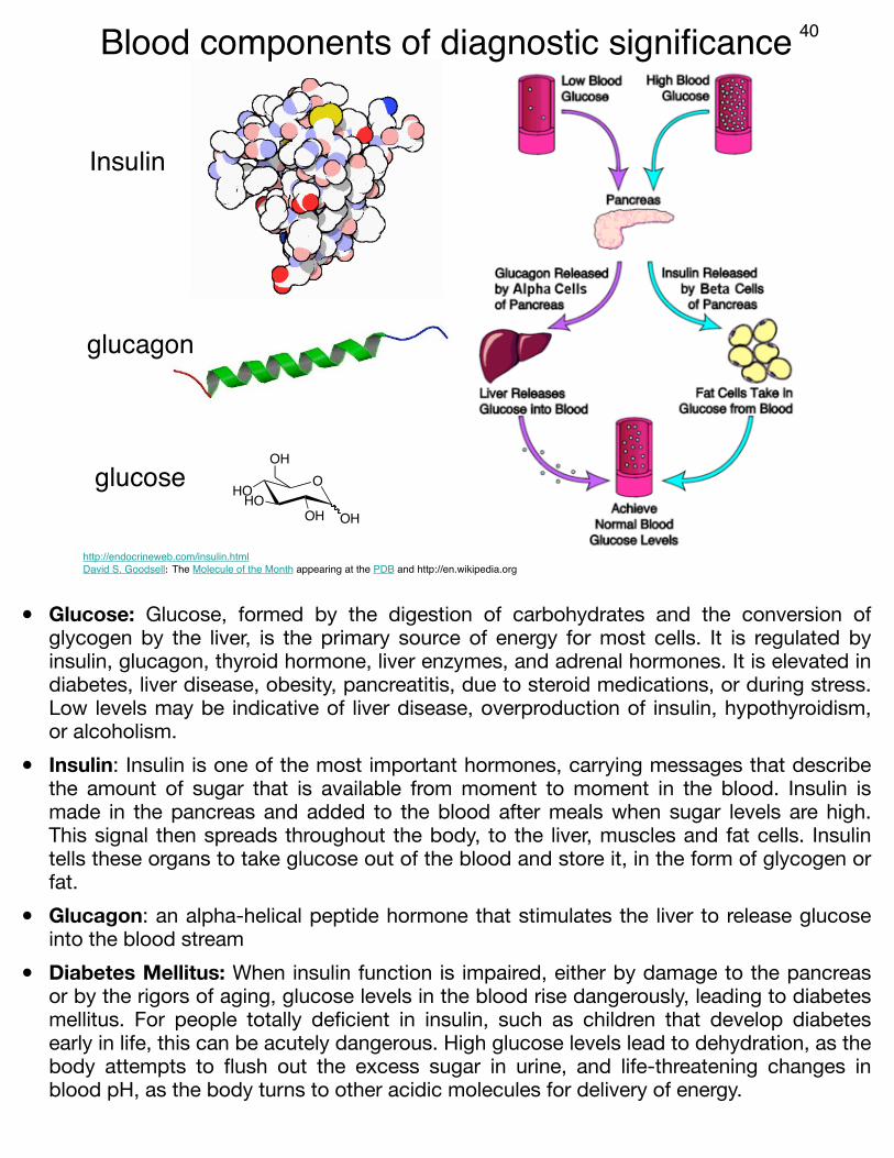
• Glucose: Glucose, formed by the digestion of carbohydrates and the conversion of glycogen by the liver, is the primary source of energy for most cells. It is regulated by insulin, glucagon, thyroid hormone, liver enzymes, and adrenal hormones. It is elevated in diabetes, liver disease, obesity, pancreatitis, due to steroid medications, or during stress. Low levels may be indicative of liver disease, overproduction of insulin, hypothyroidism, or alcoholism.
• Insulin: Insulin is one of the most important hormones, carrying messages that describe the amount of sugar that is available from moment to moment in the blood. Insulin is made in the pancreas and added to the blood after meals when sugar levels are high. This signal then spreads throughout the body, to the liver, muscles and fat cells. Insulin tells these organs to take glucose out of the blood and store it, in the form of glycogen or fat.
• Glucagon: an alpha-helical peptide hormone that stimulates the liver to release glucose into the blood stream
• Diabetes Mellitus: When insulin function is impaired, either by damage to the pancreas or by the rigors of aging, glucose levels in the blood rise dangerously, leading to diabetes mellitus. For people totally deficient in insulin, such as children that develop diabetes early in life, this can be acutely dangerous. High glucose levels lead to dehydration, as the body attempts to flush out the excess sugar in urine, and life-threatening changes in blood pH, as the body turns to other acidic molecules for delivery of energy.
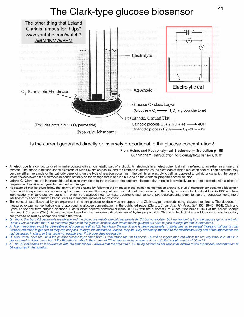
(Excludes protein but is O2 permeable)
(Glucose + O2 H2O2 + gluconolactone)
Cathodic process O2 + 2H2O + 4e- 4OH-
Or Anodic process H2O2 O2 +2H+ + 2e-
Is the current generated directly or inversely proportional to the glucose concentration?From Holme and Peck Analytical Biochemistry 3rd edition p 168
Cunningham, Introduction to bioanalytical sensors, p. 81
41
Electrolytic cell
The Clark-type glucose biosensor The other thing that Leland Clark is famous for: http://www.youtube.com/watch?
v=9MdlyM7w8PM
• An electrode is a conductor used to make contact with a nonmetallic part of a circuit. An electrode in an electrochemical cell is referred to as either an anode or a cathode. The anode is defined as the electrode at which oxidation occurs, and the cathode is defined as the electrode at which reduction occurs. Each electrode may become either the anode or the cathode depending on the type of reaction occurring in the cell. In an electrolytic cell (as opposed to voltaic or galvanic), the current which flows between the electrodes depends not only on the voltage that is applied but also on the electrical properties of the solution.
• Leland C. Clark had the ingenious idea of placing very close to the surface of the platinum electrode (by trapping it physically against the electrode with a piece of dialysis membrane) an enzyme that reacted with oxygen.
• He reasoned that he could follow the activity of the enzyme by following the changes in the oxygen concentration around it, thus a chemosensor became a biosensor. Based on this experience and addressing his desire to expand the range of analytes that could be measured in the body, he made a landmark address in 1962 at a New York Academy of Sciences symposium in which he described how "to make electrochemical sensors (pH, polarographic, potentiometric or conductometric) more intelligent" by adding "enzyme transducers as membrane enclosed sandwiches".
• The concept was illustrated by an experiment in which glucose oxidase was entrapped at a Clark oxygen electrode using dialysis membrane. The decrease in measured oxygen concentration was proportional to glucose concentration. In the published paper (Clark, L.C. Jnr. Ann. NY Acad. Sci. 102, 29-45, 1962), Clark and Lyons coined the term enzyme electrode. Clark's ideas became commercial reality in 1975 with the successful re-launch (first launch 1973) of the Yellow Springs Instrument Company (Ohio) glucose analyser based on the amperometric detection of hydrogen peroxide. This was the first of many biosensor-based laboratory analysers to be built by companies around the world.
• Q. I found that both O2 permeable membrane and the protective membrane only permeable for O2 but not protein. So I am wondering how the glucose get to react with O2?as I would assume the O2 to react with glucose at the glucose oxidase layer, which means glucose will have to pass through protective membrane.
• A. The membranes must be permeable to glucose as well as O2. Very likely the membrane is freely permeable to molecules up to several thousand daltons in size. Proteins are much larger and so they can not pass through the membrane. Indeed, they are likely covalently attached to the membrane using one of the approaches we had discussed in class, so they could not escape even if the pore sizes were larger.
• Q. Also, where does the O2 in the glucose oxidase layer come from? I understand that for Pt anode, O2 will be regenerated but where the the very initial level of O2 in glucose oxidase layer come from? For Pt cathode, what is the source of O2 in glucose oxidase layer and the unlimited supply source of O2 to it?
• A. The O2 just comes from equilibrium with the atmosphere. I believe that the amounts of O2 being consumed are very small relative to the overall bulk concentration of O2 dissolved in the solution.
Yellow Springs glucose biosensor technology
http://www.ysi.com/extranet/BTKL.nsf/447554deba0f52f2852569f500696b21/0f7ccb69a6ee82cf852569e70047cd17!OpenDocument
42
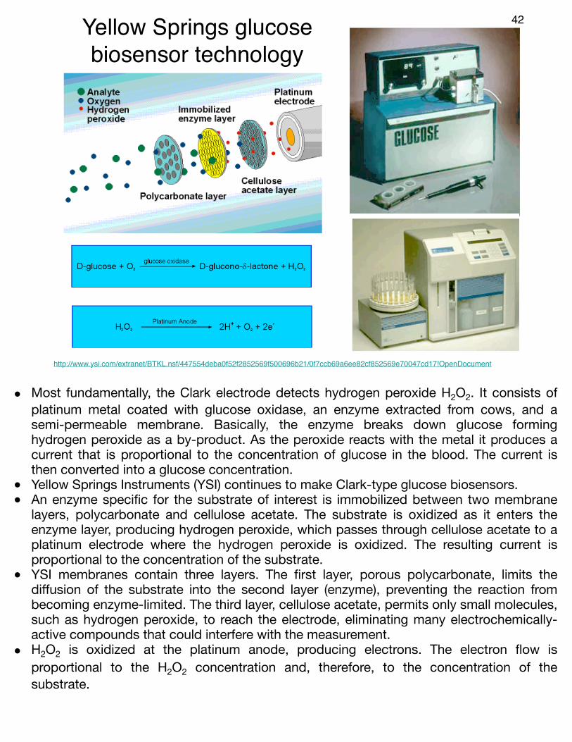
• Most fundamentally, the Clark electrode detects hydrogen peroxide H2O2. It consists of platinum metal coated with glucose oxidase, an enzyme extracted from cows, and a semi-permeable membrane. Basically, the enzyme breaks down glucose forming hydrogen peroxide as a by-product. As the peroxide reacts with the metal it produces a current that is proportional to the concentration of glucose in the blood. The current is then converted into a glucose concentration.
• Yellow Springs Instruments (YSI) continues to make Clark-type glucose biosensors.• An enzyme specific for the substrate of interest is immobilized between two membrane
layers, polycarbonate and cellulose acetate. The substrate is oxidized as it enters the enzyme layer, producing hydrogen peroxide, which passes through cellulose acetate to a platinum electrode where the hydrogen peroxide is oxidized. The resulting current is proportional to the concentration of the substrate.
• YSI membranes contain three layers. The first layer, porous polycarbonate, limits the diffusion of the substrate into the second layer (enzyme), preventing the reaction from becoming enzyme-limited. The third layer, cellulose acetate, permits only small molecules, such as hydrogen peroxide, to reach the electrode, eliminating many electrochemically-active compounds that could interfere with the measurement.
• H2O2 is oxidized at the platinum anode, producing electrons. The electron flow is proportional to the H2O2 concentration and, therefore, to the concentration of the substrate.
Improved glucose sensors using an
electron-relay mediator to help
get electrons from the FAD to the
surface
http://chem.ch.huji.ac.il/~eugeniik/electron_mediators.htm
Enzyme ‘wiring’
43
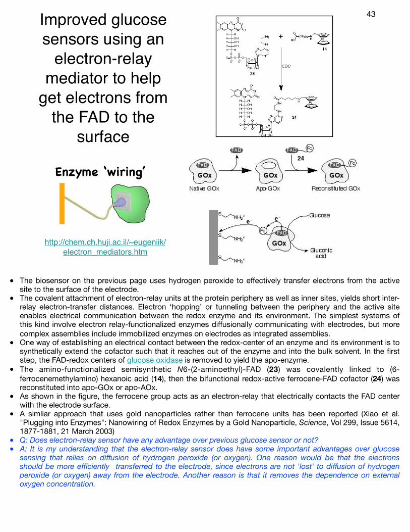
• The biosensor on the previous page uses hydrogen peroxide to effectively transfer electrons from the active site to the surface of the electrode.
• The covalent attachment of electron-relay units at the protein periphery as well as inner sites, yields short inter-relay electron-transfer distances. Electron ‘hopping’ or tunneling between the periphery and the active site enables electrical communication between the redox enzyme and its environment. The simplest systems of this kind involve electron relay-functionalized enzymes diffusionally communicating with electrodes, but more complex assemblies include immobilized enzymes on electrodes as integrated assemblies.
• One way of establishing an electrical contact between the redox-center of an enzyme and its environment is to synthetically extend the cofactor such that it reaches out of the enzyme and into the bulk solvent. In the first step, the FAD-redox centers of glucose oxidase is removed to yield the apo-enzyme.
• The amino-functionalized semisynthetic N6-(2-aminoethyl)-FAD (23) was covalently linked to (6-ferrocenemethylamino) hexanoic acid (14), then the bifunctional redox-active ferrocene-FAD cofactor (24) was reconstituted into apo-GOx or apo-AOx.
• As shown in the figure, the ferrocene group acts as an electron-relay that electrically contacts the FAD center with the electrode surface.
• A simliar approach that uses gold nanoparticles rather than ferrocene units has been reported (Xiao et al. "Plugging into Enzymes": Nanowiring of Redox Enzymes by a Gold Nanoparticle, Science, Vol 299, Issue 5614, 1877-1881, 21 March 2003)
• Q: Does electron-relay sensor have any advantage over previous glucose sensor or not?• A: It is my understanding that the electron-relay sensor does have some important advantages over glucose
sensing that relies on diffusion of hydrogen peroxide (or oxygen). One reason would be that the electrons should be more efficiently transferred to the electrode, since electrons are not 'lost' to diffusion of hydrogen peroxide (or oxygen) away from the electrode. Another reason is that it removes the dependence on external oxygen concentration.
At home finger sticks and continuous in vivo glucose monitoring
44
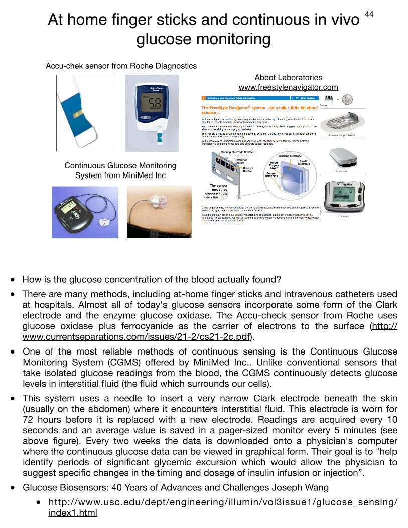
Accu-chek sensor from Roche Diagnostics
Continuous Glucose Monitoring System from MiniMed Inc
Abbot Laboratories www.freestylenavigator.com
• How is the glucose concentration of the blood actually found? • There are many methods, including at-home finger sticks and intravenous catheters used
at hospitals. Almost all of today's glucose sensors incorporate some form of the Clark electrode and the enzyme glucose oxidase. The Accu-check sensor from Roche uses glucose oxidase plus ferrocyanide as the carrier of electrons to the surface (http://www.currentseparations.com/issues/21-2/cs21-2c.pdf).
• One of the most reliable methods of continuous sensing is the Continuous Glucose Monitoring System (CGMS) offered by MiniMed Inc.. Unlike conventional sensors that take isolated glucose readings from the blood, the CGMS continuously detects glucose levels in interstitial fluid (the fluid which surrounds our cells).
• This system uses a needle to insert a very narrow Clark electrode beneath the skin (usually on the abdomen) where it encounters interstitial fluid. This electrode is worn for 72 hours before it is replaced with a new electrode. Readings are acquired every 10 seconds and an average value is saved in a pager-sized monitor every 5 minutes (see above figure). Every two weeks the data is downloaded onto a physician's computer where the continuous glucose data can be viewed in graphical form. Their goal is to "help identify periods of significant glycemic excursion which would allow the physician to suggest specific changes in the timing and dosage of insulin infusion or injection”.
• Glucose Biosensors: 40 Years of Advances and Challenges Joseph Wang• http://www.usc.edu/dept/engineering/illumin/vol3issue1/glucose_sensing/
index1.html

Long-term in vivo glucose monitoring using fluorescent hydrogel fibers
45
Y. J. Heo, H. Shibata, T. Okitsu, T. Kawanishi, S. Takeuchi, Long-term in vivo glucose monitoring using fluorescent hydrogel fibers. Proc Natl Acad Sci U S A 108, 13399 (2011).
• In this work, the authors inserted a glucose responsive fiber under the skin• The fluorescence of the fiber reports on the concentration of glucose. • The fiber contains a ‘glucose sensing’ unit composed of the fluorophore anthracene and
two boronic acids which serve as a binding site for sugars such as glucose• This sensor likely operates by a photoinduced electron transfer mechanism. In the
absence of glucose, an electron from the lone pair on the nitrogen can be donated to the excited anthracene chromophore and quench the excited state. In the presence of glucose, the conformation is distorted such that the lone pair is no longer able to donate electrons to the anthracene, and so the quenching is relieved.
• The authors demonstrated that polyethylene glycol (PEG)-bonded polyacrylamide (PAM) hydrogel fibers reduced inflammation relative to uncoated fibers
• This can be seen in the images of the mice ears, where the uncoated PAM fibre is red and inflamed after 31 days, but the PEG-coated PAM is not.
• PEG is a polymer with structure: HO-CH2-(CH2-O-CH2-)n-CH2-OH• A PEG coating is widely used for reducing immune response against foreign biomolecules
or objects placed under the skin

The future? Non-invasive monitoring. 46
Pretty sure these through-skin approaches are complete scams and vaporware!
Some approaches based on spectroscopy seem to be supported by substantial
amounts of peer-reviewed research:
www.inlightsolutions.comwww.sensysmedical.com
www.solianis.comwww.groveinstruments.com
GlucoWatch from Cygnus.
http://googleblog.blogspot.co.uk/2014/01/introducing-our-smart-contact-lens.html
Google smart contact lens for glucose sensing (Jan 2014)
• Noninvasive approaches for continuous glucose monitoring represent a promising route for obviating the challenges of implantable devices. However, it is not at all clear to me that this will be possible any time in the near future.
• One example of a non-invasive device that seemed to work (at least to some degree) is a wearable glucose monitor, the GlucoWatch from Cygnus Inc., based on the coupling of reverse iontophoretic (?) collection of glucose and biosensor functions.
• Given the obvious importance of such a device (if it were to work), there is very little reliable information available online. I suspect that this device simply does not work well enough for widespread use.
• It seems as though the most promising non-invasive technologies involve near infrared spectroscopic measurements obtained through the skin. Is it possible to measure glucose reliably through the skin? I doubt it, but there are quite a few companies who seem to think it can work. Such a device will probably not be wearable.
• Monitoring of glucose in the eye
47
26
Principles and applications of UV-visible spectroscopy
The residual from the least squares calculation is a good indicator of how well the standard spectra fit the sample spectra and is therefore a good indicator of the probable accuracy of the results.
An example of multicomponent analysis is the quantification of five hemoglobins in blood with minimum sample preparation.7 Figure 15 shows the absorption spectra of hemoglobin derivatives. This analysis was previously performed using various analytical techniques, including spectroscopy and titrations.
Figure 15Absorption spectra of hemoglobin derivatives
Other methods Other statistical approaches to multicomponent analysis include the partial least squares (PLS), principle component regression (PCR), and multiple least squares (MLS) methods. In theory, these methods offer some advantages over those described above, but in practice the calibration process is much more complex.
Sample requirements The simple simultaneous equations and least squares methods yield accurate results only if calibration is performed using pure standards or mixtures of standards
Wavelength [nm]
SulfhemoglobinOxyhemoglobinCarboxyhemoglobinHemoglobin (pH 7.0–7.4)Deoxyhemoglobin
Abso
rban
ce [A
U]
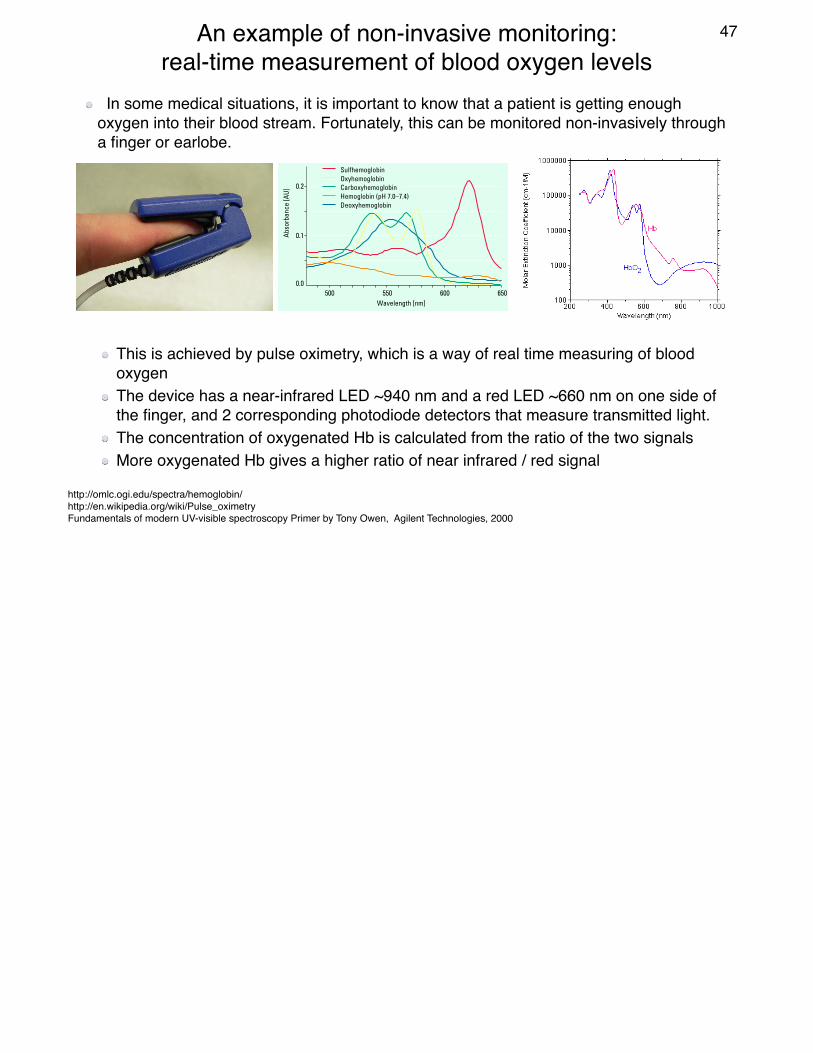
In some medical situations, it is important to know that a patient is getting enough oxygen into their blood stream. Fortunately, this can be monitored non-invasively through a finger or earlobe.
http://omlc.ogi.edu/spectra/hemoglobin/http://en.wikipedia.org/wiki/Pulse_oximetryFundamentals of modern UV-visible spectroscopy Primer by Tony Owen, Agilent Technologies, 2000
This is achieved by pulse oximetry, which is a way of real time measuring of blood oxygenThe device has a near-infrared LED ~940 nm and a red LED ~660 nm on one side of the finger, and 2 corresponding photodiode detectors that measure transmitted light.The concentration of oxygenated Hb is calculated from the ratio of the two signalsMore oxygenated Hb gives a higher ratio of near infrared / red signal
An example of non-invasive monitoring: real-time measurement of blood oxygen levels


![Determination and Analytical, Bio Analytical Methods for ... · and Analytical, Bio Analytical Methods for Its ... Therefore, most of the pharmaceutical companies have ... [12], parkinson’s](https://img.dokumen.tips/doc/110x75/5ad5f65c7f8b9a5d058dd2f4/determination-and-analytical-bio-analytical-methods-for-analytical-bio-analytical.jpg)