Embed Size (px)
Citation preview
17
2
Sucrose, Sucralose, Fructose, and some Non-Carbohydrate
High-Potency Sweeteners : Correlations Between
Hydrophobicity Patterns and AH-B-X Assignments
Abstract: MOLCAD program-mediated calculations of the molecular electrostatic potential(MEP's) profiles and of the respective lipophilicity (hydrophobicity) patterns (MLP's) on thecontact surfaces of sucrose, galacto-sucrose, sucralose, and fructose are represented incolor-coded form. Most informative with respect to the placement of the tripartite AH-B-Xglucophore are the hydrophobicity distributions, which show the lipophilic X-part to be anentire, obviously quite flexible region rather than a specific corner of the "sweetness triangle":in sucrose and sucralose encompassing the outside area of the fructofuranose moiety, in fructosethe 1- and 6-CH groups in either linked or separated form. In contrast, the hydrophilic portions 2
of these sweeteners are more compact, invariably located opposite to the hydrophobic region,and appear to contain the AH-B couple of the glucophore: the glucosyl-2- and 3-OH group insucrose and sucralose, versus the 3,4-diol grouping in fructose. Whilst absolute proof for theseassignments is still lacking, support for their relevance is derived from the sweetness ofaltogether 53 sucrose derivatives and some fructose analogs. Most remarkably, the MLP'sgenerated for the solid state conformations of some non-carbohydrate high-potency sweeteners,such as the sulfamides cyclamate, saccharin, and acesulfame, as well as structurally distinctlydifferent dipeptides, e.g. aspartame, exhibit a hydrophobicity distribution strikingly similar tothose observed for the sugars: hydrophilic and hydrophobic areas on opposite sites of themolecule. The results, particularly the lipophilicity patterns presented, sustain the notion, thatthe sweet receptor with its proteinaceous "hydrophobic cleft" – be it the same for sucrose,fructose, and non-carbohydrate sweeteners or different ones – is quite flexible in adapting tothe complementary hydrophobic region of the sweet substance. Following this "dockingprocedure", the hydrophilic AH-B area of the substrate now being in its proper position, thesweet response is elicited via hydrogen bonding to a complementary receptor-based AH-Bcouple.
[1,2] [3]The classical attempt by Shallenberger and Kier to rationalize the sweet tasteof organic compounds presumes the existence of a common AH-B-X glucophore in allsweet substances, eliciting the sweet response via interaction with a complementary [4]tripartite AH-B-X site in the taste bud receptor .
At the present state of knowledge, however, this "sweetness triangle" appears muchtoo simple to explain all of the observations, particularly when bearing in mind that [5-12]sweet taste perception is mediated by a cascade of complex biochemical processesthat are little understood at the cellular and molecular level.

18 Chapter 2
Nevertheless, the tripartite AH-B-X glucophore concept has had its merits as aunifying criterion and proved useful – despite its neglect of three-dimensional shapeand volume – in rationalizing structure-sweetness relationships in such diverseclasses of compounds as amino acids, dipeptides, sulfamides (e.g. saccharin andacesulfame), and sugars in particular, most notably the natural sweeteners sucrose (1)and fructose (2), as well as sucralose (3), a sucrose-derived high-potency sweetener.
sucrose β-D-fructose sucralose 1 2 3
The availability of advanced computer modeling techniques, their application to theelucidation of the individual conformations of carbohydrates in the vacuum and in [13-16]solution , particularly the possibility of representing various properties on the [17-19]contact surface of sugars has added a new dimension in the visual perception ofsugars. Accordingly, not only may the electropositive and electronegative areas on thesurface of a sugar molecule be reliably determined by computational methods, but the [17-19]hydrophilic and hydrophobic regions as well , which in terms of interactions withthe sweet taste receptor are apt to be of great significance.
Fig. 2-1. Location of the tripartite AH-B-X glucophore in sucrose emerging from thecomputer-generated molecular electrostatic potential (MEP's) profiles and lipophilicity [17-19]patterns (MLP's) .

Sucrose, Sucralose, Fructose, and some Non-Carbohydrate High-Potency Sweeteners 19
Incorporation of such results into structure-sweetness considerations have led to a [1-3]new allocation of the Shallenberger-Kier tripartite AH-B-X glucophore in sucrosefor the two forms likely to prevail in solution: the glucosyl-2-OH being the H-donor inthis hydrogen bond interaction with a complementary acceptor group in the receptor,entailing the glucosyl-3-OH as the H-acceptor (B site), whilst the hydrophobic X-partis an area on the "outside" of the fructose moiety (Fig. 2-1).
[1-3]Fig. 2-2. Shallenberger / Kier concept of structure sweetness relationships which was [4]assessed by Lee "to be devoid of any predictive value": a hydrogen bond donor (AH), ahydrogen bond acceptor functionality (B), and an additional hydrophobic binding point(X) are considered to be the essential structural element of all sweet tasting molecules.For sucrose, different AH-B-X-assignments have been proposed by Mathlouthi et al. in [20] [21] [22]1990 (A ) and by Hough et al. in 1989 (B and C ).
Whilst these conclusions derived from the contact surface distribution of themolecular electrostatic potential (MEP's) profiles and the molecular lipophilicity(hydrophobicity) patterns (MLP's) of sucrose appear to be reasonable, they differ fromalternative AH-B-X assignments as in Fig. 2-2, which are based on rationalizations ofthe sweetness of a sizeable number of sucrose analogs. As indicated in Fig. 2-2, thehydrophobic center X is placed at the axial 4-position of the glucose moiety, because

20 Chapter 2
of the lack of sweetness of galacto-sucrose (the 4-epimer of sucrose), the sweetness of4-deoxy-sucrose (1 x), and the enhanced sweetness of 4-chloro-4-deoxy- [21-23]galacto-sucrose (5 x) . However, alternative hydrophobic centers had to be placedat the 1'- and 6'-positions of the fructose unit to account for the increased sweetness [21-23](20 x) of the respective 1'- and 6'-chloro-deoxy-sucroses . In terms of the early [24]suggestion of Shallenberger that the presence of hydrophilic centers at differentparts of the molecule may drastically disturb their orientation pattern, therebypreventing interactions of the AH-B units with the receptor site(s), multiple centers ofhydrophobicity located in different portions of the sweetener appear to be unlikely.
In view of the fairly reliable MEP's and MLP's on which the assignment in Fig. 2-1is based, it appeared imperative to subject all sucrose derivatives of which thesweetness characteristics are known, to a thorough scrutiny with respect to the validityof the various AH-B-X assignments. This is done in the sequel based on the sweetnesscharacteristics of deoxy-, O-methyl and deoxy-halo derivatives of sucrose. In addition,the hydrophobicity patterns of fructose (2), in its β-pyranoid form, and somenon-carbohydrate sweeteners are probed as to their implications for the AH-B-Xconcept.
The Electrostatic Potential Profiles and Hydrophobicity Patterns of Sucrose
[25,26]As evidenced by solid state structural data , the conformation of sucrose in thecrystalline state is determined by two intramolecular hydrogen bonds, one between theprimary 6'-OH of fructose and the pyranoid ring oxygen of glucose (1.89 Å,cf. Fig. 2-3 A, and ball and stick model in Fig. 2-4 A), the other one between the1'-hydroxyl group and the 2-O of glucose (1.85 Å). In solution, however, particularlyin water, it is unlikely that both of these hydrogen bonds are retained. Indeed, elaborate [27-37]NMR investigations strongly attest to the disintegration of the weaker ... g f5-O HO-6 hydrogen bond by solvation. For a dimethyl sulfoxide solution a [30]competitive equilibrium between forms B and C has been deduced , with the formerpredominating.
[29,38-44]Several calculations of the energy potential surface of sucrose haveprovided additional indications concerning the relevance of forms B and C, including [45]ours using the PIMM88 force field program (see this work, Chapter 3). In Fig. 2-4,the solid state conformation of sucrose (A) is set against its PIMM-generated, lowestenergy conformation B, in which the remaining intramolecular hydrogen bond hasbeen slightly widened to 2.00 Å. For this conformation – being a realistic model forsucrose in (aqueous) solution, and hence, for the form entering into the receptor [46] [47]site – the contact surface (roughly equivalent to the solvent-accessible surface ,

Sucrose, Sucralose, Fructose, and some Non-Carbohydrate High-Potency Sweeteners 21
i.e. "how water sees the molecule") was calculated using the MOLCAD-program [48]methodology . As evident from the dotted contours in Fig. 2-4, the transition fromthe solid state conformation (A) to that likely to prevail in solution (B) results only inminor changes on the "upper" side of the molecule.
Fig. 2-3. Conformations of sucrose: (A) in the crystal as derived from neutron diffraction [25]data . For DMSO solution a competitive equilibrium B ↔ C, with B predominating has [30]been proposed .
Fig. 2-4. Contact surface (roughly equivalent to the solvent accessible surface) ofsucrose in dotted form with a stick-ball model insert, the larger balls representing oxygen [25]atoms. (A): conformation in the crystal as derived from neutron diffraction data , ... g fshowing the two intramolecular hydrogen bonds 5-O HO-6 (1.89 Å) and ... g f2-O HO-1 (1.85 Å). (B): lowest energy conformation emerging from PIMM88 force ... g ffield calculations (for vacuum), lacking the 5-O HO-6 hydrogen bond.

22 Chapter 2
[49]Calculations of the molecular electrostatic potential (MEP), i.e. the distributionof the charge density over the contact surfaces of the two sucrose conformations, were [50] [51]effected using the MOPAC program-generated AM1 -atomic charges and arerepresented in a 16 color-code ranging from red (electropositive) to violet(electronegative, cf. Fig. 2-5). These reveal only minor differences in the overallelectrostatic profile.
Fig. 2-5. Molecular electrostatic potential (MEP) of sucrose in its conformation adopted [25]in the crystal (closed and opened form with ball and stick model insert on the lefteach), and its most probable "solvated" form in solution (right each), represented on therespective contact surface in a 16 color code, red representing the positive maximum, i.e.the most electropositive potentials, violet the most negative portion of the molecule.

Sucrose, Sucralose, Fructose, and some Non-Carbohydrate High-Potency Sweeteners 23
As is clearly apparent from Fig. 2-5, each of the forms has the red, i.e. mostelectropositive area centered around the glucosyl-2-OH, which in turn appears to be [52-54]caused by the cooperative effect of the intramolecular hydrogen bond directedtowards the oxygen of this hydroxyl group. In this context, it may be noted that anexperimental verification of the validity of the MEP distribution of Fig. 2-5 is provided [55]by the behavior of DMF solutions of sucrose under electrolysis conditions : themolecule is attracted to the cathode with its most electropositive, i.e. theglucosyl-2-OH portion, and upon taking up an electron (to an alkoxyl radical anion) [56]and releasing a hydrogen atom (→ H ) leaves the glucosyl-2-oxygen deprotonated. 2
This is evidenced by the fact, that in situ alkylation or acylation (i.e. in the cathodic [57]cell) produces the 2-O-substituted sucrose derivatives with high preference .
The MEP pattern of sucrose as depicted in Fig. 2-5 may thus be considered, withconfidence, to properly represent the charge distribution in solution, and, accordingly,may be used to locate the AH-B part of the glucophore such that the AH-hydrogen islikely to be electropositive for interaction with an oppositely polarized binding site onthe receptor. Similarly, the inverse situation would have to prevail for the B portion.This reflection points towards the glucosyl-2-OH as the AH portion and the B part inits direct vicinity. However, it is seemingly unavailing to locate the hydrophobic X-siteon the basis of the MEP pattern.
Since there is ample evidence to assume that the sweet-taste receptor isproteinaceous in nature, and that interaction between a hydrophobic portion on theprotein surface with the corresponding hydrophobic portion of sucrose is involved intriggering the sweet-response, the reliable location of that is of major importance instructure-sweetness considerations. The recently advanced possibility to compute and [58] [17]visualize molecular hydrophobicity (lipophilicity) patterns (MLP's) was appliedto determine the lipophilicity profile for crystalline sucrose as depicted on the left of [59]Fig. 2-6 in an alternative 32 color-code , blue representing here the most hydrophilicand yellow-brown the respective most hydrophobic areas. As is clearly evident fromthis representation, the hydrophilic and hydrophobic portions of the molecule aredistinctly separated on opposite sides. Particularly lucid is the half-opened form withthe ball and stick-model insert, disclosing the entire outside-section of the fructosemoiety to be hydrophobic (i.e. yellow-brown), and the hydrophilic (blue) section to be [17]centered around the 3-oxygen of glucose .
Observation of the alternative sucrose conformation likely to prevail in solution(cf. Fig. 2-6, right), reveals few changes in the overall MLP, except that thehydrophobic region located at the outer side of the fructose moiety is now morecompact. In consequence, the part of sucrose amenable to engage in hydrophobic

24 Chapter 2
bonding within the sweet-taste receptor is to be assigned to an entire region in thefructose portion, rather than a specific position.
Fig. 2-6. Molecular lipophilicity pattern (MLP) of sucrose as distributed over the contactsurface in 32 colors ranging from yellow-brown (most hydrophobic) to blue (mosthydrophilic area): on the left side each, the MLP for the solid state conformation as [25]derived from neutron diffraction data , on the right the MLP for the conformationlikely to prevail in solution. Apparently, the hydrophobic and hydrophilic regions arelocated on opposite sides of the molecules.

Sucrose, Sucralose, Fructose, and some Non-Carbohydrate High-Potency Sweeteners 25
In keeping with these notions, the location of the tripartite AH-B-X glucophoreemerges in the form indicated in Fig. 2-1: the glucosyl-2-OH and 3-OH being theproton donor (AH) and proton acceptor (B) parts engaging in simultaneoushydrogen-bonding to a complementary (inverse) AH-B system on the receptor protein,whilst the hydrophobic X-part is a region centered around H-3 of fructose.
The Tripartite AH-B-X Glucophore in Sucrose Derivatives
In securing corroborative evidence for the location of the AH-B-X glucophore, thesucrose derivatives considered are limited (for elimination of steric misfits) to thosemodified either by inversion, deoxygenation, and O-methylation of individualhydroxyl groups, or by their replacement with halogens.
The most direct method of probing into the location of the AH-B-X glucophoreappears to be the replacement of a given hydroxyl group in sucrose by hydrogen andassessment of its effect on sweetness. As of now, however, sweetness data areavailable only for three of the eight mono-deoxy-sucroses, i.e. the 4-deoxy (4),6-deoxy (5) and 1'-deoxy derivatives (6) (Table 2-1). All are less sweet than [60,61]sucrose , yet the fact that sweetness is not lost altogether may be taken as anindication that none of the hydroxyl groups removed (i.e. the 4-OH and 6-OH ofglucose, and 1'-OH of fructose) occupy positions detrimental for eliciting the sweet ... g fresponse. Accordingly, the existence of the intramolecular 2-O HO-1 hydrogenbond in sucrose is obviously no prerequisite for proper interaction with the sweetreceptor, a notion that is similarly emerging from the sweetness of 1'-O-methyl- and1'-chloro-1'-deoxy derivatives of sucrose (cf. below). Unfortunately, sweetness data onthe other five deoxy-sucroses (2- and 3-deoxy in particular) or any of the 28 possibledideoxy-sucroses, which would further contribute to this issue, are not yet available.
Table 2-1. Sweetness of 4-deoxy- (4), 6-deoxy- (5), and 1'-deoxy-sucrose (6).
relative * 1 2 3 R R R sweetness refs.
sucrose OH OH OH SS – 4 H OH OH S 60
5 OH H OH S 60
6 OH OH H S 60-62
* S = sweet, SS = very sweet

26 Chapter 2
Next to the deoxygenation, O-methylation of individual OH-groups in sucroseappears to be a suitable means of probing binding sites, provided that the stericexpansion introduced by the OH → OMe conversion does not detract from the tasteassessment. Such an impairment appears to be small, if at all, as evidenced by the dataavailable for four mono- (7 – 10) and four dimethyl ethers of sucrose (11 – 14, [60-63]Table 2-2) : any of the three primary OH-groups in sucrose may be O-methylatedwithout losing sweetness, which also applies to the glucose-4-OH. In all of thesemethyl ethers the glucose-2- and 3-OH groups, i.e. the structural AH-B requirementsof Fig. 2-1, remain untouched, thus their sweetness data may be taken as anaffirmation of the glucophore assignment. Curiously, no sweetness data of sucrosederivatives methylated at O-2, O-3, or at both of these positions, which would shedfurther light on this issue, are available.
Table 2-2. Sweetness of methyl ethers of sucrose.
relative * 1 2 3 4 R R R R sweetness refs.
sucrose OH OH OH OH SS –
7 OMe OH OH OH S 63
8 OH OMe OH OH SS 60
9 OH OH OMe OH SS 61,62
10 OH OH OH OMe SS 63
11 OMe OMe OH OH S 63
12 OMe OH OH OMe S 63
13 OH OMe OH OMe SS 63
14 OH OH OMe OMe S 63
* S = sweet, SS = very sweet
The 6-position in the glucose portion of sucrose appears to be a sensitive one withrespect to the steric bulk introduced by its chemical modification. Whilst the6-deoxy (5) and 6-O-methyl (8) derivatives are as sweet as sucrose, the 6-O-acetate(15) is only slightly sweet, the 6-benzyl ether (16), the 6-benzoate (17), and6-phosphate (18) are bitter, as is the 6-chloro-6-deoxy compound (19, cf. Table 2-3).In the latter case, the obvious misfit introduced by the 6-chlorine substituent may beovercome by increasing the hydrophobicity of the fructose portion: 6'-chlorination(→ 20) removes the bitterness, and chlorination at both primary positions of thefructose portion result in a molecule (21) with enhanced sweetness [21,62,64](cf. Table 2-4) .

Sucrose, Sucralose, Fructose, and some Non-Carbohydrate High-Potency Sweeteners 27
Table 2-3. Taste characteristics of sucrose derivatives modified at the glucose C-6.
X taste refs.
sucrose OH sweet –
5 H sweet 60,62
8 OMe sweet 60,62
15 OAc slightly sweet 60,62
16 OBn bitter 60,62
17 OBz bitter 60,62
18 OPO H bitter 60,62 3 19 Cl bitter 60,62,64
Table 2-4. Taste characteristics of 6-chloro-6-deoxy-sucroses.
taste 1 2 R R (sucrose sweetness = 1) refs.
19 OH OH bitter 21,62,64
20 OH Cl not sweet 64
21 Cl Cl 25 21,62
Configurational changes in the glucose portion of sucrose seem to have a morepronounced effect on the sweetness: its 4-epimer, the "galacto-sucrose" (22) has very [63] [65]low sweetness , and the 3-epimeric analogue "allo-sucrose" (23) is tasteless . Thisclearly indicates subtle stereochemical requirements for the substrate on enteringand / or being embedded into the receptor site(s). In the case of the 3-epimer 23 thismay be rationalized via the change of the steric requirements of the AH-B site, an axial3-OH being incapable of functioning as the hydrogen bond accepting B component.
galacto-sucrose allo-sucrose 3-keto-sucrose 22 23 24

28 Chapter 2
[65]That 3-keto-sucrose (24) is sweet goes along well with the AH-B-X tripartiteglucophore assignment of Fig. 2-1, inasmuch as the 3-carbonyl function retains thehydrogen bond acceptor capabilities.
Less well comprehensible is, at first sight, the very low sweetness of [63]galacto-sucrose (22) , in which the 2-OH and 3-OH of the hexosyl portion, i.e. theAH-B functionalities suggested in Fig. 2-1, are intact. A clue as to the possible reasonsemerges from a comparison of the conformations of sucrose in solution, which features [66]a gg arrangement of the glucosyl-6-CH OH relative to the pyranoid ring 2
(Fig. 2-7 A), with the respective arrangements for its 4-epimer 22. The PIMM-programgenerated, minimum energy conformations for 22 result in the forms depicted inFig. 2-7, B – D, which only differ by the orientation of the galactosyl-6-CH OH. 2
Thereby, the gg form B, due to formation of a stabilizing intramolecular hydrogen ... g gbond 6-OH O-4 , comes out to be about 4 and 8 kJ / mol more stable than the tg (C)and gt rotamers (D), yet it is clear that this only prevails in vacuo. For solution, there is [53,67-73]abundant evidence in the literature that the sterically unfavorable 1,3-syn-interactions between 4- and 6-hydroxyl groups of a hexopyranose are evaded bysolvation, hence, rotamers C and D of Fig. 2-7 will have to be entered intostructure-sweetness considerations. Correspondingly, the sucrose conformation ofFig. 2-7 A is to be set against those of galacto-sucrose depicted in Fig. 2-7 (C, D):inversion of configuration at C-4 of sucrose entails a distinctive change in therotameric preference at the 6-OH, which is an a priori sterically sensitive position(vide supra, Table 2-3). Thus, the very low sweetness of galacto-sucrose may be takenas an indication that the C-6 substituent in the aldose portion of sucrose is not onlysensitive towards steric bulk – any significant increase resulting in the loss ofsweetness (cf. Table 2-3) – but also in its orientation to the pyranoid ring, with a gg
arrangement conceivably being favored.
Another factor responsible for the substantial decrease in sweetness on inversion ofthe sucrose-4-OH may be found in the shift of the molecular lipophilicity pattern(Fig. 2-8), of which the hydrophilic (blue) area centered around C-3 of the glucose unitis shifted to the upper side as compared to sucrose in Fig. 2-4. That, on the other hand, [64]4-chloro-4-deoxy-galacto-sucrose exhibits a 5-fold higher sweetness than sucrosecan be attributed to the hydrophobic substituent at C-4 that changes the overall shapeof the molecule and its MLP substantially (as observed, for example, for the pyranoidportion of sucralose, cf. Fig. 2-9 – 2-11 below).

Sucrose, Sucralose, Fructose, and some Non-Carbohydrate High-Potency Sweeteners 29
sucrose galacto-sucrose (gg form) (gg form)
galacto-sucrose galacto-sucrose (tg form) (gt form)
Fig. 2-7. Contact surface (in dotted form) of sucrose (A, featuring a gauche-gauche (gg) [66]arrangement for the glucosyl-6-OH relative to the pyranoid ring) as compared to thethree conformers of galacto-sucrose (22) generated by PIMM force field calculations: thegg form B (in vacuum only), stabilized by an intramolecular hydrogen bond ... g g6-OH O-4 , and the two forms relevant in solution, i.e. the trans-gauche (tg, C) andthe gauche-trans (gt, D) rotamers. The oxygens involved in the designations anddiscussion (cf. text) are accentuated by filling.

30 Chapter 2
Fig. 2-8. Hydrophobicity pattern (MLP) of the tg (left) and gt rotamers (right) ofgalacto-sucrose (22), corresponding to the forms C and D in Fig. 2-7.
Deoxy-Halo-Sucroses
Unlike the deoxy- and O-methyl-sucroses discussed so far, whose sweetness is ofabout the same or lower intensity than that of the parent sugar, some deoxy-haloderivatives of sucrose exhibit substantially enhanced sweetness, in specific instanceseven several thousand times that of sucrose. Following the discovery of the first [74]compound of this type by Hough and Phadnis in 1976 , an impressively largenumber of deoxy-halo-sucroses has been synthesized and evaluated for their potency [21-23,62,64,74-85]in sweet taste perception . The altogether 35 compounds listed in the

Sucrose, Sucralose, Fructose, and some Non-Carbohydrate High-Potency Sweeteners 31
following Tables provide a large body of experimental evidence that should berationalizable in terms of the AH-B-X conceptual assignments made in Fig. 2-1.
The AH-B-X assignment of Fig. 2-1 implies that the hydrophobic cleft of the tastereceptor protein corresponds to the hydrophobic region in the fructose moiety ofsucrose (cf. Fig. 2-6). In keeping with this notion, it may be predicted that an increaseof hydrophobicity in the fructose portion – i.e. along the yellow-brown portion in theMLP (Fig. 2-6) – favors binding to the corresponding hydrophobic site in thereceptor, and hence enhances sweetness. Indeed, the data collected for the compounds25 – 30 (Table 2-5) support this rationalization, since replacement of the fructosehydroxyl groups at the 1'-, 4'-, and / or 6'-position by chlorine or bromine uniformlyleads to compounds sweeter than sucrose.
Table 2-5. Relative sweetness of deoxy-halo-sucroses modified in the fructose portion.
relative 1 2 3 R R R sweetness refs.
sucrose OH OH OH 1 –
25 Cl OH OH 20 21,62,64,80
26 OH OH Cl 20 21,62,64 * 27 Cl Cl OH 30 81
28 Cl OH Cl 80 21,62
29 Br OH Br 80 84
30 Cl Cl Cl 100 81
[4]* In Table XIX on p. 266 of Lee's review the relative sweetness for 27 is erroneously listed as being3500.
Analysis of the sweetness characteristics of the numerous4-halo-4-deoxy-galacto-sucroses (Table 2-6) in terms of the AH-B-X assignment ofFig. 2-1 is particularly informative. A shift of the substantially hydrophilicglucosyl-4-OH of sucrose (blue area in Fig. 2-6) from the equatorial to the axialorientation (→ galacto-sucrose (22), cf. Fig. 2-8), together with the accompanying gchange in the rotameric arrangement of the 6-OH (cf. Fig. 2-7) results in a near loss [63]of sweetness . However, when placing a pronouncedly hydrophobic substituent suchas chlorine into the very same axial position, the sweetness, relative to sucrose, isenhanced by a factor of 5 (compound 31 in Table 2-6).
The comparison of the sweetness of 4-chloro-4-deoxy-galacto-sucrose (31) withthat of its analogs 32 – 41 is particularly instructive, inasmuch as the successivereplacement of the fructose 1'-, 4'-, and 6'-OH groups translates into substantially

32 Chapter 2
increased sweetness values, the 1',4',6'-tribromo analog 41 featuring a 1400 foldenhancement over 31. An essentially identical trend is observed in the4-deoxy-4-fluoro-galacto-sucroses 42 – 44 (a 10 fold increase of sweetness from the4,6'-difluoro 42 to the 4,1',6'-trifluoro compound 43), and very markedly, in the4-bromo analogs 45 – 47, in which the enhancement of sweetness perception reaches asolitary maximum.
Table 2-6. Relative sweetness (sucrose = 1) of 4-deoxy-4-halo-galacto-sucroses.
Glucose – Fructose – relative 1 2 3 a) X R R R sweetness refs.
sucrose OH OH OH OH 1 – eq 31 Cl OH OH OH 5 21,64,80
32 Cl OH OH Cl 50 84 33 Cl Cl OH OH 120 21,62
34 Cl OH Cl Cl 160 81 35 Cl Cl Cl OH 220 81 b) 3 Cl Cl OH Cl 650 78
36 Cl Br OH Br 800 84
37 Cl Cl F Cl 1000 81
38 Cl Cl Cl Cl 2200 79,82
39 Cl Cl Br Cl 3000 81 40 Cl Cl I Cl 3500 78,81
41 Cl Br Br Br 7000 81
42 F OH OH F 4 84
43 F F OH F 40 78
44 F Cl Cl Cl 200 81
45 Br Cl OH Cl 375 84
46 Br Br OH Br 800 84
47 Br Br Br Br 7500 78,81
48 I I OH I 120 78,84
a) All of these data were retrieved from the original literature as indicated by the respectivereferences. It should be noted that a number of sweetness values given in Lee's review (Adv.Carbohydr. Chem. Biochem. 1987, 45, Table XIX on p. 266) are at error, most notably the data listedfor compound 40 (7000 instead of 3500), and 41 (30 instead of 7000). – b) Sucralose.
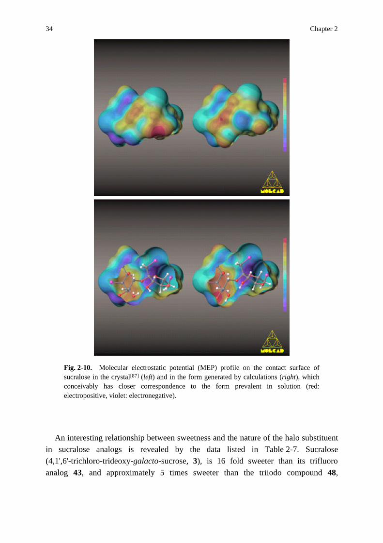
Of these compounds in Table 2-6, sucralose (3), a non-caloric high-potency [86]sweetener recently approved for food use , was selected to further probe into thehydrophobic / hydrophilic portions of the molecule by computer modeling, an intent [87]facilitated by the availability of its X-ray structure . In Fig. 2-9 the moleculargeometries and their contact surfaces are depicted for two forms of sucralose, A

Sucrose, Sucralose, Fructose, and some Non-Carbohydrate High-Potency Sweeteners 33
representing the crystalline state conformation based on X-ray data, B the state likely [88]to prevail in solution , as generated by force field optimizations. It is noteworthythat the directionality of the intersaccharidic hydrogen bond is reversed on going from ... g fthe crystal (A, 2-OH O-3 ) to the lowest energy, computer simulated form (B, ... g f2-O HO-3 ).
Fig. 2-9. Molecular geometry and dotted contact surface of sucralose , the large ballsrepresenting the three chlorine atoms at positions 4 of the hexose, and positions 1 and 6 [87]of fructose. (A): solid state conformation , (B): lowest energy conformer emergingfrom PIMM88 force field calculations. Both models differ mainly in the directionality of ... g fthe interresidue hydrogen bond 2-O O-3 , the right model corresponds closer to the [88]conformation adopted in solution .
Portraying on this contact surface the MOLCAD-generated MEP-pattern, it isevident from Fig. 2-10 that the form relevant for solution (closed and opened form onthe right each), is as pronouncedly electropositive (red) as observed for sucrose ... g f(cf. Fig. 2-5), obviously due to an analogous direction of the 2-O HO-3 hydrogenbond.
The hydrophobicity pattern pictured in Fig. 2-11, expectedly shows the two chlorineatoms in the fructose portion to be the hydrophobic center (X-site), now beingextended over the entire "outside" region of fructose (as compared to sucrose,cf. Fig. 2-6). Another obvious similarity with sucrose is the fact that hydrophobic(yellow-brown) and hydrophilic (blue) regions are located on opposite sides of themolecule, seemingly little disturbed by the third chlorine at C-4 of the pyranoid ring,which – as evident from Fig. 2-11 – is less hydrophobic than the other two.
34 Chapter 2
Fig. 2-10. Molecular electrostatic potential (MEP) profile on the contact surface of [87]sucralose in the crystal (left) and in the form generated by calculations (right), whichconceivably has closer correspondence to the form prevalent in solution (red:electropositive, violet: electronegative).
An interesting relationship between sweetness and the nature of the halo substituentin sucralose analogs is revealed by the data listed in Table 2-7. Sucralose(4,1',6'-trichloro-trideoxy-galacto-sucrose, 3), is 16 fold sweeter than its trifluoroanalog 43, and approximately 5 times sweeter than the triiodo compound 48,

Sucrose, Sucralose, Fructose, and some Non-Carbohydrate High-Potency Sweeteners 35
Fig. 2-11. Molecular lipophilicity patterns (MLP's) of sucralose in the solid state [87]structure (left), and the computer-simulated conformation (right). The reversal of the ... ... g f g fdirection of the interresidue hydrogen bond from 2-OH O-3 (left) to 2-O HO-3 g(right) results in a concentration of the hydrophilic area (blue) around O-2 . The twochlorine atoms of the fructose unit are located within the most hydrophobic (yellow-brown) region.
indicating that fluorine is too small, and iodine is too big to properly fit into thereceptor binding site(s). The best fit appears to be provided by bromo substituents inthe fructose 1'- and 6'-positions, as evidenced by the compounds 36 and 46, which are [78,84]800 times sweeter than sucrose .

36 Chapter 2
Table 2-7. Relative sweetness (sucrose = 1) of sucralose (3) analogs.
relative X Y Z sweetness refs.
43 F F F 40 78 * 3 Cl Cl Cl 650 78
36 Cl Br Br 800 84
45 Br Cl Cl 375 84
46 Br Br Br 800 84
48 I I I 120 78,84
* Sucralose
Table 2-8. Relative sweetness (sucrose = 1) of some 4,6'-dichloro substitutedgalacto-sucroses (analogs of sucralose 3) modified at C-6.
relative 1 X R sweetness refs.
* 3 OH Cl 650 78
49 Cl OH 4 80
50 Cl Cl 200 80
51 H Cl 400 78,83
52 OMe Cl 500 78,83
53 OiPr Cl not sweet 78
* Sucralose
Another probe into the effect of the 6-substituent on sweetness is provided by thesucralose analogs additionally substituted at C-6 (Table 2-8): in comparison tocompound 3, the sweetness of the 4,6,6'-trichloro isomer 49, which lacks the sweetnessenhancing 1'-chloro substituent in the hydrophobic center of the fructose portion, dropsdramatically (650 → 4), a decrease that is only partially made up by introducing that1'-chloro group (4 → 200 for 50), due to the presence of critical 6-Cl substituent(cf. Table 2-3). As expected from the discussion of 6-deoxy- and 6-O-methyl-sucrose(Table 2-1 and 2-2), modification of the 6-substituent by deoxygenation (→ compound51) and O-methylation (→ 52) has only a minor effect on the intensity of thesweetness sensation. Yet, increase of the steric bulk at C-6 from OCH (52) to 3
OCH(CH ) (53) is seemingly fatal, as sweetness is lost altogether. 3 2
Oddly enough, sucrose derivatives modified at either the glucosyl-2-OH or 3-OHgroup, like the deoxy-, O-methyl- or chloro-analogs, are not available. Sweetness dataon these have a major bearing on the AH-B assignment as in Fig. 2-1. The only

Sucrose, Sucralose, Fructose, and some Non-Carbohydrate High-Potency Sweeteners 37
examples along these lines are the two 2-chloro-2-deoxy-mannosyl analogs. The [81,82]dichloride 54 has been found to be "not sweet at all" , and the tetrachloride 55,despite its additional hydrophobicity- and, hence, sweetness-enhancing 6'-chloro [85]substituent, is "as bitter as quinine" .
[81,82]54 X = OH (not sweet at all) [85]55 X = Cl (very bitter)
This finding, along with the observation that allo-sucrose (23) is not sweet,emphasizes that there are strict steric requirements for the arrangement of the 2- and3-hydroxyl groups, and that their equatorial orientation is seemingly essential foreliciting the sweet sensation. This conclusion is also in accord with, but no absoluteproof of the AH-B assignments to the 2- and 3-OH groups of the glucosyl moiety insucrose.
By way of summary, the foregoing discussion of the sweetness of over 50derivatives or analogs of sucrose provides ample evidence for placing the hydrophobicX-site of the Shallenberger-Kier glucophore onto the outside region of the fructoserather than to other parts of the sucrose molecule. This is borne out by the MLP'spresented, and by the fact that an increase of the hydrophobicity in the fructose portioninvariably results in an enhancement of sweetness. In keeping with the notion that theX part is mainly responsible for orientation of the molecule in entering and / or beingembedded into the receptor site, it can be assumed, that this hydrophobicity-controlled"docking procedure" of the substrate into the hydrophobic cleft of the receptor isrequired for bringing the AH-B portions into the proper receptor positions to elicit thesweetness sensation via hydrogen bonding.
Placement of the hydrophobic X part into the fructose portion of sucrose leaveslittle alternative for the AH-B part: 4-deoxy- and 6-deoxy-sucrose are sweet, albeit lessthan sucrose (Table 2-1), as are the respective 4-O- and 6-O-methyl ethers(Table 2-2) – findings that render these positions most unlikely as those essential foreliciting the sweet response. In contrast, the data discussed above point towardslocation of the AH-B unit in the diol grouping made up by the glucosyl-2-OH and3-OH groups. Both are situated within the most hydrophilic region of sucrose(cf. Fig. 2-6), and in principle, each may be assigned the B (or AH) part. Here, the AHpart was assigned to the glucosyl-2-OH, mainly for the reason that the MEP pattern of

38 Chapter 2
Fig. 2-5 indicates this hydroxyl group to be the most electropositive, and hence, isconceivably better disposed for engaging its OH-proton as a donor in thesubstrate-receptor hydrogen bond.
Fig. 2-12. Location of the tripartite AH-B-X glucophore ("sweetness triangle") insucrose emerging from the computer-generated molecular electrostatic potential (MEP)profile and the molecular lipophilicity pattern (MLP).
In toto, the evidence for the assignment of the AH-B-X glucophore as depicted inFig. 2-12, though not definitive, is a most useful working hypothesis for rationalizingsweetness data in sucrose derivatives, and surely worthy of further attention. It isexpected that greater certainty will come from the sweetness data of the five remainingdeoxy-sucroses (particular the 2- and 3-deoxy compounds), and of the four missingmono-methyl ethers, especially the 2-O-methyl and 3-O-methyl-sucroses. Otherdesirable derivatives with which the validity of the assignments could be furtherprobed, are 2-epi-sucrose, i.e. its mannosyl analog, and its 3'-epimer, a psico-sucrose.
β-D-Fructopyranose: Conformations and Molecular Lipophilicity Profiles
D-Fructose crystallizes in the β-D-pyranoid form, as evidenced by solid state [89,90]structural data . Freshly prepared solutions are almost twice as sweet as sucrose [91,92](1.8 x) , but when equilibration of the β-p-form to the tautomeric β-f-, α-f-, and 93-95 [ ]α-p forms (cf. Fig. 2-13) is complete, the solution is only slightly sweeter than [91]one of sucrose of equal w / v-concentration . From this it was inferred that the twofuranoid forms are either substantially less sweet than the β-p form or devoid of sweettaste entirely, a conclusion that is supported by the parallelism of decrease ofsweetness and of β-p-form (in the equilibrium tautomeric mixture) on increasing thetemperature.

Sucrose, Sucralose, Fructose, and some Non-Carbohydrate High-Potency Sweeteners 39
Fig. 2-13. Tautomeric forms of D-fructose (2). For an equilibrated aqueous solution at [94]25°C the composition is 73 % β-p, 20 % β-f, 5 % α-f, and 2 % α-p forms , the acyclicketo-form is negligible.
In consequence, fructose-sweetness considerations are all based on the β-p form,and several assignments for the tripartite AH-B-X glucophore have been advanced: [2,93,96] [97](i) Shallenberger et al. , intuitively, and Lindley & Birch , on the basis of [97]consideration of model compounds , arrived at the anomeric 2-hydroxyl group andthe hydroxymethyl oxygen as the AH-B couple, respectively (Fig. 2-14, i). The inverse [98-100]assignment (ii) was suggested by Szarek et al. and by [20]Mathlouthi & Portmann , considering calculations of the net atomic charges and the [98] [20]relative basicities of the hydroxyl groups , and IR-data rationalizations .
Fig. 2-14. Location of the tripartite AH-B-X glucophore in β-D-fructopyranose as [2,93,96] [97] [98-100]suggested by Shallenberger and Lindley & Birch (i), by Szarek et al. and [20] [101]Mathlouthi & Portmann (ii), and by Birch et al. (iii).

40 Chapter 2
Interestingly, however, on the basis of intensity-time studies of the sweetness ofglucose and fructose, that neither showed differences between α- and β-anomers nor in [101]their apparent molar volumes, Birch et al. arrived at an entirely differentconclusion: the anomeric center of D-fructose plays no direct role in the sweetnessresponse, but rather the 3,4-diol system to which the AH-B glucophore is to beassigned (Fig. 2-14, iii).
Fig. 2-15. The three staggered rotameric forms of the hydroxymethyl group in [89,90]β-D-fructopyranose as derived from X-ray structural data (A) and from force fieldcalculations (B and C), and their respective contact surfaces (in dotted form). The tgrotamer (C), despite the unfavorable 1,3-diaxial-like interactions between the 1- and3-OH group emerges as the lowest energy conformer, due to its stabilization by an ...intramolecular hydrogen bond 1-OH O-3 (2.03 Å) in vacuo. Since this hydrogen bondwill not survive solvation with water, the tg rotamer is unlikely to be present in aqueoussolutions.

Sucrose, Sucralose, Fructose, and some Non-Carbohydrate High-Potency Sweeteners 41
For generation of the MEP's and MLP's for β-D-fructopyranose, with which theseassignments were to be probed, the relevant conformations of the hydroxymethyl 2group relative to the C -fixed pyranoid ring had to be determined. In the solid state, as 5 [89,90] [102]evidenced by X-ray structural data , the gauche-gauche (gg) arrangement ofthe primary hydroxyl group (A in Fig. 2-15) is realized. Undoubtedly, this gg rotameris one form relevant also in aqueous solution, with the minor modification though, thatthe weak intramolecular hydrogen bond circuit observed in the crystal lattice isdisintegrated, since in water the hydroxyl groups can satisfy their hydrogen bondrequirements by bonding with the solvent.
Semiempirical calculations of other conformations of 2 are encumbered with thefact that the minimum energy geometries generated represent the state in vacuo, whichmay substantially be altered on solvation with water. This applies to the conformations [98,103]emerging from very elaborate ab initio calculations and AM1-based [99]semiempirical investigations , as well as to those emanating from the more simplePIMM88 force field methodology. From the latter, the tg rotamer C (Fig. 2-15) comesout to be the global minimum energy conformation, despite the steric constraints of the1,3-diaxial-like arrangement of the 1-OH and 3-OH groups, which obviously areovercome (in vacuo) by the stabilizing effect of the intramolecular hydrogen bond ...1-OH O-3 (2.03 Å). This situation is most unlikely to prevail in water, particular in [72]view of recent molecular dynamics simulations for methyl β-D-glucopyranoside ,which convincingly proved the in vacuo minimum energy tg form not to survive inwater.
This leaves the gg and gt rotamers of β-D-fructopyranose as the molecularconformations preferred in solution, for which the contact surfaces (Fig. 2-15) and theMLP's were generated. As is evident from Fig. 2-16, both forms have their mosthydrophilic surface area (blue) centered around the fructose-4-OH, whilst thehydrophobic (yellow-brown) part(s) are associated with either of the two methylenegroups: in the gg rotamer (left entries in Fig. 2-16), the two methylene groups areconnected with a "hydrophobic band" that occupies half of the contact surface – ascontrasted by the pattern of the gt rotamer (right in Fig. 2-16), where the hydrophobicsurface areas of the 1- and 6-CH groups are separated. 2
Accordingly, the X-part of the tripartite AH-B-X glucophore can easily be located:a region (rather than a specific position) reaching from the 6-CH to the 1-CH , and, as 2 2
such, being reminiscent of the hydrophobicity pattern of sucrose (Fig. 2-6). Thus, theMLP-derived hydrophobic areas of β-D-fructopyranose appear to correlate – at leastroughly – with the X-part assignments of Fig. 2-14 that invariably were placed at the6-CH . 2

42 Chapter 2
Fig. 2-16. Molecular lipophilicity patterns (MLP's) for two conformers ofβ-D-fructopyranose (2), differing in the disposition of the hydroxymethyl group relative [102]to the pyranoid ring : the gg conformer is depicted on the left side each, the gt form onthe right. For each form, two representations were chosen, the upper corresponding intheir orientations to those of Fig. 2-15 B and C, respectively. The modelings depictedunderneath illustrate the opposite location of hydrophilic and hydrophobic regions.
Location of the AH-B entity on the basis of the MLP's of Fig. 2-16 – or thecorresponding MEP patterns not depicted here – is seemingly difficult. Yet, theconcentration of the most hydrophilic domains around the fructose-4-OH seems topoint to that position for either being the B or AH part, i.e. to the 3,4-diol grouping to

Sucrose, Sucralose, Fructose, and some Non-Carbohydrate High-Potency Sweeteners 43
represent the AH-B couple. The lipophilicity patterns obtained for the two fructose [101]conformers likely to be prevalent in solution favor Birch's proposition (iii inFig. 2-14), which designates the 3-OH and 4-OH as the AH-B part, respectively. In [103]this context, it is noteworthy that Szarek et al. found as a result of ab initio
investigations of 2 that O-4 in fructose exhibits enhanced basicity, while the secondary4- and 3-OH protons seem to be relatively acidic, only being exceeded by the primaryOH-group. These findings – in conjunction with the fact emerging from calculations [103]of molecular electrostatic potentials that "the O-4 atom would be predicted to be [103]the most attractive site for protonation" – may be taken as a hint for theimportance of the 4-OH group in respect to structure-sweetness relationships.
Fig. 2-17. Sweetness characteristics of analogs of β-D-fructopyranose (2). (SS: verysweet, LS: low sweetness)
Consideration of the few relevant fructose analogs, whose sweetness characteristicsare known (Fig. 2-17), provides no solid evidence with which a clear-cut decisionbetween the putative AH-B-assignments of Fig. 2-14 could be made. Thatβ-D-arabinose (56), 2-deoxy-fructose (57, 1,5-anhydro-D-mannitol), and the2-O-methyl derivative 58 (methyl β-D-fructopyranoside) are considerably less sweet [97]than the parent fructose advocates the anomeric hydroxyl group to play a role ineliciting sweetness. On the other hand, the fact that sedoheptulosan 59 is as sweet as [104]fructose attests to the contrary.

44 Chapter 2
The sweetness characteristics of analogs 60 – 63 tally with either of the conjecturalassignments in Fig. 2-14: the 5-hydroxyl group can be replaced by hydrogen (→ 60) [105]without loosing sweetness , and hence, as such is not an essential requirement forthe sweetness sensation. However, its steric (axial) orientation is important since itsconfigurational inversion to the 5-epimeric α-L-sorbo-pyranose (61) effects a [106]substantial decrease in sweetness , possibly by introducing a steric misfit upon [105]interaction with the receptor . Similarly, the intense sweetness of the 6-thio [107] [108](62) and 6-carba analogs (63) of fructose, although easily rationalized in termsof augmentation of the hydrophobic region within the 6-CH -1-CH band, do not 2 2
allow to differentiate between a 1,2- or 3,4-diol grouping for the AH-B couple of theglucophore.
Although further evidence is required to settle this question unequivocally, as ofnow, major significance should be attributed on the MLP's obtained for the twofructose conformers likely to prevail in solution. These (Fig. 2-16) clearly favor [101]Birch's proposal (iii in Fig. 2-14), which places the AH-B couple of theglucophore into the 3,4-diol grouping of fructose. Moreover, when focusing on theessentials contained in the lipophilicity patterns of the two fructose forms prevalent insolution (Fig. 2-16), the basic feature emerges that hydrophobic and hydrophilicregions are located on opposite sides of the molecule – a situation quite similar to theone observed for sucrose (Fig. 2-6). Thus, it may well be – and this receivesfortification from the lipophilicity profiles of a number of non-carbohydratesweeteners (see below) – that the opposite-side-distribution of hydrophobic andhydrophilic regions, the latter being capable for hydrogen bonding with the receptor, isthe principal structural feature for eliciting the sweetness response, rather than anAH-B-X "sweetness triangle".
Molecular Lipophilicity Profiles of Non-Carbohydrate, High-Potency Sweeteners
The AH-B-X glucophore concept has not only been applied to sugars, but has beenappreciated as the unifying criterion for such structurally diverse sweet substances asamino acids and a series of non-carbohydrate sweeteners such as cyclamate (64), [3]saccharin (65), acesulfame (66), and aspartame (67) . Serious reservations, however,must be advanced in regard to its general applicability, since it is known, for example,that the biologically active species of saccharin (65) and acesulfame (66) are the [109]respective anions , in which it is difficult to locate the AH entity. Furthermore, theAH-B-X concept assumes that all sweet molecules interact with the same receptor inthe same, or in an at least very similar way – an assumption which is quite [7,8]questionable. Recent evidence either points to several types of sweet receptors, or

Sucrose, Sucralose, Fructose, and some Non-Carbohydrate High-Potency Sweeteners 45
to different kinds of activations within the same one, if indeed sugars and high-potencysweeteners really elicit the sweet response via the same taste receptors. Despite thesereservations it was obviously of interest to extend the molecular modeling techniquesused above, to some representative non-carbohydrate sweeteners, as, for example, tocompounds 64 – 68.
For this purpose, the solid state conformations were retrieved from the X-ray [110] [111]structural data available for saccharin (65) , acesulfame (66) , aspartame [112] [113](67) , and the retro-inverso sweetener (68) and used to calculate the respectivecontact surfaces (Fig. 2-18 and 2-20). In the case of cyclamate (64), for which anX-ray structure is lacking, the conformation was generated by PIMM calculations.
As clearly apparent from the contact surfaces of the three sulfamido sweeteners(Fig. 2-18), their overall molecular shapes are different, although the SO NH-element 2
is placed on the left side of each compound in Fig. 2-18 to accentuate their commonstructural as well as three-dimensional feature, undoubtedly involved in eliciting thesweet response. However, when comparing their MLP's in Fig. 2-19, the similarity ofdistribution of hydrophobic and hydrophilic regions is amazing: that the sulfamidoportion is the hydrophilic part of the molecule was to be expected. That the differencesbetween a cyclohexyl ring (in cyclamate), an aromatic moiety (as in saccharin) and anacetoacetyl residue fixed in the enol form (as in acesulfame) level off to yieldhydrophobic areas closely resembling each other – in the case of 65 and 66, the twolower entries in Fig. 2-19, they are essentially identical – is most remarkable.

46 Chapter 2
Fig. 2-18. Contact surface of cyclamate (64, A), saccharin (65, B), and acesulfame (66,C) in dotted form with a ball and stick model insert. The conformation of 64 (A) wasgenerated by force field calculations, those of 65 (B) and 66 (C) were modeled according [110,111]to the X-ray structural data of the corresponding sodium or potassium salts .
Fig. 2-20. Solid state conformation and contact surfaces, based on X-ray structural [112,113] data for A: the commercial dipeptide sweetener aspartame (67, "Nutrasweet" ),and B: the intensely sweet N-L-aspartyl-N'-(2,2,5,5-tetramethylcyclopentanyl)-carbonyl-(R)-1,1-diaminomethane (68), a retro-inverso dipeptide.

Sucrose, Sucralose, Fructose, and some Non-Carbohydrate High-Potency Sweeteners 47
Fig. 2-19. The molecular hydrophobicity profiles (MLP's) in color-coded representation(yellow-brown: hydrophobic, blue: hydrophilic regions) of the sulfamide sweetenerscyclamate (64, upper middle), saccharin (65, lower left), and acesulfame (66, lower right)in closed and opened form. The MLP's are scaled separately to the range of thehydrophobicity values calculated onto the respective contact surfaces cf. Fig. 2-18.
Another striking feature is that hydrophobic and hydrophilic portions of themolecules are on opposite sites, as in the case of sucrose and fructose (cf. above).Moreover, the very same distinctive separation of hydrophilic and hydrophobic areasis observed for the dipeptide sweeteners 67 (aspartame) and 68, which appear to bequite different in their solid state conformations (ball and stick model insert in

48 Chapter 2
Fig. 2-20), yet foreshadow a basic molecular shape similarity in their contact surfaces(Fig. 2-20, in dotted contours), which fully tallies with the respective MLP's ofFig. 2-21: closely corresponding hydrophobic (yellow-brown) regions of such diverseelements as the aromatic ring of the phenylalanine portion of aspartame, and thesterically constrained tetramethylcyclopentyl group in 68.
Fig. 2-21. Lipophilicity profiles of the dipeptide sweeteners aspartame (67, left) andN-L-aspartyl-N'-(2,2,5,5-tetramethylcyclopentanyl)-carbonyl-(R)-1,1-diaminomethane [112,113](68, right) based on their crystal conformations .

Sucrose, Sucralose, Fructose, and some Non-Carbohydrate High-Potency Sweeteners 49
All of this sustains the notion that the sweet receptor – be it the same for sucrose,fructose, and non-carbohydrate sweeteners or different ones – is quite flexible inadapting to the hydrophobic portion of sweet substances, to the X part (of the tripartiteAH-B-X glucophore), which clearly is not a specific position of the molecule, but anentire region. If this hydrophobic area is the main factor governing the "dockingprocedure" of the sweet substance, i.e. directing it to and locking it into thecomplementary "hydrophobic cleft" of the receptor protein, it can well be imaginedthat, thereby, the hydrophilic area of the molecule, situated on its opposite site, andlikely to contain the AH-B portion of the Shallenberger-Kier tripartite AH-B-Xglucophore, is brought into the appropriate position to elicit the sweet response viahydrogen bonding to a complementary receptor site AH-B couple.
In summary, much remains to be learned about the intricacies of the mechanism(s)involved in activation of sweet-sensitive cells, and direct solid evidence is urgentlyrequired. Nevertheless, the incorporation of the three-dimensional shape of sweetmolecules, of their contact surfaces, and, particularly, inclusion of their MEP's andMLP's into structure-sweetness considerations has provided this field with newdynamics, not only in the visualization of the sweet molecule as such, but also of thecomplementary binding site. This unfoldment is apt to lead, via computer-aidedreceptor modeling, to more realistic structure-sweetness concepts than thosepreviously developed.


![[Sucrose][Fructose] [H3P04= · quired fructose molecule could be produced from a second molecule of G-1-P by way of glucose-6-phos-phate (G-6-P) and fructose-6-phosphate (F-6-P) as](https://img.dokumen.tips/doc/110x75/5ec3bc19478bbc53f3182a8b/sucrosefructose-h3p04-quired-fructose-molecule-could-be-produced-from-a-second.jpg)