Embed Size (px)
Citation preview

8/8/2019 2 Lasers in Medical Science 1995
http://slidepdf.com/reader/full/2-lasers-in-medical-science-1995 1/7

8/8/2019 2 Lasers in Medical Science 1995
http://slidepdf.com/reader/full/2-lasers-in-medical-science-1995 2/7
C.A. Morton, C. Whitehurst, H. Moseley et al
~
';..
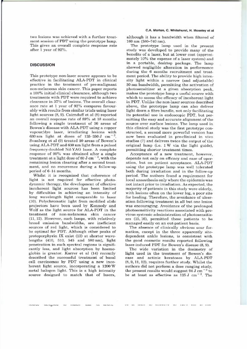
Fig. 1. Prototype lamp with timer unit and 5 mm flexible fibre bundle. The two attachments used on the end of the fibre areshown, the 11 mm perspex rod used to treat smalliesions and the 25 mm collimating lens used für larger lesions (seen
attached to fibre).
significant difference in the extent of tumour
response between the two light sources (7).The aim of the present study was to assess
the efficacy of this prototype lamp in clinical
practice in the treatment of pre-malignantnon-melanoma skin cancer.
MATERIAL AND METHODS
Patients
Ethical committee approval was obtained
für the treatment of patients with potential
precursors to non-melanoma skin cancer, by
photodynamic therapy. Patients presenting to
the Dermatology Department of the Western
Infirmary, Glasgow, with lesions of Bowen's
disease (in situ squamous cell carcinoma) or
actinic keratoses, 21 mm in diameter or less,were invited to participate in the study. No
lesion had beeil previously treated. Histologi-
cal confirrnation of the diagnosis was acquired
by performing a 4 mm punch biopsy on alllesions.
PDT light source
The prototype lamp (patent pending) incor-
parates a 300 W xenon short arc plasma
dis charge, producing a continuous wave
broadband flat spectral output across the
entire visible spectrum. The components of the
light source are as described previously (6).
The lamp used in this study can deliver 1 W
directly or 0.5 W via a flexible light guidewithin a bandwidth of 30 nm which can be
tuned to any wavelength from 300 nm to
1.1 J-lm.Infra-red emission from the source wastotally blocked using a wideband dielectric
he at filter and a non-infra-red transmitting
light guide. Zero infra-red emission up to 35 J-lm
wavelength was verified with a calorimeter.
Using appropriate filters, the spectral output
of the lamp was adjusted to a 30 nm bandwidtharound 630 nm. To broaden the treatment field
and produce uniform irradiation of lesions, an
11 mm perspex rod or 25 mm collimating teils
was attached to the 5 mm fibre bundle (Fig. 1).
The authors included at least a 10% margin
around lesions in the field of irradiation per-
mitting treatment of lesions up to 9 mm indiameter using the rod, and 21 mm in diameter
using the lens. At fluence rates of 158 mWcm - 2 für the rod and 55 mW cm - 2 für the lens,
lesions received 94-156 J cm - 2, the treatmentdose centred on 125J cm - 2 as this dose haB
beeil shown previously to be effective in amino-
laevulinic acid (ALA)-PDTusing laser (8).
~
Photosensitizer
Topical 5-aminolaevulinic acid in an oil in
water emulsion, 20% (wjw) 5-ALA (Sigma

8/8/2019 2 Lasers in Medical Science 1995
http://slidepdf.com/reader/full/2-lasers-in-medical-science-1995 3/7
Alternative Light Source tor PDT 3: Clinical Response
Table 1. Clearance of lesions after a single treatment depending on light dose. Treatment times, depending
on which end attachment to the fibre bundle was used, are also shown
Chemical Co) in Unguentum lVlerck (E. Merck
Ud), was applied to legions 4 h before illumi-
nation with the lamp. Surface crusts were
removed from the legions and the surfacegently abraded prior to 5-ALA application.
Approximately 50 mg of cream was applied per
cm2 to cover the entire irradiation field, thus
including the clinically disease-free margin.
The cream was then kept in place under an
occlusive dressing (Tegaderm, 3M) für 4 h,
after which the 5-ALA cream remaining on the
skin surface was carefully removed. The
patient was offered local anaesthetic (1% plain
lignocaine by intra dermal injection) duringtreatment.
Adverse effects, clearance and recurrence
Patients used visual analogue scales to record
pain during and over the 10 days follow-
ing PDT (with subsequent interpretation of
O<xs;3 as mild, 3<xS;7 as moderate and
7<xS;10 as severe). Lesions were examined on
completion of therapy and 24-48 hiater, then
at increasing intervals during the following
2 months. Clinical response to the first appli-cation of PDT was determined at 2 months and
a second treatment administered if legions
persisted. Monthly review of all patients wasundertaken für 12 months following clearance
to observe für recurrence and assess scarring
potential of the therapy. Post-therapy punch
biopsies were performed in legions where
doubt over clinical clearancefrecurrenceexisted.
Statistics
Comparison of the size of legions clearing
and of those not clearing after a single PDT
treatment, was performed by a Mann-WhitneyU-test. Comparisons of clearance rates depend-
ing on dosage and the apparatus used to
deliver the irradiation, were düne using a
Chi-squared test.
RESULTS
Clearance rates
Twenty legions of Bowen's disease and four
actinic keratoses (AK), in 12 patients (three
male, nine female, median age 65 years, range
43-95 years), received photodynamic therapy.
Sixteen legions were sited on the leg, six on the
forearm or hand, and two on the scalp. Themedian surface area of all treated legions was
60 mm2 (range 9-400 mm2).
Fifteen legions (12 Bowen's and 3 AK)
cleared after a single treatment with PDT
using this non-laser source. All nine remaining
legions cleared following a second treatment,2 months later. The median size of legions
clearing after Olle treatment was 56 mm2
(range 9-400 mm2), not significantly different
from the size of those legions requiring a
second treatment (median 60 mm2, range
9-380 mm2). The clinical response of Olle areaof Bowen's disease and Olle actinic keratosis is
shown in Figs 2 and 3, respectively.The effect of light dose on clearance after a
single treatment is shown in Table 1. The firstfive legions entered into the trial received 94 J
cm - 2, 75% of the intended treatment dose inorder to observe für side-effects. Subsequent
legions received 125 J cm - 2 except, as Olle
patient had seven legions, two legions received
94J cm - 2, three 125J cm - 2, and the remain-
ing two, 156 J cm -2. Three legions in this
patient did not clear with a single treatment,
Olle from each of the three dose regimens
used. Although overall a higher percentage of
legions treated with 94 J cm - 2 cleared afterOlle treatment, compared with those which
.
t II
Dose Total no. of No. of lesions treated Treatment time (min) Clearafter Olle
(J cm -2) lesions treated Rod Lens Rod Lens treatment
94 7 5 2 10 29 5
125 15 8 7 13 39 9
156 2 2 0 16 49 1

8/8/2019 2 Lasers in Medical Science 1995
http://slidepdf.com/reader/full/2-lasers-in-medical-science-1995 4/7
C.A. Morton, C. Whitehurst, H. Moseley et al
'.
(a)
(b)
Fig. 2. Plaques of Bowen's disease on leg (a) before and (b) 2 months following a single treatment with ALA-PDT using the
prototype lamp.
received 125J cm - 2, this difference was not
statistically significant.
The perspex rod, with the higher fluence rate
of 158mW cm - 2, was attached to the fibrebundle für the treatment of 15 legions, of which
10 cleared (67%) on a single treatment. The
collimating leng (fluence rate 55 mW cm - 2)
was attached to the bundle'for the treatment
of the nine larger legions, with f ive legions
clearing (56%) in this group after a singletreatment. This difference in clearance rate,
however, was not significant.
Adverse effects
~
j As treatment of the initial legions (with 94 J
cm - 2) was well tolerated, the study proceededwith the increase in dose as described. Side-
effects were similar in frequency and severity
between the different dosage groups and aretherefore listed together.
No rain was experienced by patients during
the treatment of 12 legions. Pain during PDT
was described as mild in a further 10,moderate
in one, and severe in one legion which was an

8/8/2019 2 Lasers in Medical Science 1995
http://slidepdf.com/reader/full/2-lasers-in-medical-science-1995 5/7
Alternative Light Source tor PDT 3: Clinical Response
(a)
(b)
Fig. 3. Area of actinic keratosis on scalp (a) before and (b) 2 months following a single treatment with ALA-PDT using theprototype lamp.
area of Bowen's disease that had ulcerated
prior to therapy. Only in this latter ease wasloeal anaesthesia administered. Over the 10
days following treatment, no rain was assoei-
ated with 21 treated lesions, with mild diseom-
fort lasting around 7 days deseribed in the
remaining three lesions. Whilst erythema and
oedematous swelling of the treatment Bites
were evident on eompletion of irradiation,
only three lesions proeeeded to blister (by
2 days) with Olle area of Bowen's disease sub-
sequently ulcerating. No photosensitivity
reaetions were evident following PDT.
A visible sear in the treatment field, outside
diagnostie biopsy Bites, was observed in onlythree treatment Bites, all three areas of Bowen's disease treated on the ankle and
overlying the Aehilles tendon.
Recurrence rate
During the 12 months following clinieal clear-
allee of the 24 lesions, two areas of Bowen's
disease reeurred, both at 8 months in lesionstreated with 125 J em - 2. Clearanee of these
8/8/2019 2 Lasers in Medical Science 1995
http://slidepdf.com/reader/full/2-lasers-in-medical-science-1995 6/7
two lesions was achieved with a further treat-
ment session of PDT using the prototype lamp.
This gives an overall complete rBsponse rate
after 1 year of 92%.
DISCUSSION
This prototype non-laser source appears to be
effective in facilitating ALA-PDT in clinical
practice in the treatment of pre-malignant
non-melanoma skin cancer. This paper reports
a 100% initial clinical clearance, although two
treatments with PDT were required to achieveclearance in 37% of lesions. The overall clear-
allee rate at 1 year of 92% compares favour-
ably with results from similar trials using laserlight sources (8, 9). Cairnduff et al (8) reported
an overall response rate of 89% at 18 months
following a single treatment of 36 areas of
Bowen's disease with ALA-PDT using a copper
vapourjdye laser, irradiating lesions with
630 nm light at doses of 125-250 J cm - 2.
Svanberg et al (9) treated 10 areas of Bowen's
using ALA-PDT and 630 nm light from a pulsed
frequency-doubled Nd-YAG laser. A complete
response of 90% was obtained after a single
treatment at a light dose of60 J cm -2, with the
remaining lesion clearing after a second treat-
ment, and no recurrence during a follow-up
period of 6-14 months.
Whilst it is recognized that coherence of
light is not required für effective photo-
dynamic therapy, the development of effective
incoherent light sources haB beeil limited
by difficulties in achieving an intensity of
lang wavelength light comparable to laser
(10). Polychromatic light from modified slide
projectors have beeil used by Kennedy and
Wolf as the light source für ALA-PDT in thetreatment of non-melanoma skin cancer
(11,12). However, such lamps, with relatively
broad emission bandwidths, are inefficient
sources of red light, which is considered to
be optimal für PDT. Although other peaks of
protoporphyrin IX exist (13) at shorter wave-
lengths (410, 510, 545 and 580 firn), light
penetration in such spectral regions is signifi-
cantly less, and light absorption by haemo-
globin is greater. Karrer et al (14) recentlydescribed the successful treatment of basal
cell carcinomas by PDT using a new inco-
herent light source, incorporating a 1200 W
metal halogen light. This is a high intensity
source designed to match that of lasers,
C.A. Morton, C. Whitehurst, H. Moseley et al
although it haB a bandwidth when filtered of
160 nm (580--740firn).
The prototype lamp used in the present
study was developed to provide many of the
benefits of a laser, hut at lower cast (approxi-mately 10% the expense of a laser system) and
in a portable, desktop package. The lampshowed negligible alteration in performance
during the 6 months recruitment and treat-
ment period. The ability to provide high inten-
sity light within a narrow (and adjustable)
30 nm bandwidth, permitting the activation of
photosensitizer at a given absorption peak,
makes the prototype lamp a useful source with
which to assess the efficacy of ihcoherent lightin PDT. Unlike the non-laser sources described
above, the prototype lamp can also deli ver
light down a fibre bundle, not only facilitatingits potential use in endoscopic PDT, hut per-
mitting the easy and accurate alignment of the
source over surface lesions. The lamp used in
this clinical study was the first prototype con-
structed, a second more powerful version haB
now beeil evaluated in pre-clinical in vivo
studies (7) and deli vers twice the output of the
original lamp (i.e. 1 W via the light guide),
permitting shorter treatment times.
Acceptance of a new treatment, however,
depends not only on efficacy and ease of oper-ation, hut on patient acceptance. ALA-PDT
using the prototype lamp was weIl tolerated
both during irradiation and in the follow-up
period. The authors found a requirement für
local anaesthesia only where the epidermis was
not intact prior to irradiation. As expected, the
majority of patients in this study were elderly,
with lesions orten on the lower leg, a paar sitefür healing. Therefore, the avoidance of ulcer-
ation following treatment in all hut Olle lesion
was encouraging. Avoidance of the prolonged
photosensitivity reactions associated with pre-
vious systemic administration of photosensitiz-
erB (15, 16), permitted these patients to bemanaged easily on an out-patient basis.
The absence of clinically obvious scar for-
mation, except in the three apparently site-
dependent ankle lesions, is consistent with
the good cosmetic results reported following
laser-induced PDT für Bowen's disease (8, 9).
The wide variation in the dosimetry of
light used in the treatment of Bowen's dis-
ease and actinic keratoses by ALA-PDT
(8, 9, 11, 12), requires further study. Whilst the
authors did not perform a dose ranging study,
the present results would suggest 94 J cm - 2 to
be at least as effective as 125 J cm - 2. The

8/8/2019 2 Lasers in Medical Science 1995
http://slidepdf.com/reader/full/2-lasers-in-medical-science-1995 7/7

![L 36 — Modern Physics [2] ► X-rays & gamma rays ► How lasers work Medical applications of lasers Applications of high power lasers ► Medical imaging](https://img.dokumen.tips/doc/110x75/56649dc95503460f94abf01b/l-36-modern-physics-2-x-rays-gamma-rays-how-lasers-work-.jpg)


![L 34 — Modern Physics [2] Modern physics concepts –Photons –Uncertainty principle X-rays and gamma rays Lasers –Medical applications of lasers –Applications](https://img.dokumen.tips/doc/110x75/56649e8b5503460f94b907b7/l-34-modern-physics-2-modern-physics-concepts-photons-uncertainty.jpg)


![L 36 Modern Physics [2] How lasers work Medical applications of lasers Applications of high power lasers Medical imaging techniques CAT scans MRI’s](https://img.dokumen.tips/doc/110x75/56649e7e5503460f94b8179d/l-36-modern-physics-2-how-lasers-work-medical-applications-of-lasers-applications.jpg)


![L 36 — Modern Physics [2] X-rays & gamma rays How lasers work –Medical applications of lasers –Applications of high power lasers Medical imaging techniques](https://img.dokumen.tips/doc/110x75/56649f0b5503460f94c1ea82/l-36-modern-physics-2-x-rays-gamma-rays-how-lasers-work-medical.jpg)


![L 35 — Modern Physics [2] X-rays How lasers work –Medical applications of lasers –Applications of high power lasers Medical imaging techniques –CAT scans](https://img.dokumen.tips/doc/110x75/56649d0c5503460f949e0c06/l-35-modern-physics-2-x-rays-how-lasers-work-medical-applications.jpg)