Embed Size (px)
Citation preview
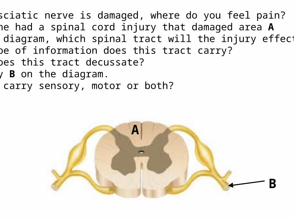
1. If your sciatic nerve is damaged, where do you feel pain?2. If someone had a spinal cord injury that damaged area A on the diagram, which spinal tract will the injury effect?3. What type of information does this tract carry?4. Where does this tract decussate?5. Identify B on the diagram. Does B carry sensory, motor or both?
A
B
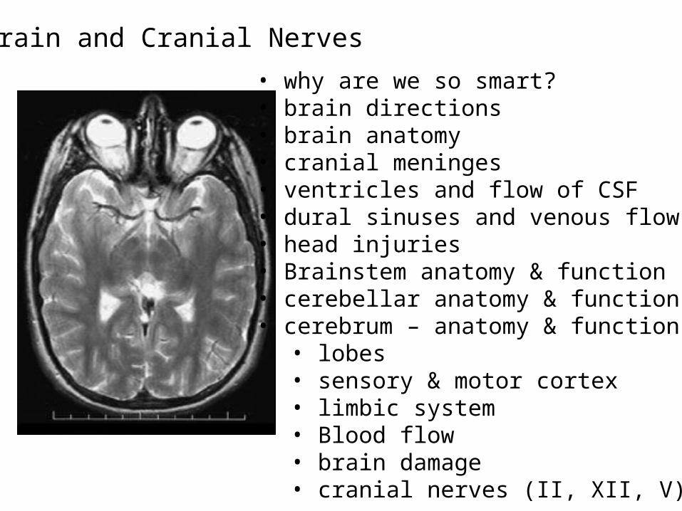
Brain and Cranial Nerves• why are we so smart?• brain directions• brain anatomy• cranial meninges• ventricles and flow of CSF• dural sinuses and venous flow• head injuries• Brainstem anatomy & function• cerebellar anatomy & function• cerebrum – anatomy & function
• lobes• sensory & motor cortex• limbic system• Blood flow• brain damage• cranial nerves (II, XII, V)

Does size matter???
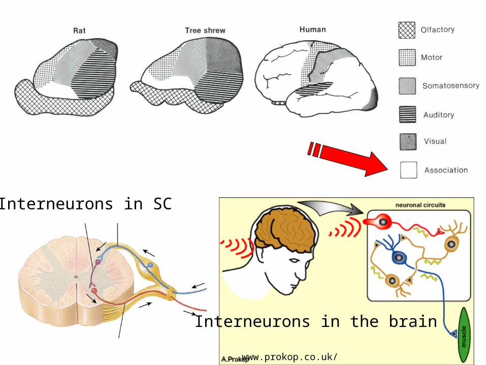
Interneurons in SC
Interneurons in the brain
www.prokop.co.uk/

anterior posterior
cerebrum
cerebellumdiencephalon
Brain stem
dorsal
ventral
Anatomical directions and parts of the brain
Posterior = dorsalAnterior = ventral
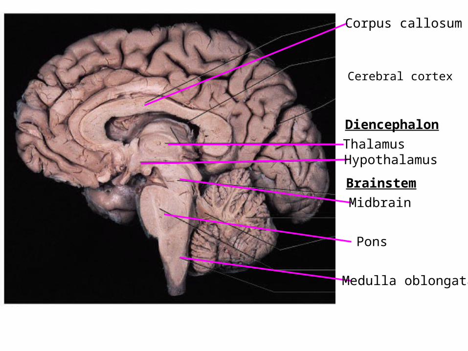
Corpus callosum
Thalamus Hypothalamus
Diencephalon
Brainstem
Midbrain
Pons
Medulla oblongata
Cerebral cortex
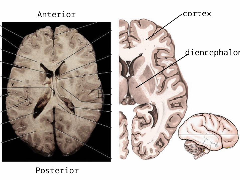
Anterior
Posterior
cortex
diencephalon

S = skinC = connective tissue **A = aponeurosisL = loose connective tissueP = periosteum
Dura mater• periosteal layer• meningeal layer• sinus (venous blood)
ArachnoidSubarachnoid space (CSF)Arachnoid villusPia mater

Meningitis = viral, bacterial, fungal infection of blood and CSF Viral >> bacterial but bacterial is a medical emergencySymptoms: fever, headache, nausea, vomiting, light sensitivity….People living in close quarters (ie dorms) should get vaccination
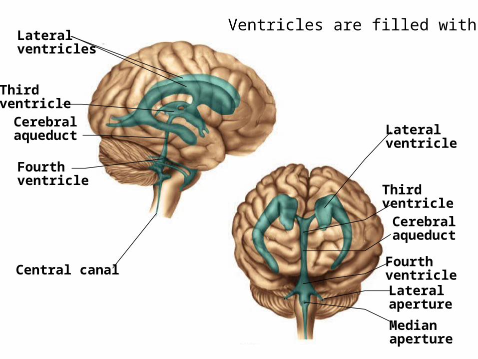
Lateralventricles
Central canal
Fourthventricle
Thirdventricle
Cerebral aqueduct Lateral
ventricle
ThirdventricleCerebralaqueduct
Lateralaperture
Fourthventricle
Medianaperture
Ventricles are filled with CSF


Choroid plexus• Ependymal cells• choroid plexus (capillary bed)
Lateral ventricle
Third ventricle
Fourth ventricle
Cerebral aqueduct
1
23
4
56
7
8
Subarachnoid space
Central canal of spinal cord
Arachnoid granulation
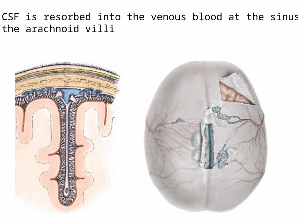
“dirty” CSF is resorbed into the venous blood at the sinusesthrough the arachnoid villi

Dural sinuses drain into the:• jugular veins•Vertebral plexus, cavernous sinus, pterygopalatine plexus• scalp infection• occlusion of sinuses• cancer metastasis
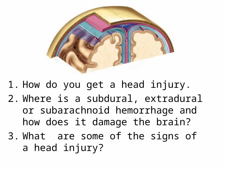
1. How do you get a head injury.2. Where is a subdural, extradural or
subarachnoid hemorrhage and how does it damage the brain?
3. What are some of the signs of a head injury?

Head injuries
Epidural hematoma• arterial bleeding• blow to head• concussion• drowsiness & coma
Subarachnoid hematoma• usually arterial• 70% due to aneurysm• rest are due to trauma• headache, stiff neck & loss of consciousness• blood in CSF
Subdural hematoma• usually venous at cerebral vein – sinus junction• creates space at dural- arachnoid junction• blow to head that jerks the brain (elderly)• trauma often forgotten

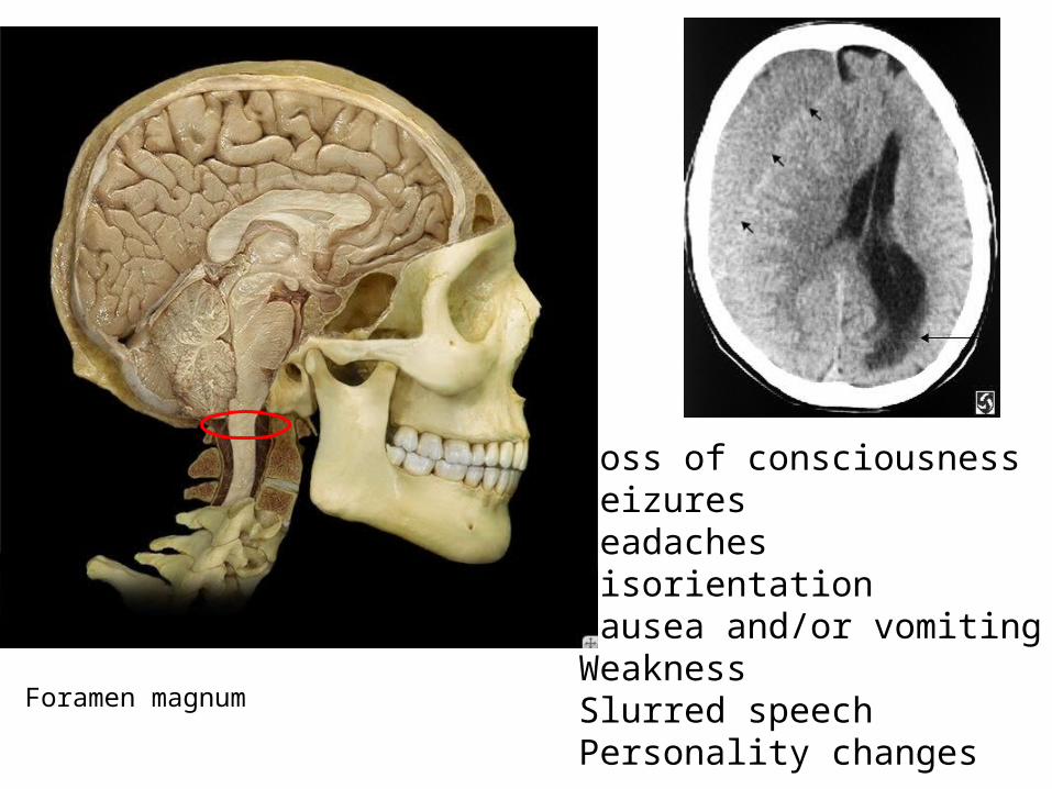
Foramen magnum
Loss of consciousnessSeizuresHeadachesDisorientationNausea and/or vomitingWeaknessSlurred speechPersonality changes

Brain stem = midbrain, pons, medulla oblongata
Medulla: controls respiration, heart rate and blood pressure!

Anterior view Dorsolateral view
thalamus
Pons
colliculi
Cerebral peduncle
Medullaoblongata
Brainstem anatomy and function

Thalamus: relay stationfor sensory input to the cortex
Hypothalamus: controls ANS & endocrine system
Diencephalon
T
T

Cerebellar function• coordinates joint movements• coordinates eye – motor movements• aids in planning, learning & storing motor movements• maintains muscle tone and posture• adjusts muscle performance during movement• damage to cerebellum
pons
ALPL
pons
Middle cerebellar peduncle

Fig. 15.9a(TE Art)Primarymotor area Motor assoc-iation areaPrimarysomestheticarea
Primarymotor areaMotor association area
Thalamus
Input to cerebellum Output from cerebellum
Sensory input
Vestibular
Auditory
Visual
Rubrospinal tract(fine motor control)
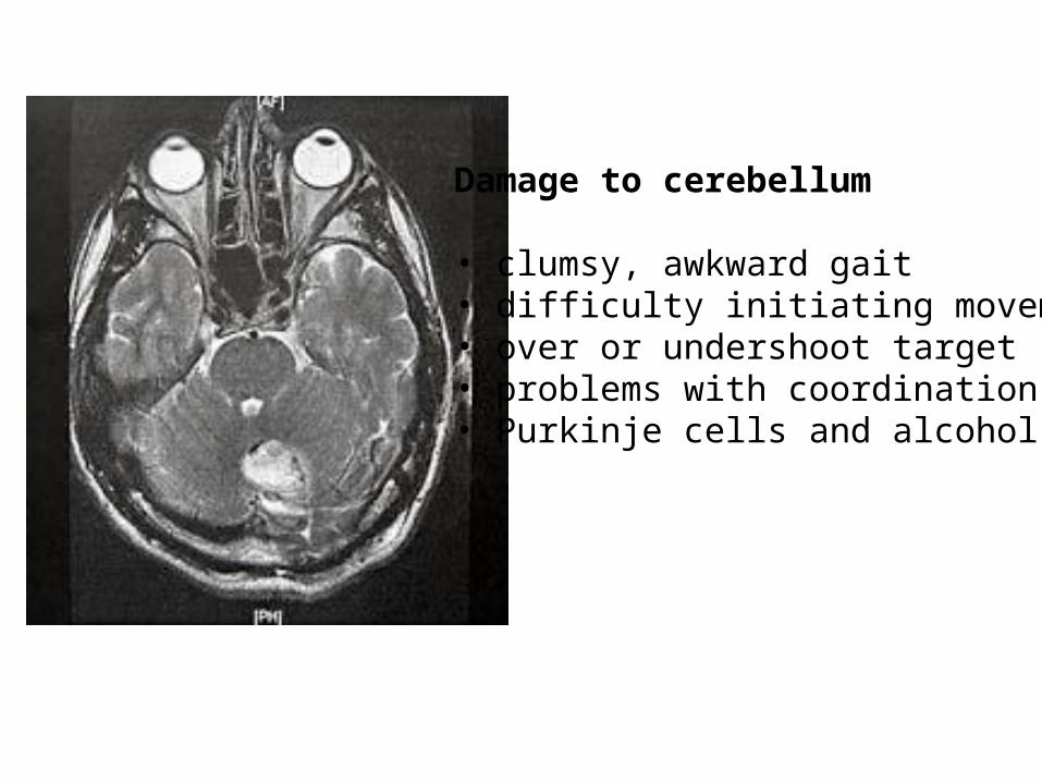
Damage to cerebellum
• clumsy, awkward gait• difficulty initiating movement• over or undershoot target• problems with coordination• Purkinje cells and alcohol

1. Identify this space (be specific)
2. Name this part of the brain.
2. The pia mater is _____ to the arachnoid mater?
4. What is in the dural sinuses?
5. How can you get brain damage from meningitis?

Fissure > sulcus

• Frontal lobe – cognition and “higher” mental processes, motor• Parietal lobe – receiving & interpreting general sensory & taste• Occipital lobe – visual information• Temporal lobe – hearing, smell, learning, memory, emotions• Insula – taste, hearing, visceral sensory info
Central sulcus
Lateral sulcus
Longitudinal fissure
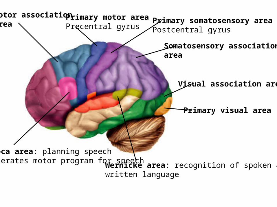
Visual association area
Primary visual area
Primary somatosensory areaPostcentral gyrus
Somatosensory associationarea
Motor associationarea
Primary motor areaPrecentral gyrus
Broca area: planning speechgenerates motor program for speech Wernicke area: recognition of spoken &
written language

Cuneate fasciculusCorticospinal tract
Primary Sensory & Motor
Areas
Precentral Gyrus (motor) Postcentral
Gyrus (sensory)
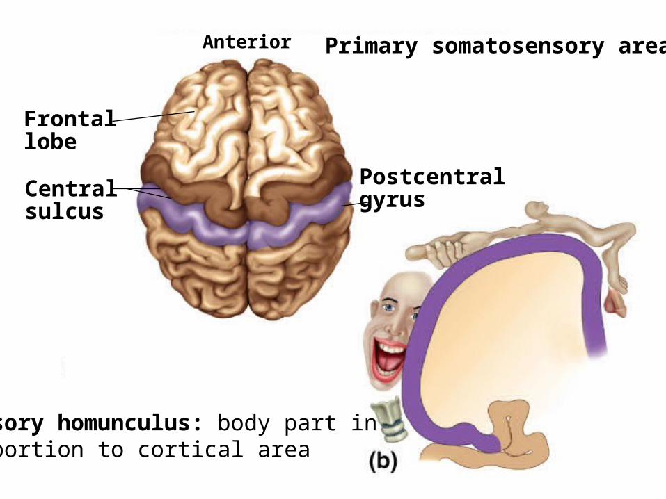
Fig. 15.19(TE Art)Anterior
Postcentralgyrus
Frontallobe
Centralsulcus
Primary somatosensory area
Sensory homunculus: body part inproportion to cortical area

Fig. 15.20(TE Art)Anterior
Precentral gyrus
Centralsulcus
Vocalizatioin
Primary Motor Area
Motor homunculus: body part inproportion to cortical area
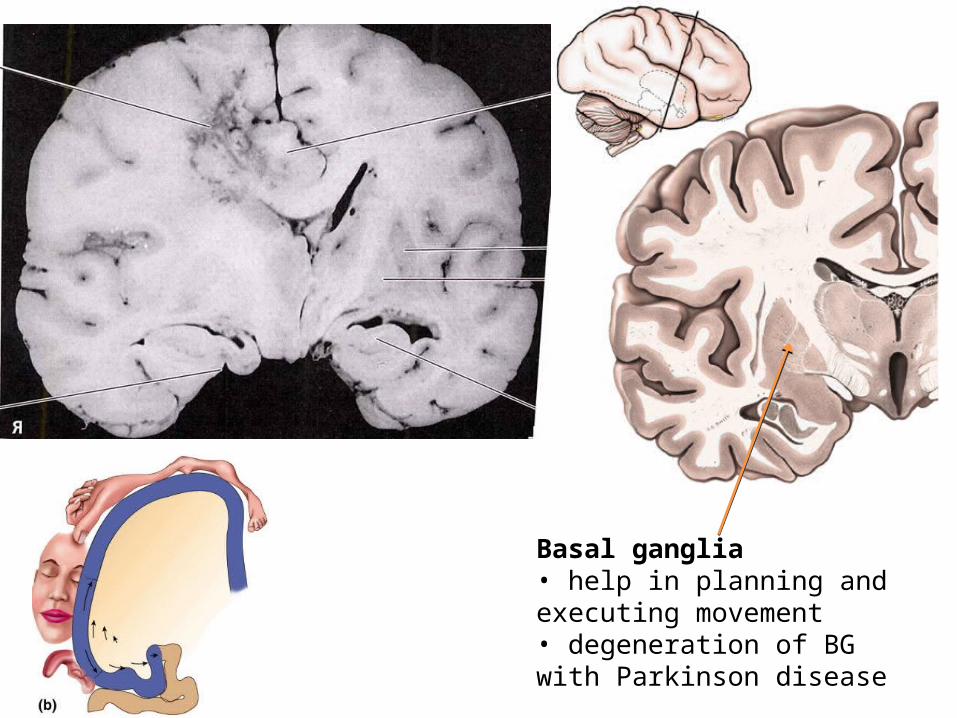
Basal ganglia• help in planning and executing movement• degeneration of BG with Parkinson disease

Prefrontal cortex
Abstract thought, judgement, foresight, responsibilitySense of socially responsible behavior, motivation
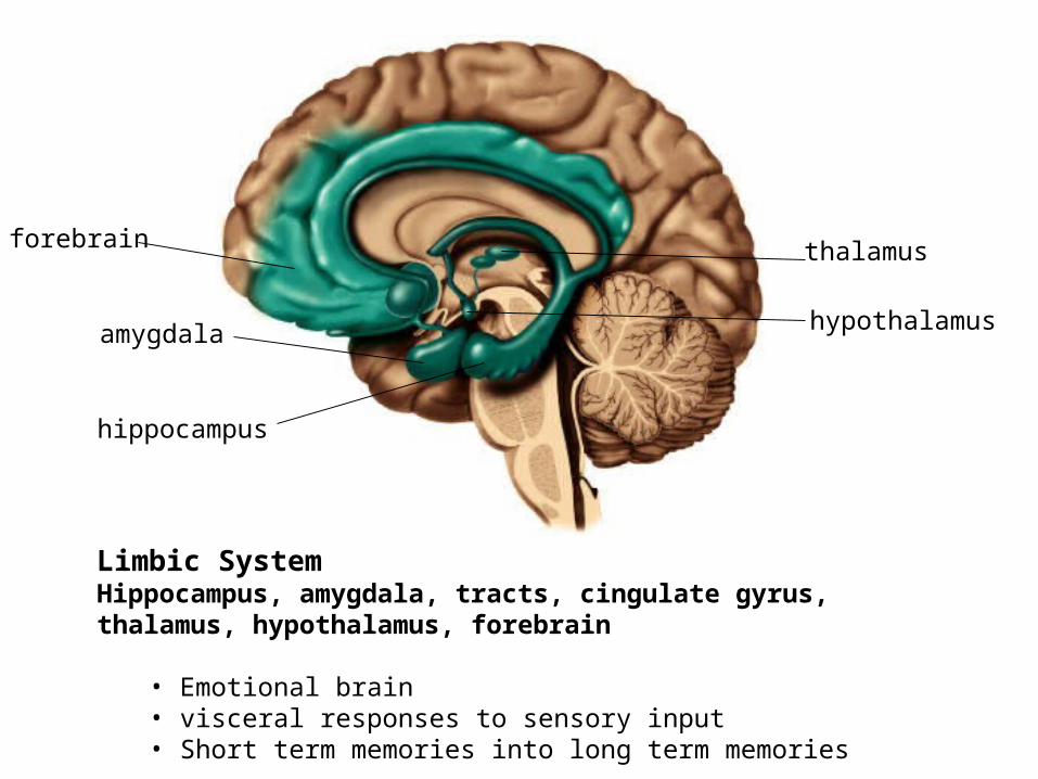
Limbic SystemHippocampus, amygdala, tracts, cingulate gyrus, thalamus, hypothalamus, forebrain
• Emotional brain• visceral responses to sensory input• Short term memories into long term memories
thalamus
hypothalamus
hippocampus
amygdala
forebrain

taste
smell
Prefrontalcortex
hearing
vision
touch
Alzheimer’s
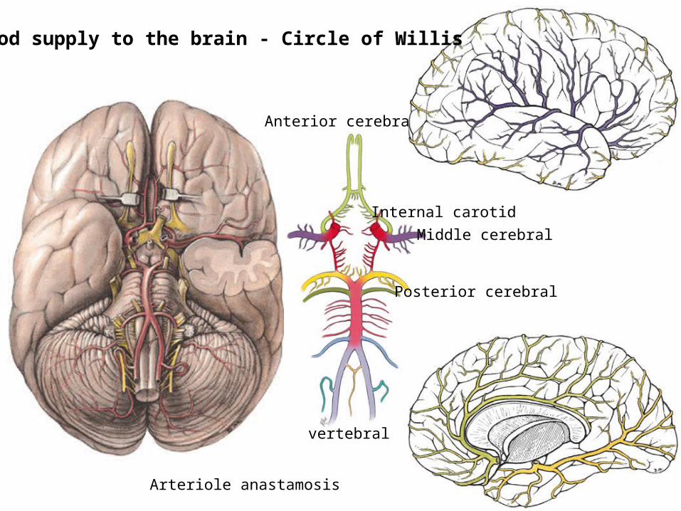
Anterior cerebral
Middle cerebral
Posterior cerebral
vertebral
Internal carotid
Blood supply to the brain - Circle of Willis
Arteriole anastamosis
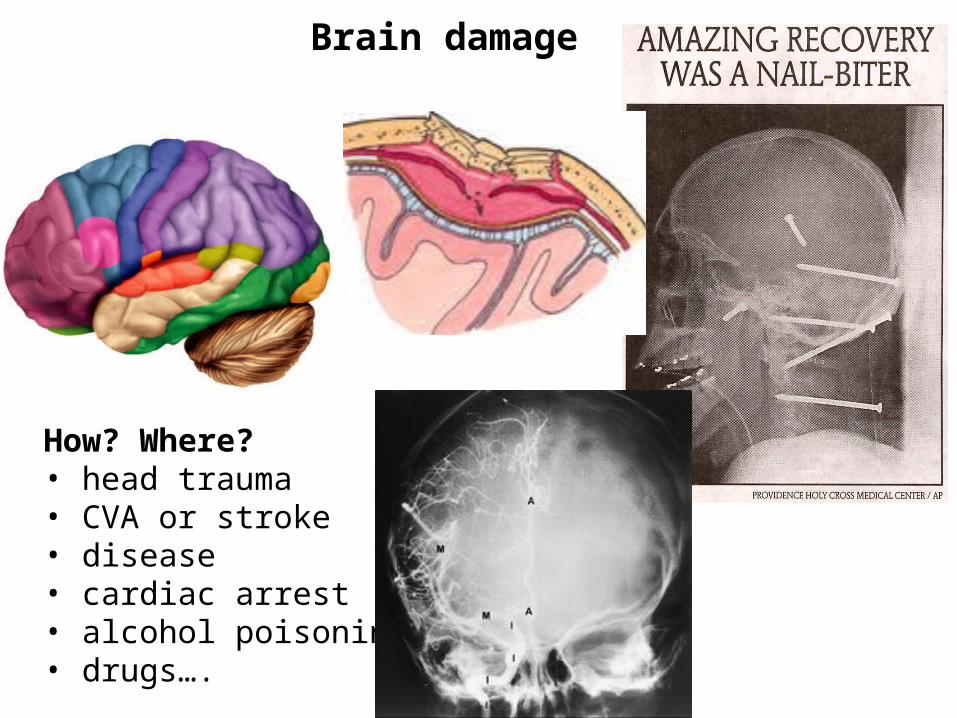
Brain damage
How? Where?• head trauma• CVA or stroke• disease• cardiac arrest• alcohol poisoning • drugs….

Coma: profound state of unconsciousness (usually eyes closed, no sleep/wake cycles)alive but unable to respond to environment (some reflexive activity)can be irreversible depending, maybe still be breathing on ownhave electrical activitycan lead to PVS
Persistent Vegetative State: severe brain damage – coma – no detectable awarenessunconscious, unresponsive, unaware (can have arousal & sleep/wake cycles)exhibit some “spontaneous” behaviors (may open & close eyes, grind teeth..)usually irreversible
Brain death: complete & irreversible cessation of brain activityno electrical activity – no CN reflexesincludes cortex & brainstemdefinition has changed (anencephaly)
Terry Shiavo

Cranial nerves
Peripheral nerves that leave at the base of the brain (instead of the spinal cord)Carry sensory & motor information to head, neck and visceraThere are 12 pair of cranial nerves I-XII (each has a name and number)Can use cranial nerves to test brain function

Cranial Nerve II – Optic NerveAn example of a purely sensory cranial nerve
CN II – receives sensory information from the retina relays info to occipital lobe

Cranial Nerve XII – Hypoglossal NerveAn example of a purely motor cranial nerve
CN XII – motor information to tongue muscles responsible for swallowing, speech, chewing

Cranial Nerve V – Trigeminal NerveAn example of a mixed cranial nerve
CN V – carries sensory information from face, teeth, gums, tongue cornea, sinuses, dura mater, test using the corneal blink reflex

CN V – motor to muscles of mastication and tensor tympani

Cranial nerves
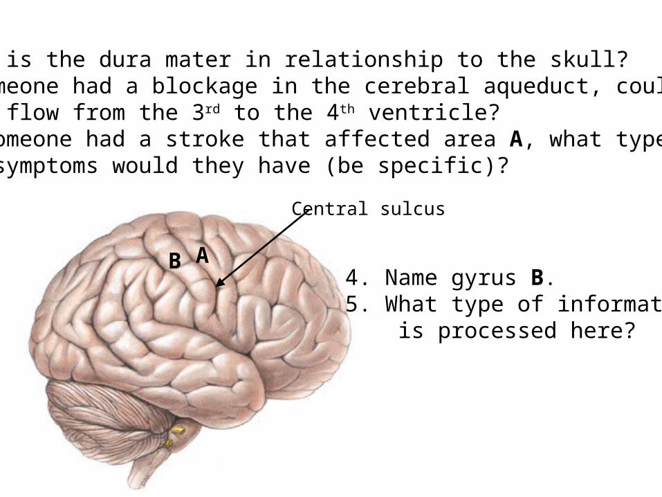
1. Where is the dura mater in relationship to the skull?2. If someone had a blockage in the cerebral aqueduct, could CSF flow from the 3rd to the 4th ventricle?3. If someone had a stroke that affected area A, what type of symptoms would they have (be specific)?
Central sulcus
A4. Name gyrus B.5. What type of information is processed here?
B